555 CALCINOSIS (A review with reports of four cases) By PETER KILBURN, M.B., M.Ch.Orth., F.R.C.S.Ed. Orthopaedic Registrar, Professorial Unit, Royal Southern Hospital, Liverpool Calcinosis is a particular example of pathological calcification, in which multiple calcareous deposits occur in the skin, subcutaneous tissues and in the more deep-seated interstitial connective tissues. The condition was described by Weber in 1878, when he reported a case of calcinosis universalis occurring in association with sclerodactylia (Weber, I878). Pathology This deposition of calcium in soft tissue is a chronic condition tending to be slowly progressive. The mechanism by which calcium salts are laid down in these abnormal sites is unknown and, despite extensive biochemical investigations, no significant departure from the normal can be detected in the large majority of patients. X-ray diffraction studies and chemical analysis indicate that deposits of calcium salts in both normal and abnormal sites have the same physical and chemical structure (Cornbleet et al., I949; Atkin- son and Weber, I938). It has been customary in the past to divide cases of calcinosis into two fairly well-defined groups, yiz. calcinosis universalis and calcinosis circumscripta (Verse, I912; Steinitz, I931). In such a classification the following points of contrast appear: Calcinosis Circumscripta This occurs most often in adult females, the ratio of females to males being approximately 6:i. In this form the deposits are limited to the skin and subcutaneous tissues of the upper extremity, especially the fingers. The condition bears a super- ficial resemblance to gout (' calcium gout'). Calcinosis Universalis This condition is uncommon and is associated with widespread deposits of calcium salts in the skin, subcutaneous tissues and fascia. The limbs bear the brunt, the head and trunk being usually spared. The generalized form occurs more frequently in children, especially males. Calcinosis has been recorded in children under the age of two by several observers (Tisdall and Erb, I924; Swanson et al., 1933). It is now felt that this division into universal and circumscript forms is undesirable, as it does not take into account the many transitional forms of the disease process, as well as carrying no diag- nostic connotation: similar types of soft-tissue calcification may occur, e.g. in hypervitaminosis D, dermatomyositis, renal insufficiency, hyper- parathyroidism and widespread carcinomatosis. The close association between soft-tissue calcifica- tion and conditions such as scleroderma (Thibierge and Weissenbach, I9I1) and Raynaud's disease (Logan, I924; Langmead, I930; Durham, 1928) has been observed for many years, but doubt has recently been cast on the association between cal- cinosis and Raynaud's disease by Cole (Cole, 1953), who considers, after a close study of the reported cases, that this, in fact, represents a developing scleroderma. Attention has been recently focused again on the question of soft-tissue calcification and its relation to the collagen diseases in a thought- provoking paper by Clayton E. Wheeler and his colleagues (Wheeler et al., 1952). These workers are not prepared to recognize idiopathic calcinosis as a clinical entity and they argue that soft-tissue calcification is always secondary to some other condition and that the primary cause can nearly always be elucidated if an adequate search is made. They classify the conditions predisposing to cal- cinosis into two main groups: (I) Tissue injury, either (a) local, due to known causes, i.e. dystrophic calcification, or (b) wide- spread tissue injury, due to unknown causes, and in this latter group are included the collagen diseases. (2) Conditions associated with abnormal calcium and/or phosphorus metabolism. In I9I9 Mme. Dejerine and her co-workers wrote a comprehensive account of soft-tissue cal- cification in patients with cord lesions (Dejerine et al., I9I9) and Liberson (I953) described soft- tissue calcification in 30 paraplegics. The mech- copyright. on 26 July 2018 by guest. Protected by http://pmj.bmj.com/ Postgrad Med J: first published as 10.1136/pgmj.33.385.555 on 1 November 1957. Downloaded from
Transcript
555
CALCINOSIS(A review with reports of four cases)
By PETER KILBURN, M.B., M.Ch.Orth., F.R.C.S.Ed.
Orthopaedic Registrar, Professorial Unit, Royal Southern Hospital, Liverpool
Calcinosis is a particular example of pathologicalcalcification, in which multiple calcareous depositsoccur in the skin, subcutaneous tissues and in themore deep-seated interstitial connective tissues.The condition was described by Weber in 1878,when he reported a case of calcinosis universalisoccurring in association with sclerodactylia (Weber,I878).Pathology
This deposition of calcium in soft tissue is achronic condition tending to be slowly progressive.The mechanism by which calcium salts are laiddown in these abnormal sites is unknown and,despite extensive biochemical investigations, nosignificant departure from the normal can bedetected in the large majority of patients. X-raydiffraction studies and chemical analysis indicatethat deposits of calcium salts in both normal andabnormal sites have the same physical andchemical structure (Cornbleet et al., I949; Atkin-son and Weber, I938).
It has been customary in the past to dividecases of calcinosis into two fairly well-definedgroups, yiz. calcinosis universalis and calcinosiscircumscripta (Verse, I912; Steinitz, I931). Insuch a classification the following points of contrastappear:
Calcinosis CircumscriptaThis occurs most often in adult females, the
ratio of females to males being approximately 6:i.In this form the deposits are limited to the skinand subcutaneous tissues of the upper extremity,especially the fingers. The condition bears a super-ficial resemblance to gout (' calcium gout').Calcinosis UniversalisThis condition is uncommon and is associated
with widespread deposits of calcium salts in theskin, subcutaneous tissues and fascia. The limbsbear the brunt, the head and trunk being usuallyspared. The generalized form occurs morefrequently in children, especially males. Calcinosis
has been recorded in children under the age of twoby several observers (Tisdall and Erb, I924;Swanson et al., 1933).
It is now felt that this division into universaland circumscript forms is undesirable, as it doesnot take into account the many transitional formsof the disease process, as well as carrying no diag-nostic connotation: similar types of soft-tissuecalcification may occur, e.g. in hypervitaminosisD, dermatomyositis, renal insufficiency, hyper-parathyroidism and widespread carcinomatosis.The close association between soft-tissue calcifica-tion and conditions such as scleroderma (Thibiergeand Weissenbach, I9I1) and Raynaud's disease(Logan, I924; Langmead, I930; Durham, 1928)has been observed for many years, but doubt hasrecently been cast on the association between cal-cinosis and Raynaud's disease by Cole (Cole, 1953),who considers, after a close study of the reportedcases, that this, in fact, represents a developingscleroderma. Attention has been recently focusedagain on the question of soft-tissue calcificationand its relation to the collagen diseases in a thought-provoking paper by Clayton E. Wheeler and hiscolleagues (Wheeler et al., 1952). These workersare not prepared to recognize idiopathic calcinosisas a clinical entity and they argue that soft-tissuecalcification is always secondary to some othercondition and that the primary cause can nearlyalways be elucidated if an adequate search is made.They classify the conditions predisposing to cal-cinosis into two main groups:
(I) Tissue injury, either (a) local, due to knowncauses, i.e. dystrophic calcification, or (b) wide-spread tissue injury, due to unknown causes, andin this latter group are included the collagendiseases.
(2) Conditions associated with abnormal calciumand/or phosphorus metabolism.
In I9I9 Mme. Dejerine and her co-workerswrote a comprehensive account of soft-tissue cal-cification in patients with cord lesions (Dejerineet al., I9I9) and Liberson (I953) described soft-tissue calcification in 30 paraplegics. The mech-
anism whereby this calcium is laid down is com-pletely unknown, the deposition of calcium saltsis not found above the level of the lesion nor dobiochemical studies show any abnormality.
It is of interest to note that Van Wagtendonkand his colleagues (Van Wagtendonk et al., I944and I947) reported a possible deficiency diseasein guinea pigs associated with extensive depositionof calcium salts when these animals were kept on arestricted diet. If cream was subsequently ad-ministered to these animals, the deposits dis-appeared. Burnett (quoted by Cecil, I955)' hasreported a syndrome of hypercalcaemia withouthypercalcuria, but with renal insufficiency andcalcinosis, in patients with peptic ulceration whoseregime consists of an excessive milk intakeassociated with absorbable alkalis. It is clear,therefore, that the deposition of calcium salts inabnormal sites represents the end stage of manypathological states.
Clinical FeaturesThe earliest symptom referable to the calcium
deposits is frequently a localized area of skintenderness and examination at this stage revealsmultiple discrete stony-hard subcutaneous nodules;these are of varying sizes and pleomorphic, butwith a tendency to be flat and to form plaques.The smaller nodules are mobile, but the larger onesare usually tethered to the deep structures. Ulcera-tion of the skin over the more superficial nodulesis a common occurrence, with the formation ofpainful shallow ulcers which exude calcareousmaterial, after which they heal with dense scarring.Limitation of joint movement may be marked,due to the thick plaques of calcium in the vicinityof the joint, together with post-ulceration scarringand muscle atrophy. The distribution of thelesions is characteristic: the deposits are mostfrequent around the elbows, knees, buttocks andshoulders, whilst the trunk, face, scalp and visceraare nearly always spared.Biochemical Studies
Estimations of serum calcium, phosphorus andalkaline phosphatase are normal in those cases ofcalcinosis associated with the collagen disorders;there is thus no evidence that disturbed levels ofthese minerals are significant aetiological factorsin the laying down of calcium in these extraordinarysites. In these cases metabolic studies are eithernormal or else show a tendency to retain calcium(Atkinson, 1938; Bauer, 1931; Brooks, I934). Ofcourse, altered serum levels of calcium and phos-phorus and alkaline phosphatase are to be foundin those cases secondary to renal or parathyroiddisease.
TreatmentUnfortunately, there is no specific treatment for
this distressing condition. Local removal of thecalcium deposits is always ineffective and is awaste of time. ACTH is worth a trial in those caseswhich are secondary to one of the collagen diseasesand Briggs and Illingworth (Briggs and Illing-worth, 1952) reported a case of calcinosis associatedwith dermatomyositis which showed regression ofthe calcium deposits after ACTH therapy. Lowcalcium diets are ineffective, as are ketogenic dietsand the administration of ammonium chloride andsodium acid phosphate.Case No. i (Figs. I to 3)Male patient, J. C. Aet. i8 years. Towards the
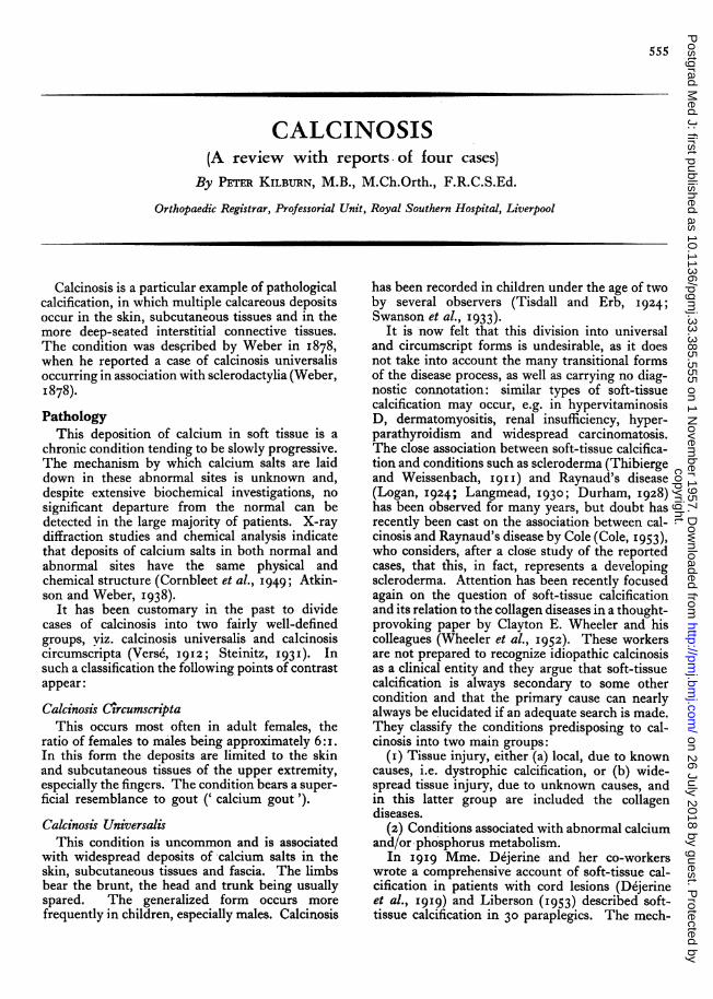
end of 1947 began to complain of pain in the legsand jaw, followed by swelling and stiffness ofjoints-knees, ankles, elbows and wrists.
Admitted to hospital 31.3.48. Was then pale,wasted and febrile with swelling and stiffness ofabove-mentioned joints and stiffness of tempero-mandibular joint and neck. Spleen, liver andlymph glands not palpable. No evidehce ofcarditis. X-ray showed decalcification of bone.Mantoux and W.R. negative. Diagnosed as rheu-matic polyarthritis.X-ray in December I949 showed extensive
generalized decalcification with patch of calcifica-tion just above left ankle. (Calcified shadows inleft renal area and over the sacrum.)
Followed, up at the out-patient clinic, whenstiffness of hips and spine was noticed. In May195I numerous subcutaneous nodules confined tothe right forearm detected (Fig. I). Patient saidthey had gradually appeared over the previousyear.
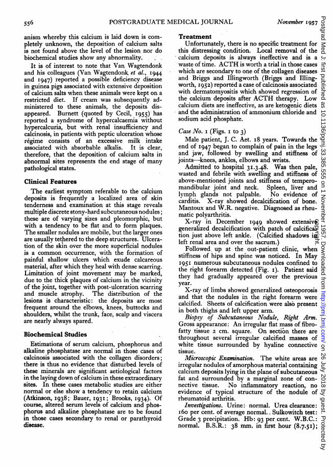
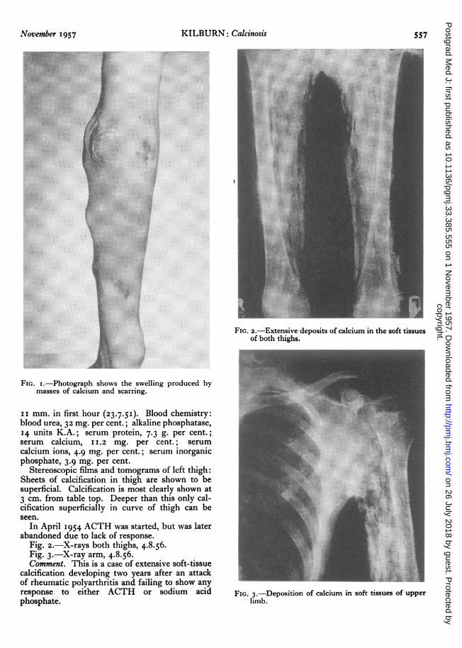
X-ray of limbs showed generalized osteoporosisand that the nodules in the right forearm werecalcified. Sheets of calcification were alsb presentin both thighs and left upper arm.
Biopsy of Subcutaneous Nodule, Right Arm.Gross appearance: An irregular flat mass of fibro-fatty tissue 2 cm. square. On sectibn there arethroughout several irregular calcified masses ofwhite tissue surrounded by hyaline connectivetissue.
Microscopic Examination. The white areas areirregular nodules of amorphous material containingcalcium deposits lying in the plane of subcutaneousfat and surrounded by a marginal zone of con-nective tissue. No inflammatory reaction, noevidence of typical structure of the nodule ofrheumatoid arthritis.
Investigations. Urine: normal. Urea clearance:I6o per cent. of average normal., Sulkowitch test:Grade 3 precipitation. Hb: 93 per cent. W.B.C.:normal. B.S.R.: 38 mm. in first hour (8.7.5I);
FIG. I.-Photograph shows the swelling produced bymasses of calcium and scarring.
ii mm. in first hour (23.7.5I). Blood chemistry:blood urea, 32 mg. per cent.; alkaline phosphatase,14 units K.A.; serum protein, 7.3 g. per cent.;serum calcium, 11.2 mg. per cent.; serumcalcium ions, 4.9 mg. per cent.; serum inorganicphosphate, 3.9 mg. per cent.
Stereoscopic films and tomograms of left thigh:Sheets of calcification in thigh are shown to besuperficial. Calcification is most clearly shown at3 cm. from table top. Deeper than this only cal-cification superficially in curve of thigh can beseen.
In April I954 ACTH was started, but was laterabandoned due to lack of response.
Fig. 2.-X-rays both thighs, 4.8.56.Fig. 3.-X-ray arm, 4.8.56.Comment. This is a case of extensive soft-tissue
calcification developing two years after an attackof rheumatic polyarthritis and failing to show anyresponse to either ACTH or sodium acidphosphate.
cm
I·1
:.i.·
:···:r
iii
Bi'
·;'·
it:.iii
·;r
·Bi ··'.· ':i
I;i' ·ly::::i:.. :I·
i:·
Ilili'" .:::i.CS.f.c.sais
*.-:: ····;
':ir
FIG. 2.-Extensive deposits of calcium in the soft tissuesof both thighs.
·.e
..i...,...
·'..1e
· .:
X...: t · ·.
.
- :. :-(::
FIG. 3.-Deposition of calcium in soft tissues of upperlimb.
FIG. 4.-Showing soft-tissue calcification and atrophicchanges in terminal phalanges.
Case No. 2W. H., female. Aet. 53 years. This patient was
admitted to hospital for gastro-intestinal investiga-tion and, in addition to her complaints of vomitingand dysphagia, she gave a history of having hadRaynaud's disease affecting both hands for 20 yearsand she had recently noticed that the digits hadbecome rather pointed and shortened and had alsobecome increasingly stiff.On examination the skin over the fingers was
white, shiny and smooth, closely bound down tothe underlying tissues, the nails were fragile, andthere were osteo-arthritic changes in the inter-phalangeal joints.
X-rays which were taken on 13.7.54 (Fig. 4)show soft-tissue calcification with atrophic changesin the terminal phalanges. A diagnosis of sclero-dactyly was made and cortisone therapy wasstarted. This resulted in a dramatic improvementin the patient's symptoms, but it was necessary todiscontinue the therapy at the patient's ownrequest, as she had become frightened after reading
*::
FIG. 5.-Showing vascular calcification and calcifica-tion of the olecranon bursa.
in the press about the dangers of cortisone therapy.The patient died of coronary occlusion on 17. 11 56.
Comment. This case illustrates digital soft-tissuecalcification developing in a long-standing case ofRaynaud's disease with secondary changes in theskin, suggesting a diagnosis of sclerodactyly.Case No. 3Male patient, E. H. H. Aet. 27 years. This
patient was first seen in the orthopaedic out-patient department in December 1953, after hehad injured his elbow.
X-rays (Fig. 5) showed vascular calcification andcalcification of the olecranon bursa. He wasadmitted to hospital and investigated.
FIG. 6.-Showing calcium deposits in soft tissues offorearm and elbow.
per cent.; serum calcium, 10.5 mg. per cent.;serum alkaline phosphatase, 26 units; serum phos-phorus, 10 mg. per cent.; serum bicarbonate,I5.7 ml. CO2 as bicarbonate; urinary calcium,0.12 g./diem.
This was taken to imply a state of renal in-sufficiency with acidosis, phosphorus retention andreactive parathyroid hyperplasia.
Biochemistry on 8.4.54. Serum bicarbonate,60 ml. C02; blood urea, 80 mg. per cent.; Na,320; K, 27.
8. I.55, patient died.Post-mortem findings were as follows: There
was widespread soft-tissue calcification presentwhich contained a white, softish material giving areaction for calcium, phosphorus and carbonate.
Right kidney: Hypoplastic and was composedof a mass of tubules with no trace of glomerularstructure.
Left kidney: Marked glomerular fibrosis withmuch interstitial fibrosis and round-celled in-filtration.
Comment. This is an example of soft-tissuecalcification occurring in association with chronicrenal failure.
Case No. 4 (Fig. 6)Female patient, B. H. Aet. nine years. At the
age of three years it was first noticed that she hadundue hardness of the muscles of both calves.This was noticed by her dancing instructor. Atthe age of four years she had a small soft swellingover the anterior aspect of the left leg, whichgradually increased in size. In June 1953 it wasincised and white chalky material was obtained.In October of that year she was seen at hospitalbecause there was a continuous discharge from thecalcified nodule. It was noticed that she wasdeveloping a similar lesion on the left buttock. AnX-ray of her skeleton at that time showed nofurther evidence of calcified lesions.A diagnosis of calcinosis universalis was made.
The biopsy, which was taken in March I954,showed perivascular plasma and mononuclear cellswith fibrosis and deposition of calcium in thefibrous tissues.
In July I954 a calcium balance study was under-taken on high and low calcium intake and with andwithout cortisone treatment. At this time she wasput on a maintenance dose of cortisone, I2.5 mg.twice daily.At that time she had some difficulty in dorsi-
flexion of the left ankle, but this improved follow-ing treatment with cortisone.By May 1955 she had deposits in both buttocks,
both legs, right knee and right elbow. A depositin the natal cleft was discharging.
In November 1955 the mass on the right buttockbegan to discharge and she was left with a largegranulating mass. She was, therefore, readmittedin May I956. She was operated on with removalof the calcified areas on the right buttock, left hipand right lower leg.On 10.9.56 the lesion on the right popliteal fossa
became painful, suggesting that secondary infec-tion had taken place. She was once more given acourse of penicillin and admitted on 11.9.56.At a second operation on I7.9.56 a mass behind
the right knee was removed. Once more there wasconsiderable bleeding. This time also the skinwas closed with great difficulty. The knee had tobe splinted in flexion for several days.
Cortisone was discontinued for a fortnight fromthe day of operation, but at the time of her dis-charge home she was taking cortisone once moreand was walking around, although extension at theknee was still limited. The wound was wellhealed.
Diagnosis. Calcinosis universalis in associationwith dermatomyositis.
Comment. This girl has shown very great im-provement in her general condition as a result ofcortisone therapy, but there has been no evidenceof resorption of the calcium deposits.
SummaryI. The clinico-pathological features of calcinosis
are discussed.2. It is emphasized that in all cases a primary
condition should be assiduously sought.3. Four cases of soft-tissue calcification are
described; in three of these cases there wasevidence of a primary collagen disorder and in thefourth case the calcification was secondary tochronic renal failure.
AcknowledgmentsI wish to express my sincere thanks to Prof.
Bryan McFarland for his very great help andinterest. I am also deeply indebted to Mr. G.Shatwell, Dr. G. S. Sanderson, Dr. S. Keidan,Mr. G. V. Osborne, Dr. R. W. Brookfield and Dr.E. L. Rubin and also Mr. A. G. O'Malley forallowing my access to their records and cases andfor their kind permission to publish their cases.
Case No. 3 has been the subject of an article byDr. R. W. Brookfield, Dr. E. L. Rubin and Dr.M. K. Alexander in the Journal of the Faculty ofRadiologists, 7, 2, 1955.
BIBLIOGRAPHY
ATKINSON, F. R. B., and WEBER, F. P. (1938), Brit. J. Derm.,o5, 267.
BAUER, W., MARBLE, A., and BENNETT, G. (I93 ), Amer. J.med. Sci., 182, 237.
BRIGGS, J. N., and ILLINGWORTH, R. S. (1952), Lancet, ii,800.
BROOKS, W. D. W. (1934), Quart. J. Med., 3, 293.BURNETT, quoted by CECIL (I955), ' A Textbook of Medicine',
Philadelphia. Saunders., p. 871.COLE, W. R. (I953), Guy's Hosp. Rep., o02, 56.CORNBLEET, T., REED, C. I., and REED, B. P. (1949), J. invest.
Derm., 13, 171.DEJERINE, MME. CEILLIER A., and DEJERINE, Y. (1919),
Rev. neurol. (Paris), 26, 399.DURHAM, R. H. (1928), Arch. intern. Med., 42, 467.LANGMEAD, F. S. (1930), Trans. med. Soc. Lond., 53, 67.LIBERSON, M. (I953), J.A.M.A., 152, IOIO.LOGAN, J. R. (1924), Arch. Radiol., 28, 55.STEINITZ, H. (193I), Ergebn. inn. Med. Kinderheilk, 39, 216.SWANSON, W. W., FORSTER, W. F., and IOB, V. (1933)
Amer. J. Dis. Child., 14, 590.THIBIERGE, G., and WEISSENBACH, R. J. (1911), Ann.
Derm. Syph. (Paris), 2, 129.TISDALL, F. F., and ERB, L. H. (1924), Amer. J. Dis. Child., 27,
28.VAN WAGTENDONK, W. J., FREED, A. M., and BALLONE,
C. E. (I944), Arch. Biochem., 5, 329.VAN WAGTENDONK, W. J., and FREED, A. M. (1947),
J. biol. Chem., 167, 225.VERSE, M. (1912), Beitr. path. Anat., 53, 2I2.WEBER, H. (1878), KorrespBl. schweiz Arz. Basel, 8, 623.WHEELER, C. E. (1952), Ann. Int. Med., 36, 1050.
THYROID DISEASE(Postgraduate Medical Journal, July I957)
Price: 3s. 9d., post free
DIAGNOSTIC PROCEDURES IN THYROID RECENT WORK ON THYROID HORMONESDISEASE J. H. Wilkinson, B.Sc., Ph.D., F.R.I.C.Russell Fraser, M.D., F.R.C.P., D.P.M.
SOME UNUSUAL MANIFESTATIONS OFTHE PLACE OF RADIOACTIVE IODINE IN THYROID DISEASETHE TREATMENT OF THYROID DISEASE W. R. Trotter, D.M., M.R.C.P.E. E. Pochin, M.D., F.R.C.P.
ANTITHYROID DRUGS CARCINOMA OF THE THYROIDJames Crooks, M.B., M.R.C.P. (Lond. and John E. Piercy, F.R.C.S., F.R.C.S.E.


![Clinical Cases in · Series: Clinical cases (Ames, Iowa) [DNLM: 1. Periodontal Diseases–therapy–Case Reports. 2. Periodontics–methods–Case Reports. WU 240] 617.6'32--dc23](https://static.documents.pub/doc/80x56/5f84edf5754ed16bc7248a59/clinical-cases-in-series-clinical-cases-ames-iowa-dnlm-1-periodontal-diseasesatherapyacase.jpg)

