Calcinosis Calcinosis Cutis: Cutis: Emerging Trends in Emerging Trends in Dermatoradiology Dermatoradiology Peter Peter Lio Lio , Harvard Medical School Year , Harvard Medical School Year - - IV IV Gillian Lieberman, MD Gillian Lieberman, MD September 2000
Transcript
CalcinosisCalcinosis Cutis: Cutis: Emerging Trends in Emerging Trends in DermatoradiologyDermatoradiology
Peter Peter LioLio, Harvard Medical School Year, Harvard Medical School Year-- IVIVGillian Lieberman, MDGillian Lieberman, MD
September 2000
2
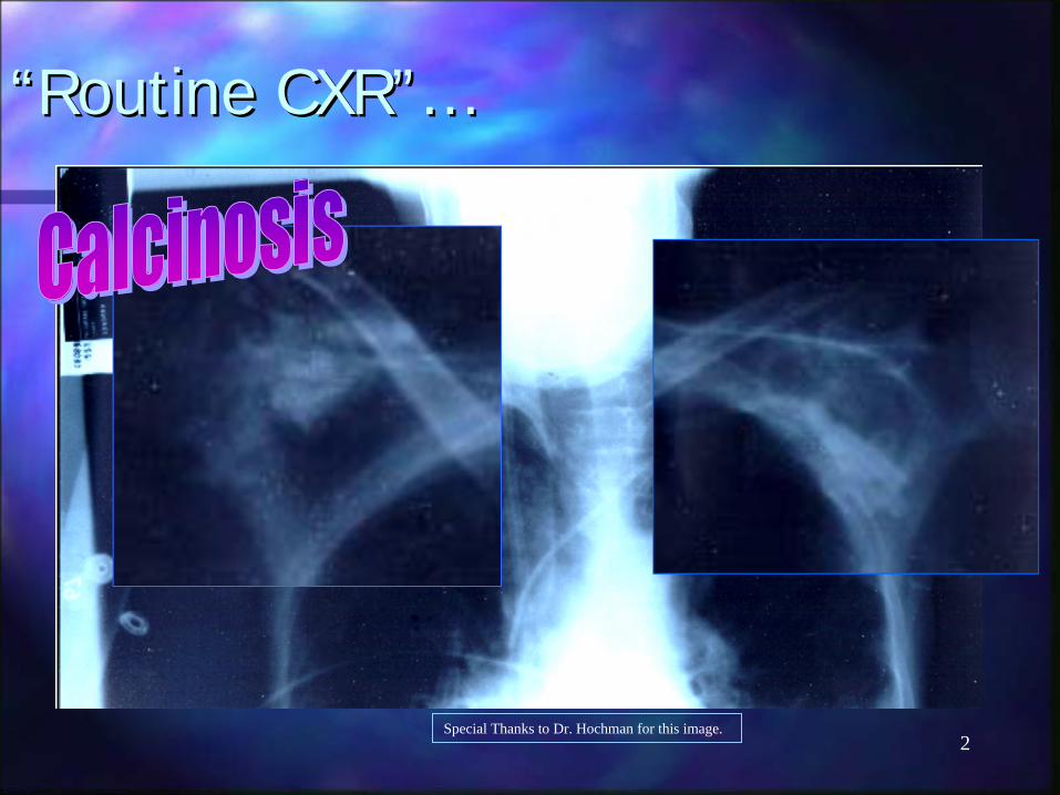
““Routine CXR”…Routine CXR”…
Special Thanks to Dr. Hochman for this image.
3
What is What is CalcinosisCalcinosis??
CalcinosisCalcinosis refers to pathologic calcification refers to pathologic calcification of the skin and soft tissues.of the skin and soft tissues.It occurs in a variety of systemic and It occurs in a variety of systemic and localized conditions.localized conditions.The deposits are generally composed of The deposits are generally composed of calcium phosphate crystals which is why we calcium phosphate crystals which is why we can see them on can see them on radiograpicradiograpic imaging.imaging.
4
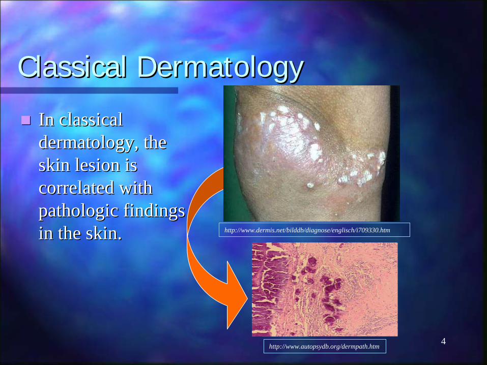
Classical DermatologyClassical Dermatology
In classical In classical dermatology, the dermatology, the skin lesion is skin lesion is correlated with correlated with pathologic findings pathologic findings in the skin.in the skin. http://www.dermis.net/bilddb/diagnose/englisch/i709330.htm
http://www.autopsydb.org/dermpath.htm
5
““DermatoradiologyDermatoradiology””
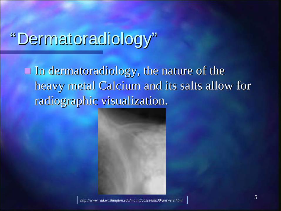
In In dermatoradiologydermatoradiology, the nature of the , the nature of the heavy metal Calcium and its salts allow for heavy metal Calcium and its salts allow for radiographic visualization. radiographic visualization.
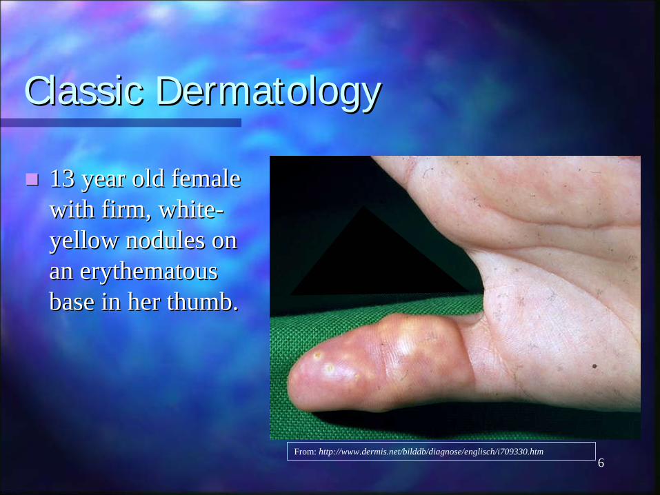
13 year old female 13 year old female with firm, whitewith firm, white--yellow nodules on yellow nodules on an an erythematouserythematousbase in her thumb.base in her thumb.
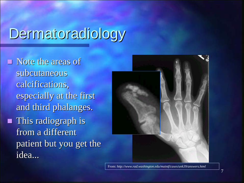
Note the areas of Note the areas of subcutaneous subcutaneous calcifications, calcifications, especially at the first especially at the first and third phalanges.and third phalanges.This radiograph is This radiograph is from a different from a different patient but you get the patient but you get the idea...idea...
CalcinosisCalcinosis: The Breakdown: The Breakdown
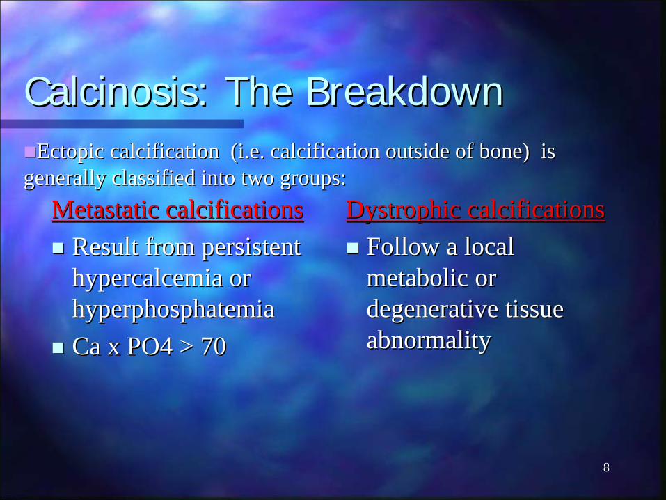
Metastatic calcificationsMetastatic calcificationsResult from persistent Result from persistent hypercalcemiahypercalcemia or or hyperphosphatemiahyperphosphatemiaCa x PO4 > 70Ca x PO4 > 70
Dystrophic calcificationsDystrophic calcificationsFollow a local Follow a local metabolic or metabolic or degenerative tissue degenerative tissue abnormalityabnormality
Ectopic calcification (i.e. calcification outside of bone) is Ectopic calcification (i.e. calcification outside of bone) is generally classified into two groups:generally classified into two groups:
9
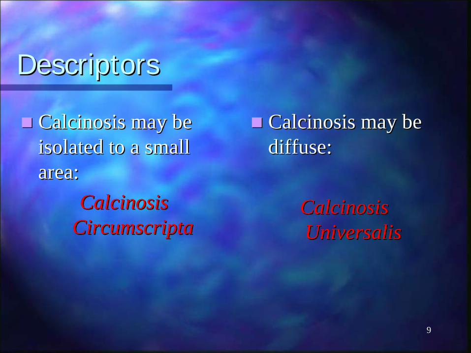
DescriptorsDescriptors
CalcinosisCalcinosis may be may be isolated to a small isolated to a small area:area:
CalcinosisCalcinosis CircumscriptaCircumscripta
CalcinosisCalcinosis may be may be diffuse:diffuse:
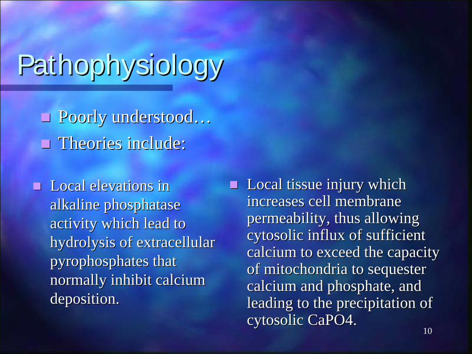
Local tissue injury which Local tissue injury which increases cell membrane increases cell membrane permeability, thus allowing permeability, thus allowing cytosoliccytosolic influx of sufficient influx of sufficient calcium to exceed the capacity calcium to exceed the capacity of mitochondria to sequester of mitochondria to sequester calcium and phosphate, and calcium and phosphate, and leading to the precipitation of leading to the precipitation of cytosoliccytosolic CaPO4.CaPO4.
Local elevations in Local elevations in alkaline alkaline phosphatasephosphataseactivity which lead to activity which lead to hydrolysis of extracellular hydrolysis of extracellular pyrophosphates that pyrophosphates that normally inhibit calcium normally inhibit calcium deposition.deposition.
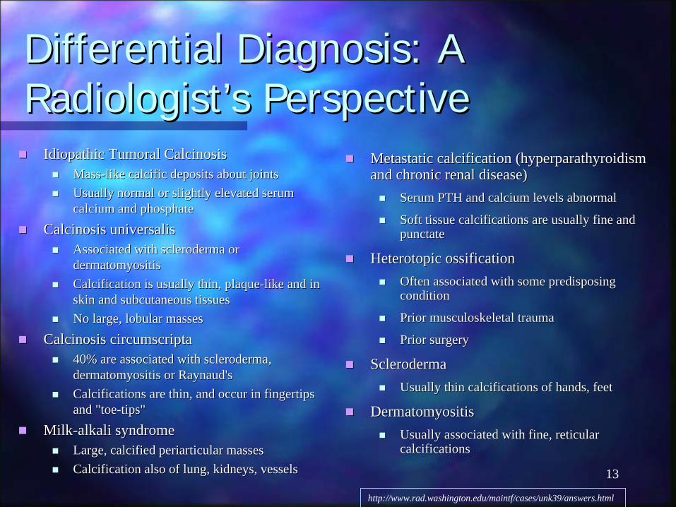
Differential Diagnosis: A Differential Diagnosis: A Radiologist’s PerspectiveRadiologist’s Perspective
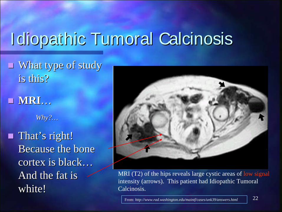
Idiopathic Idiopathic TumoralTumoral CalcinosisCalcinosisMassMass--like like calcificcalcific deposits about joints deposits about joints Usually normal or slightly elevated serum Usually normal or slightly elevated serum calcium and phosphate calcium and phosphate
CalcinosisCalcinosis universalisuniversalisAssociated with scleroderma or Associated with scleroderma or dermatomyositisdermatomyositisCalcification is usually thin, plaqueCalcification is usually thin, plaque--like and in like and in skin and subcutaneous tissues skin and subcutaneous tissues No large, lobular massesNo large, lobular masses
CalcinosisCalcinosis circumscriptacircumscripta40% are associated with scleroderma, 40% are associated with scleroderma, dermatomyositisdermatomyositis or or Raynaud'sRaynaud'sCalcifications are thin, and occur in fingertips Calcifications are thin, and occur in fingertips and "toeand "toe--tips" tips"
MilkMilk--alkali syndrome alkali syndrome Large, calcified Large, calcified periarticularperiarticular masses masses Calcification also of lung, kidneys, vessels Calcification also of lung, kidneys, vessels
Metastatic calcification (hyperparathyroidism Metastatic calcification (hyperparathyroidism and chronic renal disease) and chronic renal disease)
Serum PTH and calcium levels abnormal Serum PTH and calcium levels abnormal
Soft tissue calcifications are usually fine and Soft tissue calcifications are usually fine and punctatepunctate
HeterotopicHeterotopic ossification ossification Often associated with some predisposing Often associated with some predisposing condition condition
Menu of radiologic tests for Menu of radiologic tests for imaging ectopic calcificationsimaging ectopic calcifications
Radiographic plain filmRadiographic plain filmCT scanCT scan-- most sensitivemost sensitiveRN bone scanRN bone scanMRIMRI-- sometimessometimes
15
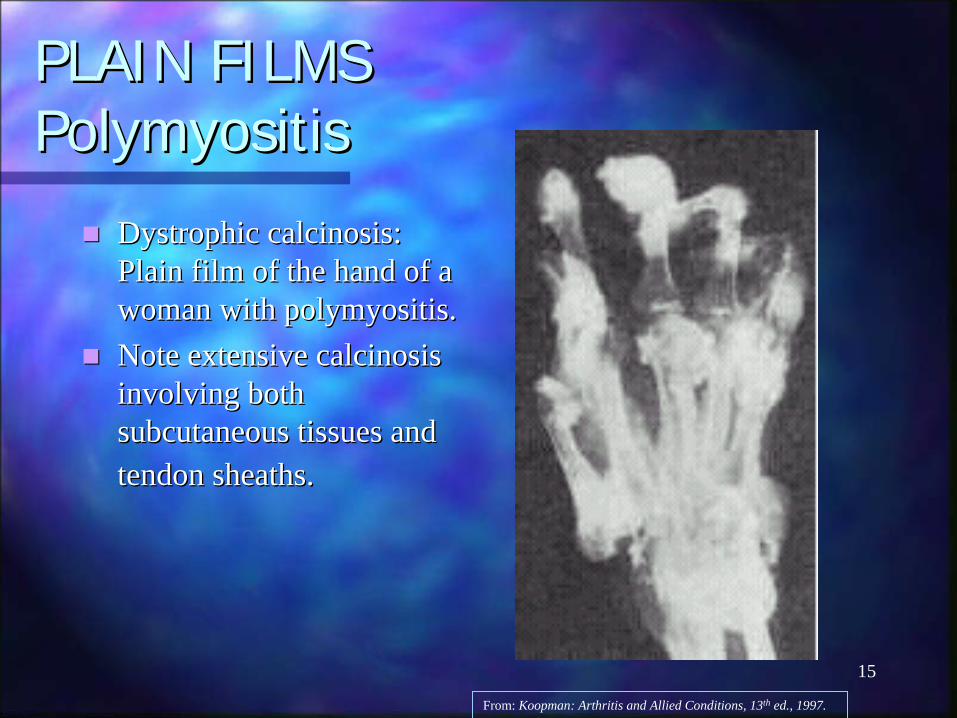
PLAIN FILMSPLAIN FILMS PolymyositisPolymyositis
Dystrophic Dystrophic calcinosiscalcinosis: : Plain film of the hand of a Plain film of the hand of a woman with woman with polymyositispolymyositis..Note extensive Note extensive calcinosiscalcinosisinvolving both involving both subcutaneous tissues and subcutaneous tissues and tendon sheaths.tendon sheaths.
From: Koopman: Arthritis and Allied Conditions, 13th ed., 1997.
16
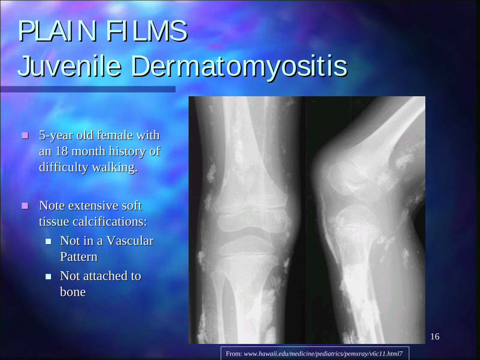
PLAIN FILMSPLAIN FILMS Juvenile Juvenile DermatomyositisDermatomyositis
55--year old female with year old female with an 18 month history of an 18 month history of difficulty walking.difficulty walking.
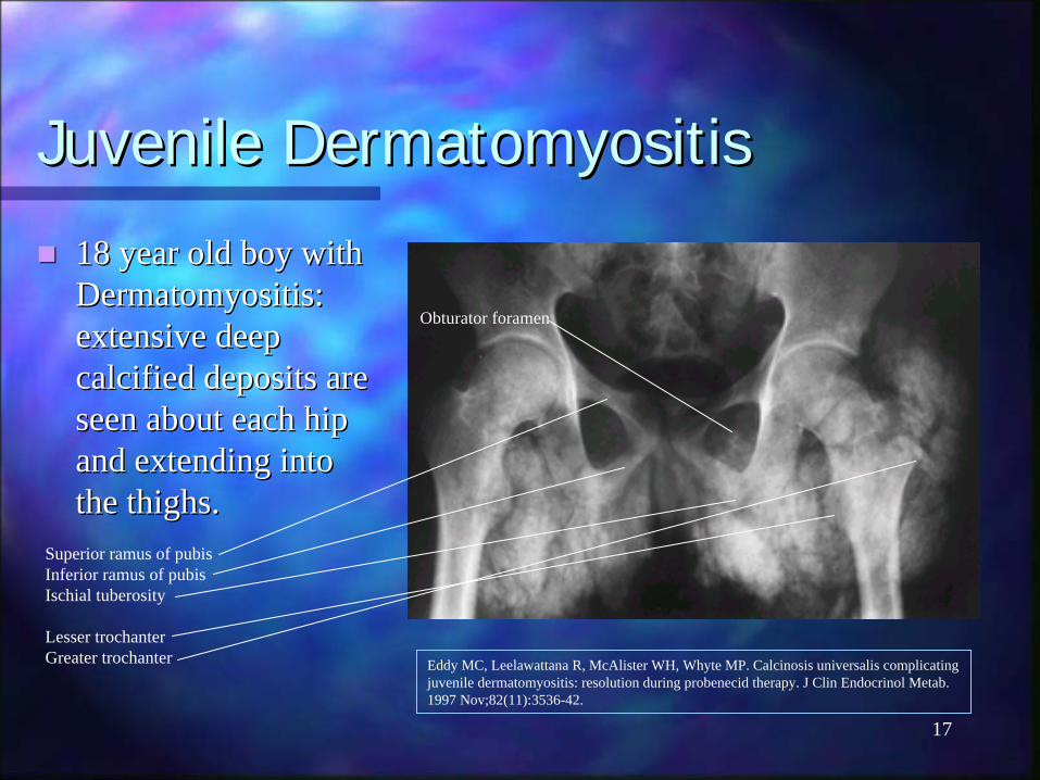
18 year old boy with 18 year old boy with DermatomyositisDermatomyositis: : extensive deep extensive deep calcified deposits are calcified deposits are seen about each hip seen about each hip and extending into and extending into the thighs.the thighs.
Superior ramus of pubis Inferior ramus of pubis Ischial tuberosity
Lesser trochanter Greater trochanter
Obturator foramen
18
Juvenile Juvenile DermatomyositisDermatomyositis
Same 18 year old Same 18 year old boy with boy with DermatomyositisDermatomyositis: : extensive deep extensive deep calcified deposits calcified deposits are seen about the are seen about the shoulder extending shoulder extending into the arm. into the arm. The clustering The clustering around joints around joints mimics mimics tumoraltumoralcalcinosiscalcinosis..
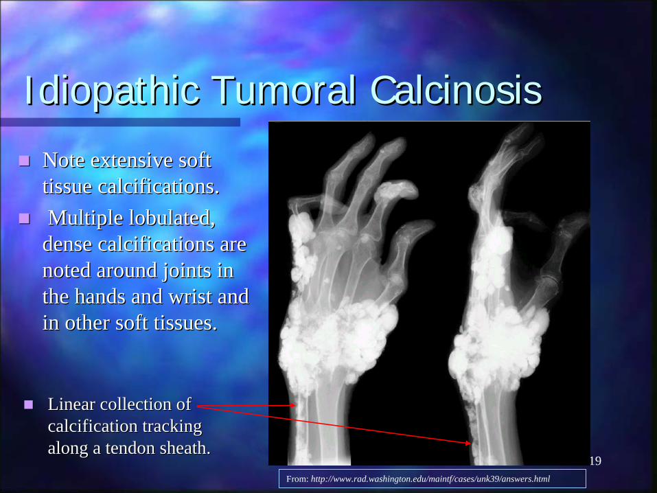
Note extensive soft Note extensive soft tissue calcifications.tissue calcifications.Multiple Multiple lobulatedlobulated, , dense calcifications are dense calcifications are noted around joints in noted around joints in the hands and wrist and the hands and wrist and in other soft tissues. in other soft tissues.
The soft tissue calcifications may be single or multiple, The soft tissue calcifications may be single or multiple, lobulatedlobulated, cystic or solid. , cystic or solid. Often occurs at hips, elbows and shoulders. Often occurs at hips, elbows and shoulders. The masses may range in size from 1 to 20 cm in diameter. The masses may range in size from 1 to 20 cm in diameter. Family history in 30 Family history in 30 -- 40 % of cases. 40 % of cases. Onset during childhood or adolescence. Onset during childhood or adolescence. May be increased prevalence in patients of African May be increased prevalence in patients of African descent. descent.
RADIONUCLIDE BONE SCANRADIONUCLIDE BONE SCAN Idiopathic Idiopathic TumoralTumoral CalcinosisCalcinosis
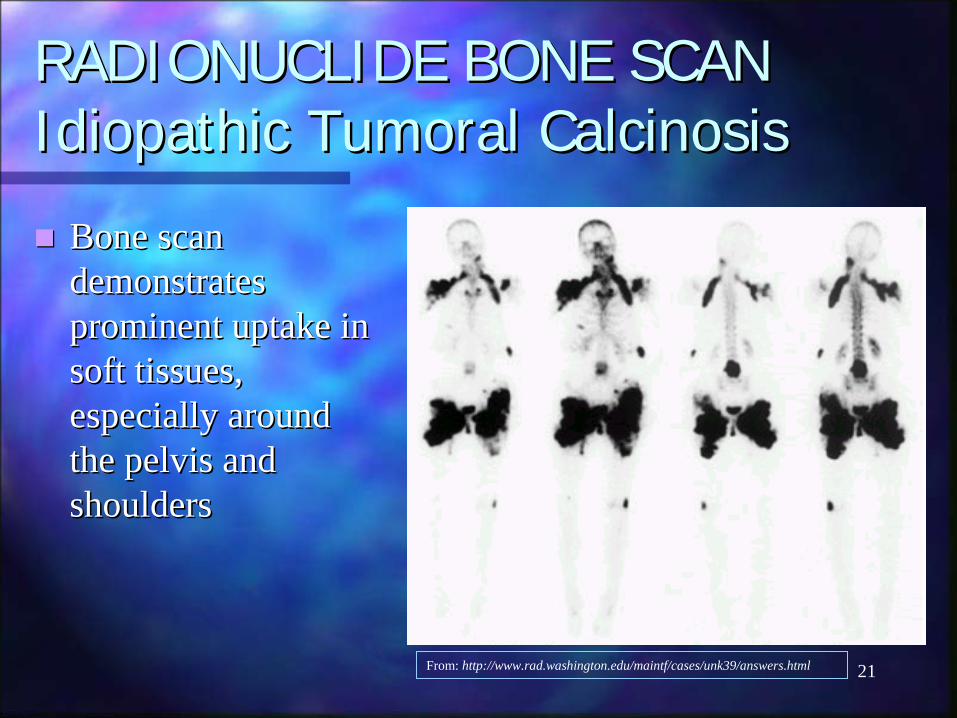
Bone scan Bone scan demonstrates demonstrates prominent uptake in prominent uptake in soft tissues, soft tissues, especially around especially around the pelvis and the pelvis and shouldersshoulders
MRI (T2) of the hips reveals large cystic areas of low signal intensity (arrows). This patient had Idiopathic Tumoral Calcinosis.
23
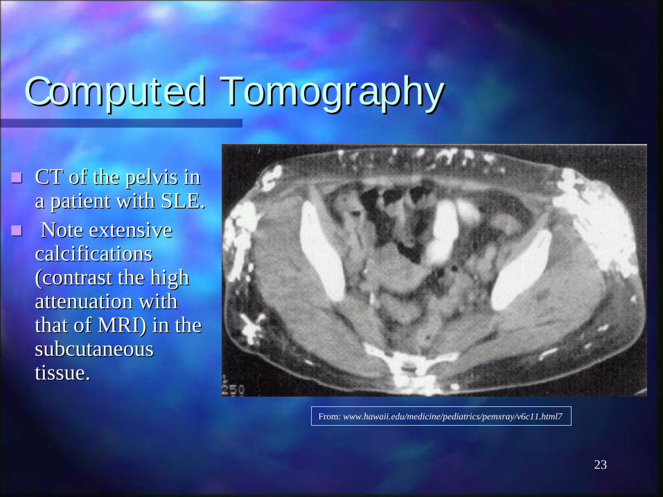
Computed TomographyComputed Tomography
CT of the pelvis in CT of the pelvis in a patient with SLE.a patient with SLE.Note extensive Note extensive calcifications calcifications (contrast the high (contrast the high attenuation with attenuation with that of MRI) in the that of MRI) in the subcutaneous subcutaneous tissue.tissue.
The patient is a 900 The patient is a 900 year old man from year old man from the the DegobahDegobahSystem…System…
25
Master YodaMaster Yoda
Courtesy of LucasFilm ®
? Scleroderma
26
Treatment of Treatment of CalcinosisCalcinosis CutisCutis
Medical treatments include:Medical treatments include:IntralesionalIntralesional corticosteroid injectioncorticosteroid injectionEtidronateEtidronate disodium (a disodium (a bisphosphonatebisphosphonate))Aluminum hydroxide (phosphateAluminum hydroxide (phosphate--binder)binder)
However, these are not very effective.However, these are not very effective.Surgical excision has been shown to be of Surgical excision has been shown to be of benefit, as it can provide symptomatic benefit, as it can provide symptomatic relief.relief.
27
SummarySummary
Soft tissue calcifications are relatively common Soft tissue calcifications are relatively common radiographic findings.radiographic findings.Dystrophic calcifications predominate.Dystrophic calcifications predominate.Often incidental finding, but can present with skin lesions Often incidental finding, but can present with skin lesions or pain.or pain.Good history is key. Good history is key. Plain film to start; other imaging can be helpful if plain Plain film to start; other imaging can be helpful if plain film is normal.film is normal.Medical treatment has variable success; surgical removal Medical treatment has variable success; surgical removal of calcification is indicated if symptomatic.of calcification is indicated if symptomatic.
28
BibliographyBibliographyKoopmanKoopman: Arthritis and Allied Conditions, 13: Arthritis and Allied Conditions, 13thth ed., 1997.ed., 1997.Richardson, M.L. Approaches to differential diagnosis in musculoRichardson, M.L. Approaches to differential diagnosis in musculoskeletal skeletal imaging: soft tissue calcifications. imaging: soft tissue calcifications. http://http://www.rad.washington.edu/mskbook/softtissueca.htmlwww.rad.washington.edu/mskbook/softtissueca.html., 1994.., 1994.Stewart, V.L. Stewart, V.L. HerlingHerling, P., , P., DalinkaDalinka, M.K. Calcification in soft tissues. JAMA , M.K. Calcification in soft tissues. JAMA 1983; 250 (1): 781983; 250 (1): 78--81.81.http://www.hawaii.edu/medicine/pediatrics/pemxray/v6c11.htmhttp://www.hawaii.edu/medicine/pediatrics/pemxray/v6c11.htmhttp://www.rad.washington.edu/maintf/cases/unk39/answers.htmlhttp://www.rad.washington.edu/maintf/cases/unk39/answers.htmlCousins, M.A. Surgical management of Cousins, M.A. Surgical management of calcinosiscalcinosis cutis cutis universalisuniversalis in systemic in systemic lupus. Arthritis Rheum 1997 Mar; 40(3): 570lupus. Arthritis Rheum 1997 Mar; 40(3): 570--2.2...