6958 Biochemistry 1987, 26, 6958-6965 Calcium Binding Domains and Calcium-Induced Conformational Transition of SPARC/BM-40/0steonectin, an Extracellular Glycoprotein Expressed in Mineralized and Nonmineralized Tissues? Jiirgen Engel,*.$ William Taylor,$ Mats Paulsson,t Helene Sage," and Brigid HoganL Department of Biophysical Chemistry, Biozentrum, CH-4056 Basel, Switzerland, Laboratory of Molecular Biology, Department of Crystallography, Birkbeck College, London WC1 E7HX, U.K., Department of Biological Structure, University of Washington, Seattle, Washington 981 95, and Laboratory of Molecular Embryology, National Institute for Medical Research, London NW7 IAA, U.K. Received March 25, 1987; Revised Manuscript Received June 12, 1987 ABSTRACT: SPARC, BM-40, and osteonectin are identical or very closely related extracellular proteins of apparent M, 43 000 (M, 33 000 predicted from sequence). They were originally isolated from parietal endoderm cells, basement membrane producing tumors, and bone, respectively, but are rather widely distributed in various tissues. In view of the calcium binding activity reported for osteonectin, we analyzed the SPARC sequence and found two putative calcium binding domains. One is an N-terminal acidic region with clusters of glutamic acid residues. This region, although neither y-carboxylated nor homologous, resembles the y-carboxyglutamic acid (Gla) domain of vitamin K dependent proteins of the blood clotting system in charge density, size of negatively charged clusters, and linkage to the rest of the molecule by a cysteine-rich domain. The other region is an EF-hand calcium binding domain located near the C-terminus. A disulfide bond between the E and F helix is predicted from modeling the EF-hand structure with the known coordinates of intestinal calcium binding protein. The disulfide bridge apparently serves to stabilize the isolated calcium loop in the extracellular protein. As observed for cytoplasmic EF-hand-containing proteins and for Gla domain containing proteins, a major conformational transition is induced in BM-40 upon binding of several Ca2+ ions. This is accompanied by a 35% increase in a-helicity. A pronounced sigmoidicity of the dependence of the circular dichroism signal at 220 nm on calcium concentration indicates that the process is cooperative. In view of its properties, abundance, and wide distribution, it is proposed that SPARC/ BM-40/osteonectin has a rather general regulatory function in calcium-dependent processes of the extra- cellular matrix. s e v e r a l model systems are available for studying the as- sembly and turnover of the extracellular matrix. One is the parietal endoderm of the mouse embryo, which synthesizes a basement membrane (Reichert's membrane) rich in type IV collagen, laminin, entactinlnidogen, and heparan sulfate proteoglycan [for review, see Hogan et al. (1984)J. These very large and complex glycoproteins are also made by the trans- plantable Engelbreth-Holm-Swarm (EHS) mouse tumor, which provides a convenient and abundant source of purified matrix components for structural and biological studies [re- viewed by Timpl et al. (1987)l. Recent work in both model systems has led to the identification of a very abundant, small glycoprotein, named SPARC (Mason et al., 1986a,b) or BM- 40 (Dziadek et al., 1986), which migrates with an apparent M, of 39 000-43 000 in sodium dodecyl sulfateplyacrylamide gel electrophoresis and contains a single oligosaccharide side chain (Hughes et al., 1987). SPARC is coded for by a single gene, and the complete amino acid sequence of the mouse protein has been deduced from the nucleotide sequence of parietal endoderm cDNAs (Mason et al., 1986a,b). This gives a predicted M, of 33 062 for the unglycosylated protein after +Supported by Grant 3.254.85 of the Swiss National Science Foun- dation and by the Medical Research Council of Great Britain. H.S. was an Established Investigator of the American Heart Association during the active phase of these studies, with support from National Institutes of Health grants. * Biozentrum. SBirkbeck College. 11 University of Washington. National Institute for Medical Research. removal of the signal sequence. The amino acid sequence of BM-40 is identical with that of SPARC in all peptides so far analyzed, covering about 50% of the total protein (Mann et al., 1987). Although SPARC was first identified as a major product of parietal endoderm cells, further studies showed that messenger RNA and/or protein are made by a variety of other cell types, including bovine endothelial cells and fibroblasts derived from fetal dermis and ligament (Sage et al., 1984; Sage, 1985, Mason et al., 1986b; Dziadek et al., 1986). The protein made by endothelial cells and isolated by Sage et al. (1984) was initially named 43K albumin binding protein; its identity or close relation with SPARC was demonstrated by Mason et al. (1986a). Moreover, there is now very good evidence that SPARC is identical with the phosphorylated calcium binding protein osteonectin, originally isolated from fetal bovine bone and dentin (Termine et al., 1981a,b). The N-terminal region of both bovine and porcine osteonectin is highly homologous to the predicted N-terminal sequence of SPARC (Romberg et al., 1985; Young et al., 1986). When this comparison is extended to the complete amino acid se- quences predicted from the cDNAs of mouse SPARC and bovine osteonectin, 92% sequence identity is observed (Young et al., 1986; M. E. Bolander and J. D. Termine, National Institutes of Health, Bethesda, MD, personal communication). Additional evidence that SPARC and osteonectin are identical comes from in situ hybridization data showing very high levels of SPARC RNA in embryonic and newborn mouse bone (osteoblasts) and teeth (odontoblasts) (Holland et al., 1987). A high-affinity calcium binding site (Kd -3 X M) was demonstrated for osteonectin, and hydroxylapatite crystal 0006-2960/87/0426-6958$01 .50/0 0 1987 American Chemical Society
Transcript
6958 Biochemistry 1987, 26, 6958-6965
Calcium Binding Domains and Calcium-Induced Conformational Transition of SPARC/BM-40/0steonectin, an Extracellular Glycoprotein Expressed in
Mineralized and Nonmineralized Tissues? Jiirgen Engel,*.$ William Taylor,$ Mats Paulsson,t Helene Sage," and Brigid HoganL
Department of Biophysical Chemistry, Biozentrum, CH-4056 Basel, Switzerland, Laboratory of Molecular Biology, Department of Crystallography, Birkbeck College, London WC1 E7HX, U.K., Department of Biological Structure, University of Washington,
Seattle, Washington 981 95, and Laboratory of Molecular Embryology, National Institute for Medical Research, London NW7 IAA, U.K.
Received March 25, 1987; Revised Manuscript Received June 12, 1987
ABSTRACT: SPARC, BM-40, and osteonectin are identical or very closely related extracellular proteins of apparent M, 43 000 ( M , 33 000 predicted from sequence). They were originally isolated from parietal endoderm cells, basement membrane producing tumors, and bone, respectively, but are rather widely distributed in various tissues. In view of the calcium binding activity reported for osteonectin, we analyzed the SPARC sequence and found two putative calcium binding domains. One is an N-terminal acidic region with clusters of glutamic acid residues. This region, although neither y-carboxylated nor homologous, resembles the y-carboxyglutamic acid (Gla) domain of vitamin K dependent proteins of the blood clotting system in charge density, size of negatively charged clusters, and linkage to the rest of the molecule by a cysteine-rich domain. The other region is an EF-hand calcium binding domain located near the C-terminus. A disulfide bond between the E and F helix is predicted from modeling the EF-hand structure with the known coordinates of intestinal calcium binding protein. The disulfide bridge apparently serves to stabilize the isolated calcium loop in the extracellular protein. As observed for cytoplasmic EF-hand-containing proteins and for Gla domain containing proteins, a major conformational transition is induced in BM-40 upon binding of several Ca2+ ions. This is accompanied by a 35% increase in a-helicity. A pronounced sigmoidicity of the dependence of the circular dichroism signal at 220 nm on calcium concentration indicates that the process is cooperative. In view of its properties, abundance, and wide distribution, it is proposed that SPARC/ BM-40/osteonectin has a rather general regulatory function in calcium-dependent processes of the extra- cellular matrix.
s e v e r a l model systems are available for studying the as- sembly and turnover of the extracellular matrix. One is the parietal endoderm of the mouse embryo, which synthesizes a basement membrane (Reichert's membrane) rich in type IV collagen, laminin, entactinlnidogen, and heparan sulfate proteoglycan [for review, see Hogan et al. (1984)J. These very large and complex glycoproteins are also made by the trans- plantable Engelbreth-Holm-Swarm (EHS) mouse tumor, which provides a convenient and abundant source of purified matrix components for structural and biological studies [re- viewed by Timpl et al. (1987)l. Recent work in both model systems has led to the identification of a very abundant, small glycoprotein, named SPARC (Mason et al., 1986a,b) or BM- 40 (Dziadek et al., 1986), which migrates with an apparent M, of 39 000-43 000 in sodium dodecyl sulfateplyacrylamide gel electrophoresis and contains a single oligosaccharide side chain (Hughes et al., 1987). SPARC is coded for by a single gene, and the complete amino acid sequence of the mouse protein has been deduced from the nucleotide sequence of parietal endoderm cDNAs (Mason et al., 1986a,b). This gives a predicted M, of 33 062 for the unglycosylated protein after
+Supported by Grant 3.254.85 of the Swiss National Science Foun- dation and by the Medical Research Council of Great Britain. H.S. was an Established Investigator of the American Heart Association during the active phase of these studies, with support from National Institutes of Health grants. * Biozentrum.
SBirkbeck College. 11 University of Washington.
National Institute for Medical Research.
removal of the signal sequence. The amino acid sequence of BM-40 is identical with that of SPARC in all peptides so far analyzed, covering about 50% of the total protein (Mann et al., 1987). Although SPARC was first identified as a major product of parietal endoderm cells, further studies showed that messenger RNA and/or protein are made by a variety of other cell types, including bovine endothelial cells and fibroblasts derived from fetal dermis and ligament (Sage et al., 1984; Sage, 1985, Mason et al., 1986b; Dziadek et al., 1986). The protein made by endothelial cells and isolated by Sage et al. (1984) was initially named 43K albumin binding protein; its identity or close relation with SPARC was demonstrated by Mason et al. (1986a). Moreover, there is now very good evidence that SPARC is identical with the phosphorylated calcium binding protein osteonectin, originally isolated from fetal bovine bone and dentin (Termine et al., 1981a,b). The N-terminal region of both bovine and porcine osteonectin is highly homologous to the predicted N-terminal sequence of SPARC (Romberg et al., 1985; Young et al., 1986). When this comparison is extended to the complete amino acid se- quences predicted from the cDNAs of mouse SPARC and bovine osteonectin, 92% sequence identity is observed (Young et al., 1986; M. E. Bolander and J. D. Termine, National Institutes of Health, Bethesda, MD, personal communication). Additional evidence that SPARC and osteonectin are identical comes from in situ hybridization data showing very high levels of SPARC RNA in embryonic and newborn mouse bone (osteoblasts) and teeth (odontoblasts) (Holland et al., 1987). A high-affinity calcium binding site (Kd -3 X M) was demonstrated for osteonectin, and hydroxylapatite crystal
0006-2960/87/0426-6958$01 .50/0 0 1987 American Chemical Society
C A Z + - B I N D I N G D O M A I N S I N S P A R C / B M - 4 0 / 0 S T E O N E C T I N V O L . 2 6 , N O . 2 2 , 1 9 8 7 6959
growth was found to be delayed by 50% at about 0.2 pM concentrations of the protein (Romberg et al., 1985). Here we report phosphorylation of SPARC/BM-40/osteonectin in cell cultures and demonstrate a large cooperative and reversible change in a-helicity upon binding of several Ca2+ ions. Structural features based on the SPARC sequence are dis- cussed, including the identification of an EF-hand calcium binding domain and putative additional calcium binding sites.
EXPERIMENTAL PROCEDURES Materials
Isolation of BM-40. Intact BM-40 was extracted from mouse EHS tumor tissue with neutral buffer containing 10 mM EDTA' and protease inhibitors [for the preparation of the extract see Paulsson et al. (1987)l. A retarded fraction from Sepharose CL-6B molecular sieve chromatography of this extract contains considerable amounts of BM-40, which was further purified by ion-exchange chromatography on DEAE-cellulose and molecular sieve chromatography on Se- phacryl S-200. The initial sample of intact BM-40 isolated from EDTA extracts was a kind gift of Dr. Rupert Timpl, Max-Planck-Institut fur Biochemie, Martinsried, FRG. Intact BM-40 migrated as a MI 40K band with less than 10% of 30K and 10K protease fragments when tested by SDS-PAGE' under reducing conditions. Nicked BM-40 was also isolated by the procedure of Dziadek et al. (1986). This material dissociates almost completely into M, 30K and 10K fragments under the same conditions (Dziadek et al., 1986) and differs from intact BM-40 also with respect to a possible loss of native conformation due to exposure to 6 M guanidine hydrochloride during extraction. Met hods
Metabolic Labeling and PYS Cell Culture. Confluent cultures of the mouse parietal endoderm cell line PYS-2 (Lehman et al., 1974) in tissue culture dishes ( 5 cm in diam- eter) were labeled for about 40 h in 2 mL of Dulbecco's minimum essential a medium without phosphate, containing 5% dialyzed fetal bovine serum and 1 mCi [32P]ortho- phosphoric acid (New England Nuclear). The medium was harvested and made 50 mM in NaF and 100 pM in sodium vanadate to inhibit phosphatase activity. SPARC was re- covered by immunoprecipitation using rabbit antipeptide serum and protein A-Sepharose (Pharmacia) as described by Mason et al. (1986a). Samples were analyzed by SDS-PAGE under reducing conditions, and phosphoamino acid analysis of the excised MI 43K SPARC band was carried out as described (Heath et al., 1986). For glycopeptide analysis the immu- nocomplex bound to protein A was incubated at 37 OC for 3 days in 0.4% Pronase (Calbiochem) in 50 mM Tris-HC1, pH 7.5, 10 mM CaC12, and 0.02% NaN3. After incubation at 90 OC for 5 min to inactivate the Pronase and centrifugation to remove the Sepharose beads, the supernatant was applied to a concanavalin A-Sepharose column (Pharmacia) in 10 mM Tris, pH 7.5, 100 mM NaC1, 1 mM MgC12, 1 mM CaCl,, 1 mM MnCl,, and 0.02% NaN3 [buffer A, Hughes et al. (1987)l. After extensive washing, glycopeptide bound to the concanavalin A was eluted with 10 mM methyl a-glucoside and 500 mM methyl a-mannoside in buffer A (Hughes et al., 1987). For analysis of tryptic cleavage fragments, PYS-2 cells were incubated overnight with 100 pCi/mL [35S]methionine
as described (Mason et al., 1986a). SPARC was recovered from the culture medium with anti-peptide serum and protein A-Sepharose. Immunocomplexes were incubated at 37 OC with 0.1-0.5 pg of trypsin (Sigma, type 111) in a 0.17 M NaCl, 3 mM KCl, 9 mM Na2HP04, and 2 mM KH2P04, pH 7.4, buffer (phosphate-buffered saline), and the reaction was stopped by adding twice concentrated SDS-PAGE sample buffer at 100 OC. SDS-PAGE was carried out as described (Cooper et al., 1981). Internal 14C-labeled markers were myosin ( M , 212000), phosphorylase b (MI 100000 and 92 500), bovine serum albumin (MI 69 000), ovalbumin (MI 46 000), carbonic anhydrase (MI 30 000), and lysozyme (MI 14 000) (Amersham International).
Circular Dichroism. A Cary 6 1 spectropolarimeter (Varian, Zug, Switzerland) was employed. Spectra were recorded in a thermostat-controlled quartz cell (Helma, Miillheim, FRG) of 1-mm path length at 20 OC. The molar ellipticity [e] expressed in deg.cm2.dmol-' was calculated on the basis of a mean residue molar mass of 110. Concentrations needed for the calculation of [e] were determined by quantitative amino acid analysis of aliquots of the sample solutions. The de- pendence of ellipticity on calcium concentration was measured with a rectangular cell of 2-mm path length which was selected for showing zero ellipticity at 220 nm when filled with buffer. A cell holder was used which assured exact reproducibility of the cell positioning after mixing. The cell temperature was maintained by the thermostat4 compartment between 20 and 22 "C. The protein was extensively dialyzed against a 10 mM Tris-HC1 and 150 mM NaC1, pH 7.4, buffer. The buffer was stirred over night with Chelex 100 resin (Bio-Rad, Richmond, CA) to remove metal ions before use, and it was acertained by atomic absorption analysis that the remaining calcium concentration was less than 10 pm. The calcium analysis was kindly performed by Dr. Hans Giinther Seiler, Institute for Inorganic Chemistry, University of Basel. The cell was filled with 0.3 mL of the protein solution (0.33 mg/mL). Aliquots (3-10 pL) of 0.5 mM EDTA in the same buffer were added, and the ellipticity was monitored for 3 min after each addition. After a plateau value was reached, a back-titration was per- formed with 3-pL aliquots of 0.5 mM CaC12 in the same buffer. Ellipticities and concentrations of the reagents in the mixture were corrected by dilution factors which did not exceed 1.15.
RESULTS Analysis of the Sequence. Some salient features of the
sequence of SPARC/BM-40/osteonectin are summarized in Figure 1. Clearly, four sequence regions of rather different characteristics can be distinguished, suggesting the presence of at least four conformationally and functionally distinct domains. Domain I, comprising the first 52 residues of the protein, features primarily two segments of 14-1 5 residues, each of which contains 7-8 glutamic acids in short clusters that impart a negative charge at physiological pH. According to secondary structure prediction these segments are a-helical. It has to be noted, however, that the a-helix is not expected to be stable at this high charge density (see Discussion).
Region I1 consists of about 85 residues exhibiting the typical features of a disulfide-stabilized domain (Doolittle, 1985). According to the secondary structure prediction in this region, short segments of p-structure alternate with @-bends. Most of the 11 cysteines are probably disulfide linked within the domain, but their uneven number suggests that one of them is connected with a Cys residue (probably Cys-247, see below) of the C-terminal region. A portion of purified BM-40 is nicked, and the small and the large fragment obtained are
6960 B I O C H E M I S T R Y E N G E L ET A L .
Ala 1 NH, /
Ip EF-Domain
I
Ip EF-Domain
Domain1 HOOC
CYS-Rich Domain II
5 10 15 20 25 30 1 A P Q Q T E V A E E I V E E E T V V E E T G V P V G A N P V
3 1 Q V E M G E F E D G A E E T V E E V V A D N P C Q N H H C K 6 1 H G K V C E L D E S N T P M C V C Q D P T S C P A P I G E P 9 1 E K V C S N D N K T F D S S C H F F A T K C T L E G T K K G
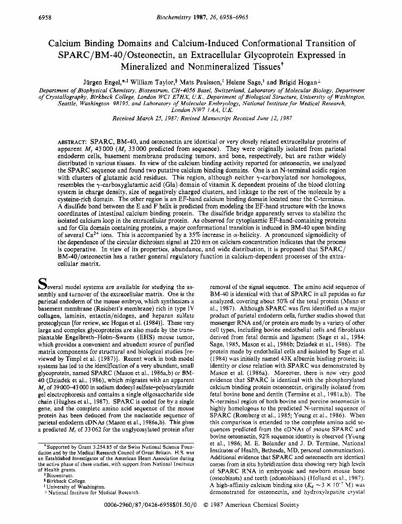
~ ~ ~ ~ K L H L D Y I G P C K Y I A P C L D S E L T E F P L R H R 1 5 1 D W L K N V L V T L Y E R D E G N N L L T E K Q K L R V K K 1 8 l I H E N E K R L E A G D H P V E L L A R D F E K N Y N n Y I Z l l F P V H W Q F G Q L D Q H P I D G Y L S H T E L A P L R A P 2 4 1 L I P B E H C T T R F F E T C D L D N D K Y I A L E E W A G 271 G G I K E Q D I N K D L V I
FIGURE 1 : Schematic representation of interesting features in the sequence of SPARC. Numbering is according to Mason et al. (1986a) without the signal peptide. The mode of linkage between Cys residues (0) is not yet established. It is suggested from the three-dimensional modeling of the EF-hand calcium binding domain IV (Figure 3) that Cys-255 and Cys-271 are connected (0- - -0). There is an uneven number of Cys residues in the Cys-rich domain 11. The presence of a disulfide bond between one of them and Cys-247 follows from the observation that cleavage at a site in region I11 leads to fragmentation only after reductive cleavage of one or more disulfide bonds (Figure 7; Sage et al., 1984; Mason et al., 1986a,b; Dziadek et al., 1986). The arrow indicates the site of endogenous cleavage in position Leu- 197-Leu-198 (Mann et al., 1987). Assuming that SPARC contains no free thiol groups, it follows that all other Cys residues are disulfide bonded within the Cys-rich domain 11. The latter contains two clusters of five positively charged residues HHCKHGK (57-63) and KKGHKLH (1 18-124), the unique glycosylation site (*) (Asn-98), and five serine residues. (A) indicates Ser -70, -82, -95, -103 and -104. In domain I toward the amino terminus there are two regions (6-20 and 32-47), each of which contains eight negatively charged residues. Segments of the sequence for which an a-helical secondary structure is predicted by the method of Chou and Fasman (1974) are indicated by cylinders. The same method predicts short segments of @-structure frequently interrupted by @-turns in the Cys-rich domain and some @-structure as well as @-turns in the rest of the molecule. Positions of all methionines (Met-33, -74, -149. -208, -238) are in- dicated (0) to show the distribution of [%]methionine in the material used for the tryptic cleavage experiment.
probably connected by this disulfide bond. The location of the endogenous cleavage site was located to Leu- 197-Leu- 198 (Mann et al., 1987) and is indicated by an arrow in Figure
1. Near both ends of the cysteine-rich domain there are clusters of closely spaced, positively charged residues, while in the central portion is the site of the single glycosylation of SPARC, Asn-98 (Mason et al., 1986a; Hughes et al., 1987). The Cys-rich domain also contains six of the seven serine residues in the protein. These serines are all potential phos- phorylation sites, including the sequence DSS ( 102-104) that is close to Asn-98 (see below).
A cysteine- and proline-free region (domain 111) is pre- dominantly a-helical (60-70%) according to the structure prediction. When all of the clearly predicted a-helical seg- ments are summed, the total a-helicity is 3 1%. This value decreases to 21% if the two Glu-rich segments in the N-ter- minal region are assumed to be in a randomly coiled confor- mation at physiological pH. The last region (IV) consists of an EF-hand calcium binding domain connected to region I11 by a non-a-helical stretch of 32 residues containing 5 prolines.
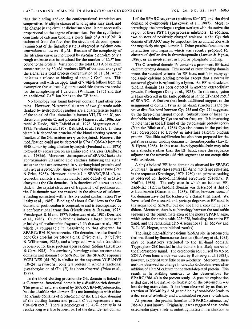
EF-Hand Ca2+ Binding Domain. In Figure 2, residue 248-275 of the SPARC sequence are compared with the consensus motif of an EF-hand calcium binding domain. This motif is based on the sequences of many cytoplasmic calcium binding proteins, of which calmodulin, parvalbumin, and in- testinal calcium binding protein are well-known examples. The sequence of domain 11, one of four EF-hand motifs in bovine intestinal calcium binding protein, is included in Figure 2 because of the high-resolution X-ray structure that is available for this protein (Szebinyi et al., 1981; Szebinyi & Moffat, 1986). The only clear deviation in the SPARC sequence from the accepted EF-hand motif is the exchange of a normally conserved glycine by lysine (Figure 2). Lysine residues in this position are, however, observed in the recently discovered putative EF-hand domains of another extracellular protein, fibrinogen (Dang et al., 1985). An interesting feature of the EF-hand sequence in SPARC is the Occurrence of the two Cys residues in positions of the E and F helices which are occupied by hydrophobic residues in the EF motifs of cytoplasmic proteins. In the intestinal calcium binding protein the hy- drophobic side chains of these residues (Leu-53 and Leu-69) are in close contact, and Cys residues in these positions can easily be connected by a disulfide bond (Figure 3). Con- nection of Cys-255 and Cys-271 by a disulfide bond in the EF calcium binding domain of SPARC leaves Cys-247 as a likely candidate for disulfide bridging to one of the cysteine residues in the Cys-rich domain. This assignment is supported by recent fragmentation studies (Mann et al., 1987). The model in Figure 3 was also used to verify that glycine-59 in intestinal E-Helix Loop F-Helix
hXXhhXXhDXDXDGXIDX-XEhXXhhXXh
TTRFFETCDLDNDKYIAL-EEWAGCFGIK 248-275 * *
L 5
fibrinogen chain precursor, human NGMQps-nrpNDNDKFEGNCAEQDOSOWWM 334-363
,rat NGMHZSWNDNDKFEGNCAEQDPSGWWM 334-363
,sea lamprey NGMLpSEPERmYEGSCAEQDpGGwwM 330-358
FIGURE 2: Comparison of a sequence region in SPARC with the consensus sequence for the EF-hand calcium binding domain (Kretsinger, 1979) and related motifs in intestinal calcium binding protein (Fullmer & Wassermann, 1981) and fibrinogen (Dang et al., 1985). In the EF-hand motif Asp (D) can be replaced by Am (N), and Glu (E) can be replaced by Asp. "h" denotes a hydrophobic residue, and X indicates any residue. Other variations are possible (Kretsinger, 1979, 1980). A deviation of the SPARC sequence from the central portion (boldface letters) of the EF-hand motif is the Occurrence of lysine (K) instead of the normally conserved glycine (G) (asterisks). Lysines in this position are however observed in the putative E F hands of fibrinogen y chains (Dang et al., 1985). Residues that are identical in fibrinogen and SPARC are underlined. The other deviation, alanine (A) instead of D (asterisks), is not inconsistent with an EF hand, since there is only a weak preference for an oxygen-containing side chain at this position (Szebenyi & Moffat, 1986). Residues Leu-53 and Leu-69 (arrows) are in close proximity in the three-dimensional structure of intestinal calcium binding protein (Szebinyi et al., 1981; Szebinyi & Moffat, 1986) and were substituted by cysteines in Figure 3 to test for the possibility of a disulfide bridge formation in this position.
C A * + - B I N D I N G D O M A I N S I N S P A R C / B M - 4 0 / 0 S T E O N E C T I N V O L . 2 6 , N O . 2 2 , 1 9 8 7 6961
FIGURE 3: Three-dimensional model of an EF-hand calcium binding domain that is stabilized by a disulfide bond. The structure of one of the calcium binding domains in intestinal calcium binding protein (49-70, see Figure 2) was displayed according to the coordinates of Szebinyi et al. (1981) taken from the Brookhaven library (Dayhoff & Baker, 1972). Residues Leu-53 and Leu-69 were replaced by Cys residues which occur in equivalent positions in the EF motif of SPARC (Figure 2). By adjustment of the side chains of the two Cys residues without changes in the positions of their C, atoms, formation of a disulfide bond (S-S) was easily possible. The bond was slightly stretched (S-S distance of 3 8, and both C-S-S angles greater than 90°), but a slight relative movement of the a-helices would enable an ideal bond geometry to be attained. The diagram was produced by the molecular modeling program FRODO (Jones, 1978). The a-carbon positions are emphasized as filled dots, adjacent to which are indicated the names and numbers of certain residues. The two sulfur atoms are represented as open circles, and the proposed bond between them is dashed. The main chain runs from bottom right to left, and its termini are indicated “n” and “c”, respectively.
calcium binding protein can be replaced by lysine without steric interference of the newly introduced side chain with the rest of the structure.
Secondary Structure and Conformational Changes Moni- tored by Circular Dichroism. BM-40 was extensively dialyzed against Chelex-treated 10 mM Tris-HC1, pH 7.4, and 150 mM NaCl buffer, which according to atomic absorption mea- surements contained less than 10 pM calcium. Circular di- chroism spectra of intact BM-40 recorded at this low con- centration of free calcium (Figure 4) were identical with spectra measured in the same buffer containing 1 mM calcium. The pronounced shoulder at 220 nm in the spectrum of BM-40 indicates 2530% a-helical content. Upon removal of calcium by addition of EDTA to a total concentration of 1 mM the mean residue ellipticity at 220 nm decreases by 35%, indicating a corresponding decrease of the a-helix content to a value of 16-19%. This change is completely reversible upon readdition of calcium, and the cycle can be repeated 3 times without any significant change of the spectra of saturated and free protein. Nicked BM-40 purified from 6 M guanidine hydrochloride extracts exhibits a similar spectrum and calcium-induced change, but the amplitude as well as the magnitude of the change is smaller.
The change of ellipticity was plotted as a function of EDTA or calcium concentration for intact SPARC/BM-40/osteo- nectin (Figure 5). A cooperative process is indicated by the sigmoidal curve. Data points collected upon addition of EDTA match the points obtained in the back-titration with calcium. As the helix-promoting effect of calcium may in part be due to a compensation of negative charges, we tested whether a similar effect could be induced by lowering the pH. Intact
200 210 220 230 240 250
A l n m )
FIGURE 4: Circular dichroism spectra of BM-40 in the presence and absence of calcium. The spectrum of intact BM-40 (-) was measured in Chelex-treated 10 mM Tris-HC1 and 150 mM NaCI, pH 7.4. Concentration of free calcium was less than 10 pM. A decrease of negative ellipticity by 35% at 220 nm was observed (arrow) when EDTA was added to a final total concentration of 1 mM (- - -). This change was completely reversed by addition of calcium to a total concentration of 2 mM (2-fold excess over EDTA). The spectrum of nicked BM-40 purified from 6 M guanidine hydrochloride extracts (.-) had a similar shape but smaller amplitude, and the changes upon removal and addition of EDTA were less pronounced (change of about 10% at 220 nm, arrow). The protein concentration was 0.33 mg/mL (1 1 pM), and the temperature was 20 OC. The spectrum of adult bovine asteonectin extracted from bone (-.-) was taken from Romberg et al. (1985). According to these authors, this spectrum is not in- fluenced by addition of EDTA or calcium.
- [ C U I , OJMI
200 150 i n n 50 n
[ E D T A l 0 [ ? M I - FIGURE 5: Calcium-dependent conformational transition in intact BM-40. The change of ellipticity at 220 nm (see arrow in Figure 4) was monitored as a function of EDTA added to the protein solution (0). At the mean molar residue ellipticity of -5600 degcm*.dmol-’ characteristic for the calcium-dependent protein (at 190 pM EDTA), the effect was reversed by successive addition of calcium (upper ordinate, 0) . For protein concentration and buffer conditions see legend, Figure 4.
BM-40 was dissolved in a 100 mM NaCl solution containing 1 mM EDTA, and the pH was adjusted by addition of dilute acetic acid. No change of [e],, = -5600 deg.cm2.dmol-’ was observed between pH 7 and pH 6. When the pH was lowered, however, a sharp sigmoidal increase of negative ellipticity was observed. At pH 5.1 the value was [e] = -6500. This increase corresponds to one-third of the total change observed upon calcium addition. Between pH 5.1 and pH 4.85 the protein precipitated from the solution but redissolved when the pH was readjusted to pH 5.1.
Phosphorylation. Bovine osteonectin was reported to be phosphorylated (Uchiyama et al., 1986). By incubating PYS
6962 B IOC H E M IS T R Y
A B C
212) l 6 O 0 1 I t
46* a 4 8 12 16 20 24 28 30
Fraction number
FIGURE 6: Phosphorylation of SPARC secreted by PYS cells. Panel A: PYS cells were incubated with [32P]orthophosphate for 42 h, and SPARC was immunoprecipitated from the culture medium with anti-peptide serum before analysis by 1 Wo SDSPAGE under reducing conditions. Panel B: The M, 43K band shown in panel A was excised and phosphoamino acid analysis carried out as described (Heath et al., 1986). The positions of the internal markers phosphoserine (SerP), phosphothreonine (ThrP), and phosphotyrosine (TyrP) on the thin- layer chromatogram are shown. Autoradiography detected phosp- hoserine and a trace of phosphothreonine (apparent on original au- toradiogram). The spot to the right of the TyrP marker is an artifact. Panel C: Phosphorylation of SPARC glycopeptide. SPARC was immunoprecipitated from the culture medium of PYS cells that were incubated for 42 h with [32P]orthophosphate. Digestion proceeded for 3 days at 37 O C with 0.4% Pronase in a 50 mM Tris, pH 7.5, 10 mM CaCI2, and 0.02% NaN3 buffer. The sample was then chro- matographed on a column of concanavalin A-Sepharose as described (Hughes et al., 1987). In this experiment 83% of the radioactivity was eluted from the column in 10 mM Tris, pH 7.5, and 0.1 M NaN3 (buffer A), while 17% was eluted with 10 mM methyl a-glucoside (aMG) in buffer A. A 500 mM methyl a-mannoside (aMM) con- centration released no further peak of radioactivity. The average values over five experiments were 86% and 1496, respectively. In other experiments it was shown that the phosphorylated glycopeptide moieties eluted with 10 mM methyl a-glucoside have an average 12-1 5 amino acid residue (Hughes et al., 1987).
cells with [32P]orthophosphate, we also show that SPARC carries this posttranslational modification. Most of the label is recovered in the form of phosphoserine, with only a trace of phosphothreonine and no phosphotyrosine (Figure 6B). The SPARC sequence contains seven serine residues, six of which are in the Cys-rich domain (Figure 1). At present, it is not known whether all of these residues are phosphorylated. However, there is evidence for phosphorylation of at least one residue close to the glycosylation site at Asn-98, on a Pro- nase-resistant peptide which is bound to concanavalin A-Se- pharose and eluted with 10 mM methyl a-glucoside (Figure 6C). From chromatography before and after treatment with endo-F this peptide is estimated to contain between 12 and 15 amino acid residues (Hughes et al., 1987) and would in- clude the sequence DSS (102-104). This sequence and the sequence DSE (139-141) are also found in the protein statherin in which the serines are phosphorylated (Schlesinger & Hay, 1977).
Tryptic Cleavage. Studies with bovine endothelial M, 43K protein had shown that trypsin cleaved the molecule to a major M , 30K fragment and smaller peptides after reduction, al- though the complex migrated as the intact protein on SDS- PAGE under nonreducing conditions (Sage et al., 1984). In contrast, initial tryptic cleavage experiments with SPARC had generated 30K and 10K fragments that dissociated on SDS- PAGE, even under nonreducing conditions [Mason et al. (1 986a) and data not shown]. This apparent discrepancy is resolved in the experiment shown in Figure 7 in which milder trypsin cleavage is used. Under these conditions, 30K and 1 OK fragments held together by one or more disulfide bond(s) were
E N G E L
Mins incubation 0 5 30 60 120 0 5 30 60 120
69 b
46 b
c) 30b
'9
s' %
14 b
E T A L .
R NR
FIGURE 7: Trypsin cleavage of SPARC. ["SI Methionine-labeled SPARC was digested with 0.1 pg of trypsin at 37 OC for 0, 5, 30, 60, and 120 min before analysis on a 10-1596 gradient SDS-poly- acrylamide gel under reducing (left-hand panel) and nonreducing (right-hand panel) conditions.
obtained. Two lines of evidence suggest that the larger fragment encompasses the Cys-rich domain. First, it contains the glycosylation site of SPARC (Mason et al., 1986a). Second, when this fragment is formed as a result of more extensive trypsin cleavage, its mobility increases under non- reducing conditions (data not shown).
DISCUSSION SPARC/BM-40/osteonectin contains two potential calcium
binding domains, an EF-hand domain at the C-terminus (domain IV) and a glutamic acid rich domain near the N- terminus (domain I). Upon binding of calcium, the protein undergoes a large reversible conformational change corre- sponding to an increase in a-helicity of about 35%. Compa- rable transitions have been observed for small cytoplasmic calcium binding proteins which consist of several EF-hand domains. The best studied example is calmodulin [Martin and Bayley (1986) and references cited therein]. Since the EF- hand domain in SPARC/BM-40/osteonectin consitutes only a small fraction of the molecule, the large increase in a-helicity is difficult to explain by a local change in the EF-hand domain alone but is suggestive of a mjaor conformational rearrange- ment involving, for example, the glutamic acid rich domain I. The two glutamic acid rich sequences in the N-terminal region, in which on average 50% of the residues carry a neg- ative charge, are expected to assume a randomly coiled con- formation at physiological pH. This behavior is similar to that of poly(r;glutamic acid), which is randomly coiled at pH values greater than 5.5 and ionic strength 0.2, due to charge repulsion. The onset of the pH-induced coil - a-helix tran- sition observed for BM-40 before it precipitates at pH 4.8-5.0 agrees with that observed for poly(L-glutamic acid) (Wada, 1960; Applequist & Breslow, 1963). Precipitation in this pH range has also been observed for poly(L-glutamic acid) (Schuster, 1965). A coil to a-helix transition of the glutamic acid rich regions in domain I upon binding of calcium would quantitatively account for the observed change in circular dichroism. The observed change in mean molar ellipticity of
= 2700 deg.cm2-dmol-' corresponds to a conversion of 25-30 residues from a coiled to an a-helical conformation (Chen et al., 1972), consistent with the total number of 29 residues in the glutamic acid rich clusters in domain I of SPARC.
The pronounced sigmoidicity of the calcium-induced tran- sition curve indicates that more than one Ca2+ ion binds and
C A 2 + - B I N D I N G D O M A I N S I N S P A R C / B M - 4 0 / O S T E O N E C T I N V O L . 2 6 , N O . 2 2 , 1 9 8 7 6963
that the binding and/or the conformational transition are cooperative. Multiple classes of binding sites may exist, and the change in the circular dichroism signal is not necessarily proportional to the degree of saturation. For the equilibrium constants of calcium binding a lower limit of K >lo5 M-' is estimated from the fact that the circular dichroism spectra reminiscent of the liganded state is observed at calcium con- centrations as low as 10 pM. Because of the complexity of the titration curve as monitored by circular dichroism only a rough estimate can be obtained for the number of Ca2+ ions bound to the protein. Variation of the total EDTA or calcium concentration by 80 pM produces 80% of the total change of the signal at a total protein concentration of 11 pM, which indicates a release or binding of about 7 Ca2+ ions. This compares well with an upper limit of 9 which follows from the assumption that at least 2 glutamic acid side chains are needed for the complexing of 1 calcium (Williams, 1977) and that an additional Ca2+ ion binds to the E F hand.
No homology was found between domain I and other pro- teins. However, N-terminal clusters of two glutamic acids flanked by hydrophobic residues are a characteristic feature of the so-called Gla' domains in factors VII, IX and X, pro- thrombin, protein C, and protein S (Hagen et al., 1986; Ku- rachi & Davie, 1982; Enfield et al., 1978; Magnusson et al., 1975; Fernlund et al., 1978; Dahlback et al., 1986a). In these vitamin K dependent proteins of the blood clotting system, a fraction of the Glu residues are y-carboxylated. However, this modification could not be detected in SPAC/BM-40 from the EHS tumor by using alkaline hydrolysis (Femlund et al., 1973) followed by separation on an amino acid analyzer (Dahlback et al., 1986b). Moreover, the sequence of SPARC lacks the approximately 20 amino acid residues following the signal sequence that are conserved in y-carboxylated proteins and may act as a recognition signal for the modifying enzyme (Pan & Price, 1985). However, domain I in SPARC/BM-40/os- teonectin exhibits a similar number and density of negative charges as the Gla domains. It is therefore of interest to note that, in the crystal structure of fragment 1 of prothrombin, the Gla domain was not resolved in the absence of calcium, a finding consistent with a flexible coiled conformation (Tu- linsky et al., 1985). Binding of about 6 Ca2+ ions to the Gla domain of prothrombin is cooperative and is accompanied by a conformational transition (Hendriksen & Jackson, 1975; Prendergast & Mann, 1977; Nelsestuen et al., 1981; Deerfield et al., 1986). Calcium binding induces a large increase in a-helicity of prothrombin fragment 1 (Nelsestuen et al., 198 1) which is comparable in magnitude to that observed for SPARC/BM-40/osteonectin. Gla domains are also found in bone Gla proteins (or osteocalcins) (Price et al., 1977; Price & Williamson, 1985), and a large coil - a-helix transition is observed for these proteins upon calcium binding (Hauschka & Carr, 1982). No convincing homology exists between these domains and domain I of SPARC, but the SPARC sequence VCELDES (64-70) is similar to the sequence VCELNVS (18-24) in swordfish bone Gla protein in which a fractional y-carboxylation of Glu (E) has been observed (Price et al., 1977).
In the blood clotting proteins the Gla domain is linked to a C-terminal functional domain by a disulfide-rich domain. This general feature is shared by SPARC/BM-40/osteonectin, but the disulfide-rich domain I1 is not homologous with either the kringle domains of prothrombin or the EGF-like domains of the clotting factors and protein C but represents a new Cys-rich motif. There is homology of 30-42% identity in 24 residue long overlaps between part of the disulfide-rich domain
I1 of the SPARC sequence (positions 83-107) and the third domain of ovomucoids (Laskowski et al., 1987). Most in- terestingly, the homologous region overlaps with the active site region of these PST 1 type protease inhibitors. In addition, two clusters of positively charged residues in the Cys-rich domain of SPARC may be important for an interaction with the negatively charged domain I. Other possible functions are interaction with heparin, which was recently proposed for clusters of similar size in thrombospondin (Lawler & Hynes, 1986), or an involvement in lipid or phosphate binding.
The C-terminal domain IV contains a prominent EF-hand calcium binding domain. This second calcium binding domain meets the standard criteria for EF-hand motifs in many cy- toplasmic calcium binding proteins except that a normally conserved glycine is replaced by a lysine. An EF-hand calcium binding domain has been detected in another extracellular protein, fibrinogen (Dang et al., 1985). In this case, lysine is again observed in the same position as in the EF-hand motif of SPARC. A feature that lends additional support to the assignment of domain IV as an EF-hand structure is the pu- tative disulfide bond between Cys-255 and Cys-271 suggested by the three-dimensional model. Substitutions of large hy- drophobic residues by Cys are rather frequent. It is interesting to note that in the EF hand of the cytoplasmic protein sorcin (Van der Blick et al., 1986) Cys also occurs in the position that corresponds to Leu-69 in intestinal calcium binding protein. Disulfide stabilization has also been proposed for the putative calcium binding domains in thrombospondin (Lawler & Hynes, 1986). In this case, the polypeptide chain may take on a structure other than the EF hand, since the sequence adjacent to the aspartic acid rich segment are not compatible with a-helices.
A single isolated EF-hand domain as observed for SPARC is unusual. In most cases several EF-hand motifs are repeating in the sequence (Kretsinger, 1979, 1980) and pairwise packing is observed in three-dimensional structures (Szebinyi & Moffat, 1986). The only case in which an isolated EF- hand-like calcium binding domain was described is that of a-lactalbumin (Stuart et al., 1986). Often, however, some of the domains have lost the features of a classical EF hand. We have looked for a second and perhaps degenerate EF hand in the sequence of SPARC but did not find a convincing can- didate. Moreover, there is no homology between the nucleotide sequence of the penultimate exon of the mouse SPARC gene, which codes for amino acids 228-278, including the entire EF hand, and the remaining cDNA sequence (J. H. McVey and B. L. M. Hogan, unpublished results).
The single high-affinity calcium binding site in osteonectin that was found by fluorescence titration (Romberg et al., 1985) may be tentatively attributed to the EF-hand domain. Tryptophan-268 located in this domain is a likely source of the fluorescence signal. The osteonectin solubilized by 0.5 M EDTA from bone which was used by Romberg et al. (1985), however, exhibited very little or no a-helicity. Moreover, these authors observed no change in circular dichroism even after addition of 10 mM calcium to the metal-depleted protein. This result is in striking contrast to the observations for SPARC/BM-40 in the present study. A possible explanation is that part of the native conformation of the osteonectin was lost during extraction. It has been observed by us that ex- traction of BM-40 by 6 M guanidine hydrochloride leads to a decrease of a-helicity and a diminished response to calcium.
At present, the precise function of SPARC/osteonectin/ BM-40 is not known. Originally, it was suggested that bovine osteonectin plays a role in initiating matrix mineralization in
6964 B I O C H E M I S T R Y E N G E L E T A L .
Research Fellow of the Royal Society, which enabled him to work at the National Institute for Medical Research, London, from May to October 1986.
REFERENCES Applequist, J., & Breslow, J. L. (1963) J. Am. Chem. SOC.
85, 2869. Chen, Y. H., Yang, Y. T., & Martinez, H. M. (1972) Bio-
chemistry 11, 4120-4131. Chou, P. Y., & Fasman, G. P. (1974) Biochemistry 13,
Cooper, A. R., Kurkinen, M., Taylor, A., & Hogan, B. L. M.
Dahlback, B., Lundwall, A., & Stenflo, J. (1986a) Proc. Natl.
Dahlback, B., Lundwall, A., & Stenflo, J. (1986b) J. Biol.
Dang, C. V., Ebert, R. F., & Bell, W. R. (1985) J. Biol. Chem.
Dayhoff, M. O., & Baker, W. C. (1972) in Atlas of Protein Sequence and Structure, National Biomedical Research Foundation, Washington, DC.
Deerfield, D. W., Berkowitz, P., Olson, D. L., Wells, S., Hoke, R. A., Koehler, K. A,, Pedersen, L. G., & Hiskey, R. G. (1986) J . Biol. Chem. 261, 4833-4839.
Dixit, V. M., Galvin, N. J., O’Rourke, K. M., & Frazier, W. A. (1986) J. Biol. Chem. 261, 1962-1968.
Doolittle, R. F. (1 985) Trends Biochem. Sci. (Pers. Ed.) I O ,
Drouven, B. J., & Evans, C. H. (1986) J. Biol. Chem. 261,
Dziadek, M., Paulsson, M., Aumailley, M., & Timpl, R. (1986) Eur. J. Biochem. 161, 455-464.
Enfield, D. L., Ericsson, L. H., Fujikawa, K., Walsh, K. A., Neurath, H., & Titani, K. (1980) Biochemistry 19,
Fernlund, P., Stenflo, J., Roepstorff, P., & Thomsen, J. (1975)
Fernlund, P., Stenflo, J., & Tufvesson, A. (1978) Proc. Natl.
Fullmer, C . S. , & Wassermann, R. H. (1981) J. Biol. Chem.
Hagen, F. S. , Gray, C. L., OHara, P., Grant, F. J., Saari, G. C., Woodbury, R. G., Hart, C. E., Insley, M., Kisiel, W., Kurachi, K., & Davie, E. W. (1986) Proc. Natl. Acad. Sci.
Hauschka, P. V., & Carr, S . A. (1982) Biochemistry 21,
Heath, J. K., Mahadevan, L., & Foulkes, J. G. (1986) EMBO
Hendriksen, R. A., & Jackson, C. M. (1975) Arch. Biochem.
Hogan, B. L. M., Barlow, D. P., & Kurkinen, M. (1984) Ciba
Holland, P. W. H., Harper, S. J., McVey, J. H., & Hogan,
Hughes, R. C., Taylor, A,, Sage, H., & Hogan, B. L. M.
Jackson, C. M., & Nemerson, Y. (1980) Annu. Rev. Biochem.
Jones, T. A. (1978) J. Appl. Crystallogr. 11, 268-272. Kretsinger, R. H. (1979) Adv. Cyclic Nucleotide Res. 1 1 ,
Kretsinger, R. H. (1980) CRC Crit. Rev. Biochem. 8, 119-174. Kurachi, K., & Davie, E. W. (1982) Proc. Natl. Acad. Sci.
222-226.
(1981) Eur. J. Biochem. 119, 189-197.
Acad. Sci. U.S.A. 83, 4199-4203.
Chem. 261, 5111-5115.
260, 9713-9719.
23 3-23 7.
11792-11797.
65 9-667.
J . Biol. Chem. 250, 6125-6133.
Acad. Sci. U.S.A. 75, 5889-5892.
256, 5669-5674.
U.S.A. 83, 2412-2416.
2538-2547.
J. 5, 1809-1814.
Biophys. 170, 149-159.
Found. Symp. 108, 60-74.
B. L. M. (1987) J. Cell Biol. (in press).
(1987) Eur. J. Biochem. 163, 57-65.
49, 765-8 1 1.
1-26.
U.S.A. 79, 6461-6464.
bone and teeth by binding to type I collagen and hydroxyl- apatite (Termine et al., 1981a,b). Studies by a number of laboratories using different techniques have provided evidence for osteonectin/BM-40/SPARC protein and/or messenger RNA expression by a wide variety of nonmineralized tissues (Wasi et al., 1984; Sage, 1985; Mason et al., 19866 Tung et al., 1985; Young et al., 1986; Dziadek et al., 1986; Holland et al., 1987). Among other things, these results establish a correlation between high levels of SPARC expression and synthesis of basement membrane components (laminin, col- lagen IV, etc.). These observations clearly demand a reas- sessment of the original role proposed for osteonectin. One possibility, based on the ability of low concentrations of os- teonectin to delay hydroxylapatite-seeded crystal growth (Romberg et al., 1985), is that the protein plays a role in preventing rather than promoting matrix mineralization. This function may be important, for example, in tissues where there may be a risk of ectopic calcification (Russell et al., 1986) and during the early stages of fetal bone marphogenesis when a balance presumably needs to be struck between de novo as- sembly of the type I collagen fibrils in the matrix and de- position of hydroxylapatite crystals. As osteocytes and bone mature, SPARC/BM-40/osteonectin expression declines (Holland et al., 1987), a process that might allow crystal growth to proceed in an orderly way.
Another possibility is that the large conformational tran- sition induced in SPARCIBM-40 by the binding of several Ca2+ ions is involved in some regulatory function involving or mediated by either domains within the same molecule or in- teraction with other proteins. Intramolecular regulation is exemplified by the blood clotting proteins, which show cal- cium-mediated binding to lipids and a modulation of the protease activity by conformational changes in the Gla domain (Jackson & Nemerson, 1980). There are numerous studies that emphasize the requirement of an intact and native Gla domain for these functions [Hendriksen and Jackson (1975) and Morita and Jackson (1986) and references cited therein]. Calcium binding to the EF-containing domain of fibrinogen is believed to regulate fibrin polymerization, binding of fi- brinogen to platelet membranes, and cross-linking with tissue factor (Dang et al., 1985). Less clearly understood is the functional significance of the large calcium-induced changes in the dimensions of thrombospondin (Dixit et al., 1986).
Troponin C, calmodulin, and other cytoplasmic EF-hand- containing proteins mediate the effect of calcium to other proteins with which they interact, and some proteins of this class also act as buffer proteins for calcium (Kretsinger, 1979, 1980). By analogy, SPARC/BM-40/osteonectin may exert similar functions in the extracellular space in which some processes, for example, the assembly of laminin (Yurchenco et al., 1985; Paulsson et al., 1987) and collagen fibril formation (Drouven & Evans, 1986), are calcium-dependent. Studies are under way to estimate the ability of SPARCIBM-40 to bind to components of the extracellular matrix, to phospho- lipids, and to cell surfaces and to measure the binding constants and number of metal ions bound to the protein and their dependence on phosphroylation. Furthermore, in view of the homology with ovomucoids, the possibility of a protease in- hibitor function is being explored.
ACKNOWLEDGMENTS We thank Dr. R. Timpl for his gift of intact BM-40, Dr.
J. Stenflo for performing the Gla analysis, Dr. H. G. Seiler for the calcium analysis, and Dr. N. M. Green for helpful advice and discussions. We also thank Amanda Taylor and Sarah Conolly for technical assistance. J.E. was a Guest
C A * + - B I N D I N G D O M A I N S I N S P A R C / B M - 4 0 / 0 S T E O N E C T I N V O L . 2 6 , N O . 2 2 , 1 9 8 7 6965
Laskowski, M., Jr., Ikunoshin, K., Ardelt, W., Cook, J., Denton, A., Empie, M. W., Kohr, W. J., Park, S. J., Parks, K., Schatzley, B. L., Schoenberger, 0. L., Tashiro, M., Vichot, G., Whatley, H. E., Wieczorek, A., & Wieczorek, M. (1987) Biochemistry 26, 202-221.
Lehman, J. M., Speers, W. C., Swarzendruber, D. E., & Purie, G. B. (1974) J . Cell. Physiol. 84, 13-28.
Magnusson, S. , Sottrup-Jensen, L., Petusen, T. E., & Claeys, H. (1975) in Boerhaave Symposium on Prothrombin and Related Coagulation Factors (Hemker, H. C., & Veltkamp, J. J., Eds.) pp 25-46, Leiden University Press, Leiden.
Mann, K., Deutzmann, R., Paulsson, M., & Timpl, R. (1987) FEBS Lett. (in press).
Martin, S . R., & Bayley, P. M. (1986) Biochem. J . 238,
Mason, I. J., Taylor, A., Williams, J. G., Sage, H., & Hogan, B. L. M. (1986a) EMBO J . 5 , 1465-1472.
Mason, I. J., Murphy, D., Kunke, M., Francke, U., Elliott, R. W., & Hogan, B. L. M. (1986b) EMBO J. 5 1831-1837.
Morita, T., & Jackson, C. M. (1986) J . Biol. Chem. 261,
Nelsestuen, G. L., Resnick, R. M., Wei, G. J., Pletcher, C. H., & Bloomfield, V. A. (1981) Biochemistry 20,351-358.
Pan, L. C., & Price, P. A. (1985) Proc. Natl. Acad. Sci. U.S.A.
Paulsson, M., Aumailley, M., Deutzmann, R., Timpl, R., Beck, K., & Engel, J. (1987) Eur. J . Biochem. 166, 11-19.
Prendergast, F. G., & Mann, K. G. (1977) J. Biol. Chem. 252,
Price, P. A., & Williamson, M. K. (1985) J . Biol. Chem. 260,
Price, P. A., Otsuka, A. S. , & Poser, J. W. (1977) in Calcium Binding Proteins and Calcium Function (Wassermann, R. H., Corradino, R. A,, Carafoli, E., Kretsinger, R. H., MacLennan, D. H., & Sigel, F. L., Eds.) pp 333-337, North-Holland, New York.
Romberg, R. W., Werness, P. G., Lollar, P., Lawrence Riggs, B., & Mann, K. G. (1985) J . Biol. Chem. 260,2728-2736.
Russell, R. G. G., Caswell, A. M., Hearn, P. R., & Sharrard, R. M. (1986) Br. Med. Bull. 42, 435-446.
1635-1 648.
485-490.
4015-4023.
82, 6109-6113.
840-850.
1497 1-14975.
Sage, H. (1985) Biochemistry 24, 7430-7440. Sage, H., Johnson, C., & Bornstein, P. (1984) J . Biol. Chem.
Schlesinger, D. H., & Hay, D. I. (1977) J . Biol. Chem. 252,
Schuster, T. M. (1965) Biopolymers 3, 681-684. Stuart, D. I., Acharya, K. R., Walker, N. P. C., Smith, S . G.,
Lewis, M., & Phillips, D. C. (1986) Nature (London) 324,
Szebinyi, D. M. E., & Moffat, K. (1986) J. Biol. Chem. 261,
Szebinyi, D. M. E., Obendorf, S . K., & Moffat, K. (1981) Nature (London) 244, 327-332.
Termine, J. D., Kleinman, H. K., Whitson, S . W., Conn, K. M., McGarvey, J. L., & Martin, G. R. (1981a) Cell (Cambridge, Mass.) 26, 99-105.
Termine, J. D., Belcourt, A. B., Conn, K. M., & Kleinman, H. K. (1981b) J . Biol. Chem. 256, 10403-10408.
Timpl, R., Paulsson, M., Dziadek, M., & Fujiwara, S. (1987) Methods Enzymol. 145, 363-391.
Tufvesson, A. (1978) Proc. Natl . Acad. Sci. U.S.A. 75,
Tulinsky, A,, Park, C. H., & Rydel, T. J. (1985) J . Biol.
Tung, P. S . , Domenicucci, C., Wasi, S., & Sodek, J. (1985)
Uchiyama, A., Suzuki, M., Lefteriou, B., & Glimcher, M.
Van der Blick, A. M., Meyers, M. B., Biddler, J. L., Hes, E.,
Wada, A. (1960) Mol. Phys. 3, 409-41 1. Wasi, S . , Otsuka, K., Yao, K.-L., Tung, P. S . , Aubin, J. E.,
& Sodek, J. (1984) Can. J. Biochem. Cell Biol. 62,470-477. Williams, R. J. P. (1977) in Calcium Binding Proteins and
Calcium Function (Wassermann, R. H., Corradino, R. A., Carafoli, E., Kretsinger, R. H., MacLennan, D. H., & Sigel, F. L., Eds.) pp 3-11, North-Holland, New York.
Young, M. F., Bolander, M. E., Day, A. A,, Ramis, C. I., Robey, P. G., Yamada, Y., & Termine, J. D. (1 986) Nucleic Acids Res. 14, 4483-4497.
Yurchenco, P. D., Tsilibary, E. C., Charonis, A. S . , & Furthmayr, H. (1985) J . Biol. Chem. 260, 7636-7644.