Page 1
Calreticulin in kidney function and disease: chronic low level of
calreticulin impairs Ca2+ homeostasis leading to mitochondrial
dysfunction and chronic renal injury
In partial fulfillment of the requirements for the degree of
“Doctor rerum naturalium (Dr. rer. nat.)”
Division of Mathematics and Natural Sciences
Georg-August University Goettingen
Submitted by
Asima Bibi
From Gujrat, Pakistan
Goettingen, 2012
Page 2
Supervisor: Prof. Dr. Hassan Dihazi
Department of Nephrology and Rheumatology,
University Medical Center,
Georg-August University, Goettingen, Germany.
Reviewer 1: Prof. Dr. Sigrid Hoyer-Fender
Johann-Friedrich-Blumenbach-Institute of Zoology and Anthropology,
Georg-August University, Goettingen, Germany.
Reviewer 2: Prof. Dr. Jürgen Brockmöller
Department of Clinical Pharmacology,
University Medical Center,
Georg-August University, Goettingen, Germany.
Date of oral examination: 11/10/2012
Page 3
DECLARATION
I hereby declare that the Ph.D. thesis entitled “Calreticulin in kidney function and disease:
chronic low level of calreticulin impairs Ca2+
homeostasis leading to mitochondrial dysfunction
and chronic renal injury” has been written independently, with no other sources than quoted,
and no portion of the work referred to in the thesis has been submitted in support of an
application for another degree.
Asima Bibi
Page 4
To the loving memory of my Taia Aba (Uncle)
Dr. Muhammad Fazal
Page 5
v
Table of Contents
List of Abbreviations ....................................................................................................... viii
List of Tables .................................................................................................................... xii
List of Figures .................................................................................................................. xiii
1. General Introduction .................................................................................................. 1
1.1 Chronic kidney diseases ................................................................................................................ 2
1.1.1 Etiopathology of CKD .............................................................................................................. 3
1.1.2 Risk factors of CKD .................................................................................................................. 4
1.2 Endoplasmic reticulum .................................................................................................................. 5
1.2.1 ER protein folding and ER resident proteins .......................................................................... 5
1.2.2 ER stress.................................................................................................................................. 6
1.2.3 Intracellular Ca2+ homeostasis ................................................................................................ 8
1.3 Calreticulin ................................................................................................................................... 10
1.3.1 Structure of calreticulin ........................................................................................................ 11
1.3.2 Functions of calreticulin ....................................................................................................... 13
1.4 Objectives ................................................................................................................................ 17
2. Calreticulin is crucial for Ca2+ homeostasis mediated adaptation and survival of thick
ascending limb of Henle’s loop cells under osmotic stress ............................................... 18
2.1 Abstract ....................................................................................................................................... 19
2.2 Introduction ................................................................................................................................. 20
2.3 Materials and Methods ............................................................................................................... 23
2.3.1 Cell line and culture procedure ............................................................................................ 23
2.3.2 Osmotic stress experiments ................................................................................................. 23
2.3.4 Protein extraction and estimation ....................................................................................... 24
2.3.5 In-gel digestion and mass spectrometry analysis of protein spots ...................................... 25
2.3.6 Western blot analysis ........................................................................................................... 26
2.3.8 MTT cell viability assay ......................................................................................................... 26
2.3.9 Ca2+ measurements .............................................................................................................. 27
2.3.10 Quantitative real-time PCR ................................................................................................. 28
2.3.11 Construction of CALR expression and CALR siRNA vectors and cellular transfection ........ 28
Page 6
vi
2.3.12 Indirect immunofluorescence staining ............................................................................... 29
2.3.13 Antibodies........................................................................................................................... 29
2.4 Results ......................................................................................................................................... 30
2.4.1 ER Ca2+ binding proteins and osmotic stress ........................................................................ 30
2.4.2 CALR and Ca2+ homeostasis under osmotic stress ............................................................... 37
2.4.3 CALR and cell death under osmotic stress ........................................................................... 40
3. Reduced calreticulin level results in oxidative stress mediated mitochondrial damage
and kidney injury ............................................................................................................ 49
3.1 Abstract ....................................................................................................................................... 50
3.2 Introduction ................................................................................................................................. 51
3.3 Materials and Methods ............................................................................................................... 54
3.3.1 Animals ................................................................................................................................. 54
3.3.2 Morphometric analysis of kidneys ....................................................................................... 54
3.3.3 Histological analysis of kidneys ............................................................................................ 54
3.3.4 Immunohistological analysis of kidneys ............................................................................... 55
3.3.5 Electron microscopy ............................................................................................................. 55
3.3.6 Protein extraction, precipitation and estimation ................................................................. 56
3.3.7 2-D gel electrophoresis (2-DE) .............................................................................................. 56
3.3.8 In-gel digesion and mass spectrometery analysis ................................................................ 57
3.3.9 Bioinformatic Analyses ......................................................................................................... 58
3.3.10 Western blot analysis ......................................................................................................... 58
3.3.11 Isolation of mitochondria ................................................................................................... 58
3.3.12 Cytochrome c oxidase activity assay .................................................................................. 59
3.3.13 Data analysis ....................................................................................................................... 59
3.3.14 Antibodies........................................................................................................................... 59
3.4 Results ......................................................................................................................................... 59
3.4.1 Low Calr level results in progressive kidney damage in Calr +/- mice ................................... 59
3.4.3 Ultrastructural analysis shows glomerular and tubular cell damage in Calr+/- mice ............ 64
3.4.4 Enhanced expression of ECM proteins in advanced kidney injury in Calr+/- mice ................ 66
3.4.5 ER stress pathway is not operative in Calr+/- mice kidney damage ...................................... 67
3.4.6 Comparative proteomic analysis show strong metabolic dysregulation in Calr+/- mice
kidneys ........................................................................................................................................... 70
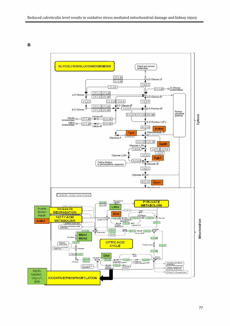
3.4.7 Alteration of energy metabolism in Calr+/- mice kidneys ..................................................... 75
3.4.8 Chronic low levels of Calr induces kidney injury through oxidative stress induction .......... 78
Page 7
vii
3.4.9 Activation of iNos dimerization in Calr+/- mice ..................................................................... 80
3.4.10 Mitochondrial damage in Calr+/- mice ................................................................................ 81
3.5 Discussion .................................................................................................................................... 88
4. Summary ................................................................................................................. 95
Bibliography .................................................................................................................. 100
Appendix ....................................................................................................................... 114
Acknowledgements ....................................................................................................... 120
Curriculum Vitae ............................................................................................................ 122
Page 8
viii
List of Abbreviations
∆: Mutant
2D DIGE: 2 dimensional differential in gel electrophoresis
ACN: Acetonitrile
ACTB: β-actin
ALS: Amyotrophic lateral sclerosis
ANOVA: Analysis of variance
ATP: Adenosine trisphosphate
GBM: Glomerular basement membrane
BSA: Bovine serum albumin
Ca2+: Calcium ions
Calr: Calreticulin mouse
CALR: Calreticulin rabbit, human
cDNA: Complementary DNA
CHAPS: 3-[(3-cholamidopropyl)dimethylammonio]-1-propanesulfonate
CKD: Chronic kidney diseases
Cnx: Calnexin
Cox: Cytochrome c oxidase
CT: Threshold cycle
DMEM: Dulbecco's modified Eagle's medium
DMF: Dimethylformamide
DMSO: Dimethyl sulfoxide
DNA: Deoxyribonucleic acid
dNTPs: Deoxyribonucleotides
Page 9
ix
DTT: Dithiothreitol
ECM: Extra-cellular matrix
EF-2: Elongation factor 2
eif2α: Eukaryotic translation initiation factor-2α subunit
EMT: Epithelial to mesenchymal transition
ER: Endoplasmic reticulum
ERAD: ER-associated degradation
Erp72: Endoplasmic reticulum protein 72
ESI-QTOF-MS: Electrospray ionization time of flight mass spectrometry
ESRD: End stage renal disease
Ezr: Ezrin
FCS: Fetal calf serum
FITC: Fluorescein isothiocyanate
Fn1: Fibronectin
FSP1: Fibroblast specific protein 1
G: Gravitational (unit of centrifugation)
GFR: Glomerular filteration rate
Grp78: Glucose regulated protein 78
H&E: Hematoxylin and eosin
H2O2: Hydrogen peroxide
HCl: Hydrochloric acid
HE: Heparin
HRP: Horse radish peroxidase
IC: Interstitial cells
IEF: Iso-electric focusing
Page 10
x
IgA: Immunoglobulin-A
IMCD: Inner medullary collecting duct
iNos: Induced nitric oxide synthase
InsP3: Inositol 1,4,5-trisphosphate receptor
IPG: Immobilised pH gradient
kDa: Kilo dalton
KEGG: Kyoto Encyclopedia of Genes and Genomes
Lam: Laminin
LC: Liquid chromatography
LDH: Lactate dehydrogenase
mGA: Mean glomerular area
mMA. Mean mesengial area
MS: Mass spectrometery
NaCl: Sodium chloride
NADH: Nicotinamide adenine dinucleotide
NCX: Na+ Ca2+ exchanger
NMR: Nuclear magnetic resonance
NO: Nitric oxide
OD: Optical density
OxPhos: Oxidative phosphorylation
P: Probability
PAGE: Polyacrylamide gel electrophoresis
PAS: Periodic acid shift
PBS: Phosphate buffer saline
PCR: Polymerase chain reaction
Page 11
xi
PDI: Protein disulphide-isomerase
PMCA: Plasma membrane calcium pump
PMSF: Phenylmethanesulfonylfluoride or phenylmethylsulfonyl fluoride
Prdx1: Peroxiredoxin 1
RNA: Ribonucleic acid
ROS. Reactive oxygen species
RT: Reverse transcriptase
SD: Standard deviation
SDS: Sodium dodecyl sulfate
SERCA: Sarco/endoplasmic reticulum Ca2+-ATPase
siRNA: Small interfering RNA
Sod: Superoxide dismutase
STD: Standard
TALH: Thick ascending limb of Henle’s loop
TBS-T: Tris boric acid-tween
TCA: Tricarboxylic acid cycle
TFA: Trifluoroacetic acid
TG: Thapsigargin
TGFβ1: Transforming growth factor beta 1
TJ: Tight junctions
TM: Tunicamycin
UPR: Unfolded protein response
WT: Wild type
Zn2+: Zinc ions
Page 12
xii
List of Tables
Table 2.1: Differentially regulated proteins in TALH-NaCl cells compared to TALH-STD cells. ............. 44
Table 3.1: Proteins differentially regulated in the kidneys of WT and Calr+/- mice ............................... 86
Page 13
xiii
List of Figures
Figure 1.1: The ER stress response pathway. .......................................................................................... 7
Figure 1.2: Schematic representation of intracellular calcium homeostasis. ....................................... 10
Figure 1.3: A model of linear and 3D structure of Calreticulin.............................................................. 13
Figure 1.4: Calreticulin-Calnexin cycle representing the proper folding of glycoproteins in ER. .......... 15
Figure 2.1: 2D gel electrophoresis expression of differentially regulated proteins under osmotic stress
conditions. ..................................................................................................................................... 34
Figure 2.2: Time dependent expression changes of ER Ca2+ binding proteins under varied osmotic
stress conditions. ........................................................................................................................... 36
Figure 2.3: Impact of hyperosmotic stress on expression of ER Ca2+ binding proteins under TM
(5µg/ml) induced ER stress ............................................................................................................ 37
Figure 2.4: Time dependent increase of free Ca2+ in TALH cells exposed to hyper-osmotic stress. ..... 38
Figure 2.5: Impact of HE and hyperosmotic stress on expression of proteins and cell viability in TALH
cells. ............................................................................................................................................... 39
Figure 2.6: Effect of overexpression of CALR on cell viability of TALH cells under hyperosmotic stress.
....................................................................................................................................................... 41
Figure 2.7: Knockdown of CALR enhances the resistance of TALH cells to hyperosmotic NaCl stress. 43
Figure 3.1: Morphometric analysis of embryonic and adult kidneys of Calr KO mice. ......................... 61
Figure 3.2: Progressive structural alterations in Calr+/- mice. ............................................................... 63
Figure 3.3: Electron microscopy analysis of Calr+/- and WT kidneys. .................................................... 65
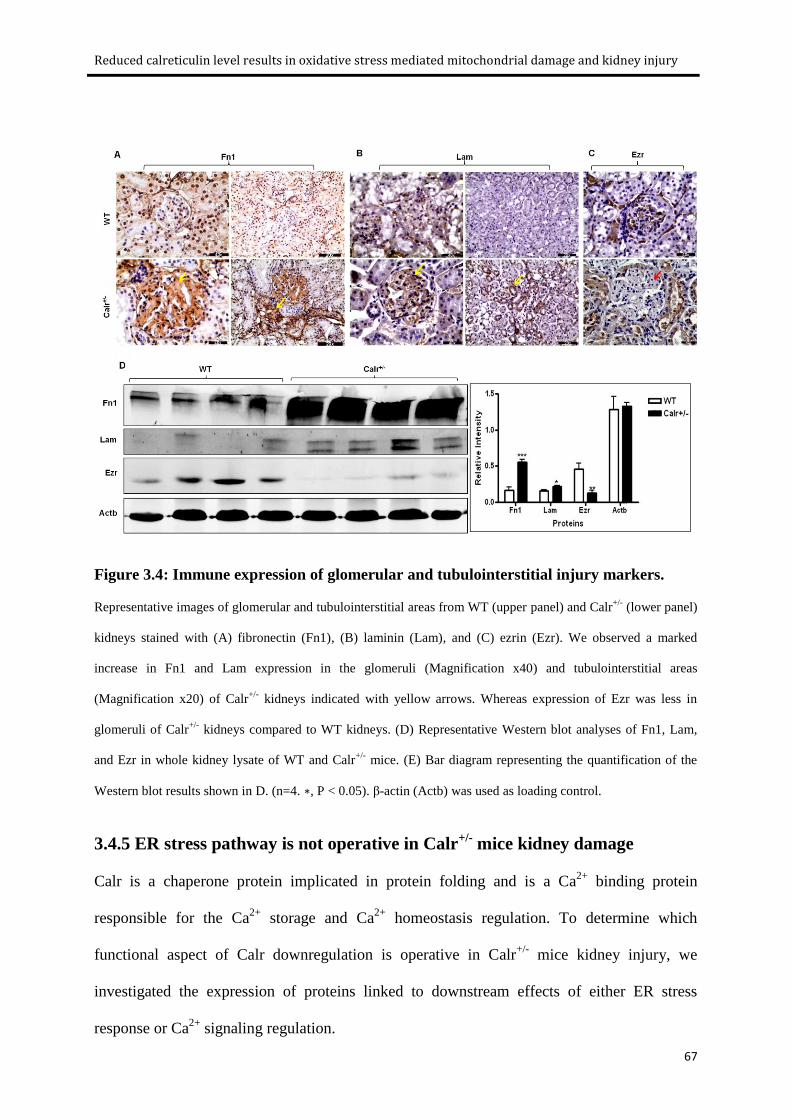
Figure 3.4: Immune expression of glomerular and tubulointerstitial injury markers. .......................... 67
Figure 3.5: Effects of low Calr level on expression of ER stress marker and EF-hand Ca2+ binding
proteins. ........................................................................................................................................ 70
Figure 3.6: 2D gel map expression of differentially regulated proteins in Calr+/- mice kidneys
compared to WT. ........................................................................................................................... 72
Figure 3.7: Gene Ontology (GO) classification of differentially regulated proteins by DAVID
Bioinformatics. .............................................................................................................................. 74
Figure 3.8: Energy metabolism pathways. ............................................................................................ 78
Figure 3.9: Induction of oxidative stress in Calr+/- mice kidneys. .......................................................... 79
Figure 3.10: Activation of iNos in Calr+/- mice kidneys. ......................................................................... 81
Figure 3.11: Electron micrographs demonstrating mitochondrial damage in Calr+/- mice. .................. 84
Figure 4.1: Schematic representation of potential pathway of low calrericulin level in the progression
of renal injury. .............................................................................................................................................. 99
Page 14
1
1. General Introduction
Page 15
General Introduction
2
1.1 Chronic kidney diseases
Kidney, a major homeostatic organ or highly specialized “Natural filters” of the body, mainly
functions to remove waste products, excess of water and salts from the blood and excretes
them outside the body in the form of urine. The kidneys filter about 180 liters of blood every
day and produce about two liters of urine. The kidneys also produce certain hormones such as
erythropoietin, which stimulates the bone marrow to make red blood cells, renin which
regulates blood pressure, calcitriol the active form of vitamin D, which helps maintain Ca2+
for bones and for normal chemical balance in the body. Loss of renal function is a life
threatening event due to accumulation of wastes in the blood and consequent body damage.
Chronic loss of kidney function or chronic kidney disease (CKD) is becoming a major public
health problem worldwide affecting 7.2% of the global adult population with the number
dramatically increasing from 23.4% to 35.8% in the elderly persons aged over 64 years
(Zhang & Rothenbacher, 2008). However, results from an epidemiological survey of chronic
kidney disease in population of older adults in Germany also showed prevalence of CKD in
17.4% subjects aged 50-74 which increased with age and peaked 23.9% in age of 70–74 years
(Zhang et al, 2009). CKD is associated with outcomes such as progression to end-stage renal
disease (ESRD), development of cardiovascular disease, hospitalization, and death in
community-based populations (Go et al, 2004; Orantes et al, ; Schiffrin et al, 2007; Tonelli et
al, 2006; Weir). Progressive nature of CKD to end stage renal failure, a condition requiring
dialysis or renal transplantation for long-term survival is putting an extensive load on global
health care costs (Hossain et al, 2009; Lysaght, 2002; Meguid El Nahas & Bello, 2005; Zhang
& Rothenbacher, 2008).
Page 16
General Introduction
3
1.1.1 Etiopathology of CKD
The nephron, the structural and functional unit of the kidney is progressively damaged in
many chronic kidney diseases starting with either glomerular or tubular injury. Despite the
start, most renal diseases eventually converge into common histopathological impairments
such as glomerulosclerosis and tubulointerstitial fibrosis leading to progressive functional
deterioration of the renal system (Fogo, 2006; Lopez-Novoa et al, 2010; Meguid El Nahas &
Bello, 2005).
Glomerulosclerosis
Glomerulosclerosis is thought to have a central pathogenetic role in the progression from
chronic glomerulopathies to end-stage renal disease (Klahr et al, 1988). It frequently
complicates most renal diseases and is characterized by progressive remodeling of the
glomerular structure such as thickening of the glomerular basement membrane, expansion of
mesangium, podocyte damage and disruption of glomerular filteration machinery.
Microinflammation of endothelial cells is the early sign of glomerular injury leading to
activation and release of a wide range of cytokines and growth factors from mesengial cells
(Cybulsky et al, 2010). Under the influence of growth factors, especially transforming growth
factor beta 1 (TGFβ 1), mesangial cells regress to an embryonic mesenchymal phenotype
capable of excessive production and accumulation of extracellular matrix (ECM) such as
fibronectin ultimately causing glomerular mesangial expansion and fibrosis (El-Nahas, 2003;
Hohenadel & Van der Woude, 2004). These structural impairments are associated with
proteinuria, disturbed glomerular filteration rate (GFR), tubule damage and fibrosis (Levey &
Coresh, 2011; Lopez-Novoa et al, 2010). Stress states, such as sustained hypertension, nitric
oxide and oxidative stress are commonly known to implicate in glomerulosclerosis
(Modlinger et al, 2004; Oberg et al, 2004; Okada et al, 2012).
Page 17
General Introduction
4
Tubulointerstitial fibrosis
Despite the primary cause, many renal diseases also lead to tubulointerstitial fibrosis.
Inflammation, proliferation, apoptosis, and fibrosis are hallmarks of tubulointerstitial fibrosis
(Zeisberg et al, 2000). Direct attack of disease or indirectly due to proteinuria from
glomerular damage initiates the inflammation of tubular cells and interstitial fibroblasts and
myofibroblasts associated with an increased synthesis and release of matrix proteins (Lopez-
Novoa et al, 2010). Progressive deposition of harmful connective tissue in interstitial spaces
of the kidney together with epithelial to mesenchymal transition (EMT) of tubular epithelial
cells, are directing to apoptosis based tubular atrophy and the formation of atubular glomeruli.
(Carew et al, ; Efstratiadis et al, 2009; Meguid El Nahas & Bello, 2005; Radisky et al, 2007;
Zeisberg et al, 2000). Continuing injury, inflammation, and fibroblast activation, ECM
deposition and proliferation lead to irreversible fibrosis.
1.1.2 Risk factors of CKD
Hypertension (Barri, 2008; Tedla et al, 2011), and diabetes (Bash et al, 2008; Pyram et al,
2011) are the two mainly discussed causes of kidney disease worldwide. Cytokines
(Schulman, 2012), kidney infections (Barsoum, 2006), urinary obstruction or blockage with
kidney stones (Rule et al, 2009), oxidative stress and hypoxia (Mimura & Nangaku, 2010),
and salt-induced renal injury (Mimran & du Cailar, 2008; Susic & Frohlich, 2012; Tuomilehto
et al, 2001) are also known as some of the potential risk factors of CKD. Progressive kidney
injury also develops in many hereditary disorders such as atherosclerosis (Boykin et al, 2011;
Kottgen et al, 2010; Vehaskari, 2011). Regardless of the underlying cause, CKD is
characterized by appearance of glomerulosclerosis, and tubulointerstitial fibrosis with
subsequent progression toward end stage renal disease (ESRD) (Meguid El Nahas & Bello,
2005).
Page 18
General Introduction
5
In the last few decades, the progression of the disease process is well documented. Much
interest has focused on investigating potential mechanisms to prevent or reverse the damage.
However, the intracellular mechanisms responsible for renal disease initiation leading to
complete damage are mostly not well understood. There is an immense need to explore the
approaches to minimize the risks of renal diseases. Over the past few decades, intensive
investigations of the molecular and cellular mechanisms revealed the association of ER
function alteration in normal kidney structure and function, with the early-onset and
pathogenesis of renal diseases (Cunard & Sharma, 2011; Cybulsky et al, 2010; Hebert &
Molinari, 2007; Inagi, 2009; Inagi et al, 2008; Liu et al, 2008).
1.2 Endoplasmic reticulum
The ER is a perinuclear, cytoplasmic compartment comprising membranous network of
branching tubules and flattened sacs. It is mainly recognized as a protein-folding factory
involved in synthesis, proper folding, trafficking, and modification of proteins, degradation of
proteins, as well as for synthesis of steroids, cholesterol, and other lipids. Ca2+
storage and
Ca2+
signaling regulation is another basic important role of ER in cell (Baumann & Walz,
2001; Bedard et al, 2005; Inagi, 2009; Nauseef et al, 1995). Importantly, the ER contains
numerous molecular chaperones and catalysts to aid in the ER functions.
1.2.1 ER protein folding and ER resident proteins
Newly synthesized proteins translocate to ER, where they are covalently modified and attain
their correctly folded three dimensional conformation through ER resident chaperones
including BiP /Grp 78, calreticulin (CALR), calnexin (Cnx), Grp94 and the thiol
oxidoreductases PDI and ERp57, all involved in generating conformationally competent and
functional proteins (Bedard et al, 2005; Brodsky & Skach, ; Ellgaard & Helenius, 2003;
Page 19
General Introduction
6
Kleizen & Braakman, 2004). Each of these proteins follows their distinctive chaperon system
for specific types of proteins.
1.2.2 ER stress
Environmental insults like ischemia, glucose deprivation, oxidative stress, osmotic stress or
genetic mutation can cause expression regulation of ER chaperone proteins. This expression
changes the result in aberrant ER function due to inefficient protein folding (Buchberger et al,
2010; Chevet et al, 2001; Wu & Kaufman, 2006; Yoshida, 2007; Zhao & Ackerman, 2006).
Improper protein folding results in accumulation of misfolded proteins leading to ER stress
and induction of ER stress response pathways (Figure 1.1). Misfolded proteins are corrected
by either activation of unfolded protein response (UPR) (Hetz, 2012; Ron & Walter, 2007;
Wu & Kaufman, 2006), a coordinated stress response that upregulates the capacity of the ER
to process abnormal proteins or ER quality control CALR -Cnx cycle (Discussed later). UPR
is an adaptive mechanism that targets the transcription regulation of proteins which can
restore the proper folding of proteins through induction of chaperone such as Grp78 (Lee,
1992; Lee, 2007; Zhang et al, 2010).
Page 20
General Introduction
7
Figure 1.1: The ER stress response pathway.
ER stress leads to accumulation of unfolded proteins in ER resulting in induction of four responses. A: Induction
of ER chaperones such as Calr, Grp78 and Erp57 to correctly fold the misfolded proteins and avoid protein
aggregates (Hong et al, 2004). B: Translation attenuation which reduces ER load by turning down the general
translation (Lee do et al, 2010) C: ERAD is the ER quality-control system which detects and exposes to
cytosolic proteasomal degradation of the misfolded proteins through ubiquitylation . D: apoptosis of cells in
which severe and prolonged ER stress extensively impairs the ER functions and threatens the integrity of the
organism (Timmins et al, 2009). ER: endoplasmic reticulum, ERAD: ER-associated degradation. Adapted from
Araki et al. (Araki et al, 2003).
Accumulating data suggest a pathophysiological role of ER stress in renal diseases. Patient
biopsies and animal models of kidney diseases demonstrate the implication of ER stress in the
development and progression of both glomerular and tubular injuries (Chiang et al,2011;
Inagi, 2009; Inagi et al, 2008). ER stress is also associated with many risk factors of CKD
such as hypertension, diabetes, hypoxia/ischemia and genetic disorders giving a possible
mechanistic link between disease mediators and final diseased state (Lindenmeyer et al, 2008;
Page 21
General Introduction
8
Okada et al, 2012; Yoshida, 2007). In vitro studies further show an expression regulation of
ER chaperones along with UPR activation in renal cells treated with cytokines, oxidative
stress, or osmotic stress mediators (Bibi et al, 2011; Dihazi et al, 2005; Dihazi et al, 2011;
Eltoweissy et al, 2011; Lindenmeyer et al, 2008; Yoshida, 2007). Using proteomics, Dihazi
and coworkers demonstrated a clear correlation between upregulation of ER stress–related
proteins and the fibrosis phenotype highlighting an important role of ER proteins in fibrosis
progression (Dihazi et al, 2011).
1.2.3 Intracellular Ca2+ homeostasis
Ca2+
is an universal signal transduction element. Free intracellular Ca2+
is the physiologically
active form of Ca2+
(Means & Rasmussen, 1988). It plays an important role in the regulation
of diverse cellular processes from contraction, secretion, gene transcription, cell growth and
movement to cell differentiation and death (Berridge, 1993). Maintenance of a constant
luminal level of Ca2+
is also essential for the post-translational processing, folding and export
of proteins (Verkhratsky, 2007). Therefore, the maintenance of free Ca2+
to certain critical
limits called intracellular Ca2+
homeostasis is of prime importance in the cell to keep it
functioning normally. Intracellular Ca2+
homeostasis refers to a cytosolic concentration as low
as ~100 nM compared to 10,000 folds more in extracellular environment. Figure 1.2
represents the simple pathways and organelles involved in the intracellular Ca2+
homeostasis.
ER, being a major intracellular Ca2+
store plays an important role in the regulation of
intracellular Ca2+
homeostasis (Berridge, 1993). Ca2+
signalling between ER and cytoplasm is
tightly regulated by ER membrane Ca2+
entry and exit channels. Ca2+
enters the ER through
SERCA, a Ca2+
pump that transfers Ca2+
from the cytosol to the lumen of the SR/ER at the
expense of ATP hydrolysis (Kubala, 2006), whereas InsP3 (inositol 1,4,5-trisphosphate
Page 22
General Introduction
9
receptor) and ryanodine receptors are used for Ca2+
release from the ER (Arendshorst & Thai,
2009; Vanderheyden et al, 2009). Moreover, ER luminal Ca2+
is also in homeostasis with total
ER Ca2+
concentration (up to 1 mM) and the free ER Ca2+
concentration (200 μM). ER
resident proteins, in particular the molecular chaperones and folding enzymes; Cnx, CALR,
BiP, Grp94, and PDI have both high- and low-affinity Ca2+
-binding sites and are responsible
for mediating intracellular Ca2+
dynamics (Michalak et al, 2002). Because of the sheer
abundance and number of Ca2+
-binding sites, CALR is considered the most important protein
for Ca2+
storage and buffering. It binds to over 50% of ER luminal Ca2+
(Nakamura et al,
2001) and engages in intracellular Ca2+
homeostasis due to two Ca2+
binding domains with
different affinities and capacities. The protein is involved in a variety of cellular processes and
functions from cell to organ level (discussed later in part 1.3).
Page 23
General Introduction
10
Figure 1.2: Schematic representation of intracellular calcium homeostasis.
A schematic representation of intracellular calcium homeostasis mechanism showing tightly regulated Ca2+
concentrations across the cell and cellular components like organelles and channels that may take part in this
regulation. [Ca2+
]i: calcium concentration, iNCX: Na+ Ca
2+ exchanger, MNCX: mitochondrial Na
+ Ca
2+
exchanger, PMCA: plasma membrane calcium pump, MPT: mitochondrial permeability pore, RyR: ryanodine,
Ins(1,4,5)P3R: inositol-1,4,5-trisphosphate receptors, SERCA: sarco-endoplasmic reticulum Ca2+
ATPase.
Adapted from Popi Syntichaki and Nektarios Tavernarakis (Syntichaki & Tavernarakis, 2003).
1.3 Calreticulin
CALR also known as high-affinity Ca2+
binding protein, Calreguiin, Erp60, CRP55, CAB-63
and CaBP3 and calsequestrin-like protein (Michalak et al, 1992) is an endoplasmic reticulum
resident protein. The protein was first identified in the 70s as a Ca2+
binding protein in
Page 24
General Introduction
11
skeletal muscle sarcoplasmic reticulum (Ostwald & MacLennan, 1974). Fifteen years later,
with advances in molecular biology, two groups, Koch and Michalak, isolated simultaneously
the cDNA encoding this Ca2+
-binding protein (Kottgen et al, 2010; Michalak et al, 1992).
The authors named this protein calreticulin (Kottgen et al, 2010). Since then, CALR emerged
as a ubiquitously expressed protein in a wide range of species and in almost al1 cell types
studied. CALR is highly conserved protein with over 90% amino acid identity existing
between human, rabbit, rat and mouse forms of the protein (Michalak et al, 1992).
1.3.1 Structure of calreticulin
CALR, is a 46 kDa (400 amino acid residues) ER Ca2+
binding chaperon. Biochemical and
structural studies have demonstrated three distinct structural domains of CALR: the amino-
terminal N-domain, the middle P-domain, and the carboxyl-terminal C-domain.The protein
also contains a cleavable amino acid signal sequence at the beginning of N-terminal directing
the protein to ER and an ER retention/retrieval signal at the C-terminal (Fliegel et al, 1989;
Kottgen et al, 2010; Mesaeli et al, 1999). (Figure 1.3A)
N-domain: The N-domain (residues 1–170) is an extremely conserved and highly folded
globular domain composed of eight antiparallel β-strands (Opas et al, 1996) as shown in
Figure 1.3B. The N-domain of CALR also has a lectin binding site and a polypeptide binding
site (Kapoor et al, 2003; Leach et al, 2002). It also binds with protein disulphide-isomerase
(PDI) and ERp57 mediated by Zn2+
(Baksh et al, 1995; Leach et al, 2002; Michalak et al,
1999; Pollock et al, 2004). N-domain has a binding site for rubella virus RNA, a putative
phosphorylation site and a segment which binds to steroid hormone receptors and the
cytoplasmic domains of integrin α subunits and is recently known to have a single high-
affinity Ca2+
binding site (Chouquet et al, 2011; Kozlov et al, 2010; Pocanschi et al, 2011).
Page 25
General Introduction
12
P-domain: The middle P-domain (residues 170–285) of CALR is a proline rich domain. The
P domain is also known as “extended arm” based on its three-dimensional structure obtained
by NMR technique (Figure 1.3B). Moreover, this hairpin loop is also known to interact with
Erp57 (Ellgaard et al, 2002; Martin et al, 2006). The P-domain also binds Ca2+
with high
affinity (Kd =1 μM) and low capacity (approximately 1 mol of Ca2+
per mol of protein)
(Baksh et al, 1995; Tjoelker et al, 1994). The P domain, having a lectin binding site, together
with the N-domain is involved in the chaperoning of nascent polypeptides (Pocanschi et al,
2011; Vassilakos et al, 1998). (Figure 1.3)
C-domain: The C-domain (residues 285– 400) of CALR is highly acidic Ca2+
binding and
storage domain (Figure 1.3B). Depending on negatively charged residues of the C-domain,
protein binds Ca2+
with low affinity (Kd =2 mM) and high capacity (approximately 25 mol of
Ca2+
per mol of protein) (Baksh et al, 1995; Mesaeli et al, 1999) and works as a Ca2+
-buffer in
ER. The C-domain of CALR is also known to regulate the protein-protein interactions of
CALR with PDI, Erp57 and other chaperones (Michalak et al, 1999).
Page 26
General Introduction
13
Figure 1.3: A model of linear and 3D structure of Calreticulin.
(A) Linear representation of CALR domains. (B) 3D model of the CALR domains. Adapted from Gelebart et al.
(Gelebart et al, 2005).
1.3.2 Functions of calreticulin
CALR is a multi-functional Ca2+
binding chaperon of ER. It plays two main functions in ER
as a chaperon and as a Ca2+
binding and storage protein. CALR is also found in several other
sub-cellular locations: the cell surface, cytoplasm, and ECM (Gold et al, 2010). The presence
Page 27
General Introduction
14
of CALR, a protein with ER retention signal KDEL, in other cell compartments was a
mystery. However, Afshar and coworkers (Afshar et al, 2005) demonstrated the
retrotranslocation process of CALR, which is safe from proteasomal degradation. Many
extracellular function of CALR have been reported including roles in immunogenic cell death
in cancer, cellular adhesion, cell migration, phagocytosis, inflammation, cell signaling, and
enhancing wound healing (Gold et al, 2010). Cytosolic CALR is also involved in certain
processes such as adhesion, gene expression, translation and nuclear export (Hsu et al, 2005).
Calreticulin, an ER quality control protein
CALR functions as a molecular chaperone in the folding of many proteins and especially
glycoproteins. The property of CALR to help other proteins to fold correctly and become
functional, assigns the protein as a quality control unit in ER. The majority of growing
polypeptides aspargine side chains bind to glycans and are translocated to ER in N-
glycosylated form and are correctly folded into functional transportable forms as shown in
Figure 1.4. Briefly, Glucosidase I and Glucosidase II are two independent enzyme systems,
which bring these unfolded nascent proteins to monoglucosylated form by trimming two
terminal glucose residues. Chaperon systems of ER, which recognize and fold specifically N-
linked monoglucosylated proteins comprises of CALR, Cnx and Erp57. CALR and Cnx are
homologous lectin molecular chaperones in ER. Their central P-domain binds to the
hydrophilic N-linked monoglucosylated glycans of unfolded and misfolded proteins in ER
leading to their proper, functionl and transportable folded conformations (Hebert & Molinari,
2007; Kapoor et al, 2003; Meunier et al, 2002; Trombetta & Helenius, 1998). On the other
hand, misfolded proteins are degraded through ERAD system. In contrast, folding is
significantly impaired in CALR or Cnx-deficient cells having accelerated folding with an
accumulation of misfolded proteins (Hebert & Molinari, 2007).
Page 28
General Introduction
15
Figure 1.4: Calreticulin-Calnexin cycle representing the proper folding of glycoproteins
in ER.
Adapted from Ellgard and Helenius (Ellgaard & Helenius, 2003). EDEM: ER degradation-enhancing 1,2-
mannosidase-like protein, ERAD: ER-associated degradation
Page 29
General Introduction
16
Calreticulin and Ca2+
homeostasis regulation
ER, being a major store of intracellular Ca2+
, exerts a key role in the complex and precise
mechanism of Ca2+
signalling and homeostasis. The ER lumen Ca2+
storage capacity is
enhanced by Ca2+
-binding proteins. CALR is one of the most important Ca2+
binding proteins
of ER. CALR plays a critical role in the regulation of intracellular Ca2+
homeostasis directly
through Ca2+
storage capacity of ER. Earlier studies with overexpression of CALR in various
cell lines show increased ER Ca2+
storage capacity with almost no impact on protein folding
(Bastianutto et al, 1995; Bibi et al, 2011; Mery et al, 1996; Opas et al, 1996). On the other
hand, downregulation and deficiency of CALR decreases the ER Ca2+
storage (Bibi et al,
2011; Coe & Michalak, 2009; Michalak et al, 1999). It also controls the Ca2+
homeostasis
through store operated Ca2+
influx. CALR interacts with Ca2+
entry and exit channels called
SERCA and IP3R and modulates Ca2+
influx by controlling the extent of inositol 1,4,5-
trisphophate-induced Ca2+
store depletion (Mery et al, 1996; Michalak et al, 2002; Xu et al,
2000). Michalak et al. showed that CALR knockout is lethal due to impaired cardiac
development (Michalak et al, 1999). They further demonstrated that this impairment of heart
development is due to Ca2+
homeostasis regulation and not because of chaperon function of
protein.
Short-term increase in Ca2+
is an essential signal for vitally important cell processes whereas,
long-term increase in Ca2+
leads to irreversible impairment of cellular functions and/or
structure, up to cell death. There is convincing evidence that sustained increase in intracellular
Ca2+
alters cell functions and is associated with various diseases such as diabetes mellitus,
hypertension, Alzheimer`s disease, neurodegenerative disorders, cardiac ischemia, and
atherosclerosis and renal diseases (Chan et al, 2009; Lajdova et al, 2009; Rivera et al, 1996;
Vamvakas & Anders, 1990; Zile & Gaasch, 2011). The role of intracellular Ca2+
homeostasis
Page 30
General Introduction
17
disturbances has also been discussed in some renal diseases but very little is known about its
the role in normal kidney function and mechanisms undergoing in renal impairments.
1.4 Objectives
The general aim of our group is to understand the molecular mechanisms, which are involved
in renal function or lead a normal functioning kidney towards disease state. In this regard we
have undertaken proteomic screening of several renal cell line models exposed to different
physiological conditions, such as osmotic stress, oxidative stress, and cytokines. Moreover,
proteome of renal cells derived from fibrotic human kidney were also compared to healthy
renal cells. All these studies highlighted the involvement of a group of ER resident proteins
mainly CALR, Grp78, Erp72 and Erp57 in kidney injury. The present work is focused on one
of these proteins, CALR and its physiological importance in renal structure and function,
specifically through the following aims:
i) To investigate the role of CALR in renal cells functions and adaptation specifically, the
potential mechanism of CALR downregulation under conditions of osmotic stress. This is
addressed in Chapters 2.
ii) To examine the in vivo role of chronic low level of CALR in kidney structure and function.
Especially, to analyze the intracellular signaling pathways that regulates the development of
chronic kidney injury in mice with chronic low level of CALR. This aim is addressed in
Chapters 3.
Page 31
18
2. Calreticulin is crucial for Ca2+ homeostasis
mediated adaptation and survival of thick
ascending limb of Henle’s loop cells under
osmotic stress
Asima Bibi, Nitin K. Agarwal, Gry H. Dihazi, Marwa Eltoweissy, Phuc Van Nguyen,
Gerhard A. Mueller, Hassan Dihazi
The International Journal of Biochemistry and Cell Biology, 43 (2011): 1187-97.
Page 32
Calreticulin is crucial for Ca2+ homeostasis mediated adaptation and survival of thick ascending limb of
Henle’s loop cells under osmotic stress
19
2.1 Abstract
The thick ascending limb of Henle’s loop (TALH) is normally exposed to variable and often
very high osmotic stress and involves different mechanisms to counteract this stress. ER
resident calcium ions (Ca2+
) binding proteins especially calreticulin (CALR) play an
important role in different stress balance mechanisms. To investigate the role of CALR in
renal epithelial cells adaptation and survival under osmotic stress, two-dimensional
fluorescence difference gel electrophoresis combined with mass spectrometry and functional
proteomics were performed. CALR expression was significantly altered in TALH cells
exposed to osmotic stress, whereas renal inner medullary collecting duct cells and interstitial
cells exposed to hyperosmotic stress showed no significant changes in CALR expression.
Moreover, a time dependent downregulation of CALR was accompanied with continuous
change in the level of free intracellular Ca2+
. Inhibition of the Ca2+
release, through IP3R
antagonist, prevented CALR expression alteration under hyperosmotic stress, whereas the cell
viability was significantly impaired. Overexpression of wild type CALR in TALH cells
resulted in significant decrease in cell viability under hyperosmotic stress. In contrast, the
hyperosmotic stress did not have any effect on cells overexpressing the CALR mutant,
lacking the Ca2+
-binding domain. Silencing CALR with siRNA significantly improved the
cell survival under osmotic stress conditions. Taken together, our data clearly highlight the
crucial role of CALR and its Ca2+
-binding role in TALH adaptation and survival under
osmotic stress.
Page 33
Calreticulin is crucial for Ca2+ homeostasis mediated adaptation and survival of thick ascending limb of
Henle’s loop cells under osmotic stress
20
2.2 Introduction
The osmoregulation of the body is one of the most controlled physiological mechanisms,
regulated by a balance of hydration and solute concentrations (Bourque, 2008). The kidney is
one of the main organs of the body which maintain osmolality. As a consequence of this, the
kidney cells are exposed to very hyper-osmotic environment compared to the rest of the body
(Marsh & Azen, 1975). The thick ascending limb of Henle’s loop (TALH) segment is the part
of the kidney nephron, which plays a vital role in urinary concentration mechanism by
generating concentrated urine in antidiuresis and dilutes urine in water diuresis.
Hyperosmolality affects numerous cellular functions and causes cell cycle delay and apoptosis
in renal cells (Burg et al, 2007; Michea et al, 2000). To study the adaptive changes under
variable osmotic stress conditions in this segment of the kidney, the TALH-cell line from
rabbit kidney provides a unique tool. The ability at the cellular level to alter gene expression
and metabolic activity in response to changes in the osmotic environment provides an
additional regulatory mechanism. TALH cells adapt to an increased levels of NaCl by
morphological shrinkage (Grunewald et al, 2001). These morphological adaptations are
accompanied by dramatic change in the proteome of the cells. Especially the downregulation
of the ER Ca2+
binding chaperones like calreticulin (CALR), Erp72, and GRP78 is debatable,
since such a reaction of a protein with chaperone function is quite unlikely under stress
conditions in TALH cells (Dihazi et al., 2005). The aim of the current study is to understand
the role of CALR in terms of Ca2+
homeostasis regulation in the adaptation mechanism of
TALH cells under osmotic stress. CALR is a 46 kDa protein, which is ubiquitously expressed
in nearly all cells of higher organisms (Mesaeli et al, 1999). It is subdivided into three
structural and functional regions: a highly conserved N-domain, a proline-rich P-domain and a
Page 34
Calreticulin is crucial for Ca2+ homeostasis mediated adaptation and survival of thick ascending limb of
Henle’s loop cells under osmotic stress
21
very acidic C-domain, which binds Ca2+
with high capacity and low affinity. Different cellular
functions have been characterized for CALR, intracellular as well as extracellular. Mainly due
to its ability to bind monoglucosylated high mannose oligosaccharides, CALR plays an
important role as a lectin-like chaperon by binding to incompletely folded proteins that
contain one terminal glucose on N-linked oligosaccharides, retaining the protein inside the ER
until proper folding (Peterson et al, 1995). Directing proper conformation of misfolded
proteins and glycoproteins under stress conditions, CALR, is generally induced as stress
response protein to protect the cells against various toxic insults (Ihara et al, 2005; Little &
Lee, 1995; Liu et al, 1997; Marber et al, 1995; Morris et al, 1997; Sugawara et al, 1993) and
is involved in various cellular functions and signaling, including apoptosis, stress responses,
organogenesis, and transcriptional activity (Michalak et al, 2002). Ca2+
is an important
signaling molecule and stored mainly in the lumen of the ER. Fluctuations of the ER luminal
Ca2+
concentration result in disturbance of intracellular Ca2+
homeostasis. Intracellular Ca2+
homeostasis has received considerable attention
as a cell death signal and as an activator of gene expression (Nicotera et al, 1992; Nicotera &
Orrenius, 1998). CALR due to its Ca2+
binding C-domain and accumulation of large amounts
of Ca2+
without an excessive increase in the free ER intraluminal Ca2+
concentration was
proved to regulate the intracellular Ca2+
homeostasis and ER Ca2+
storage capacity (Fliegel et
al, 1989; Gelebart et al, 2005; Nakamura et al, 2001; Treves et al, 1990; Vassilakos et al,
1998). Additionally, CALR appears to play an essential role in the development of heart and
brain since CALR-deficient mice develop embryonic lethality due to decreased ventricular
wall thickness, whereas cells derived from CALR knockout embryos have impaired Ca2+
homeostasis (Gelebart et al, 2005). Intracellular Ca2+
concentration (Ca2+
) also plays an
important role in the signal transduction processes within the TALH cells and regulates the
Page 35
Calreticulin is crucial for Ca2+ homeostasis mediated adaptation and survival of thick ascending limb of
Henle’s loop cells under osmotic stress
22
transepithelial transport of sodium across the renal epithelial tubular cells (Friedman et al,
1981; Taylor & Windhager, 1979).
Page 36
Calreticulin is crucial for Ca2+ homeostasis mediated adaptation and survival of thick ascending limb of
Henle’s loop cells under osmotic stress
23
2.3 Materials and Methods
2.3.1 Cell line and culture procedure
The epithelial cell line used in these experiments was derived from a rabbit kidney’s outer
medulla. Cultured cells were immortalized by SV 40 early region DNA (Bartek et al, 1991).
They showed a high degree of differentiation and specialization and provided a suitable model
to study TALH cell function in vitro. The TALH cell line was maintained as a monolayer
culture in DMEM (Gibco) including 5.5 mmol/l d-glucose supplemented with 10% fetal calf
serum (Roche), 1% MEM nonessential amino acids, 1% l-glutamine and 1%
Penicillin/Streptomycin (Gibco). Cells were routinely cultured in 75 cm2 tissue culture flasks
(Falcon) at 37 °C in a humidified 5% CO2/95% air atmosphere.
2.3.2 Osmotic stress experiments
After reaching 70% confluence, TALH cells cultivated in 300 mosmol/kg medium (TALH-
STD) were stressed with 600 mosmol/kg NaCl medium. TALH-cell lines exhibiting a high
resistance to osmolality (600 mosmol/kg) (TALH-NaCl) were established. The osmolality
was adjusted with 3 M NaCl solution and was controlled routinely. Later on, the TALH-NaCl
cells, which were growing for a long time in hyperosmolality NaCl medium (600 mosmol/kg)
were transferred back to hypoosmotic medium (300 mosmol/kg) in a time dependent manner
for 12, 24, 48 and 72 h. All osmotic stress experiments were repeated at least three times.
Isolation of IMCD and IC cells
Inner medullary collecting duct (IMCD) and interstitial cells (IC) were isolated from rat
kidney by following the protocol of Grupp et al. (Grupp et al, 1998).
Page 37
Calreticulin is crucial for Ca2+ homeostasis mediated adaptation and survival of thick ascending limb of
Henle’s loop cells under osmotic stress
24
2.3.4 Protein extraction and estimation
75% confluent cultures were scraped and washed three times with PBS with the
corresponding osmolality (300 or 600 mosmol/kg). The cells were harvested by centrifugation
at 200×g for 10 min, the pellet was treated with 0.3-0.5 mL lysis buffer (9.5 M urea, 2%
CHAPS (w/v), 2% ampholytes (w/v), 1% DTT, 10 mM PMSF). Ampholytes, DTT, pepstatin
(to a final concentration of 1.4 µM), and complete from Roche Diagnostic (according to the
manufacturer’s protocol) were added before use. To remove the cell debris, sample
centrifugation was carried out at 13,000×g and 4ºC for 45 min. Supernatant was recentrifuged
at 13,000×g and 4 °C for an additional 45 min to get maximal purity. The resulting samples
were used immediately or stored at -80ºC until use. Protein concentration was estimated
according to Bradford (1976), using bovine serum albumin as a standard.
2.5. Two-dimensional fluorescence difference gel electrophoresis (2D DIGE) Protein
extraction was performed as described above. The resulting pellet was solubilized in labeling
buffer (30 mM Tris–HCl pH 8.5, 9.5 M urea, 2% CHAPS, 10mM PMSF), centrifuged (5 min,
13,000 × g) and the protein concentration of the supernatant was determined as described
above. For the fluorescence labeling, each dye was freshly dissolved in anhydrous N,N-
dimethylformamide (DMF) (Sigma–Aldrich, St. Louis, USA) to a stock solution containing
1000 pmol/µl. One volume of CyDye solution was added to 1.5 volumes of high grade DMF,
to make a 400 pmol CyDye solution. For minimal labeling 400 pmol of the amine-reactive
cyanine dyes Cy3 and Cy5 was added respectively to 50 µg proteins from each TALH-STD
and TALH-NaCl, following the manufacturer’s protocol (GE Healthcare). The labeling
reaction was carried out at 4ºC in the dark for 30 min and the reaction was terminated by
addition of 10 nmol lysine at 4ºC in the dark for 10 min. Equal volumes of 2× sample buffer
(30mM Tris-HCl pH 8.5, 9.5 M urea, 2% CHAPS, 10 mM PMSF, 130 mM DTT and 2%
Page 38
Calreticulin is crucial for Ca2+ homeostasis mediated adaptation and survival of thick ascending limb of
Henle’s loop cells under osmotic stress
25
ampholytes 3-10) were added to each of the labeled protein samples. To avoid the dye-
specific protein labeling, every pair of protein samples from two independent cell extract
preparations was processed in duplicate while swapping the dyes. Thereby, four replicate gels
were obtained which allowed monitoring regulation factors down to two-fold changes. 50µg
of an internal standard consisting of a mixture of all cell samples under investigation were
labeled with 400 pmol Cy2 and included on all gels to facilitate gel matching, thereby
eliminating experimental variation. The three differentially labeled fractions were pooled.
Rehydration buffer (8 M urea, 1% CHAPS, 13 mM DTT and 1% ampholytes 3-10) was added
to make a total volume of 185 µl prior to IEF. The 2-DE was performed with 11cm 3–10 IPG
strips. The CyDye-labeled gels were scanned at 50µm resolution on a Fuji FLA5100 scanner
(Fuji Photo, Kanagawa, Japan) with laser excitation light at 473nm and long pass emission
filter 510LP (Cy2), 532nm and long pass emission filter 575LP (Cy3), and 635nm and long
pass emission filter 665LP (Cy5). Fluorescent images were acquired in 16-bit TIFF files
format. Spot matching across gels and normalization based on the internal standard was
performed with Delta2D software (Decodon, Greifswald, Germany). To analyze the
significance of protein regulation, a Student’s t-test was performed, and statistical significance
was assumed for p values <0.01. For protein visualization, 2-DE was poststained with
colloidal Coomassie blue (Roti-Blue) overnight. Differentially regulated proteins were
excised and processed for identification by MS.
2.3.5 In-gel digestion and mass spectrometry analysis of protein spots
Differentially expressed spots were manually excised from the gels and in-gel digestion, mass
spectrometry analysis and protein identification with database search was performed as
described by Dihazi et al. (Dihazi et al, 2005).
Page 39
Calreticulin is crucial for Ca2+ homeostasis mediated adaptation and survival of thick ascending limb of
Henle’s loop cells under osmotic stress
26
2.3.6 Western blot analysis
In order to confirm the protein expression differences during 2-D DIGE analysis, Western
blot analysis was performed for the proteins of interest according to a standard protocol of
Towbin et al. (Towbin et al, 1979).
2.3.7 Tunicamycin (TM), heparin (HE) and thapsigargin (TG) treatment.
TALH-cells were cultured in 96-well microtiter plates at a concentration of 5x10³ cells per
well (for cell viability assay) and to 70% confluence in 75 cm² tissue culture flasks (for
Western blot analysis). A stock solution of TM, an ER stress inducer was prepared by
dissolving in DMSO. Heparin, an IP3R antagonist and blocker was used to block the IP3R. A
low molecular weight, water soluble heparin which can enter the cell was purchased from
Sigma. It was dissolved in culture media. Cells were treated with a concentration of 5 µM
TM and 25 µM HE alone and coupled with NaCl stress separately, for 24 hours compared to
control groups with normal and NaCl stress media. Cells grown in normal media also received
equivalent volumes of DMSO as a control. After 24 hours of treatments cells cultured in 96
well plates were further processed for MTT cell viability assay and samples were collected for
Western blot analysis.
A stock solution of Thapsigargin (Sigma), a SERCA inhibitor was prepared by dissolving in
DMSO and a concentration of 0.3 µM was used to treat the TALH-cells cultured in 6-well
plate in a time dependent manner for 0-50 min. mRNA samples were collected for RT-PCR.
2.3.8 MTT cell viability assay
Cell viability was tested using cell Proliferation Kit I (MTT), a colorimetric assay for the non-
radioactive quantification of cell proliferation and viability (Roche Applied Bioscience,
Mannheim, Germany). Cells were plated in 200 µl ofmedium at a concentration of 5×10³
Page 40
Calreticulin is crucial for Ca2+ homeostasis mediated adaptation and survival of thick ascending limb of
Henle’s loop cells under osmotic stress
27
cells per well in 96-well microtiter plates (tissue culture grade, Falcon) and MTT cell viability
was performed according to manufacturer’s protocol. GraphPad Prism 4 software (GraphPad
Software Inc., San Diego, CA) was used for statistical analysis. Comparisons of two groups
were conducted using paired two-tailed t-test. A one-way ANOVA test was performed for
comparisons among multiple groups, and statistical significance was set at p < 0.05. All
assays were performed using at least three separate experiments in triplicate, and data were
expressed as mean±SD in comparison to untreated cells (controls).
2.3.9 Ca2+ measurements
Imaging of intracellular free Ca2+
Cells grown on cover slides were incubated with 2.5 mM probenecid (an inhibitor of organic
ion transport by blocking multidrug resistance-associated proteins) for 30 min at 37 °C in
standard medium. Loading of cells with fura-2/AM (Invitrogen) was performed according to
Vamvakas et al. (Vamvakas & Anders, 1990) in 3 ml standard medium for cover slides in
four well plates respectively, both containing fura-2/AM in a final concentration of 8 µM, 2.5
mM probenecid and 1:1000 Pluronic® F-127. After loading the cells for 1 h at room
temperature, the samples were washed two times with standard medium containing 2.5 mM
probenecid to prevent leakage of fluorescent dye. Subsequently, cells were allowed to
incubate for 30 min at room temperature to deesterify fura-2/AM dye. The cover slides were
removed from the well plates and imaging was carried out at 37 °C on the stage of an inverted
microscope (Zeiss, Oberkochen) equipped for epifluorescence with objectives ranging from
magnifications of 10× to 100× with oil-immersion.
Measurement of intracellular free Ca2+
with FlexStation
Page 41
Calreticulin is crucial for Ca2+ homeostasis mediated adaptation and survival of thick ascending limb of
Henle’s loop cells under osmotic stress
28
Measurement of free Ca2+
was also made with a fluorescence microplate reader (FlexStation,
Molecular devices). Cells were plated in 200 µl of medium at a concentration of 5×10³ cells
per well in 96-well microtiter plates (tissue culture grade, Falcon). Fura-2/AM was loaded as
described above. Fura-2/AM fluorescence was measured by illuminating the cells with an
alternating 340/380 nm light every 5 s. Fluorescence intensity was measured at 510 nm.
Automated pipette was settled for the addition of 45 ml of thapsigargin to remove
extracellular Ca2+
. Changes in intracellular Ca2+
concentration are presented as the change in
the ratio of fluorescence intensity for excitation at 340 and 380 nm.
2.3.10 Quantitative real-time PCR
Short-term stress dependent CALR mRNA expression levels were determined by quantitative
real-time PCR. Briefly, total RNA was isolated from TALH-STD and TALH-NaCl cells
exposed for different times to NaCl stress with the column-based RNeasy Mini Kit (Qiagen,
Hilden) according to the manufacturer’s protocol.RNA was transcribed using the
SuperScriptTM
II RNase H-Reverse Transcriptase Kit. PCR was performed with a PCR kit
(Invitrogen) according to the manufacturer’s directions. Primer sequences were as follows:
Rabbit-CALR forward, 5’-GAA ATC GAC AAC CCC GAG TA-3’; reverse, 5’-CCT CGT
CCT GCT TGT CTT TC-3’ (MWG Biotech, Ebersberg D). Quantitative real-time PCR was
carried out on an Mx3000P PCR system (Stratagene, Amsterdam). Reaction conditions were
adopted according to Hsu et al. (Hsu et al. 2005).
2.3.11 Construction of CALR expression and CALR siRNA vectors and
cellular transfection
The construction procedure of wild type (WT-CALR), mutant without the Ca2+
binding C-
domain (∆CALR) and CALR siRNA vectors has been provided in detail in supplementary
Page 42
Calreticulin is crucial for Ca2+ homeostasis mediated adaptation and survival of thick ascending limb of
Henle’s loop cells under osmotic stress
29
data. All constructs were verified by sequencing. The transfection was performed using
transfection reagent Lipofectamine 2000TM
(Invitrogen) according to manufacturer’s standard
protocol. In brief, 2 µg of plasmids and 8 µl of Lipofectamine 2000TM
were added to 100 µl
OptiMEM (Gibco). The mixture was gently mixed, incubated at room temperature for 20 min,
and then added drop-wise to TALH cells cultured to approximately 80% confluence in 100-
mm plates. The analysis of the transfection was carried out after three days of incubation.
After 24 h, transfection media was changed with selection media for stable transfection. Cells
were maintained in the selection medium for 14 days to achieve stable transfection and
assessed for CALR expression by Western blot and immunofluorescence staining.
2.3.12 Indirect immunofluorescence staining
For the indirect immunofluorescence staining 10×103 cells from each TALH-STD and
TALH-NaCl were cultivated overnight in 16-well chamber slides. The medium was removed
and the cells were washed twice with PBS-buffer. Fixation of the cells was carried out for 30
min at −20 °C with methanol/acetone (1:1, v/v). The fixed cells were blocked with 1:5 normal
goat serum (DAKO)/PBSbuffer for 1 h and incubated with primary antibodies overnight.
Alexa Fluor labeled goat anti-rabbit antibody was used as secondary antibodies. The
incubation was performed for 60 min at room temperature in the dark. Thereafter the samples
were counterstained with DAPI in mounting medium. Afterwards samples were analyzed with
immunofluorescence microscopy (Carl Zeiss Axiovert S100TV).
2.3.13 Antibodies
Rabbit anti-Erp72 polyclonal antibody was from Stressgen, mouse anti-β-actin monoclonal
antibody and rabbit anti- GRP78/BiP polyclonal antibody were from Sigma, mouse anti-
CALR monoclonal antibody was from BD Bioscience. Anti-CALR was purified from rat liver
Page 43
Calreticulin is crucial for Ca2+ homeostasis mediated adaptation and survival of thick ascending limb of
Henle’s loop cells under osmotic stress
30
according the procedure of Nguyen Van and Soling (Nguyen Van, 1989). Alexa Fluor dye
conjugated to secondary anti-rabbit antibody and Alexa dye conjugated to secondary anti-
mouse antibody were from Molecular Probes. Anti-flag antibody was from Sigma.
2.4 Results
2.4.1 ER Ca2+ binding proteins and osmotic stress
In order to understand the molecular mechanism of TALH cells adaptation and survival under
osmotic stress, cell extracts were prepared from TALH-STD and TALH-NaCl cells. The
protein extracts were subjected to DIGE analysis. The 2D DIGE images were analyzed with
the Delta2D software (Decodon); interesting protein spots were excised and analyzed by mass
spectrometry. The proteins were identified using MASCOT Database. 2D DIGE coupled with
mass spectrometry analysis showed that many proteins were differently expressed in the
stressed TALH-NaCl cells compared to TALH-STD cells. Among these differentially
expressed proteins, a group of ER resident proteins, GRP78, Erp72 and especially CALR,
were downregulated in TALH-NaCl cells as reaction on hyperosmotic stress. In contrast,
other ER stress proteins like the heat shock proteins, HSP 70 and HSP 90 were found to be
upregulated (Figure 2.1A) (Table 2.1).
In order to further investigate the effects of osmolality changes on the expression of these
proteins in TALH cells, TALH-NaCl cells were exposed to hypoosmotic stress by culturing
the cells back in isoosmotic medium (300 mosmol/kg). To assess the time dependent effect of
osmolality changes on protein expression the cell were harvested after 24 and 48 h upon
incubation in hypoosmotic medium. The protein extract were subjected to 2D DIGE analysis.
TALH-STD was used as a control (Figure 2.1B). Quantitative analysis of the protein spots
Page 44
Calreticulin is crucial for Ca2+ homeostasis mediated adaptation and survival of thick ascending limb of
Henle’s loop cells under osmotic stress
31
revealed that the expression of CALR, GRP78 and Erp72 increased progressively after the
transfer of the TALH-NaCl cells to isoosmotic medium and achieves the level of the proteins
in TALH-STD after 48 h of incubation (Figure 2.1C).
Page 45
Calreticulin is crucial for Ca2+ homeostasis mediated adaptation and survival of thick ascending limb of
Henle’s loop cells under osmotic stress
32
Page 46
Calreticulin is crucial for Ca2+ homeostasis mediated adaptation and survival of thick ascending limb of
Henle’s loop cells under osmotic stress
33
Page 47
Calreticulin is crucial for Ca2+ homeostasis mediated adaptation and survival of thick ascending limb of
Henle’s loop cells under osmotic stress
34
Figure 2.1: 2D gel electrophoresis expression of differentially regulated proteins under
osmotic stress conditions.
(A) Downregulation of ER Ca2+
binding proteins under hyperosmotic stress conditions: dual color 2-D DIGE
images of proteins extracted from TALH-STD control and TALH-NaCl cells. Cy3-labeled proteins are shown in
green color (TALH-STD) and Cy5-labeled proteins are in red color (TALH-NaCl), whereas protein spots in
yellow color are present in both samples. (B) 2D DIGE proteome analysis of the TALH-STD and TALH-NaCl
cultured in hypoosmotic medium in a time dependent manner for 0 h (i), 24 h (ii) and 48 h (iii). Cy3 labeled
proteins are false colored in blue (TALH-STD) and Cy5 labeled proteins are false colored in orange (TALH-
NaCl). (C) Enlargement of the gel regions of interest showing protein spots found to be differentially expressed:
(i) CALR, (ii) Erp72, (iii) GRP78 and (iv) ACTB (β-actin). The protein expression quantification for selected
proteins is given in form of bar diagrams. Expression of the same protein was quantified under different hypo-
osmotic conditions shown in the form of black bar while control is shown in the form of white bar. Results are
given as the means±SD from three independent DIGE experiments.
To validate the data obtained from 2D DIGE and protein identification, we confirmed the
regulation profiles of the three differentially expressed key proteins by Western blot, namely
CALR, GRP78 and Erp72. The Western blot analysis showed a downregulation of these
proteins when TALH-STD cells were transferred to hyperosmotic NaCl medium in a time
Page 48
Calreticulin is crucial for Ca2+ homeostasis mediated adaptation and survival of thick ascending limb of
Henle’s loop cells under osmotic stress
35
dependent manner from 12 to 72 h (Figure 2.2A). In contrast, the TALH-NaCl cells showed
an upregulation of these proteins when they were cultured back to hypoosmotic standard
medium in a time dependent manner from 12 to 72 h (Figure 2.2B). To compare the results
obtained with TALH cells under hyperosmotic stress, similar experiments were performed
with IMCD and IC primary cells, which were isolated from rat kidney. After three passages of
cell culture, the cells were subjected to NaCl stress for 72 h and samples were collected for
Western blot analysis. In contrast to TALH cells, IMCD and IC cells showed that CALR
expression was not affected with osmotic stress in both cell types. Whereas Erp72 was found
to be upregulated in IMCD cells, IC cells showed no regulation of Erp72 under hyperosmotic
stress (Figure 2.2C).
Page 49
Calreticulin is crucial for Ca2+ homeostasis mediated adaptation and survival of thick ascending limb of
Henle’s loop cells under osmotic stress
36
Figure 2.2: Time dependent expression changes of ER Ca2+
binding proteins under
varied osmotic stress conditions.
Western blot analysis of ER Ca2+
binding proteins (CALR, Erp72 and GRP78) found to be differentially
expressed in time dependent manner under osmotic stress. Protein expression was investigated with respective
antibodies for CALR, Erp72 and GRP78, while ACTB was kept as control: (A) TALH-STD cells cultivated in
hyper-osmotic stress of NaCl for 24, 48 and 72 h. (B) TALH-NaCl cells cultivated back to hypo-osmotic
medium for 24, 48 and 72 h. (C) IC and IMCD kidney cells under control and exposed to NaCl stress for 72 h.
To further characterize the role of the downregulation of these proteins for the TALH cell
survival under hyperosmotic stress, we investigated the impact of upregulation of these
proteins on cells subjected to osmotic stress. As expected TALH cell treated with TM resulted
in ER-stress reflected in upregulation of CALR, GRP78 and Erp72. Cells exposed to a
combination of tynicamycin and hyperosmotic stress showed a downregulation of the three
Page 50
Calreticulin is crucial for Ca2+ homeostasis mediated adaptation and survival of thick ascending limb of
Henle’s loop cells under osmotic stress
37
investigated proteins (Figure 2.3A) accompanied by a significant increase in cell death
revealed by the cell viability assay (Figure 2.3B).
Figure 2.3: Impact of hyperosmotic stress on expression of ER Ca2+
binding proteins
under TM (5µg/ml) induced ER stress
(A) Western blot analysis of the expression changes of CALR, GRP78 and Erp72 in TALH-STD cells treated
with TM and exposed to NaCl stress for 72 h. ACTB was kept as control. (B) MTT cell viability assay, 5000
cells/well were cultured in 96 well cell culture plates, incubated with NaCl stress or 5µM TM or both for 72 h.
The cell viability was measured and plotted in the form of bar diagrams with the cell treatment on x-axis and cell
viability on y-axis. TM: tunicamycin ns, non significant and ** shows significance.
2.4.2 CALR and Ca2+ homeostasis under osmotic stress
To investigate the impact of stress on Ca2+
store in ER, TALH cells growing in isoosmotic
medium were transferred in hyperosmotic environment and the ER- Ca2+
release was
monitored using fura-2/AM fluorescence dye, fluorescence microscopy and AnalySIS
software. 10 min after stress application, a significant increase in ER- Ca2+
release could be
detected (Figure 2.4A). A time dependent increase of Ca2+
release could be observed: the Ca2+
release was 1.6-fold higher after 20 min and 1.8 after 30 min of incubation in hyperosmotic
medium when compared to the cell in isoosmotic one (Figure 2.4B). Parallel to Ca2+
imaging,
RT-PCR was carried out for CALR. The increase in CALR threshold cycles (CT) confirmed
Page 51
Calreticulin is crucial for Ca2+ homeostasis mediated adaptation and survival of thick ascending limb of
Henle’s loop cells under osmotic stress
38
an alteration in CALR-expression under osmotic stress. Parallel to the increase in Ca2+
release, a time dependent downregulation of CALR could be measured (Figure 2.4C). RT-
PCR analysis of CALR expression from TALH cells treated with thapsigargin and exposed to
hyperosmotic stress revealed an upregulation of CALR in the first 50 min as showed by a
diminution in CT, while this decrease was stabilized afterwards. The decrease in CT value
reflects the upregulation of CALR after thapsigargin treatment in contrast to downregulation
of CALR when exposed to NaCl stress (Figure 2.4D). As reaction on Ca2+
loss upon
thapsigargin treatment, the cells increase the expression of CALR to prevent excessive
attenuation in Ca2+
and to rescue the homoeostasis.
Figure 2.4: Time dependent increase of free Ca2+
in TALH cells exposed to hyper-
osmotic stress.
(A) Control cells in isoosmotic medium (STD), and cells exposed to NaCl stress for 10 and 20 min. (B)
Quantitative analysis of fluorescence intensity in fura-2/AM-stained TALH cells after osmotic stress treatment.
Results are given as the means±SD from three independent experiments. (C) Quantitative real-time PCR for the
mRNA of CALR in TALH cells from 0 to 70min in NaCl stress. (D) Real-time PCR analysis of CALR
Page 52
Calreticulin is crucial for Ca2+ homeostasis mediated adaptation and survival of thick ascending limb of
Henle’s loop cells under osmotic stress
39
expression in TALH cells treated with 0.3µM TG. The bar diagram showed the CALR mRNA in the form of CT
value on y-axis while time is plotted on x-axis. TG: thapsigargin.
To investigate the impact of Ca2+
release inhibition on ER Ca2+
binding protein expression
and cell viability under osmotic stress, TALH cell treated with 25µM heparin, to block the
IP3R mediated Ca2+
release, were exposed to hyperosmotic stress with NaCl. Cells treated
with heparin alone showed no pronounced effect on the expression of CALR. In contrast, we
could demonstrate a significant downregulation of GRP78 and Erp72 in TALH cells under
NaCl stress with and without heparin treatment (Figure 2.5A). In comparison CALR was only
downregulated in TALH cells under NaCl stress without heparin. Cells treated with heparin or
heparin combined with NaCl stress showed no significant effect on CALR expression (Figure
2.5A). To determine the effect of alteration of Ca2+
traffic combined with osmotic stress on
cell viability and proliferation, MTT test was carried out with TALH cells exposed to
different conditions of heparin treatment with and without NaCl stress. The cell viability
assay revealed a significant increase in cell death of heparin treated cells upon exposition to
NaCl stress compared to NaCl or heparin treatment separately (Figure 2.5B).
Figure 2.5: Impact of HE and hyperosmotic stress on expression of proteins and cell
viability in TALH cells.
Page 53
Calreticulin is crucial for Ca2+ homeostasis mediated adaptation and survival of thick ascending limb of
Henle’s loop cells under osmotic stress
40
(A) Western blot analysis of the TALH-STD cells treated with HE and exposed to NaCl stress for 72 h compared
to untreated control cells for the expression of CALR, Erp72, and GRP78 while ACTB was kept as control. (B)
MTT cell viability assay, 5000 cells/well cultured in 96 well cell culture plates, incubated with NaCl stress or
25µM heparin or both. The cell viability (%) was measured from values obtained from the assay and plotted in
form of bar diagrams with the cell treatment on x-axis and cell viability on y-axis. HE: heparin.
2.4.3 CALR and cell death under osmotic stress
CALR is an ER- Ca2+
binding protein, to investigate the impact of CALR due to its Ca2+
binding capacity on cell adaptation and survival in hyperosmotic stress, vectors expressing
WT-CALR and ∆CALR (mutant without the Ca2+
binding site) were constructed and
transfected into TALH cells separately. Intracellular localization of CALR and ∆CALR was
examined by indirect immunofluorescence staining. As shown in Figure 2.6A, CALR showed
a perinuclear reticular pattern in all cases, including the control and gene-transfected cells,
although the signal intensity was increased in the transfectants compared to the control cells.
Moreover, the Figure 2.6A also shows the transfection efficiency analyzed with anti-flag
antibody against the flagged CALR transfected cells. To assess whether the increase in CALR
expression interfered with ER Ca2+
storing capacity and Ca2+
homeostasis, we measured the
free intracellular Ca2+
in cells transfected with WT-CALR and ∆CALR compared with
control TALH cells. FlexStation and imaging analyses showed almost same basal levels of
free intracellular Ca2+
in both WT-CALR and ∆CALR transfected cells. However
thapsigargin induced a significant increase in free intracellular Ca2+
in cells overexpressing
WT-CALR compared to ∆CALR and control, whereas the difference between ∆CALR and
control was not significant (Figure 2.6B, Supplemental Figure 2.1). These results revealed the
higher Ca2+
storing capacity of WT-CALR compared to CALR and that the Ca2+
buffering
capacity of the cells is directly correlated to CALR level in ER. The cell viability assay was
Page 54
Calreticulin is crucial for Ca2+ homeostasis mediated adaptation and survival of thick ascending limb of
Henle’s loop cells under osmotic stress
41
performed with WT-CALR and ∆CALR transfected cells exposed to hyperosmotic NaCl
stress compared to non-transfected control cells. We observed a significant decrease in cell
viability (%) in cells overexpressing WT-CALR under NaCl stress conditions compared to
control non-stressed conditions. Moreover these cells also showed significant increase in cell
death compared to cells over-expressing ∆CALR with no Ca2+
binding region (almost 70%)
and non-transfected cells exposed to NaCl stress (almost 70%) (Figure 6C).
Figure 2.6: Effect of overexpression of CALR on cell viability of TALH cells under
hyperosmotic stress.
(A) Immunolocalization of transfected and endogenous CALR in TALH cells. (i) Endogenous CALR. (ii)
Transfected CALR with anti-flag antibody. (iii) Transfected WT-CALR. (iv) Transfected mutant CALR
(∆CALR). (B) Quantitative analysis of free Ca2+
using FlexStation in cells overexpressing WT-CALR and
∆CALR. Transfected cells were loaded with fura-2/AM dye. Free Ca2+
wasmeasured in terms of fluorescence
intensity of fura-2/AM at basal and TG induced levels. Results are given as the means±SD from three
independent experiments. (C) MTT cell viability assay, TALH cells transfected with WT-CALR and ∆CALR
Page 55
Calreticulin is crucial for Ca2+ homeostasis mediated adaptation and survival of thick ascending limb of
Henle’s loop cells under osmotic stress
42
were cultured to approximately 70% confluence in 96-well culture plates. After 24 h, cells were incubated to
hyperosmotic NaCl medium for 72 h and proceded for MTT cell viability assay. The cells expressing ∆CALR
showed significant less cell death compared to cells overexpressing WT-CALR under hyperosmotic stress
conditions. Results are given as the means ±SD from three independent experiments. TG: thapsigargin.
To further evaluate the role of CALR expression in TALH cells under hyperosmotic stress,
we knocked down the expression of CALR in TALH cells with siRNA vector. Western blot
analysis and immunofluorescence staining showed an efficient reduction in endogenous
CALR protein levels compared to control (Figure 2.7A and B). Interestingly, the knockdown
of CALR led to significant reduction in cell death under hyperosmotic stress condition
compared to control cells under the same conditions (Figure 2.7C).
Page 56
Calreticulin is crucial for Ca2+ homeostasis mediated adaptation and survival of thick ascending limb of
Henle’s loop cells under osmotic stress
43
Figure 2.7: Knockdown of CALR enhances the resistance of TALH cells to
hyperosmotic NaCl stress.
(A) Immunofluorescence staining of CALR in non-transfected cells (i) and cells transfected with siRNA vector
for the knockdown of CALR (ii) showing the knockdown of CALR. (B) Western blot analysis of TALH-STD,
cells transfected only with vector (transfected control) and cells transfected with siRNA vector targeting CALR
(TALH-CALR siRNA) against CALR antibody showing almost 100% knockdown of CALR in siRNA
transfected cells compared to controls. (C) MTT cell viability assay, cell viability assay was performed to access
the effect of knockdown of CALR with siRNA on the viability of TALH cells under hyperosmotic NaCl stress.
The cells with CALR knockdown showed significant decrease in cell death after 24 and 48 h NaCl stress
compared to TALH cells under NaCl stress as a control. Results are given as the means±SD from three
independent experiments.
Page 57
Calreticulin is crucial for Ca2+ homeostasis mediated adaptation and survival of thick ascending limb of
Henle’s loop cells under osmotic stress
44
Table 2.1: Differentially regulated proteins in TALH-NaCl cells compared to TALH-
STD cells.
Spot Name of protein Gene Name Uniprot Accession
MS/MS
Score
Nominal
Mass (KDa)
Up/down
regulation
Calreticulin CALR P15253 215 48.274 ↓
2 78 kDa glucose-regulated protein GRP78 P20029 325 72.422 ↓
3 Protein disulfide isomerase A4 Erp72 P08003 98 71.973 ↓
4 Heat shock protein 70 Hsp70-1 Q9MYS2 110 57.484 ↑
5 Heat shock protein HSP 90-alpha HSP90AA1 P30946 72 46.811 ↑
6 Vimentin Vim Q6S5G2 144 68.946 ↑
7 Alpha-enolase Eno1 P17182 165 47.140 ↑
8 Creatine kinase B-type Ckb Q04447 85 42.713 ↑
Page 58
Calreticulin is crucial for Ca2+ homeostasis mediated adaptation and survival of thick ascending limb of
Henle’s loop cells under osmotic stress
45
2.5 Discussion
In recent years, CALR was described to play a role in many biological systems, including
functions outside the ER, indicating that the protein is a multi-process molecule. Regulation
of Ca2+
homeostasis and ER Ca2+
buffering by CALR might be the key for the explanation of
its multi-process property. CALR due to its chaperon function is generally induced as a stress
response protein to correct misfolded proteins. In proteomic analysis of cellular response to
osmotic stress in TALH cells, Dihazi et al. described the downregulation of CALR under
hyperosmotic stress as part of the osmotic stress resistant in kidney cells (Dihazi et al, 2005).
In contrast to TALH cells, IMCD cells and IC of kidney showed no regulation under
hyperosmotic stress conditions. Furthermore renal fibroblast cell lines subjected to
hyperosmotic stress showed a significant upregulation of ER-stress proteins, e.g. CALR,
GRP78 and Erp72 (Dihazi et al, 2011). This revealed a TALH cells specific role of CALR
downregulation. In the present study, we investigated the role of downregulation of CALR in
TALH cells adaptation to osmotic stress. Reversible regulation of CALR in TALH-NaCl
under hypoosmotic stress conditions showed that the downregulation of the protein is a part of
the cell resistance to osmotic stress. Furthermore, TM treatment of TALH cells strengthens
our theory of the non-chaperon function of CALR under osmotic stress conditions. TM is an
antibiotic that inhibits N-linked glycosylation of proteins leading to accumulation of
misfolded proteins in the endoplasmic reticulum. Incorrect folding of proteins in the ER
causes ER stress and upregulation of ER stress proteins, e.g. CALR and GRP78 (Elbein,
1987). Upregulation of CALR under TM treatment did not have any significant impact on cell
viability, whereas TM treatment combined with hyperosmotic stress which resulted in
significant reduction in the cell viability. CALR is one of the major Ca2+
buffering chaperones
Page 59
Calreticulin is crucial for Ca2+ homeostasis mediated adaptation and survival of thick ascending limb of
Henle’s loop cells under osmotic stress
46
in the endoplasmic reticulum. It plays a critical role in Ca2+
signaling in the endoplasmic
reticulum lumen and has significant impacts on many Ca2+
dependent pathways (Coe &
Michalak, 2009). CALR is involved in regulation of intracellular Ca2+
homoeostasis and ER
Ca2+
capacity. Regulation of Ca2+
homeostasis and ER Ca2+
buffering by CALR might be the
key to explain its multiprocess properties (Coe & Michalak, 2009; Fliegel et al, 1989;
Nakamura et al, 2001; Treves et al, 1990). Time dependent increase in the intensity of free
intracellular Ca2+
coupled with continuous decrease of mRNA levels of CALR under NaCl
stress revealed that CALR expression is interconnected with Ca2+
homeostasis. Our results
suggested that the unusual downregulation of a protein with chaperon function under stress
condition is necessary to free Ca2+
from ER store and to increase cytosolic Ca2+
levels to
inhibit the excessive NaCl transport across the plasma membrane. Despite the increased free
Ca2+
ions, it does not elicit cell death directly like excessive Na+, which may damage cells by
direct osmotic effects (Nicotera & Orrenius, 1998) . Further, a recent study on murine renal
epithelial cells showed that Ca2+
inhibits the Na+ transport (Sugawara et al, 1993) and changes
in cytosolic Ca2+
levels play a critical role in the regulation of transepithelial sodium
transport. This suggest the involvement of a process of coupled Na+/Ca
2+ exchange across the
plasmamembrane by the sodium gradient (Friedman et al, 1981; Taylor & Windhager, 1979).
Therefore, it is possible that cytosolic Ca2+
buffering system consisting of mitochondria,
endoplasmic reticulum, and Ca2+
binding proteins may also play a role in this control system.
Inositol 1,4,5-trisphosphate receptor (IP3R) is an intracellular Ca2+
release channel on the
endoplasmic reticulum of all types of cells and controls via Ca2+
mobilization which
ultimately attributed to a perturbation in intracellular Ca2+
, the Ca2+
homeostasis (Elbein,
1987; Kottgen et al, 2010; Thastrup et al, 1990). Heparin is an IP3R antagonist and potentially
blocks the IP3 mediated release of Ca2+
from endoplasmic reticulum (Walensky & Snyder,
Page 60
Calreticulin is crucial for Ca2+ homeostasis mediated adaptation and survival of thick ascending limb of
Henle’s loop cells under osmotic stress
47
1995). Our study revealed that blocking the Ca2+
release by IP3R antagonist had no impact on
the expression of CALR. In contrast, emptying the ER Ca2+
stores by inhibition of SERCA
pumps with thapsigargin resulted in upregulation of CALR. This highlights direct correlation
between Ca2+
signaling and CALR expression alteration. On the other hand, Camacho and
Lechleiter reported that CALR expression influence the IP3R mediated Ca2+
signaling by
inhibiting the repetitive intracellular Ca2+
waves in ER (Camacho & Lechleiter, 1995). The
fact that heparin significantly increased the cell death under NaCl stress conditions, supported
the theory, that downregulation of CALR accompanied with Ca2+
signaling regulation is
essential for the cell survival under hyperosmotic stress. The further evidence for the
involvement of CALR due to its Ca2+
storage capacity under osmotic stress was provided by
overexpression of WT-CALR and ∆CALR, without the Ca2+
binding C-domain in TALH
cells. It is evident that overexpression of CALR increases ER Ca2+
storage capacity and Ca2+
buffering power of the ER lumen with increased intracellular free Ca2+
on induction (Xu et
al., 2000; Bastianutto et al., 1995; Mery et al., 1996). The C-domain of CALR is a highly
acidic region that binds 20–50 mol of Ca2+
per mole of protein and has been shown to be the
major site of Ca2+
storage within the endoplasmic reticulum. The work done on the expression
of the high capacity Ca2+
-binding domain of CALR suggested that ectopic expression of the
CALR C-domain increases Ca2+
stores (Wyatt et al, 2002). Cells overexpressing WT-CALR
binds Ca2+
and prevents the Ca2+
release from ER to cytosol when exposed to NaCl. In
contrast, cells overexpressing ∆CALR cannot bind efficiently Ca2+
resulting in increased
release of Ca2+
from ER under NaCl stress conditions. This allowed a faster adaptation to
hyperosmotic stress conditions. Similar effects were described on Xenopus oocytes by Xu et
al. that deletion mutant with an increase in intracellular free Ca2+
, requires the CALR high
capacity Ca2+
-binding domain to reduce the elevations of Ca2+
ions due to Ca2+
influx (Xu et
Page 61
Calreticulin is crucial for Ca2+ homeostasis mediated adaptation and survival of thick ascending limb of
Henle’s loop cells under osmotic stress
48
al, 2000). Moreover, knockdown of CALR with siRNA showed no significant impact of
hyperosmotic stress on cell viability compared to control. Increased osmotic stress resistance
in cells expressing ∆CALR or in siRNA CALR knockdown cells confirmed the role of CALR
in cell survival under NaCl stress. Taken together, the results directly support the notion that
CALR plays a crucial role in the adaptation and survival of TALH cells under hyperosmotic
NaCl stress conditions due to its Ca2+
binding and storage capacity. The presented data are
good basis for in vivo studies to highlight the role of CALR and Ca2+
signaling in the onset
and progression of kidney diseases.
Page 62
49
3. Reduced calreticulin level results in
oxidative stress mediated mitochondrial
damage and kidney injury
Asima Bibi, Hassan Dihazi
Page 63
Reduced calreticulin level results in oxidative stress mediated mitochondrial damage and kidney injury
50
3.1 Abstract
Calreticulin (Calr) is an important endoplasmic reticulum resident calcium binding protein.
Recently, our work showed that calreticulin expression alteration is involved in the
functioning of renal cells coupled with disturbances in Ca2+
homeostasis. The aim of the
present study was to investigate if there is any critical role of Calr level in the renal function
and in onset and progression of kidney diseases. The chronic physiological low level of Calr
was achieved by using heterozygote Calr mice (Calr+/-
). Histological analysis illustrated that
low expression of Calr caused progressive renal injury in Calr+/-
mice as evidenced by an age-
dependent development of the glomerulosclerosis and tubulointerstitial damage. Upregulation
of the cytosolic calcium buffering proteins with almost no significant change in ER stress
proteins was observed in the kidneys of 40 wk old Calr+/-
mice, ruling out ER stress and
suggesting disturbance of intracellular calcium homeostasis as a causal factor for the renal
injury. Further proteomic analysis revealed expression alterations in proteins associated with
oxidative stress, energy production and mitochondrial damage. Here, especially the significant
downregulation of Sod1 coupled with irregular, aggregated immunohistochemical expression
could only be observed in the kidneys of heterozygote mice. High magnification electron
microscopy analysis displayed the enlarged, swollen and vacuolated mitochondria confirming
the mitochondrial damage in Calr+/-
mice kidneys. Decrease in activity of cytochrome c
oxidase in isolated intact mitochondria further confirmed the impairments of mitochondria
and energy metabolism in Calr+/-
kidneys.
Consequently, our findings suggest that chronic low level of Calr results in downregulation of
Sod1 accompanied with increase in oxidative stress and mithondiral damage. This plays an
aggravating role in the progression of renal injury throughout chronic kidney disease.
Page 64
Reduced calreticulin level results in oxidative stress mediated mitochondrial damage and kidney injury
51
3.2 Introduction
Chronic kidney disease (CKD) is becoming a major public health problem worldwide
affecting 7.2% of the global adult population (Zhang & Rothenbacher, 2008). Despite the
start, most renal diseases eventually converge into common histopathological impairments
such as glomerulosclerosis and tubulointerstitial fibrosis leading to progressive functional
deterioration of renal system (Meguid El Nahas & Bello, 2005). In the last few decades,
progression of the disease process is well documented. Much interest has focused on
investigating potential mechanisms to prevent or reverse the damage. However, the
intracellular mechanisms responsible for renal disease initiation leading to complete damage
are mostly not well understood. Accumulating evidence from focus on the molecular and
cellular mechanisms of CKD, including our previous studies, revealed a pathophysiologic
involvement of ER, especially ER Ca2+
binding proteins in renal disease progression (Bibi et
al, 2011; Dihazi et al, 2011; Eltoweissy et al, 2011; Lindenmeyer et al, 2008; Yoshida, 2007).
Therefore, ER Ca2+
binding proteins have become an area of interest to understand the
possible links in renal disease initiation and progression. In the present study, we will focus
on one of the major Ca2+
binding proteins, calreticulin (Calr), and its potential role in the
progression of kidney injury.
Calr is an ubiquitously expressed ER resident Ca2+
binding chaperon. Biochemical and
structural studies have demonstrated three distinct structural and functional domains of Calr;
the amino-terminal lectin binding N-domain for chaperone function of the protein, the middle
proline rich P-domain assisting in both Ca2+
storage and chaperone activity, and the carboxyl-
terminal, highly acidic Ca2+
binding and storing C-domain followed by an ER
retention/retrieval signal on C-terminal (Fliegel et al, 1989; Kottgen et al, 2010; Mesaeli et al,
1999). Within ER, Calr plays two important functions; as a chaperon in ER quality control
Page 65
Reduced calreticulin level results in oxidative stress mediated mitochondrial damage and kidney injury
52
and binding to high concentration of ER luminal Ca2+
in ER Ca2+
storage and buffering.
Consistent with Ca2+
storing property, expression (up or down) studies of Calr show direct
correlation of Calr expression with ER Ca2+
storage capacity (Bastianutto et al, 1995; Bibi et
al, 2011; Martin et al, 2006; Michalak et al, 1999; Opas et al, 1996) . In addition to storage of
Ca2+
, Calr is also known to modulate Ca2+
signalling and homeostasis through store operated
Ca2+
influx from plasma membrane. It interacts with Ca2+
entry and exit channels SERCA and
IP3R and modulates Ca2+
influx by controlling the extent of inositol 1,4,5-trisphophate-
induced Ca2+
store depletion (Mery et al, 1996; Michalak et al, 2002; Xu et al, 2000).
A major breakthrough in Calr research was made in 1999, when Mesaeli et al. showed that
Calr deficiency in mice is lethal and homozygote animals mostly die between E12/E15 due to
impaired heart development (Michalak et al, 1999). Further studies showed development of
cardiomyopathy, exencephaly, and omphalocele in Calr deficient mice. Calr is also stated to
perform an anti-oxidative role in protecting human type II alveolar epithelial cells against
hypoxic injury (Xu et al, 2000). Many extracellular functions of Calr have been reported
including roles in immunogenic cell death in cancer, cellular adhesion, cell migration,
phagocytosis, inflammation, cell signaling, and enhancing wound healing (Gold et al, 2010).
Additionally, we have recently demonstrated that Calr level is playing important role in the
functioning and survival of renal cells through Ca2+
homeostasis (Bibi et al, 2011).
Since the generation of Calr KO mice in 1999, most of the work has been done at different
embryonic stages, whereas viable Calr heterozygotes has not been enough investigated. In the
present study, we have focused on the viable Calr heterozygote mice. The aim of the study
was to analyze the impact of chronic low level of Calr on kidney structure and function.
Results obtained showed a significant effect of low Calr level on the development of kidney
injury. Proteomic screening further highlighted the impact of Calr low level, through Sod1
Page 66
Reduced calreticulin level results in oxidative stress mediated mitochondrial damage and kidney injury
53
repression mediated oxidative stress induction and mitochondrial damage in the progression
of kidney injury.
Page 67
Reduced calreticulin level results in oxidative stress mediated mitochondrial damage and kidney injury
54
3.3 Materials and Methods
3.3.1 Animals
Calreticulin heterozygote (Calr+/-
) and wild type (WT) littermate mice in identical C57BL/6J
genetic backgrounds were obtained from Prof. Marek Michalak, University of Alberta,
Edmonton, Alberta, Canada. Mice were bred under specific-pathogen-free housing conditions
and genotyped as previously described in Michalak et al. (Michalak et al, 1999). A total of 25
Calr+/-
and 25 WT mice were sacrificed. For embryonic studies, ages of embryos subject to
analysis were given as embryonic day (E). The presence of a copulation plug was defined at
E0.5. Embryos were removed from euthanized mothers, analyzed and genotyped at E17.5. To
access morphological and further biochemical analyses of adult kidney, three time points of
average age 15, 30 and 40 weeks (wk) were decided. All experimental procedures were
performed according to the German animal care and ethics legislation and were approved by
the local government authorities.
3.3.2 Morphometric analysis of kidneys
Immediately after cervical dislocation, the freshly excised kidneys from embryos (WT, Calr+/-
and Calr-/-) and adult mice (WT, Calr+/-
) were quickly removed, cleaned of surrounding fat,
washed in sterile saline solution, and weighed. Kidneys were dissected along sagittal section
for macroscopic and microscopic analyses of the renal injury in Calr+/-
mice. The macroscopic
differences in Calr+/-
kidneys compared to WT controls were recorded using a Nikon D5000
Camera. Data were recorded from all the 50 mice used in the present study.
3.3.3 Histological analysis of kidneys
Freshly excised embryonic and adult kidneys were immediately fixed overnight in a freshly
prepared 5% paraformaldehyde solution. Fixed kidneys were processed for paraffin
Page 68
Reduced calreticulin level results in oxidative stress mediated mitochondrial damage and kidney injury
55
embedding and sectioning using standard procedures. 3 µm thick tissue sections were stained
with PAS reagent and hematoxylin-eosin, separately for light microscopic examination and
histological evaluation. Histological analysis was performed with ImageJ software as
described by Rangan and Tesch (Rangan & Tesch, 2007). Briefly, the mean glomerular areas
(mGA) of at least 30 glomeruli tuft /animal group were measured. PAS-positive material in
each of these glomeruli was quantified and expressed as the mean mesangial area (mMA).
3.3.4 Immunohistological analysis of kidneys
Immunostaining of deparaffinized and rehydrated sections was performed to detect the
expression of several proteins. Following antigen retrieval pretreatment in 0.01 M citric acid
using Braun Electrical steamer for 25 min, endogenous peroxidase was inactivated with 3%
H2O2 in PBS for 10 min at room temperature in the dark. Sections were blocked with 10%
goat serum in PBS for 1 h and incubated with primary antibodies overnight at 4ºC. Primary
antibodies were detected with HRP labeled secondary antibody for 1 h at room temperature
(GE Healthcare). For negative controls tissue sections were incubated only with the secondary
antibody. The detection reaction was developed with 3,3-diaminobenzidine (Sigma) for 10
min at room temperature in the dark. Nuclei were counterstained with hematoxylin before
examination. All tissue sections were dehydrated in graded alcohols and xylene and
embedded in mounting solution Entellan (Merck).
Some primary antibodies were also detected with fluorescence Alexa 555–conjugated goat
anti-rabbit or Alexa 488–conjugated goat anti-mouse secondary antibody (Invitrogen) as
recommended. Slides were rinsed and mounted with Vectashield 4,6-diamidino-2-
phenylindole (DAPI) (Vector Laboratories) for visualization of nuclei.
3.3.5 Electron microscopy
Page 69
Reduced calreticulin level results in oxidative stress mediated mitochondrial damage and kidney injury
56
For ultrastructural electron microscopy, 4mm3 kidney samples were taken from three mice
per group, fixed in Karnovsky solution. After dehydration in graded series of ethanol, tissue
samples were cleared in propylene oxide, and embedded in epoxy resin as described
previously (Girgert et al). Ultrathin sections (70 nm) were prepared (Reichert-Jung Ultracut E;
Leica, Wetzlar, Germany) and examined under an electron microscope (LEO 906E; Zeiss,
Oberkochen, Germany).
3.3.6 Protein extraction, precipitation and estimation
Kidneys were homogenized in buffer containing Tris-HCl 50 mmol/L (pH 7.4), 1% Triton
X-100, 100 mmol/L NaCl and protease inhibitors. After incubation for 30 min at 4°C, kidney
tissue homogenates were centrifuged two times at 14,000 rpm for 30 min, and the supernatant
was collected. To reduce the salt contamination and to enrich the proteins, protein
precipitation was performed Whole tissue homogenate was precipitated by methanol-
chloroform as previously described by (Dihazi et al, 2005). The precipitation eliminates
lipids, nucleotides, and salts, which improves the resolution of 2D gel analysis (Gorg et al,
1997). Protein concentration was measured according to Bradford assay (Bradford, 1976),
using bovine serum albumin as a standard.
3.3.7 2-D gel electrophoresis (2-DE)
2-D gel electrophoresis (2-DE) analysis of Calr+/-
kidneys compared to WT kidneys was
performed according to Dihazi et al., 2011. Briefly, a total protein concentration of 150 μg in
rehydration buffer (8 M urea, 1% CHAPS, 1% DTT, 0.2% ampholytes, and a trace of
bromphenol blue) was loaded on 11-cm IPG strips pH 5-8 from Bio-Rad (Hercules, CA)
using passive rehydration at 20 °C. Isoelectric focusing was performed using the Protean IEF
cell (Bio-Rad) for 50,000 Vh. After equilibration, IPG strips were loaded on 12% BisTris
Page 70
Reduced calreticulin level results in oxidative stress mediated mitochondrial damage and kidney injury
57
Criterion precast gels (Protean Xi, Bio-Rad) and run at 200 V for second dimension
separation of proteins.
For image analysis, 2D gels were fixed in a solution containing 50% methanol and 12% acetic
acid for 2 h and stained with Flamingo fluorescent gel stain (Bio-Rad, Hercules, CA, USA)
for minimum 5 h. After staining, gels were scanned at 50 μm resolution on a Fuji FLA5100
scanner. The digitalized images were analyzed; spot matching across gels and normalization
were performed using Delta2D 3.4 (Decodon, Braunschweig, Germany). In order to ensure
that the same spot area was quantified in all gels, a master gel was created by fusing all gel
images with the maximum intensity option selected in Delta2D. To analyze the significance
of protein regulation, a Student's t-test was performed, and statistical significance was
assumed for P values less than 0.01.
3.3.8 In-gel digesion and mass spectrometery analysis
Significantly regulated spots were excised from the gels and tryptic in-gel digestion and
peptide extraction were performed as previously described by Dihazi et al. (Dihazi et al,
2011). Briefly, gel spots were rinsed twice in 25 mM ammonium bicarbonate (amBic) and
once in water, shrunk with 100% acetonitrile (ACN) for 15 min, and dried in a Savant
SpeedVac for 20–30 min. All excised spots were incubated with 12.5 ng/μl sequencing grade
trypsin (Roche Molecular Biochemicals, Basel, CH) in 25 mM amBic overnight at 37 °C.
Peptide extraction was carried out twice using first 50% CAN/1% trifluoroacetic acid (TFA)
and then 100% ACN. All extracts were pooled, and the volume was reduced using SpeedVac.
Tryptic peptides were subjected to mass spectrometric sequencing using a Q-TOF Ultima
Global mass spectrometer (Micromass, Manchester, UK) equipped with a nanoflow ESI Z-
spray.
Page 71
Reduced calreticulin level results in oxidative stress mediated mitochondrial damage and kidney injury
58
Protein identification was carried out with Mascot search engine against MSDB and
Swissprot databases through using a peptide mass tolerance and fragment tolerance of 0.5 Da.
3.3.9 Bioinformatic Analyses
To examine potential protein function categories and pathways of significantly regulated
proteins, we performed bioinformatic analysis using a public protein software named DAVID
Functional Annotation Bioinformatics Microarray Analysis (http://david.abcc.ncifcrf.gov/).
3.3.10 Western blot analysis
Western blot analyses were performed according to Towbin et al. (Towbin et al, 1979). Equal
amount of proteins (50-75 µg) were separated by polyacrylamide gel electrophoresis (SDS-
PAGE) and transferred on nitrocellulose membranes (Amersham Pharmacia Biotech,
Buckinghamshire, UK). The membranes were blocked in 5% non-fat dry milk in Tris buffer
and incubated with the indicated primary antibody at 4ºC overnight. To visualize the protein
bands, flourescence labeled secondary antibody was used. To confirm equal protein loading,
the blots were reprobed with β-actin (Actb).
3.3.11 Isolation of mitochondria
Kidney mitochondria were isolated from average 40 weeks old WT and Calr+/-
mice following
the mitochondria isolation kit for issues and cultured cells protocol (amsbio, UK). 150 mg of
mice kidneys were minced and homogenized with a Douncer homogenizer in 2 ml
mitochondrial isolation buffer provided with kit. The suspension was centrifuged at 600 xg
for 10 min and the resulting supernatant at 10,000 g for 15 min at 4ºC. After centrifugation,
the mitochondrial pellet was collected from the lower interface and washed in mitochondrial
isolation buffer by repeating the above centrifugation steps. Isolated mitochondrial pellet were
Page 72
Reduced calreticulin level results in oxidative stress mediated mitochondrial damage and kidney injury
59
either resuspended in mitochondria storage buffer for intact mitochondria for functional assay
or lysed with mitochondria lysis buffer for Western blot analysis of mitochondrial proteins.
3.3.12 Cytochrome c oxidase activity assay
Cytochrome c oxidase (Cox) activity was determined in intact isolated mitochondria from
kidney tissues using the Cox Kit according to the manufacturer’s instructions (Mitochondrial
activity assay kit, amsbio, UK). The colorimetric assay is based on the observation that a
decrease in absorbance at 550 nm of ferrocytochrome c is caused by its oxidation to
ferricytochrome c by Cox.
3.3.13 Data analysis
All blots were quantified using the ImageJ software. Graphpad prism was used for graphical
presentation and analysis by Student’s t-distribution. Results are expressed as the average of
three or more independent experiments. Results are presented as the mean±SD of at least
three independent experiments. Differences were considered statistically significant when p<
0.05.
3.3.14 Antibodies
Monoclonal rabbit anti-Fn1, anti-Lamc1, anti-Grp78, anti-Park7, and mouse anti- Actb
antibodies were from Sigma. Polyclonal rabbit anti- Ddit3/Chop and anti-Sod1 antibodies
were from Abnova. Rabbit monoclonal anti- Pvalb, anti-Cam, anti-Prdx6, anti- Nos1, anti-
Phb, anti-Vdac1 antibodies were from Abcam. Rabbit anti-Hsp47 monoclonal antibody was
purchased from Sigma.
3.4 Results
3.4.1 Low Calr level results in progressive kidney damage in Calr +/- mice
Page 73
Reduced calreticulin level results in oxidative stress mediated mitochondrial damage and kidney injury
60
As reported earlier Calr gene knockout is embryonic lethal and Calr-/- embryos die during
embryonic stages with multiple developmental defects especially heart development (Mesaeli
et al., 1999). To investigate the role of Calr in kidney development, viable and fertile
heterozygote Calr+/-
mice were mated. Viable embryos were obtained at E17.5 and kidneys
were excised. Gross morphological analysis showed significant reduction in the size of the
Calr-/-
embryos. However, phenotype of the kidneys of the Calr+/-
embryos did not show any
significant differences compared to WT (Figure 3.1A). H&E staining of E17.5 kidney
sections further revealed severe developmental defects in Calr-/-
especially the formation of
comma- and S-shape was impaired compared to WT. Moreover, the ureter bud formation
seems to be affected resulting in less developed kidneys. In contrast, the staining of the
kidneys from Calr+/-
embryos showed normal renal embryonic structures (comma and S-
shapes), but their number was reduced compared to WT (Figure 3.1B) revealing a
disadvantage in kidney development of Calr+/-
embryos.
Our previous studies (Dihazi et al., 2005, Bibi et al 2011) revealed that chronic reduction of
Calr could play a significant role in kidney function. Despite the major differences in kidney
development, we investigated adult Calr+/-
mice to study the chronic low level of Calr in
structure and function of the kidney. Morphological examination of freshly excised adult
kidneys from 5 different animals from 12, 30 and 40 wk old mice showed a progressive
deterioration of kidneys with development of hypertrophy (Figure 3.1C). 70% of Calr+/-
animals at an average age of 40 wk showed severely affected kidneys with remarkable
morphological differences compared to young Calr+/-
and WT controls of same age. 10%
Calr+/-
mice of total Calr+/-
animals displayed hemizygous kidneys with one missing kidney
(Fig 3.1D) whereas 20% showed heterozygous kidneys with one hypoplastic kidney.
Page 74
Reduced calreticulin level results in oxidative stress mediated mitochondrial damage and kidney injury
61
Figure 3.1: Morphometric analysis of embryonic and adult kidneys of Calr KO mice.
(A) Whole mount views of control and mutant fetuses of the indicated genotypes of WT, Calr+/-
and Calr -/-
at E17.5 in upper lane with corresponding kidneys in lower lane. The size and gross morphology of Calr+/-
embryos and kidneys are comparable to WT whereas, Calr-/-
shows significant morphological alteration with
remarkable reduced size. (B) Histological staining of embryonic kidney section with H&E stain shows similar
structures in Calr+/-
and WT kidneys compared to severely affected Calr-/-
kidneys. (C) Gross morphology of
kidneys from 15, 30 and 40 weeks Calr+/-
mice (from left to right) from external sight (upper lane) showing a
progressive enlargement and impairment of kidneys. Bar diagram represents the significant increase in kidney
weight at 40 wk of age. Longitudinal sections of kidneys showing progressive internal impairments (lower lane)
indicated with arrows (yellow) in Calr+/-
mice. Genotypes are indicated at the top. wk: weeks
3.4.2 Calr+/-
mice develop progressive glomerulosclerosis and tubulointerstitial damage
Histological examination of kidneys from 15 to 40 wk old mice revealed the development of
progressive pathological changes in both glomerular tufts and interstitial tubular parts of
Calr+/-
kidneys compared to WT (Figure 3.2A-D). Consistent with macroscopic examination,
kidneys from 15 wk old Calr+/-
mice showed indistinguishable changes resulting in normal
Page 75
Reduced calreticulin level results in oxidative stress mediated mitochondrial damage and kidney injury
62
prenatal and postnatal nephrogenesis in Calr+/-
mice. At 30 wk, glomeruli of Calr+/-
mice
demonstrated prominent mesangial expansion with increased matrix deposition. However, in
Calr+/-
mice of 40 wk of age, more advanced glomerular damage with characteristic sclerotic
lesions evolved (Figure 3.2A). In addition to glomeruloscerosis, tubulointersitial area was also
severely affected at this age with a significant number of dilated, atrophic and necrotic tubules
with expanded lumen (Figure 3.2B). Measurement of mean glomerular area and volume, as a
parameter for overall glomerular architecture, showed a progressive increase in the
glomerular size from 15 to 40 wk (Figure 3.2C). Similar results were also observed when
measuring the mean mesangial area. Moreover, at advanced stage expansion in mesangial
matrix was manifested with significant increase in the PAS-positive area (Figure 3.2D). The
histological impairments were confirmed with classical H&E staining (Supplemental Figure
3.1A).
Page 76
Reduced calreticulin level results in oxidative stress mediated mitochondrial damage and kidney injury
63
Figure 3.2: Progressive structural alterations in Calr+/-
mice.
Page 77
Reduced calreticulin level results in oxidative stress mediated mitochondrial damage and kidney injury
64
Paraffin embedded kidney sections (3 µm) were stained with PAS to compare the kidney structures of Calr+/-
at
15 wk, 30 wk and 40 wk of age. (A) Glomerular histopathology analysis. The pictures display representative
glomeruli of PAS-stained sections from Calr+/-
mice (lower lane) and WT mice (upper lane). At 15 wk of age,
size and structure of glomerulus is comparable with WT mice. At 30 wk of age, size of glomeulus is significantly
enlarged with little deposition of mesangial matrix compared to WT. At 40 wk of age, glomerular damage is
highly significant showing expansion of mesangium with accumulation of PAS-positive material indicated with
arrowhead in Calr+/-
mice (Magnification x40). (B) Tubulointerstitial analysis. The pictures show progressive
tubulointerstitial necrosis in Calr+/-
mice (lower lane) compared to WT mice (upper lane). At 40 wk of age,
tubules are damaged with necrotic debris and PAS positive brush borders indicated with asterisks (Magnification
x20) and one shown in higher magnification at the corner. (C) Bar diagram show an increase in mGA in the
kidneys of 30 and 40 wk old Calr+/-
mice in comparison to that of young Calr+/-
and WT mice of same age (P <
.05). (D) Bar diagram show a significant increase in mMA in 40 wk old Calr+/-
mice compared to young Calr+/-
mice of 15 and 30 wk old and WT of same age. The data shown are mean ± SE (n = 30 glomeruli per group,
P<0.05). PAS: periodic acid shift, mGA: mean glomerular area, mMA: mean mesangial area.
3.4.3 Ultrastructural analysis shows glomerular and tubular cell damage in
Calr+/- mice
To investigate the structural changes in Calr+/-
mice, electron microscopy analysis was
performed. As expected, the electron microscopy results were consistent with the light
microscopic observations. Ultrastructural analysis revealed significant alterations in 40 wk old
Calr+/-
mice compared to WT mice (Figure 3.3A-E) and young Calr+/-
mice kidneys
(Supplemental Figure 3.2). Ultrastructural changes in Calr+/-
mice were characterized by a
significant mesangial sclerosis, marked and irregular thickening of the glomerular basement
membrane and enlarged vacuolated podocytes with foot process broadening and effacement.
In addition to glomerular abnormalities, damage of the renal tubules was noted as focal loss of
the brush border of the epithelial lining of proximal renal tubules and disturbance of tight
junctions. These data indicate that a critical level of Calr is necessary to maintain glomerular
and tubular architecture.
Page 78
Reduced calreticulin level results in oxidative stress mediated mitochondrial damage and kidney injury
65
Figure 3.3: Electron microscopy analysis of Calr+/-
and WT kidneys.
Kidney section from 40-weeks-old WT and Calr+/-
mice were assessed by electron microscopy. Representative
electron microscopic images show damaged structures in Calr+/-
(lower panel) compared with normal structures
in WT (upper panel). (A) At lower magnification (10 µm), changes in glomerular matrix of Calr+/-
mice became
visible with extensive accumulation of ECM compared to WT (*). (B) Irregular glomerular basement membrane
indicated with GBM compared to normal in WT (10 µm). (C) Vacuolated podocyte indicated with red asterisks
(2 µm). (D) Extensive podocyte foot process indicated with black arrows (1 µm) in Calr+/-
mice. (E)
Representative micrographs of proximal tubular cells showing disturbance of tight junctions and brush borders in
Page 79
Reduced calreticulin level results in oxidative stress mediated mitochondrial damage and kidney injury
66
Calr+/-
mice indicated with TJ and BB compared to WT (10 µm). GBM: basement membrane, BB: brush borders,
TJ: tight junction
3.4.4 Enhanced expression of ECM proteins in advanced kidney injury in
Calr+/- mice
Following the histological findings of kidney damage, immuno-histochemical analysis of
kidneys of 40 wk old Calr +/-
mice demonstrated the enhanced expression of ECM proteins in
both glomeruli and tubulointerstitial parts. A strong deposition of Fn1 was observed in the
mesangium of glomeruli and interstitial areas of Calr +/-
compared to WT controls (Fig 3.4A).
Immunofluorescence staining of Fn1 confirming the deposition of protein in expanded
mesangium of Calr+/-
kidney glomeruli and interstitial spaces is provided in supplementary
data (Supplemental Figure 3.1B). Besides the higher expression of Fn1, Lam expression was
also significantly enhanced in Calr +/-
mice kidneys (Figure 3.4B). However, no significant
immunoreaction was observed for Ezr, a podocyte marker, in glomeruli of Calr+/-
kidneys
compared to control confirming the severe damage of podocytes (Fig 3.4C). Western blot
analysis confirmed the expression of these kidney injury markers in total protein extract of 4
different Calr+/-
mice kidneys, whereas WT mice kidneys were kept as control (Fig 3.4D).
These data indicate that Calr level is critical for maintaining an intact kidney as well as in the
progression of kidney injury.
Page 80
Reduced calreticulin level results in oxidative stress mediated mitochondrial damage and kidney injury
67
Figure 3.4: Immune expression of glomerular and tubulointerstitial injury markers.
Representative images of glomerular and tubulointerstitial areas from WT (upper panel) and Calr+/-
(lower panel)
kidneys stained with (A) fibronectin (Fn1), (B) laminin (Lam), and (C) ezrin (Ezr). We observed a marked
increase in Fn1 and Lam expression in the glomeruli (Magnification x40) and tubulointerstitial areas
(Magnification x20) of Calr+/-
kidneys indicated with yellow arrows. Whereas expression of Ezr was less in
glomeruli of Calr+/-
kidneys compared to WT kidneys. (D) Representative Western blot analyses of Fn1, Lam,
and Ezr in whole kidney lysate of WT and Calr+/-
mice. (E) Bar diagram representing the quantification of the
Western blot results shown in D. (n=4. ∗, P < 0.05). β-actin (Actb) was used as loading control.
3.4.5 ER stress pathway is not operative in Calr+/- mice kidney damage
Calr is a chaperone protein implicated in protein folding and is a Ca2+
binding protein
responsible for the Ca2+
storage and Ca2+
homeostasis regulation. To determine which
functional aspect of Calr downregulation is operative in Calr+/-
mice kidney injury, we
investigated the expression of proteins linked to downstream effects of either ER stress
response or Ca2+
signaling regulation.
Page 81
Reduced calreticulin level results in oxidative stress mediated mitochondrial damage and kidney injury
68
Immunohistochemical staining showed that intensity of ER chaperon Grp78 expression was
not significantly altered (Figure 3.5A). Western blot and 2D gel analyses further confirmed
that expression of Grp78 is unchanged demonstrating the absence of ER stress in Calr+/-
mice
(Figure 3.5D, Figure 3.6B). In addition to ER chaperon Grp78, Hsp47 and downstream
proteins of ER stress pathway, chop and eif2α-phospho were also not altered in Calr+/-
mice
compared to WT mice (Figure 3.5E). All these results depict that ER stress is not operative in
Calr+/-
mice kidney damage.
Ca2+
homeostasis is an important cellular phenomenon. Increase in free intracellular Ca2+
is
associated with disturbance of Ca2+
homeostasis. Alterations in free intracellular Ca2+
level
result in the expression regulation of a group of EF-hand cytosolic Ca2+
binding proteins.
Here, we examined the expression of some of the EF-hand Ca2+
binding proteins; S100a4, Pv
and Cam with immunohistochemistry and immunoblotting in the Calr+/-
kidneys. The S100a4
was highly expressed in tubular epithelial cells of Calr+/-
kidneys compared to WT (Figure
3.5B). Specific staining of distal convoluted tubules with Pv showed tubular damage in terms
of decrease in tubular lumen and damaged tubular walls (Figure 3.5C). Expression of Cam
was disturbed with overall nonspecific staining compared to specific tubular staining in WT
mice (Figure 3.5D). Western blot analysis further confirmed the altered expression of calcium
binding proteins. Actb was kept as control (Figure 3.5E).
Page 82
Reduced calreticulin level results in oxidative stress mediated mitochondrial damage and kidney injury
69
Page 83
Reduced calreticulin level results in oxidative stress mediated mitochondrial damage and kidney injury
70
Figure 3.5: Effects of low Calr level on expression of ER stress markers and EF-hand
Ca2+
binding proteins.
Representative images for immunohistochemical staining of Grp78, S100a4, Pv and Cam are shown in 40 wk
old Calr+/-
(upper lane) and WT (lower lane) mice kidney sections. (A) Grp78 staining is showing no significant
expression changes (B) S100a4 is overexpressed in epithelial tubular cells highlighted with arrows, (C) Pv
staining shows an expression decrease in tubular lumen and damaged tubular wall indicated with arrows (D)
Cam expression alters from highly specific tubular expression in WT (indicated with arrows) to overall
nonspecific expression in Calr+/-
mice kidney section. (Magnification: x20). (E) Immunoblotting of Grp78, chop,
eif2α-phospho, Hsp47, S100a4, Pv, and Cam was performed for kidney lysate of Calr+/-
and WT mice. Actb was
used as loading control. Bar diagram represents the quantification of the Western blot results shown in D. (n=4.
∗, P < 0.05).
3.4.6 Comparative proteomic analysis show strong metabolic dysregulation in
Calr+/- mice kidneys
To investigate the mechanism behind the kidney injury in Calr+/-
mice, proteomic analysis
was performed. Kidneys were obtained from both WT and Calr +/-
mice at an average age of
40 weeks and homogenized. Proteins were extracted and purified from both WT and Calr+/-
mice kidney homogenates, and separated by 2-D gel electrophoresis as described in Methods
part. For comparison, three independent 2-DE images of each protein extract from three
independent WT and Calr+/-
mice were selected for statistical analysis. Significantly regulated
proteins in Calr+/-
mice kidneys as compared to their corresponding control (Delta 2D
analysis, see Materials and Methods) were excised from gels, in-gel digested with trypsin, and
prepared for mass spectrometric (MS/MS) analysis. Proteins were identified by the sequence
databases search using Mascot.
The low Calr level results in statistically significant changes (p<0.05) in the expression of 65
proteins, obtained from WT and Calr+/-
kidneys. By mass spectrometry we identified about 50
of the differentially expressed proteins in the whole lysate. Among them, 22 protein spots
Page 84
Reduced calreticulin level results in oxidative stress mediated mitochondrial damage and kidney injury
71
were downregulated, and 28 were up regulated (Table 3.1). A proteome map from three
independent experiments labeled with differentially regulated proteins is presented in Figure
3.6A. Some of the interesting proteins spots are highlighted in higher magnifications with
their expression quantification (Figure 3.6B).
Page 85
Reduced calreticulin level results in oxidative stress mediated mitochondrial damage and kidney injury
72
Figure 3.6: 2D gel map expression of differentially regulated proteins in Calr+/-
mice
kidneys compared to WT.
(A) Overlapping 2-DE expression map of WT and Calr+/-
kidneys. Blue spots indicate higher expression in WT
mice samples than in Calr+/-
mice samples. Orange spots indicate the reverse. Overlapping spots are shown in
black. (B) Magnified images of regions of interest showing differentially regulated proteins in the Calr+/-
mouse
kidney. The protein expression quantification for selected proteins is given in form of bar diagrams. Results are
given as the means ± SD of the percentage volume of spot from at least three independent experiments
(P < 0.05).
Each identified protein was assigned to cellular components, functional categories and
biological processes based on the Gene Ontology annotation system using the DAVID
Functional Annotation Bioinformatics Microarray Analysis (http://david.abcc.ncifcrf.gov/).
Interestingly, the largest part of the identified proteins was found to be located in the
mitochondrion (Figure 3.7A). Functional analysis with DAVID Bioinformatics tool classified
the differentially regulated proteins into 24 different functional protein categories according to
Gene Ontology annotation, some proteins belonging to more than one category due to their
Page 86
Reduced calreticulin level results in oxidative stress mediated mitochondrial damage and kidney injury
73
multifunctional properties (Supplemental Figure 3.3). Proteins categories belonging to
mitochondria are shown (Figure 3.7B). Classification of proteins according to biological
processes illustrates that many of the identified proteins were assigned into energy
metabolism, response to oxidative stress and mitochondria dysfunction (Figure 3.7C). Taken
together, the proteomic data provides converging evidence for perturbations in a number of
key metabolic pathways and evidence for kidney injury.
Page 87
Reduced calreticulin level results in oxidative stress mediated mitochondrial damage and kidney injury
74
Figure 3.7: Gene Ontology (GO) classification of differentially regulated proteins by
DAVID Bioinformatics.
Categorization was achieved by correlating GO identification numbers corresponding to cellular component and
biological process with the regulated proteins. Values in figures presented the ratio distribution of proteins found
in that respective category, (A) identified proteins categorized based upon their cellular component, (B)
identified mitochondrial proteins categorized based upon their functional category. Gene names for proteins
indicated in blue are downregulated, whereas, the ones in orange are upregulated in Calr+/-
compared to WT mice
(C) identified proteins categorized based upon their biological processes.
Page 88
Reduced calreticulin level results in oxidative stress mediated mitochondrial damage and kidney injury
75
3.4.7 Alteration of energy metabolism in Calr+/- mice kidneys
Life is the interplay between structure and energy. Classification of differentially regulated
proteins into functional categories and pathway analysis demonstrated that the majority of
them were enzymes that catalyze reactions in intermediary energy metabolism (Figure 3.8A).
For example, 5 enzymes of the cytoslic resident pathways glycolysis/gluconeogenesis were
upregulated. Ldhb, an enzyme for anaerobic respiration was upregulated. However, in
contrast to cytosolic energy pathways, the majority of enzymes belonging to mitochondrial
energy production were downregulated. Among these, an important oxidative energy
metabolism pathway is pyruvate metabolism, which is a link between glycolysis and TCA
cycle. Enzymes of the pyruvate metabolism were altered with 4 downregulated enzymes and
2 upregulated. In addition to carbohydrate metabolism, 3 enzymes of fatty-acid oxidation,
another major pathway for oxidative energy metabolism, were also downregulated in Calr+/-
mice kidneys. End products of carbohydrate and fatty acid metabolism enter the TCA cycle to
produce NADH. Here, 4 enzymes of TCA cycle were significantly regulated. Among them, 3
were downregulated and 1 was upregulated. TCA cycle mainly reduces NAD+ to NADH,
which enters the electron transport chain for ATP production on the basis of oxidative
phosphorylation. Our proteomic data further demonstrated the downregulation of 3 enzymes
of electron transport chain, which might results in diminished oxidative phosphorylation
(Figure 3.8B). All these results showed that Calr+/-
mice have reduced oxidative energy
metabolism enzymes, one might expect them to exhibit low energy levels leading to signs of
energetic stress.
Page 89
Reduced calreticulin level results in oxidative stress mediated mitochondrial damage and kidney injury
76
Figure 3.8 continued….
Page 90
Reduced calreticulin level results in oxidative stress mediated mitochondrial damage and kidney injury
77
B
Page 91
Reduced calreticulin level results in oxidative stress mediated mitochondrial damage and kidney injury
78
Figure 3.8: Energy metabolism pathways.
(A) Pathway analysis of regulated proteins using DAVID Bioinformatics showing majority of pathways related
to energy metabolism (B) The KEGG (http://www.genome.jp/kegg/pathway.html) #00010 pathway
diagrams shows the major carbohydrate metabolic pathways including glycolysis/gluconeogenesis, pyruvate
metabolism, citric acid/TCA cycle, fatty acid metabolism, and Val-Leu-Ile degradation. Enzymes in orange color
denote upregulated, while in green color denote downregulated.
3.4.8 Chronic low levels of Calr induces kidney injury through oxidative stress
induction
Excessive ROS production or inefficient antioxidant system are known as major causes of
oxidative stress in the target cells and tissues. Our proteomic analysis revealed that low Calr
level results in impairment of the antioxidant system of kidney through significant
downregulation (2.6 fold) of an important antioxidant enzyme, Sod1 in Calr+/-
mice.
Furthermore, significant upregulation (˃2 fold) of a group of proteins called peroxiredoxins;
Prxd1, Prxd2 and Prdx6 validated the occurrence of oxidative stress. Peroxiredoxins (Prdxs)
work as a cellular redox control via their ability to eliminate organic hydroperoxides. Their
upregulation in cells and tissues under oxidative stress conditions is known as one of the
cellular recovery responses after oxidative damage (Ishii & Yanagawa, 2007). Furthermore,
the significant upregulation (>2 fold) of another oxidative stress response protein Park7
(Figure 3.6B), as shown by proteomic data, confirmed the high oxidative stress level in Calr+/-
mice.
Western blot analysis from the kidney lysate of each of the four different Calr+/-
and WT mice
further confirmed the significant downregulation of antioxidant Sod1 and upregulation of
Prdx6 and Park7 proteins in all Calr+/-
mouse kidney lysates on individual basis compared to
the WT mice. The expression of Actb, kept as a protein loading control, was unchanged
(Figure 3.9A). Immunohistochemical analysis of Sod1 further demonstrated anomalous
Page 92
Reduced calreticulin level results in oxidative stress mediated mitochondrial damage and kidney injury
79
expression of this protein in Calr+/-
mice kidneys. Sod1 is expressed in irregular aggregate
form in Calr+/-
mice compared to homogenous distribution in WT mice kidneys. Sod1
aggregates could be better observed by co-staining of Sod1 with ubiquitin (Figure 3.9B).
Immunoflourescence staining of Prdx6 showed an enhanced expression in Calr+/-
kidneys
(Figure 3.9B). These results indicate that the Calr+/-
mice kidneys, due to ineffective
antioxidant system, were subjected to oxidative stress leading to renal injury.
Figure 3.9: Induction of oxidative stress in Calr+/-
mice kidneys.
(A) Western blot analysis of oxidative stress related proteins; Sod1, Prdx6, and Park7 were performed for kidney
lysate of Calr+/-
and WT mice. Actb was used as loading control. Bar diagram representing the quantification of
the Western blot results shown in D. (n=4. ∗, P < 0.05). (B) Left panel: Immunohistofluorescence staining show
uneven Sod1 staining in Calr+/-
mice indicated with black arrows compared to uniform staining in WT mice.
Middle panel: immunofluorescnce staining of Sod1 coupled with ubiquitin further confirmed the presence of
Page 93
Reduced calreticulin level results in oxidative stress mediated mitochondrial damage and kidney injury
80
uneven Sod1 aggregates indicated with red arrows compared to overlapped Sod1 and ubiquitin staining in WT
indicated with yellow arrows. Right panel: immunofluorescence staining of Prdx6 showing an enhanced
expression of the protein in Calr+/-
kidneys. Magnification: x40-100.
3.4.9 Activation of iNos dimerization in Calr+/- mice
Nitric oxide synthase plays a critical role in ROS generation, mitochondrial function and
signaling during inflammation. Overproduction of nitric oxide (NO) by inducible nitric oxide
synthase (iNos) has been implicated in the pathogenesis of many disorders. It is well known
that iNos is functional only in its dimer form (Kolodziejski et al, 2003). Immunochemical
staining of iNos in Calr+/-
showed no significant expression changes compared to WT kidneys
(Figure 3.10A). In contrast, Western blot analysis showed an induction of higher molecular
weight iNos dimer in Calr+/-
mice compared to lower molecular weight inactive monomer in
WT mice (Figure 3.10B). These results provide evidence for the involvement of of nitric
oxide stress in Calr+/-
kidney.
Page 94
Reduced calreticulin level results in oxidative stress mediated mitochondrial damage and kidney injury
81
Figure 3.10: Activation of iNos in Calr+/-
mice kidneys.
(A) Immunohistochemical (upper lane) and immunofluorescence staining of iNos shows no significant change
in expression of protein in Calr+/-
compared to WT wih lower magnificantion (x20). However, zoomed tubules
shown at corners represent an expression alteration of iNos in Calr+/-
compared to WT (Magnification: x40). (B)
Western blot analysis of iNos was performed for kidney lysates of Calr+/-
and WT mice. Actb was used as
loading control. Bar diagram representing the quantification of the MM and DM of iNos Western blot results
shown in B (n=4. ∗, P < 0.05). MM: monomer, DM: dimer.
3.4.10 Mitochondrial damage in Calr+/- mice
In order to examine the effect of oxidative stress on intracellular organelles, we used high
magnification electron microscopy analysis. Interestingly, ultrastructural examination of
kidney tissues showed profound alterations in mitochondrial morphology and number in both
glomerular and tubular cells in Calr+/-
mice kidneys compared to WT ones. In comparison to
Page 95
Reduced calreticulin level results in oxidative stress mediated mitochondrial damage and kidney injury
82
normal mitochondrial structures of WT kidney cells (Figure 3.11A-B), the Calr+/-
kidney
mitochondria displayed vacuole like structures with prominent loss of cristae and inner
mitochondrial membrane (Figure 3.11C). The latter varied widely in size and shape, from
small and rounded to markedly enlarged and swollen with disorganized and fragmented
cristae in podocytes (Figure 3.11D). Moreover, proximal tubular cells also showed swelling of
several mitochondria with regression of their cristae and an increased number of mitochondria
with loss of other cellular structures (Figure 3.11E).
Examination of electron micrographs from kidneys of Calr+/-
mice also revealed the presence
of mitochondrial autophagy in some tubular cells. A number of mitochondria were observed
enclosed in vacuoles with clear cristae (Figure 3.11F). Progressive degradation in some places
with presence of myelin like structures (Figure 3.11G) provides further evidence of autophagy
of mitochondria. In contrast to autophagy, certain tubular cells were densely packed with
mitochondria (Figure 3.11H).
To investigate the possible expression alteration of proteins associated with mitochondria
damage, mitochondria from WT and Calr+/-
mice kidneys were isolated and lysed as described
under “Materials and Methods.” The expressions of soluble mitochondrial proteins were
quantified using Western blot analysis. The data showed a significant downregulation of outer
membrane channel Vdac1; whereas Phb a mitochondrial chaperon and stress induced protein
was upregulated in Calr+/-
mice kidneys compared to control. Cat, a mitochondrial oxidative
stress marker was also upregulated (Figure 3.11I). In addition, fluorescence staining of Cat
showed a perturbed expression with clear translocation to nuclei (Figure 3.11J).
In addition to structural impairments coupled with protein alterations, we performed Cox
activity assay with intact isolated mitochondria to evaluate the effects of the low Calr level on
kidney mitochondrial ETC function (Figure 3.11K). Cox or complex IV of the mitochondrial
Page 96
Reduced calreticulin level results in oxidative stress mediated mitochondrial damage and kidney injury
83
electron transport chain is the primary site of cellular oxygen consumption and, as such, is
central to oxidative phosphorylation and the generation of ATP. The data showed that a
decrease in the enzyme activity occurred in Calr+/-
mice exhibiting the mitochondrial
dysfunction leading to reduced energy metabolism.
Page 97
Reduced calreticulin level results in oxidative stress mediated mitochondrial damage and kidney injury
84
Figure 3.11: Electron micrographs demonstrating mitochondrial damage in Calr+/-
mice.
A-B: Representative electron micrographs for ultrastructural morphology of mitochondria from WT kidney. (A)
a podocyte showing normal mitochondria pointed with white asterisks (B) Higher magnification image of
normal WT mitochondria indicated with white arrows. C-H: Representative electron micrographs for
ultrastructural morphology of mitochondria from Calr+/-
kidney (C) a podocyte with damaged vacuolated
mitochondria highlighted with red asterisks (D) Higher magnification image of a podocyte illustrating
mitochondrial swelling and damage with disordered cristae indicated with red arrows (E) Mitochondrial swelling
in a proximal tubular cell indicated with arrow (F) a number of mitochondria are enclosed in a membranous
network in tubular cell (G) progressive autophagous damage of mitochondria enclosed in a vacuolated structure
pointed with arrow (H) Robust number of mitochondria in certain tubular cells indicated with arrow. (I) Western
blot analysis of mitochondrial proteins; Vdac1, Phb and Cat from isolated mitochondrial lysate of WT and Calr+/-
kidneys. Ponso stained Cellulose membrane is used as a PLC. Quantification of protein expression is shown in
bar diagram. (J) Immunoflourescence staining of Cat: Immunofluorescence staining of Cat coupled with
ubiquitin shows enhanced expression in glomerulus (left panel indicated with box) and nuclear translocation in
Page 98
Reduced calreticulin level results in oxidative stress mediated mitochondrial damage and kidney injury
85
proximal tubules of Calr+/-
kidney (right panel indicated with box) compared to WT kidney (upper row). (K)
Quantification of cytochrome c oxidase activity. Intact mitochondria were isolated for the quantification of
cytochrome c oxidase activity. Comparison of respiratory activity between Calr+/-
and WT kidneys revealed
about 50% decrease in mitochondrial activity in Calr+/-
compared to WT kidneys. Results are given as the
means ± SD of the percentage volume of spot from at least three independent experiments (P < 0.05). PLC:
protein loading control.
Page 99
Reduced calreticulin level results in oxidative stress mediated mitochondrial damage and kidney injury
86
Table 3.1: Proteins differentially regulated in the kidneys of WT and Calr+/-
mice
Spot Name of protein
Gene
Name
Uniprot
Accession
MS/MS
Score
Nominal Mass
(KDa)
Fold
change
1 Abhydrolase domain-containing protein 14B Abhd14b Q8VCR7 99 22,451 2,06 ↓
2 Alcohol dehydrogenase [NADP+] Akr1a1 Q9JII6 262 36,587 3.49 ↑
3 Aldehyde dehydrogenase, mitochondrial Aldh2 P47738 63 56,538 3.31 ↑
4 Alpha-enolase Eno1 P17182 110 47,141 2.84 ↑
5 Aminoacylase-1 Acy1 Q99JW2 113 45,781 4,50 ↓
6 ATP synthase subunit alpha, mitochondrial Atp5a1 Q03265 236 59,753 3,49 ↓
7 ATP synthase subunit beta, mitochondrial Atp5b P56480 505 56,300 2,04 ↓
8 Catalase Cat P24270 54 59,795 1.92 ↑
9
Cytochrome b-c1 complex subunit 1,
mitochondrial Uqcrc1 Q9CZ13 58 52,852 1,91 ↓
10
Delta-1-pyrroline-5-carboxylate dehydrogenase,
mitochondrial. Alh4a1 Q8CHT0 83 61,841 1.69 ↑
11
Dihydrolipoyllysine-residue acetyltransferase
component of pyruvate dehydrogenase complex,
mitochondrial Dlat Q8BMF4 110 67,942 5.8 ↑
12
Dihydrolipoyllysine-residue succinyltransferase
component of 2-oxoglutarate dehydrogenase
complex, mitochondrial Dlst Q9D2G2 98 22,23 6,82 ↓
13 Electrogenic sodium bicarbonate cotransporter 1 Nbc1 O88343 43 121,484 3,47 ↓
14 Electron transfer flavoprotein subunit beta Etfb Q9DCW4 50 27,623 3,95 ↓
15 Enoyl-CoA hydratase, mitochondrial Echs1 Q8BH95 384 31,474 3,53 ↓
16 Ezrin Ezr P26040 95 69,407 1.91 ↓
17 Fibrinogen beta chain Fgb Q8K0E8 132 54,753 2,42 ↓
18 Fructose-bisphosphate aldolase B Aldob Q91Y97 271 39,507 2.18 ↑
19 Fructose-bisphosphate aldolase B Aldob Q91Y97 71 39,507 3.25 ↑
20 Glutamate dehydrogenase 1, mitochondrial Glud1 P26443 79 61,337 2.16 ↑
21 Glutathione synthetase Gss P51855 67 52,247 3.03 ↑
22 Glyceraldehyde-3-phosphate dehydrogenase Gapdh P16858 194 35,810 2.13 ↑
23 Heat shock cognate 71 kDa protein Hspa8 P63017 158 68,779 2.16 ↑
24 Heat shock protein beta-8 Hspb8 Q9JK92 151 21,533 2.19 ↑
25 Hemoglobin subunit beta-1 Hbb-b1 P02088 461 15.840 4,24 ↓
26
Hydroxyacyl-coenzyme A dehydrogenase,
mitochondrial Hadh Q61425 366 34,464 2.00 ↓
27 Ketohexokinase Khk P97328 91 32,75 1.65 ↑
28 L-lactate dehydrogenase A chain Ldha P06151 77 36,499 2,18 ↓
Page 100
Reduced calreticulin level results in oxidative stress mediated mitochondrial damage and kidney injury
87
29 L-lactate dehydrogenase B chain Ldhb P16125 190 36,572 1.75 ↑
30 Malate dehydrogenase, cytoplasmic Mdh1 P14152 50 36,511 3,58 ↓
31 Malate dehydrogenase, mitochondrial Mdh2 P08249 75 35,611 2.62 ↓
32
Mitochondrial peptide methionine sulfoxide
reductase Msra Q9D6Y7 96 25,988 2,38 ↓
33
Na(+)/H(+) exchange regulatory cofactor NHE-
RF3 Nherf3 Q9JIL4 126 56,499 1.74 ↑
34
NADH dehydrogenase [ubiquinone] iron-sulfur
protein 8, mitochondrial Ndufs8 Q8K3J1 123 24,038 1,86 ↓
35 Origin recognition complex subunit 3 Orc3 Q9JK30 81 82,342 1.96 ↑
36 Peroxiredoxin-1 Prdx1 P35700 120 22,176 3.25 ↑
37 Peroxiredoxin-2 Prxd2 Q5M9N9 73 21,791 2.74 ↑
38 Peroxiredoxin-6 Prdx6 O08709 141 24,871 4.66 ↑
39 Phosphoglycerate kinase 1 Pgk1 P09411 103 44,55 2.81 ↑
40 Protein DJ-1 PARK7 Q99LX0 160 20,021 2.91 ↑
41
Pyridine nucleotide-disulfide oxidoreductase
domain-containing protein 2 Pyroxd2 Q3U4I7 175 62,685 4.73 ↓
42 Serum albumin Alb P07724 92 68,693 3.59 ↑
43
Short-chain specific acyl-CoA dehydrogenase,
mitochondrial Acads Q07417 165 44,89 2,81 ↓
44 Sorbitol dehydrogenase Sord Q64442 193 38,249 7,13 ↓
45 Stress-70 protein, mitochondrial Grp75 P38647 128 73,528 2.19 ↑
46 Superoxide dismutase [Cu-Zn] Sod1 P08228 143 15,943 2,60 ↓
47
Toll/interleukin-1 receptor domain-containing
adapter protein Tirap Q99JY1 203 26,035 2.59 ↑
48 Triosephosphate isomerase Tpi1 P17751 262 32,192 2.90 ↑
49 60 kDa heat shock protein, mitochondrial Hspd1 P63038 236 60,955 1.82 ↑
50 78 kDa glucose-regulated protein Grp78 P20029 110 72,422 1,01
Page 101
Reduced calreticulin level results in oxidative stress mediated mitochondrial damage and kidney injury
88
3.5 Discussion
In this study, we presented the first report for the potential role of reduction of Calr level in
triggering renal injury leading to CKD. Morphological analyses of Calr+/-
mice indicate a
progressive development of kidney injury with marked structural defects such as
glomerulosclerosis and tubulointerstitial fibrosis at advanced stage complement with
previously reported symptoms of CKD. Glomerulosclerosis is consistent with progressive
increase in glomeular volume, mesangial expansion and deposition of ECM, whereas
tubulointerstitial fibrosis is characterized by tubular necrosis with deposition of ECM in
interstitial spaces. Ultrastructural analysis further demonstrates GBM defects, vacuolated
podocyte along with foot process effacement and loss of tubular brush borders. Severely
impaired expression of Ezr, a podocyte marker, in Calr+/-
kidneys further confirms severe
podocyte damage (Hsu et al, 2005). Moreover, expression of S100a4 is extremely enhanced
in tubular epithelial cells. S100a4 is also known as FSP1 (fibroblast specific protein 1)
expressed specifically in fibrosing cells and involved in the development of fibrosis (Strutz et
al, 1995). Finally, the distinct and dramatic renal phenotypes observed with Calr+/-
mice
suggest that balanced expression of Calr is pivotal in renal health and establish its role in the
pathogenesis of renal disease.
Calr is mainly involved in two major functions; as a chaperon and as a Ca2+
binding protein
within ER (Coe & Michalak, 2009). Therefore, the progression of kidney injury in Calr+/-
mice can be anticipated from either improper protein folding or Ca2+
cytotoxicity.
Malfunctioning of chaperons result in accumulation of misfolded proteins leading to ER
stress. ER stress is peer reviewed to play a pathophysiological role in several renal diseases
(Chiang et al, ; Cybulsky et al, 2010; Inagi, 2009; Inagi, 2010). It is also known that ER stress
results in the activation of UPR, a coordinated stress response that upregulates the capacity of
Page 102
Reduced calreticulin level results in oxidative stress mediated mitochondrial damage and kidney injury
89
the ER to process abnormal folded proteins (Hetz, 2012; Ron & Walter, 2007). As an adaptive
mechanism, UPR further targets the transcription regulation of proteins, which can restore the
proper folding of proteins such as Grp78 (Lee, 2007) or phosphorylation of eukaryotic
translation initiation factor-2α subunit (eif2α), which decreases the ER load by turning down
the general translation (Lee do et al, 2010). However, in the present study, expression of
Grp78 and eif2α-phospho are not significantly changed excluding the role of ER stress in
renal damage. In addition, prolonged ER stress followed by extended UPR is also known to
play hazardous role via triggering cellular apoptosis and Chop/Gadd153 is induced as a
proapoptotic signal (Araki et al, 2003). The unaltered expression of Chop in Calr+/-
kidneys
further rules out the role of ER stress in renal damage.
Calr is also known as a major Ca2+
buffering protein of ER. Another hypothesis for the
involvement of Calr in renal injury can be because of decreased Ca2+
buffering through Calr
within ER resulting in increased free reactive intracellular Ca2+
. Calr+/-
mice showed a
significant upregulation of a group of EF-hand cytoslic Ca2+
binding proteins such as Cam, Pv
and S100a4. These proteins, that contain EF-hand motifs, are Ca2+
sensors and are
mainly involved in Ca2+
buffering in the cytosol. Upregulation of these proteins suggests an
involvement of disturbance of free intracellular Ca2+
levels in Calr+/-
kidney injury (Cioffi,
2011). Previous studies have shown similar results, namely that regulation of expression of
Calr leads to altered ER Ca2+
buffering capacity with almost no impact on protein folding
(Bastianutto et al, 1995; Bibi et al, 2011; Opas et al, 1996).
The application of proteomics, a combination of sophisticated techniques including 2D gel
electrophoresis, image analysis, mass spectrometry, amino acid sequencing, and bio-
informatics, provides major opportunities to elucidate disease mechanisms and to identify
new diagnostic markers and therapeutic targets (Chambers et al, 2000; Vidal et al, 2005). In
Page 103
Reduced calreticulin level results in oxidative stress mediated mitochondrial damage and kidney injury
90
the present study, we used proteomics to further reveal the molecular mechanisms associated
with renal injury in Calr+/-
kidneys. Proteomics identification coupled with bioinformatics
analysis characterizes the involvement of oxidative stress, mitochondrial dysfunction and
energy metabolism in the worsening of kidneys in Calr+/-
mice.
Oxidative stress is known as a major culprit in the progression of chronic kidney diseases
(Djamali, 2007; Forbes et al, 2008). Generally, oxidative stress is the result of an imbalance
between generation of free radicals and radical scavenging antioxidant systems. Therefore, the
degree of imbalance defines the degree of oxidative stress (Droge, 2002; Finkel & Holbrook,
2000). Our proteomic analysis of kidneys of Calr+/-
mice compared to WT mice revealed
regulation of various proteins related to induction of or induced by oxidative stress. Mainly 3
out of 6 isoforms of peroxiredoxins are upregulated in our Calr+/-
mice kidneys. Peroxiredoxin
along with thioredoxin comprise an important anti-oxidative system which is sensitive to ROS
accumulation (Michalak et al, 2002). Induction of ROS sensitive redox system confirms that
oxidative stress is operative in the deterioration of kidneys in Calr+/-
mice (Immenschuh &
Baumgart-Vogt, 2005).
The superoxide dismutase (Sod) family is a major antioxidant system (Kojima et al, 2012).
Sod1 is an important antioxidant widely distributed in the tissues and represents 90% of the
total Sod activity which protects a range of tissues from various oxidative stresses (Fridovich,
1997). Proteomic analysis further revealed a significant decrease in Sod1expression in Calr+/-
mice. Downregulation of Sod1 might cause vulnerability to oxidative stress mediated renal
injury in Calr+/-
mice. Several studies have already reported downregulation of Sod1 as a
causal link between oxidative stress and progressive renal injury (Inagi et al, 2008; Kapoor et
al, 2004; Vaziri et al, 2003; Vaziri & Rodriguez-Iturbe, 2006; Wyatt et al, 2002). Knockout
studies also indicate that elimination of the Sod1 gene is associated with a variety of renal
Page 104
Reduced calreticulin level results in oxidative stress mediated mitochondrial damage and kidney injury
91
pathological conditions, including acceleration of diabetic renal injury (DeRubertis et al,
2007), ischemia/reperfusion-induced acute renal failure (Yamanobe et al, 2007) salt
sensitivity and hypertension in hydronephrosis (Carlstrom et al, 2011).
In addition to downregulation, we showed for the first time an irregular expression of Sod1 in
the form of aggregates or inclusion bodies in kidneys of Calr+/-
mice compared to
homogenous distribution in WT mice kidneys. To our knowledge, such inclusion bodies have
been discussed in the neurodegenerative disease amyotrophic lateral sclerosis (ALS) ((Shaw,
2005) but not in any kidney disease. 20% of the familial ALS cases have a mutation of the
Sod1 gene and are characterized by progressive degeneration of motor neurons (Wijesekera &
Leigh, 2009). Despite the exact mechanism of action, the SOD1 aggregates have been
proposed to play a cytotoxic role by reducing the availability of other essential intracellular
proteins (Bruening et al, 1999), interfering normal intracellular mechanisms such as
proteasome degradation (Allen et al, 2003), oxidative stress (Johnston et al, 2000) or by
interacting with cellular organelles like mitochondria leading to dysfunction (Faes &
Callewaert, 2011). In a recent study, downregulation of Calr in Sod1 mutant mice further
demonstrated a link between these two proteins (Bernard-Marissal et al, 2012).
Mitochondria play vital roles in energy production, metabolism, apoptosis, necrosis,
intracellular signaling and Ca2+
homeostasis. They are quite sensitive cellular organelles
particularly, because of their capacity to change morphology, number and function in
response to cellular stressors and diseases including diabetes, neurodegenerative diseases and
cancer. Distribution of differentially regulated proteins in Calr+/-
mice according to cellular
localization and functional categories also showed that 36 % of the regulated proteins belong
to mitochondria and mitochondrial functions (Figure 3.7). Electron microscopy further
revealed that the mitochondria are vacuolated and dilated with disorganized cristae and
Page 105
Reduced calreticulin level results in oxidative stress mediated mitochondrial damage and kidney injury
92
damaged inner membrane, which is in agreement with neuronal mitochondrial damage in
ALS (Echaniz-Laguna et al, 2002; Higgins et al, 2003; Meunier et al, 2002; Song et al, 2012;
Vande Velde et al, 2011). Upregulation of mitochondrial chaperones Grp75, Hsp60 and Phb
further confirmed the mitochondrial damage in Calr+/-
mice kidneys.
Chronic low level of Calr coupled with consistent increase in free intracellular Ca2+
might
play a toxic role leading to mitochondrial damage in Calr+/-
mice through dimerization of iNos
in its active dimer form. Active iNos is known to produce NO. Under pathological conditions,
NO might react with O2 to produce peroxynitrite (ONOO−) species which modifies proteins
leading to mitochondrial dysfunction (Radisky et al, 2007; Sandhu et al, 2005). Moreover,
accumulation of mitochondrial reactive oxygen species as a result of Sod1 downregulation,
might lead to oxidative damage and mitochondrial dysfunction. Oxidative stress is
inseparably linked to mitochondrial dysfunction, as mitochondria are both generators of and
targets for reactive species (Andreyev et al, 2005; Balaban et al, 2005; Maleki et al, 2012;
Small et al, 2012). Previous studies have also shown mitochondrial damage as an important
phenomenon related to Sod1 down regulation, Sod1 knockout (Jang et al, 2010), or Sod1
mutation (Faes & Callewaert, 2011; Magrane et al, 2012).
Mitochondria are considered the powerhouse of the cell and play a central role in energy
metabolism because of producing more than 80% of the cellular energy. Therefore,
mitochondrial dysfunction, as a consequence of calcium load and oxidative stress can lead to
impaired energy metabolism in Calr+/-
mice kidneys. With the use of the KEGG pathways, a
very consistent view related to energy metabolism showed a significant downregulation of
enzymes belonging to mitochondrial resident energy pathways such as the oxidative
phosphorylation (OxPhos) pathway. On the other hand, cytosolic resident energy production
with glycolysis pathway is enhanced. It is well known that the mitochondrial respiratory chain
Page 106
Reduced calreticulin level results in oxidative stress mediated mitochondrial damage and kidney injury
93
together with ATP synthase constitutes the OxPhos machinery, which produces 15 times more
energy equivalents in the form of ATP than the glycolytic pathway does (Huttemann et al,
2012). Therefore, enhancement of glycolysis might be unable to fulfill cellular energy needs
in Calr+/-
kidneys leading to starved cellular conditions. Decreased cytochrome c oxidase
activity further confirmed the loss of proper mitochondrial function leading to energy crisis in
Calr+/-
mice kidneys. Ca2+
concentration changes are known to directly correlate to changes in
mitochondrial energy metabolism and ATP production through interaction with OxPhos and
electron transport chain enzymes (Glancy & Balaban, 2012; Griffiths & Rutter, 2009).
Moreover, it can also act through the decrease in Vdac1 expression, which is well established
to regulate the energy balance of mitochondria and the entire cell by serving as a common
pathway for metabolite exchange between mitochondria and cytoplasm (Shoshan-Barmatz et
al, 2008). As Vdac1 is absolutely required for PINK1/Parkin-mediated selective autophagy of
damaged mitochondria, its downregulation further accelerate the accumulation of necrotic and
damaged toxic mitochondria in intracellular environment leading to cell apoptosis (Geisler et
al, 2010).
Earlier studies showed that cellular ability to utilize different metabolic pathways in support
of energy production is critical for survival under stress, and if compromised, activates the
programmed cell death and dies by autophagy; a phenomenon whereby cells can digest
themselves from within (Sandhu et al, 2005). Calr+/-
mice kidneys show similar results with
presence of mitochondrial autophagy and robust increase in number.
In summary, we demonstrated that low level of Calr is responsible for the impairment of
entire pathways involved in oxidative stress, mitochondrial structure and function, and energy
metabolism at the protein level and is linked to the pathology of renal injury of Calr +/- mice.
Page 107
Reduced calreticulin level results in oxidative stress mediated mitochondrial damage and kidney injury
94
Our observations suggest a notion that chronic low level of Calr favors conditions for the
onset and progression of chonic kidney disease.
Page 109
Summary
96
Chronic kidney disease (CKD) is becoming a major public health problem worldwide. The
persistent progression of CKD is postulated to result from a self-perpetuating vicious cycle of
events activated after initial injury. Being a major excretory and homeostatic organ of the
body, kidney is continuously exposed to toxic wastes, excess of water and ions. In an attempt
to understand the molecular mechanisms, which lead a normal functioning kidney towards
disease state, proteomic screening of renal cells under various physiological conditions such
as osmotic stress, oxidative stress and cytokines were performed. The data highlighted the
expression regulation of an endoplasmic reticulum resident Ca2+
binding protein, calreticulin.
Within endoplasmic reticulum (ER), calreticulin plays important function as a chaperon
directing proper conformation of proteins, as well as a major ER Ca2+
binding protein, which
controls cytosolic and ER Ca2+
levels. The purpose of this study was to investigate the
potential role of calreticulin and mechanisms connecting this protein in regulating the renal
cells function and progression of renal injury.
In vitro investigations described in Chapter 2 using two-dimensional fluorescence difference
gel electrophoresis combined with mass spectrometry analysis revealed an expression
alteration of calreticulin in renal cells under osmotic stress conditions. It was also found that
downregulation of calreticulin is combined with continuous change in the level of free
intracellular Ca2+
. On the other hand, inhibition of the Ca2+
release, through IP3R antagonist,
prevented calreticulin expression alteration under hyperosmotic stress, whereas the cell
viability was significantly impaired. An increase in ER Ca2+
storage with decreased cell
viability was observed in cells overexpressing wild type calreticulin compared to no
significant change in Ca2+
level and viability in cells overexpressing mutant calreticulin,
lacking the Ca2+
binding domain. Furthermore, free Ca2+
level and cell survival were
significantly improved under osmotic stress conditions by silencing calreticulin with siRNA.
Page 110
Summary
97
Taken together, our data clearly highlight the crucial role of calreticulin in renal cells
functioning and survival through modulating Ca2+
homeostasis under osmotic stress
conditions.
The work presented in Chapter 3 was performed with adult heterozygote Calr+/-
mice having
chronic low level of calreticulin to further investigate the in vivo impact of downregulation of
calreticulin on kidney structure and function. A progression of renal injury evidenced by
development of glomerulosclerosis and tubulointerstitial damage was observed in histological
analysis of Calr+/-
mice kidneys from different age groups. The significant overexpression of
cytosolic Ca2+
binding proteins with an insignificant alteration of ER stress proteins, suggested
the role of intracellular Ca2+
homeostasis disturbance in renal impairments in Calr+/-
mice. It
was also found that endoplasmic reticulum stress protein markers are not significantly
induced. Proteomic analysis further highlighted the role of oxidative stress and mitochondrial
dysfunction in renal injury in Calr+/-
mice kidneys. Especially, the reactive oxidative species
scavenging enzyme, Sod1 expression was not only significantly downregulated but also
showed irregular aggregates with immunohistochemical staining. Ultrastructural analysis
further indicated significantly impaired mitochondrial morphology characterized by enlarged,
swollen mitochondria with disturbed membranous structures in Calr+/-
mice. These
morphological changes were accompanied by biochemical abnormalities with specific
decreases in the activity of cytochrome c oxidase of the mitochondrial electron transfer chain.
Consequently, the oxidative stress together with mitochondrial damage and energy imbalance
resulted in kidney injury in Calr+/-
mice. A diagram summarizing the results of this chapter is
provided in in Figure 4.1.
In conclusion, the work presented in this thesis, revealed for the first time, the role of
calreticulin in renal cells function and in the progression of chronic kidney injury. The study
Page 111
Summary
98
also points out that low level of calreticulin mediated Ca2+
homeostasis disturbances impacts
the mitochondrial morphology, function and expression of Sod1. It will be interesting to
investigate the exact mechanism by which calreticulin modulates Sod1 downregulation, at the
molecular level. This should provide more concentrated foci for future experimentation.
However, our findings highlighted a new potential mechanism of the progression of CKD and
encourage new directions in CKD research, which in turn should have impact on treatment
approach, diagnosis and prevention of CKD.
Page 112
Summary
99
Figure 4.1: Schematic representation of potential pathway of low calrericulin level in the
progression of renal injury.
Low expression of calreticulin results in the elevated cytosolic Ca2+
level. The present study revealed the
overexpression of cytosolic Ca2+
buffering proteins; Pv, Cam and S100a4 as a result of increase in Ca2+
level. We
further observed that low calreticulin level results in the dimerization of iNos, downregulation of Sod1 and
expression alteration of proteins related to cellular energy metabolism. Based on the present data and literature,
we hypothesized that downregulation of Sod1 results in accumulation of ROS, which may lead to a pathological
alteration in mitochondrial function, favouring more ROS generation, and oxidative stress. Oxidative stress in
turn results in accumulation of ECM, mitochondrial necrosis and energy imbalance; all these impairments finally
converge to renal injury. Red arrows show the up- or downregulation. Broken line indicates the supposed link.
Ca2+
: calcium ions, Cam: calmodulin, ECM: extracellular matrix, ER: endoplasmic reticulum, ETC: electron
transport chain, iNOS: induced nitric oxide synthase, M: mitochondria, , NO: nitric oxide, Pv: parvalbumin,
ROS: reactive oxidative species, Sod1: superoxide dismutase 1, TCA: tricarboxylic cycle.
Page 113
100
Bibliography
Page 114
Bibliography
101
Afshar N, Black BE, Paschal BM (2005) Retrotranslocation of the chaperone calreticulin from the endoplasmic reticulum lumen to the cytosol. Mol Cell Biol 25: 8844-8853 Allen S, Heath PR, Kirby J, Wharton SB, Cookson MR, Menzies FM, Banks RE, Shaw PJ (2003) Analysis of the cytosolic proteome in a cell culture model of familial amyotrophic lateral sclerosis reveals alterations to the proteasome, antioxidant defenses, and nitric oxide synthetic pathways. J Biol Chem 278: 6371-6383 Andreyev AY, Kushnareva YE, Starkov AA (2005) Mitochondrial metabolism of reactive oxygen species. Biochemistry (Mosc) 70: 200-214 Araki E, Oyadomari S, Mori M (2003) Impact of endoplasmic reticulum stress pathway on pancreatic beta-cells and diabetes mellitus. Exp Biol Med (Maywood) 228: 1213-1217 Arendshorst WJ, Thai TL (2009) Regulation of the renal microcirculation by ryanodine receptors and calcium-induced calcium release. Curr Opin Nephrol Hypertens 18: 40-49 Baksh S, Burns K, Andrin C, Michalak M (1995) Interaction of calreticulin with protein disulfide isomerase. J Biol Chem 270: 31338-31344 Balaban RS, Nemoto S, Finkel T (2005) Mitochondria, oxidants, and aging. Cell 120: 483-495 Barri YM (2008) Hypertension and kidney disease: a deadly connection. Curr Hypertens Rep 10: 39-45 Barsoum RS (2006) Chronic kidney disease in the developing world. N Engl J Med 354: 997-999 Bartek J, Bartkova J, Kyprianou N, Lalani EN, Staskova Z, Shearer M, Chang S, Taylor-Papadimitriou J (1991) Efficient immortalization of luminal epithelial cells from human mammary gland by introduction of simian virus 40 large tumor antigen with a recombinant retrovirus. Proc Natl Acad Sci U S A 88: 3520-3524 Bash LD, Selvin E, Steffes M, Coresh J, Astor BC (2008) Poor glycemic control in diabetes and the risk of incident chronic kidney disease even in the absence of albuminuria and retinopathy: Atherosclerosis Risk in Communities (ARIC) Study. Arch Intern Med 168: 2440-2447 Bastianutto C, Clementi E, Codazzi F, Podini P, De Giorgi F, Rizzuto R, Meldolesi J, Pozzan T (1995) Overexpression of calreticulin increases the Ca2+ capacity of rapidly exchanging Ca2+ stores and reveals aspects of their lumenal microenvironment and function. J Cell Biol 130: 847-855 Baumann O, Walz B (2001) Endoplasmic reticulum of animal cells and its organization into structural and functional domains. Int Rev Cytol 205: 149-214 Bedard K, Szabo E, Michalak M, Opas M (2005) Cellular functions of endoplasmic reticulum chaperones calreticulin, calnexin, and ERp57. Int Rev Cytol 245: 91-121
Page 115
Bibliography
102
Bernard-Marissal N, Moumen A, Sunyach C, Pellegrino C, Dudley K, Henderson CE, Raoul C, Pettmann B (2012) Reduced calreticulin levels link endoplasmic reticulum stress and Fas-triggered cell death in motoneurons vulnerable to ALS. J Neurosci 32: 4901-4912 Berridge MJ (1993) Inositol trisphosphate and calcium signalling. Nature 361: 315-325 Bibi A, Agarwal NK, Dihazi GH, Eltoweissy M, Van Nguyen P, Mueller GA, Dihazi H (2011) Calreticulin is crucial for calcium homeostasis mediated adaptation and survival of thick ascending limb of Henle's loop cells under osmotic stress. Int J Biochem Cell Biol 43: 1187-1197 Bourque CW (2008) Central mechanisms of osmosensation and systemic osmoregulation. Nat Rev Neurosci 9: 519-531 Boykin S, Diez-Roux AV, Carnethon M, Shrager S, Ni H, Whitt-Glover M (2011) Racial/ethnic heterogeneity in the socioeconomic patterning of CVD risk factors: in the United States: the multi-ethnic study of atherosclerosis. J Health Care Poor Underserved 22: 111-127 Bradford MM (1976) A rapid and sensitive method for the quantitation of microgram quantities of protein utilizing the principle of protein-dye binding. Anal Biochem 72: 248-254 Brodsky JL, Skach WR Protein folding and quality control in the endoplasmic reticulum: Recent lessons from yeast and mammalian cell systems. Curr Opin Cell Biol 23: 464-475 Bruening W, Roy J, Giasson B, Figlewicz DA, Mushynski WE, Durham HD (1999) Up-regulation of protein chaperones preserves viability of cells expressing toxic Cu/Zn-superoxide dismutase mutants associated with amyotrophic lateral sclerosis. J Neurochem 72: 693-699 Buchberger A, Bukau B, Sommer T (2010) Protein quality control in the cytosol and the endoplasmic reticulum: brothers in arms. Mol Cell 40: 238-252 Burg MB, Ferraris JD, Dmitrieva NI (2007) Cellular response to hyperosmotic stresses. Physiol Rev 87: 1441-1474 Camacho P, Lechleiter JD (1995) Calreticulin inhibits repetitive intracellular Ca2+ waves. Cell 82: 765-771 Carew RM, Wang B, Kantharidis P The role of EMT in renal fibrosis. Cell Tissue Res 347: 103-116 Carlstrom M, Persson AE, Larsson E, Hezel M, Scheffer PG, Teerlink T, Weitzberg E, Lundberg JO (2011 ) Dietary nitrate attenuates oxidative stress, prevents cardiac and renal injuries, and reduces blood pressure in salt-induced hypertension. Cardiovasc Res 89: 574-585 Chambers G, Lawrie L, Cash P, Murray GI (2000) Proteomics: a new approach to the study of disease. J Pathol 192: 280-288 Chan CS, Gertler TS, Surmeier DJ (2009) Calcium homeostasis, selective vulnerability and Parkinson's disease. Trends Neurosci 32: 249-256
Page 116
Bibliography
103
Chevet E, Cameron PH, Pelletier MF, Thomas DY, Bergeron JJ (2001) The endoplasmic reticulum: integration of protein folding, quality control, signaling and degradation. Curr Opin Struct Biol 11: 120-124 Chiang CK, Hsu SP, Wu CT, Huang JW, Cheng HT, Chang YW, Hung KY, Wu KD, Liu SH (2011) Endoplasmic reticulum stress implicated in the development of renal fibrosis. Mol Med 17: 1295-1305 Chouquet A, Paidassi H, Ling WL, Frachet P, Houen G, Arlaud GJ, Gaboriaud C (2011) X-ray structure of the human calreticulin globular domain reveals a peptide-binding area and suggests a multi-molecular mechanism. PLoS One 6: e17886 Cioffi DL, Barry, C. J. and Stevens, T. (2011) Role of Calcium as a Second Messenger in Signaling: A Focus on Endothelium. Textbook of Pulmonary Vascular Disease 1: 261-272 Coe H, Michalak M (2009) Calcium binding chaperones of the endoplasmic reticulum. Gen Physiol Biophys 28 Spec No Focus: F96-F103 Cunard R, Sharma K (2011) The endoplasmic reticulum stress response and diabetic kidney disease. Am J Physiol Renal Physiol 300: F1054-1061 Cybulsky AV, Takano T, Papillon J, Kitzler TM, Bijian K (2010) Endoplasmic reticulum stress in glomerular epithelial cell injury. Am J Physiol Renal Physiol 301: F496-508 DeRubertis FR, Craven PA, Melhem MF (2007) Acceleration of diabetic renal injury in the superoxide dismutase knockout mouse: effects of tempol. Metabolism 56: 1256-1264 Dihazi H, Asif AR, Agarwal NK, Doncheva Y, Muller GA (2005) Proteomic analysis of cellular response to osmotic stress in thick ascending limb of Henle's loop (TALH) cells. Mol Cell Proteomics 4: 1445-1458 Dihazi H, Dihazi GH, Mueller C, Lahrichi L, Asif AR, Bibi A, Eltoweissy M, Vasko R, Mueller GA (2011) Proteomics characterization of cell model with renal fibrosis phenotype: osmotic stress as fibrosis triggering factor. J Proteomics 74: 304-318 Djamali A (2007) Oxidative stress as a common pathway to chronic tubulointerstitial injury in kidney allografts. Am J Physiol Renal Physiol 293: F445-455 Droge W (2002) Free radicals in the physiological control of cell function. Physiol Rev 82: 47-95 Echaniz-Laguna A, Zoll J, Ribera F, Tranchant C, Warter JM, Lonsdorfer J, Lampert E (2002) Mitochondrial respiratory chain function in skeletal muscle of ALS patients. Ann Neurol 52: 623-627 Efstratiadis G, Divani M, Katsioulis E, Vergoulas G (2009) Renal fibrosis. Hippokratia 13: 224-229 El-Nahas AM (2003) Plasticity of kidney cells: role in kidney remodeling and scarring. Kidney Int 64: 1553-1563
Page 117
Bibliography
104
Elbein AD (1987) Inhibitors of the biosynthesis and processing of N-linked oligosaccharide chains. Annu Rev Biochem 56: 497-534 Ellgaard L, Bettendorff P, Braun D, Herrmann T, Fiorito F, Jelesarov I, Guntert P, Helenius A, Wuthrich K (2002) NMR structures of 36 and 73-residue fragments of the calreticulin P-domain. J Mol Biol 322: 773-784 Ellgaard L, Helenius A (2003) Quality control in the endoplasmic reticulum. Nat Rev Mol Cell Biol 4: 181-191 Eltoweissy M, Muller GA, Bibi A, Nguye PV, Dihazi GH, Muller CA, Dihazi H (2011) Proteomics analysis identifies PARK7 as an important player for renal cell resistance and survival under oxidative stress. Mol Biosyst 7: 1277-1288 Faes L, Callewaert G (2011) Mitochondrial dysfunction in familial amyotrophic lateral sclerosis. J Bioenerg Biomembr 43: 587-592 Finkel T, Holbrook NJ (2000) Oxidants, oxidative stress and the biology of ageing. Nature 408: 239-247 Fliegel L, Burns K, Opas M, Michalak M (1989) The high-affinity calcium binding protein of sarcoplasmic reticulum. Tissue distribution, and homology with calregulin. Biochim Biophys Acta 982: 1-8 Fogo AB (2006) Progression versus regression of chronic kidney disease. Nephrol Dial Transplant 21: 281-284 Forbes JM, Coughlan MT, Cooper ME (2008) Oxidative stress as a major culprit in kidney disease in diabetes. Diabetes 57: 1446-1454 Fridovich I (1997) Superoxide anion radical (O2-.), superoxide dismutases, and related matters. J Biol Chem 272: 18515-18517 Friedman PA, Figueiredo JF, Maack T, Windhager EE (1981) Sodium-calcium interactions in the renal proximal convoluted tubule of the rabbit. Am J Physiol 240: F558-568 Geisler S, Holmstrom KM, Treis A, Skujat D, Weber SS, Fiesel FC, Kahle PJ, Springer W (2010) The PINK1/Parkin-mediated mitophagy is compromised by PD-associated mutations. Autophagy 6: 871-878 Gelebart P, Opas M, Michalak M (2005) Calreticulin, a Ca2+-binding chaperone of the endoplasmic reticulum. Int J Biochem Cell Biol 37: 260-266 Girgert R, Martin M, Kruegel J, Miosge N, Temme J, Eckes B, Muller GA, Gross O Integrin alpha2-deficient mice provide insights into specific functions of collagen receptors in the kidney. Fibrogenesis Tissue Repair 3: 19 Glancy B, Balaban RS (2012) Role of mitochondrial Ca2+ in the regulation of cellular energetics. Biochemistry 51: 2959-2973
Page 118
Bibliography
105
Go AS, Chertow GM, Fan D, McCulloch CE, Hsu CY (2004) Chronic kidney disease and the risks of death, cardiovascular events, and hospitalization. N Engl J Med 351: 1296-1305 Gold LI, Eggleton P, Sweetwyne MT, Van Duyn LB, Greives MR, Naylor SM, Michalak M, Murphy-Ullrich JE (2010) Calreticulin: non-endoplasmic reticulum functions in physiology and disease. Faseb J 24: 665-683 Gorg A, Obermaier C, Boguth G, Csordas A, Diaz JJ, Madjar JJ (1997) Very alkaline immobilized pH gradients for two-dimensional electrophoresis of ribosomal and nuclear proteins. Electrophoresis 18: 328-337 Griffiths EJ, Rutter GA (2009) Mitochondrial calcium as a key regulator of mitochondrial ATP production in mammalian cells. Biochim Biophys Acta 1787: 1324-1333 Grunewald RW, Fahr M, Fiedler GM, Jehle PM, Muller GA (2001) Volume regulation of thick ascending limb of Henle cells: significance of organic osmolytes. Exp Nephrol 9: 81-89 Grupp C, Troche I, Steffgen J, Langhans S, Cohen DI, Brandl L, Muller GA (1998) Highly specific separation of heterogeneous cell populations by lectin-coated beads: application for the isolation of inner medullary collecting duct cells. Exp Nephrol 6: 542-550 Hebert DN, Molinari M (2007) In and out of the ER: protein folding, quality control, degradation, and related human diseases. Physiol Rev 87: 1377-1408 Hetz C (2012) The unfolded protein response: controlling cell fate decisions under ER stress and beyond. Nat Rev Mol Cell Biol 13: 89-102 Higgins CM, Jung C, Xu Z (2003) ALS-associated mutant SOD1G93A causes mitochondrial vacuolation by expansion of the intermembrane space and by involvement of SOD1 aggregation and peroxisomes. BMC Neurosci 4: 16 Hohenadel D, van der Woude FJ (2004) Gene expression in diabetic nephropathy. Curr Diab Rep 4: 462-469 Hong M, Luo S, Baumeister P, Huang JM, Gogia RK, Li M, Lee AS (2004) Underglycosylation of ATF6 as a novel sensing mechanism for activation of the unfolded protein response. J Biol Chem 279: 11354-11363 Hossain MP, Goyder EC, Rigby JE, El Nahas M (2009) CKD and poverty: a growing global challenge. Am J Kidney Dis 53: 166-174 Hsu WM, Hsieh FJ, Jeng YM, Kuo ML, Chen CN, Lai DM, Hsieh LJ, Wang BT, Tsao PN, Lee H et al (2005) Calreticulin expression in neuroblastoma--a novel independent prognostic factor. Ann Oncol 16: 314-321 Huttemann M, Helling S, Sanderson TH, Sinkler C, Samavati L, Mahapatra G, Varughese A, Lu G, Liu J, Ramzan R et al (2012) Regulation of mitochondrial respiration and apoptosis through cell signaling: cytochrome c oxidase and cytochrome c in ischemia/reperfusion injury and inflammation. Biochim Biophys Acta 1817: 598-609
Page 119
Bibliography
106
Ihara Y, Kageyama K, Kondo T (2005) Overexpression of calreticulin sensitizes SERCA2a to oxidative stress. Biochem Biophys Res Commun 329: 1343-1349 Immenschuh S, Baumgart-Vogt E (2005) Peroxiredoxins, oxidative stress, and cell proliferation. Antioxid Redox Signal 7: 768-777 Inagi R (2009) Endoplasmic reticulum stress in the kidney as a novel mediator of kidney injury. Nephron Exp Nephrol 112: e1-9 Inagi R (2010) Endoplasmic reticulum stress as a progression factor for kidney injury. Curr Opin Pharmacol 10: 156-165 Inagi R, Kumagai T, Nishi H, Kawakami T, Miyata T, Fujita T, Nangaku M (2008) Preconditioning with endoplasmic reticulum stress ameliorates mesangioproliferative glomerulonephritis. J Am Soc Nephrol 19: 915-922 Ishii T, Yanagawa T (2007) Stress-induced peroxiredoxins. Subcell Biochem 44: 375-384 Jang YC, Lustgarten MS, Liu Y, Muller FL, Bhattacharya A, Liang H, Salmon AB, Brooks SV, Larkin L, Hayworth CR et al (2010) Increased superoxide in vivo accelerates age-associated muscle atrophy through mitochondrial dysfunction and neuromuscular junction degeneration. Faseb J 24: 1376-1390 Johnston JA, Dalton MJ, Gurney ME, Kopito RR (2000) Formation of high molecular weight complexes of mutant Cu, Zn-superoxide dismutase in a mouse model for familial amyotrophic lateral sclerosis. Proc Natl Acad Sci U S A 97: 12571-12576 Kapoor M, Ellgaard L, Gopalakrishnapai J, Schirra C, Gemma E, Oscarson S, Helenius A, Surolia A (2004) Mutational analysis provides molecular insight into the carbohydrate-binding region of calreticulin: pivotal roles of tyrosine-109 and aspartate-135 in carbohydrate recognition. Biochemistry 43: 97-106 Kapoor M, Srinivas H, Kandiah E, Gemma E, Ellgaard L, Oscarson S, Helenius A, Surolia A (2003) Interactions of substrate with calreticulin, an endoplasmic reticulum chaperone. J Biol Chem 278: 6194-6200 Klahr S, Schreiner G, Ichikawa I (1988) The progression of renal disease. N Engl J Med 318: 1657-1666 Kleizen B, Braakman I (2004) Protein folding and quality control in the endoplasmic reticulum. Curr Opin Cell Biol 16: 343-349 Kojima T, Wakamatsu TH, Dogru M, Ogawa Y, Igarashi A, Ibrahim OM, Inaba T, Shimizu T, Noda S, Obata H et al (2012) Age-related dysfunction of the lacrimal gland and oxidative stress: evidence from the Cu,Zn-superoxide dismutase-1 (Sod1) knockout mice. Am J Pathol 180: 1879-1896 Kolodziejski PJ, Rashid MB, Eissa NT (2003) Intracellular formation of "undisruptable" dimers of inducible nitric oxide synthase. Proc Natl Acad Sci U S A 100: 14263-14268
Page 120
Bibliography
107
Kottgen A, Pattaro C, Boger CA, Fuchsberger C, Olden M, Glazer NL, Parsa A, Gao X, Yang Q, Smith AV et al (2010 ) New loci associated with kidney function and chronic kidney disease. Nat Genet 42: 376-384 Kozlov G, Pocanschi CL, Rosenauer A, Bastos-Aristizabal S, Gorelik A, Williams DB, Gehring K (2010) Structural basis of carbohydrate recognition by calreticulin. J Biol Chem 285: 38612-38620 Kubala M (2006) ATP-binding to P-type ATPases as revealed by biochemical, spectroscopic, and crystallographic experiments. Proteins 64: 1-12 Lajdova I, Spustova V, Oksa A, Chorvatova A, Chorvat D, Jr., Dzurik R (2009) Intracellular calcium homeostasis in patients with early stages of chronic kidney disease: effects of vitamin D3 supplementation. Nephrol Dial Transplant 24: 3376-3381 Leach MR, Cohen-Doyle MF, Thomas DY, Williams DB (2002) Localization of the lectin, ERp57 binding, and polypeptide binding sites of calnexin and calreticulin. J Biol Chem 277: 29686-29697 Lee AS (1992) Mammalian stress response: induction of the glucose-regulated protein family. Curr Opin Cell Biol 4: 267-273 Lee AS (2007) GRP78 induction in cancer: therapeutic and prognostic implications. Cancer Res 67: 3496-3499 Lee do Y, Lee KS, Lee HJ, Kim do H, Noh YH, Yu K, Jung HY, Lee SH, Lee JY, Youn YC et al (2010) Activation of PERK signaling attenuates Abeta-mediated ER stress. PLoS One 5: e10489 Levey AS, Coresh J (2011) Chronic kidney disease. Lancet 379: 165-180 Lindenmeyer MT, Rastaldi MP, Ikehata M, Neusser MA, Kretzler M, Cohen CD, Schlondorff D (2008) Proteinuria and hyperglycemia induce endoplasmic reticulum stress. J Am Soc Nephrol 19: 2225-2236 Little E, Lee AS (1995) Generation of a mammalian cell line deficient in glucose-regulated protein stress induction through targeted ribozyme driven by a stress-inducible promoter. J Biol Chem 270: 9526-9534 Liu G, Sun Y, Li Z, Song T, Wang H, Zhang Y, Ge Z (2008) Apoptosis induced by endoplasmic reticulum stress involved in diabetic kidney disease. Biochem Biophys Res Commun 370: 651-656 Liu H, Bowes RC, 3rd, van de Water B, Sillence C, Nagelkerke JF, Stevens JL (1997) Endoplasmic reticulum chaperones GRP78 and calreticulin prevent oxidative stress, Ca2+ disturbances, and cell death in renal epithelial cells. J Biol Chem 272: 21751-21759 Lopez-Novoa JM, Martinez-Salgado C, Rodriguez-Pena AB, Lopez-Hernandez FJ (2010) Common pathophysiological mechanisms of chronic kidney disease: therapeutic perspectives. Pharmacol Ther 128: 61-81 Lysaght MJ (2002) Maintenance dialysis population dynamics: current trends and long-term implications. J Am Soc Nephrol 13 Suppl 1: S37-40
Page 121
Bibliography
108
Magrane J, Sahawneh MA, Przedborski S, Estevez AG, Manfredi G (2012) Mitochondrial dynamics and bioenergetic dysfunction is associated with synaptic alterations in mutant SOD1 motor neurons. J Neurosci 32: 229-242 Maleki S, Sepehr R, Staniszewski K, Sheibani N, Sorenson CM, Ranji M (2012) Mitochondrial redox studies of oxidative stress in kidneys from diabetic mice. Biomed Opt Express 3: 273-281 Marber MS, Mestril R, Chi SH, Sayen MR, Yellon DM, Dillmann WH (1995) Overexpression of the rat inducible 70-kD heat stress protein in a transgenic mouse increases the resistance of the heart to ischemic injury. J Clin Invest 95: 1446-1456 Marsh DJ, Azen SP (1975) Mechanism of NaCl reabsorption by hamster thin ascending limbs of Henle's loop. Am J Physiol 228: 71-79 Martin V, Groenendyk J, Steiner SS, Guo L, Dabrowska M, Parker JM, Muller-Esterl W, Opas M, Michalak M (2006) Identification by mutational analysis of amino acid residues essential in the chaperone function of calreticulin. J Biol Chem 281: 2338-2346 Means AR, Rasmussen CD (1988) Calcium, calmodulin and cell proliferation. Cell Calcium 9: 313-319 Meguid El Nahas A, Bello AK (2005) Chronic kidney disease: the global challenge. Lancet 365: 331-340 Mery L, Mesaeli N, Michalak M, Opas M, Lew DP, Krause KH (1996) Overexpression of calreticulin increases intracellular Ca2+ storage and decreases store-operated Ca2+ influx. J Biol Chem 271: 9332-9339 Mesaeli N, Nakamura K, Zvaritch E, Dickie P, Dziak E, Krause KH, Opas M, MacLennan DH, Michalak M (1999) Calreticulin is essential for cardiac development. J Cell Biol 144: 857-868 Meunier L, Usherwood YK, Chung KT, Hendershot LM (2002) A subset of chaperones and folding enzymes form multiprotein complexes in endoplasmic reticulum to bind nascent proteins. Mol Biol Cell 13: 4456-4469 Michalak M, Corbett EF, Mesaeli N, Nakamura K, Opas M (1999) Calreticulin: one protein, one gene, many functions. Biochem J 344 Pt 2: 281-292 Michalak M, Milner RE, Burns K, Opas M (1992) Calreticulin. Biochem J 285 ( Pt 3): 681-692 Michalak M, Robert Parker JM, Opas M (2002) Ca2+ signaling and calcium binding chaperones of the endoplasmic reticulum. Cell Calcium 32: 269-278 Michea L, Ferguson DR, Peters EM, Andrews PM, Kirby MR, Burg MB (2000) Cell cycle delay and apoptosis are induced by high salt and urea in renal medullary cells. Am J Physiol Renal Physiol 278: F209-218 Mimran A, du Cailar G (2008) Dietary sodium: the dark horse amongst cardiovascular and renal risk factors. Nephrol Dial Transplant 23: 2138-2141 Mimura I, Nangaku M (2010
Page 122
Bibliography
109
) The suffocating kidney: tubulointerstitial hypoxia in end-stage renal disease. Nat Rev Nephrol 6: 667-678 Modlinger PS, Wilcox CS, Aslam S (2004) Nitric oxide, oxidative stress, and progression of chronic renal failure. Semin Nephrol 24: 354-365 Morris JA, Dorner AJ, Edwards CA, Hendershot LM, Kaufman RJ (1997) Immunoglobulin binding protein (BiP) function is required to protect cells from endoplasmic reticulum stress but is not required for the secretion of selective proteins. J Biol Chem 272: 4327-4334 Nakamura K, Zuppini A, Arnaudeau S, Lynch J, Ahsan I, Krause R, Papp S, De Smedt H, Parys JB, Muller-Esterl W et al (2001) Functional specialization of calreticulin domains. J Cell Biol 154: 961-972 Nauseef WM, McCormick SJ, Clark RA (1995) Calreticulin functions as a molecular chaperone in the biosynthesis of myeloperoxidase. J Biol Chem 270: 4741-4747 Nguyen Van PP, F; and Soling, HD, (1989) J Biol Chem 264: 17494-17501 Nicotera P, Bellomo G, Orrenius S (1992) Calcium-mediated mechanisms in chemically induced cell death. Annu Rev Pharmacol Toxicol 32: 449-470 Nicotera P, Orrenius S (1998) The role of calcium in apoptosis. Cell Calcium 23: 173-180 Oberg BP, McMenamin E, Lucas FL, McMonagle E, Morrow J, Ikizler TA, Himmelfarb J (2004) Increased prevalence of oxidant stress and inflammation in patients with moderate to severe chronic kidney disease. Kidney Int 65: 1009-1016 Okada Y, Sim X, Go MJ, Wu JY, Gu D, Takeuchi F, Takahashi A, Maeda S, Tsunoda T, Chen P et al (2012) Meta-analysis identifies multiple loci associated with kidney function-related traits in east Asian populations. Nat Genet 44: 904-909 Opas M, Szewczenko-Pawlikowski M, Jass GK, Mesaeli N, Michalak M (1996) Calreticulin modulates cell adhesiveness via regulation of vinculin expression. J Cell Biol 135: 1913-1923 Orantes CM, Herrera R, Almaguer M, Brizuela EG, Hernandez CE, Bayarre H, Amaya JC, Calero DJ, Orellana P, Colindres RM et al Chronic kidney disease and associated risk factors in the Bajo Lempa region of El Salvador: Nefrolempa study, 2009. MEDICC Rev 13: 14-22 Ostwald TJ, MacLennan DH (1974) Isolation of a high affinity calcium-binding protein from sarcoplasmic reticulum. J Biol Chem 249: 974-979 Peterson JR, Ora A, Van PN, Helenius A (1995) Transient, lectin-like association of calreticulin with folding intermediates of cellular and viral glycoproteins. Mol Biol Cell 6: 1173-1184 Pocanschi CL, Kozlov G, Brockmeier U, Brockmeier A, Williams DB, Gehring K (2011) Structural and functional relationships between the lectin and arm domains of calreticulin. J Biol Chem 286: 27266-27277
Page 123
Bibliography
110
Pollock S, Kozlov G, Pelletier MF, Trempe JF, Jansen G, Sitnikov D, Bergeron JJ, Gehring K, Ekiel I, Thomas DY (2004) Specific interaction of ERp57 and calnexin determined by NMR spectroscopy and an ER two-hybrid system. Embo J 23: 1020-1029 Pyram R, Kansara A, Banerji MA, Loney-Hutchinson L (2011) Chronic kidney disease and diabetes. Maturitas 71: 94-103 Radisky DC, Kenny PA, Bissell MJ (2007) Fibrosis and cancer: do myofibroblasts come also from epithelial cells via EMT? J Cell Biochem 101: 830-839 Rangan GK, Tesch GH (2007) Quantification of renal pathology by image analysis. Nephrology (Carlton) 12: 553-558 Rivera VM, Clackson T, Natesan S, Pollock R, Amara JF, Keenan T, Magari SR, Phillips T, Courage NL, Cerasoli F, Jr. et al (1996) A humanized system for pharmacologic control of gene expression. Nat Med 2: 1028-1032 Ron D, Walter P (2007) Signal integration in the endoplasmic reticulum unfolded protein response. Nat Rev Mol Cell Biol 8: 519-529 Rule AD, Bergstralh EJ, Melton LJ, 3rd, Li X, Weaver AL, Lieske JC (2009) Kidney stones and the risk for chronic kidney disease. Clin J Am Soc Nephrol 4: 804-811 Sandhu JK, Sodja C, McRae K, Li Y, Rippstein P, Wei YH, Lach B, Lee F, Bucurescu S, Harper ME et al (2005) Effects of nitric oxide donors on cybrids harbouring the mitochondrial myopathy, encephalopathy, lactic acidosis and stroke-like episodes (MELAS) A3243G mitochondrial DNA mutation. Biochem J 391: 191-202 Schiffrin EL, Lipman ML, Mann JF (2007) Chronic kidney disease: effects on the cardiovascular system. Circulation 116: 85-97 Schulman G (2012) A nexus of progression of chronic kidney disease: tryptophan, profibrotic cytokines, and charcoal. J Ren Nutr 22: 107-113 Shaw PJ (2005) Molecular and cellular pathways of neurodegeneration in motor neurone disease. J Neurol Neurosurg Psychiatry 76: 1046-1057 Shoshan-Barmatz V, Keinan N, Zaid H (2008) Uncovering the role of VDAC in the regulation of cell life and death. J Bioenerg Biomembr 40: 183-191 Small DM, Coombes JS, Bennett N, Johnson DW, Gobe GC (2012) Oxidative stress, anti-oxidant therapies and chronic kidney disease. Nephrology (Carlton) 17: 311-321 Song W, Song Y, Kincaid B, Bossy B, Bossy-Wetzel E (2012) Mutant SOD1(G93A) triggers mitochondrial fragmentation in spinal cord motor neurons: Neuroprotection by SIRT3 and PGC-1alpha. Neurobiol Dis Sonnichsen B, Fullekrug J, Nguyen Van P, Diekmann W, Robinson DG, Mieskes G (1994) Retention and retrieval: both mechanisms cooperate to maintain calreticulin in the endoplasmic reticulum. J Cell Sci 107 ( Pt 10): 2705-2717
Page 124
Bibliography
111
Strutz F, Okada H, Lo CW, Danoff T, Carone RL, Tomaszewski JE, Neilson EG (1995) Identification and characterization of a fibroblast marker: FSP1. J Cell Biol 130: 393-405 Sugawara S, Takeda K, Lee A, Dennert G (1993) Suppression of stress protein GRP78 induction in tumor B/C10ME eliminates resistance to cell mediated cytotoxicity. Cancer Res 53: 6001-6005 Susic D, Frohlich ED (2012) Salt consumption and cardiovascular, renal, and hypertensive diseases: clinical and mechanistic aspects. Curr Opin Lipidol 23: 11-16 Syntichaki P, Tavernarakis N (2003) The biochemistry of neuronal necrosis: rogue biology? Nat Rev Neurosci 4: 672-684 Taylor A, Windhager EE (1979) Possible role of cytosolic calcium and Na-Ca exchange in regulation of transepithelial sodium transport. Am J Physiol 236: F505-512 Tedla FM, Brar A, Browne R, Brown C (2011) Hypertension in chronic kidney disease: navigating the evidence. Int J Hypertens 2011: 132405 Thastrup O, Cullen PJ, Drobak BK, Hanley MR, Dawson AP (1990) Thapsigargin, a tumor promoter, discharges intracellular Ca2+ stores by specific inhibition of the endoplasmic reticulum Ca2(+)-ATPase. Proc Natl Acad Sci U S A 87: 2466-2470 Timmins JM, Ozcan L, Seimon TA, Li G, Malagelada C, Backs J, Backs T, Bassel-Duby R, Olson EN, Anderson ME et al (2009) Calcium/calmodulin-dependent protein kinase II links ER stress with Fas and mitochondrial apoptosis pathways. J Clin Invest 119: 2925-2941 Tjoelker LW, Seyfried CE, Eddy RL, Jr., Byers MG, Shows TB, Calderon J, Schreiber RB, Gray PW (1994) Human, mouse, and rat calnexin cDNA cloning: identification of potential calcium binding motifs and gene localization to human chromosome 5. Biochemistry 33: 3229-3236 Tonelli M, Wiebe N, Culleton B, House A, Rabbat C, Fok M, McAlister F, Garg AX (2006) Chronic kidney disease and mortality risk: a systematic review. J Am Soc Nephrol 17: 2034-2047 Towbin H, Staehelin T, Gordon J (1979) Electrophoretic transfer of proteins from polyacrylamide gels to nitrocellulose sheets: procedure and some applications. Proc Natl Acad Sci U S A 76: 4350-4354 Treves S, De Mattei M, Landfredi M, Villa A, Green NM, MacLennan DH, Meldolesi J, Pozzan T (1990) Calreticulin is a candidate for a calsequestrin-like function in Ca2(+)-storage compartments (calciosomes) of liver and brain. Biochem J 271: 473-480 Trombetta ES, Helenius A (1998) Lectins as chaperones in glycoprotein folding. Curr Opin Struct Biol 8: 587-592 Tuomilehto J, Jousilahti P, Rastenyte D, Moltchanov V, Tanskanen A, Pietinen P, Nissinen A (2001) Urinary sodium excretion and cardiovascular mortality in Finland: a prospective study. Lancet 357: 848-851
Page 125
Bibliography
112
Vamvakas S, Anders MW (1990) Perturbation of calcium homeostasis as a link between acute cell injury and carcinogenesis in the kidney. Toxicol Lett 53: 115-120 Vande Velde C, McDonald KK, Boukhedimi Y, McAlonis-Downes M, Lobsiger CS, Bel Hadj S, Zandona A, Julien JP, Shah SB, Cleveland DW (2011 ) Misfolded SOD1 associated with motor neuron mitochondria alters mitochondrial shape and distribution prior to clinical onset. PLoS One 6: e22031 Vanderheyden V, Devogelaere B, Missiaen L, De Smedt H, Bultynck G, Parys JB (2009) Regulation of inositol 1,4,5-trisphosphate-induced Ca2+ release by reversible phosphorylation and dephosphorylation. Biochim Biophys Acta 1793: 959-970 Vassilakos A, Michalak M, Lehrman MA, Williams DB (1998) Oligosaccharide binding characteristics of the molecular chaperones calnexin and calreticulin. Biochemistry 37: 3480-3490 Vaziri ND, Dicus M, Ho ND, Boroujerdi-Rad L, Sindhu RK (2003) Oxidative stress and dysregulation of superoxide dismutase and NADPH oxidase in renal insufficiency. Kidney Int 63: 179-185 Vaziri ND, Rodriguez-Iturbe B (2006) Mechanisms of disease: oxidative stress and inflammation in the pathogenesis of hypertension. Nat Clin Pract Nephrol 2: 582-593 Vehaskari VM (2011) Genetics and CKD. Adv Chronic Kidney Dis 18: 317-323 Verkhratsky A (2007) Calcium and cell death. Subcell Biochem 45: 465-480 Vidal BC, Bonventre JV, S IHH (2005) Towards the application of proteomics in renal disease diagnosis. Clin Sci (Lond) 109: 421-430 Walensky LD, Snyder SH (1995) Inositol 1,4,5-trisphosphate receptors selectively localized to the acrosomes of mammalian sperm. J Cell Biol 130: 857-869 Weir MR Recognizing the link between chronic kidney disease and cardiovascular disease. Am J Manag Care 17 Suppl 15: S396-402 Wijesekera LC, Leigh PN (2009) Amyotrophic lateral sclerosis. Orphanet J Rare Dis 4: 3 Wu J, Kaufman RJ (2006) From acute ER stress to physiological roles of the Unfolded Protein Response. Cell Death Differ 13: 374-384 Wyatt SE, Tsou PL, Robertson D (2002) Expression of the high capacity calcium-binding domain of calreticulin increases bioavailable calcium stores in plants. Transgenic Res 11: 1-10 Xu W, Longo FJ, Wintermantel MR, Jiang X, Clark RA, DeLisle S (2000) Calreticulin modulates capacitative Ca2+ influx by controlling the extent of inositol 1,4,5-trisphosphate-induced Ca2+ store depletion. J Biol Chem 275: 36676-36682 Yamanobe T, Okada F, Iuchi Y, Onuma K, Tomita Y, Fujii J (2007) Deterioration of ischemia/reperfusion-induced acute renal failure in SOD1-deficient mice. Free Radic Res 41: 200-207
Page 126
Bibliography
113
Yoshida H (2007) ER stress and diseases. Febs J 274: 630-658 Zeisberg M, Strutz F, Muller GA (2000) Role of fibroblast activation in inducing interstitial fibrosis. J Nephrol 13 Suppl 3: S111-120 Zhang QL, Koenig W, Raum E, Stegmaier C, Brenner H, Rothenbacher D (2009) Epidemiology of chronic kidney disease: results from a population of older adults in Germany. Prev Med 48: 122-127 Zhang QL, Rothenbacher D (2008) Prevalence of chronic kidney disease in population-based studies: systematic review. BMC Public Health 8: 117 Zhang Y, Liu R, Ni M, Gill P, Lee AS (2010 ) Cell surface relocalization of the endoplasmic reticulum chaperone and unfolded protein response regulator GRP78/BiP. J Biol Chem 285: 15065-15075 Zhao L, Ackerman SL (2006) Endoplasmic reticulum stress in health and disease. Curr Opin Cell Biol 18: 444-452 Zile MR, Gaasch WH (2011 ) Abnormal calcium homeostasis: one mechanism in diastolic heart failure. J Am Coll Cardiol 58: 155-157
Page 128
Appendix
115
Construction of CALR-expression and CALR-siRNA vectors
The pCMV2 flag-tag-CALR construct was generated by insertion of flag DNA fragment into
pVCM2 with the KDEL retention signal at the 3’-end. In brief, the flag-KDEL-construct
(generated by PCR) was inserted into the MluI-XbaI-sites of pCMV2
ACGCGTAGATCTAAGCTTGATTACAAGGACGATGACGATAAGATCGATTRRSKLD
YKDDDDKIDAGTGAGAAGGACGAGCTATGAATCGATAGATCTTGAAAGCTTAGT
GAGSEKDE L * ID S E * KLSEAAA GAT GAG CTT TGA TCT AGA KDEL * SR
* Bold underline: flag peptide sequence; faint underline: Ristriction sites MluI and XbaI
CALR fragment was generated by PCR using the sense primer (5’-AAG CTT GAA TTC
CCT CGG CCC GCC ATGCTCCTT TCGGTG CCG CTC-3’), and antisense primer (without
EKDEL coded nucleotides and flanked by MluI site): (5’-CATAGCACGCGTTGATGTATC
CTCTTCACCAG-3’) and inserted into pCMV2-flag-KDEL via EcoRI and MluI.
The production of CALR without the calcium binding site (∆CALR) was carried out by
standard PCR procedure using the same sense primer for CALR as above and the antisense
primer (5’-GC GAATTC AAGCTTCTACAGCTCATCCTTCTGCTTGTCCTTCATCTGCT
TCTC-3’). The insertion of ∆CALR into the pCMV2 was performed via EcoRI (Sonnichsen
et al, 1994).
siRNA oligonucleotides specific for the knockdown of CALR expression (sense strand: 5’-
ACCTCGGCGATCAGGAGAAAGATAAATCAAGAGTTTATCTTTCTCCTGATCGCCT
T3’),(antisense strand: 3’-CAAAAAGGCGATCAGGAGAAAGATAAACTCTTGATTTAT-
CTTTCTCCTGATCGCCG-5’), were designed in our laboratory and synthesized by Eurofins
MWG Operon. siRNA vector was constructed by ligating oligonucleotides in psiRNAh7SK
Page 129
Appendix
116
neo vector (Invivogen). Stable clones were selected by adding 1 mg/ml Neomycin. All
constructs were verified by sequencing. TALH-cells cultured to approximately 80%
confluence were transfected with siRNA containing vector for the knockdown of CALR using
transfection reagent Lipofectamine 2000™ (Invitrogen) according to standard protocol of
manufacturer.
Supplemental Figure 2.1: Measurement of free intracellular Ca2+
in TALH-cells
overexpressing WT-CALR and mutant CALR compared to TALH-STD cells.
Cells grown on cover slides were loaded with fura-2/AM in a final concentration of 8 µM.
Imaging was carried out at 37 °C on the stage of an inverted microscope for the measurement
of free intracellualr Ca2+
in terms of fluorescence intensity emitted by fura-2.AM.
Page 130
Appendix
117
Supplemental Figure 3.1: Histological analysis of Calr+/-
mice kidneys. (A) Paraffin
embedded kidney sections (3µm) were stained with H&E to compare the kidney structures of
Calr+/-
at age of 40 wk with WT mice at the same age. (B) Immunofluorescence staining of
Fn1 of 40 wk old WT and Calr+/-
kidneys.
Page 131
Appendix
118
Supplemental Figure 3.2: Electron microscopy analysis of young Calr+/-
mice kidneys.
Kidney section from 12-weeks-old WT and Calr+/-
mice were assessed by electron
microscopy. Representative electron microscopic images show normal structures in young
Calr+/-
(A) lower magnification shows no deposition of ECM (10µm) (B) Normal glomerular
basement membrane indicated with GBM and podocyte foot process indicated with arrow
(2µm). (C) Podocyte with normal mitochondria (1µm). (E) Representative micrographs of
proximal tubular cells from showing normal TJ ans BB in 12 wk Calr+/-
mice (10µm). GBM:
glomerular basement membrane, BB: brush borders, TJ: tight junction
Supplemental Figure 3.3: Functional classification of differentially regulated proteins on
the basis of functional protein categories. Distribution of all differentially regulated
proteins, according to the Gene Ontology (GO) annotation system, was performed using the
DAVID database bioinformatic resources (http://david.abcc.ncifcrf.gov/).
Page 133
120
Acknowledgements
In the name of Allah, the most Merciful, the most Gracious. I am thankful to Allah Almighty,
that after four year’s journey, I finally arrived at the end of the road. But this was not a lonely
trip, and I would like to thank a number of great people who helped me to achieve this goal.
First, I would like to express my gratitude to my supervisor, Prof. Dr. Hassan Dihazi for
giving me opportunity to work as a member of “AG Dihazi”. He has been a great source of
inspiration and motivation throughout my PhD period, and without whom, this thesis would
not have been a reality. I have enjoyed being taught and guided by him, and I hope that this
project serves as the foundation for many more years of collaboration.
This thesis owes a great deal to the support and guidance of Prof. Dr. Sigrd Hoyer-Fender and
Prof. Dr. med. Jürgen Brockmöller. I would like to take opportunity to thank them for
accepting to be my supervisors and the interest they have shown throughout in my work.
Special thanks go to Elke Brünst-Knoblich, who not only guided and helped me in lab work
but also was very helpful in private issues throughout my stay in Germany. I would also like
to thank to my research colleagues and friends Marwa, Gry, Ivana, Constanze and Diana for
their support, guidance and friendship. I am proud to have such fellows who deserve my high
appreciation. I would also like to thank to Prof. Dr. Gerhard A. Müller, who kindly provided
me opportunity to stay in Department of Nephrology, University Medical Center Goettingen.
I am thankful to Jenny Krugel for providing support in Electron Microscopy, Dr. Abdul
Rahman Asif for Proteomic Analysis and Dr. Phuc Von Nugvan for providing guidance in
Molecular Biology work. I would also like to thank to Prof. Dr. Michalak, Alberta, Canada
who kindly provided “Calreticulin knockout mice” for my PhD project.
Page 134
Acknowledgements
121
None of the work offered here would have been possible without the financial support
afforded from Higher Education Commission of Pakistan and German Academic Exchange
Service (DAAD). I am very thankful to both of these organizations for their support.
Friends, I am so fortunate to have you all. I am thankful to Rehana, Mubeen, Zia bhai,
Tayyab, Aneela and many others for their support, company and entertainment to make my
PhD period, a memorable time of my life. Thanks to my very dear and near friend, Zakia for
her never-ending emotional and moral support. I would also like to mention my little sweet
and cute friend Khadija Zia for her lovely chit chats.
I would like to thank to a very special category. They are friends but I find them more like a
family in Goettingen. Maliha, Shahid bhai, Seemin aapi, Abid bhai, Sana, Imran bhai, and
very sweet and cute Mashal, Eshal and Hamza, for their love, support, homelike environment
and a lot more during my stay in Germany. Especially our memorable tours; I can never
forget those fantastic moments.
Very special thanks go to all my family members in Pakistan; my mother, sisters, brothers,
sisters-in-law, brothers-in-law, nieces and nephews. In all aspects of my life they have
provided me endless support encouragement and love. In particular, I am deeply grateful to
my brother Waqar Alam for being a pillar of support and love, during all stressful moments of
my PhD project.
Asima
Page 135
122
Curriculum Vitae
Personal Data
Name
Place and date of Birth
Country of Origin
Email
Asima Bibi
Gujrat, 02-04-1983
Pakistan
[email protected]
Academic Career
2008-2012 PhD (Biology), George August University, Goettingen, Germany
2006-2008
M.Phil (Zoology), University of Agriculture, Faisalabad, Pakistan
2004-2006
M.Sc. (Zoology), University of Agriculture, Faisalabad, Pakistan
2002-2004 B.Sc (Zoology, Botany and Chemistry), Uiversity of Punjab, Lahore,
Pakistan
Research presentations/Published abstarcts:
Bibi A., Müller GA., and Dihazi H. Nuclear translocation of ER calcium binding
proteins under hyper osmotic stress in TALH cells. Kongress der Gesellschaft für
Nephrologie, page 432 Nieren-und Hochdruck-ankheiten, 38/9 September 2009,
Göttingen, Germany
Bibi, A., Dihazi, G.H., Van Nguyen, P., Mueller, G.A., El-Toweissy, M., Dihazi, H.,
Role of calreticulin (CRT) by regulating calcium homeostasis in the osmotic stress
adaption of thick ascending limb of Henle’s loop (TALH) cells, XLVII ERA-EDTA
Congress–II DGfN Congress, June, 25–28, 2010, Nephrol. Dial. Transpl. Vol. 3
Supplement 3 iii315, Munich, Germany.
El-Toweissy, M., Mueller, G.A., Bibi, A., Dihazi, H., Oxidative stress pathways in
the pathogenesis of renal fibrosis: the role of the vasoactive compound angiotensin II
(ANGII) and the platelet derived growth factor (PDGF), XLVII ERA-EDTA
Congress–II DGfN Congress, June, 25–28, 2010, Nephrol. Dial. Transpl. Vol.3
Supplement 3 iii314, Munich, Germany.
Oral presentation: Bibi A., Calreticulin (CALR) expression and calcium homeostasis
regulation plays important role in the osmotic stress adaptation of thick ascending
limb of Henle’s loop (TALH) cells, Göttinger Transporttage 2010, October 16-
17.2010, Göttingen, Germany.
Page 136
Curriculum Vitae
123
Poster presentation in Göttingen Proteomic Forum, 18 November 2010, Göttingen,
Germany.
Eltoweissy M., Müller GA., Bibi A., and Dihazi H. Proteomics analysis identify
PARK7 as an important player for renal cell resistance and survival under oxidative
stress. Deutsche Physiologische Gesellschaft Regensburg, March 26-29 2011,
Regensburg, Acta Physiol. March 2011 Volume 201 Suppl. 682 page 248.
Eltoweissy, M., Dihazi, G.H., Bibi, A., Mueller, G.A., and Dihazi, H., Proteomics
characterization of cell model with renal fibrosis phenotype: Osmotic stress as fibrosis
triggering factor. Deutsche Physiologische Gesellschaft Regensburg, March 26-29
2011, Regensburg, Acta Physiol. March 2011 Volume 201 Suppl. 682 page 144.
Eltoweissy, M., Mueller, G.A., Bibi, A., Van Nguyen, P., Dihazi, G.H., Mueller, C.A.,
and Dihazi, H., Functional proteomics analyses identify PARK7 as an important
player for renal cell resistance and survival under oxidative stress, 10th
HUPO World
Congress 2011, September 4-7, 2011, Page 502, Geneva, Switzerland.
Bibi, A., Buchmeier, B., Eltoweissy, M., Dihazi, G.H., Mueller, G.A., and Dihazi, H.,
Expression and reorganization of vimentin play a vital role in osmotic stress resistance
of renal cells, 3. Jahrestagung der Deutchen Gessellschaft für Nephrologie 2011,
DGfN Heft 03/2011, September 10-13, 2011, P273, Berlin, Germany.
List of Publications:
Bibi, A., Agarwal, N.K., Dihazi, G.H., El Toweissy, Nguyen, P.V., Mueller, G.A. and
Dihazi, H., Calreticulin is crucial for calcium homeostasis mediated adaptation and
survival of thick ascending limb of Henle’s loop cells under osmotic stress. Int. J.
Biochem. Cell. Biol., (2011),
Dihazi, H., Dihazi, G.H., Mueller, A.C., Lahrichi, L., Asif, A.R., Bibi, A., El
Toweissy, M., Vasko, R. and Mueller, G.A., Proteomics characterization of cell
model with renal fibrosis phenotype: Osmotic stress as fibrosis triggering factor, J.
Proteomic., (2011), 74:304-318.
El Toweissy, M., Mueller, G.A., Bibi, A., Nguyen, P.V., Dihazi, G.H., Mueller, A.C.
and Dihazi, H., Proteomics analysis identifies PARK7 as an important player for renal
cell resistance and survival under oxidative stress, Mol. BioSyst., (2011), 7(4):1277-
88.
Buchmaier, B., Bibi, A., Dihazi, G.H., Mueller, G.A., El Toweissy, M., Renal cells
express different forms of vimentin, the independent expression regulation alteration
of these forms is important in cell resistance to osmotic stress and apoptosis. Plos
One, (In revision).
Dihazi, G.H., Bibi, A., Jahn, O., Nolte, J., Mueller, G.A.,v Engel, W., Dihazi, H.,
Impact of the antiproliferative agent ciclopirox olamine treatment on stem cells
Page 137
Curriculum Vitae
124
proteome, World J. Stem Cells, (In revision).
Reduced calreticulin level results in oxidative stress mediated mitochondrial damage
and kidney injury (in process).
Key qualifications:
Certificate for Laser capture microdissection coupled with mass spectrometery,
Eurokup Training School, May 25-27, 2009, Rotterdam, Netherlands.
Certificate for MALDI imaging mass spectrometery, Eurokup Training School,
October 12-14, 2010, Helsinki, Finland.
Introduction to laboratory animal science, February 21-25, 2011, Central animal
facility, University Medical center, Goettingen, Germany.
Distinction and awards:
Scholarship award for doctoral studies in Germany by Higher education commission
of Pakistan (HEC) (2008-2012).
First position holder in class through the period of 2004-2008 in University of
Agriculture, Faisalabad, Pakistan.
Refernec:
Prof. Dr. Hassan Dihazi
Department of Nephrology and Rheumatology,
University Medical Center Goettingen, Germany.
Telefon: +49 551-39-91221
Fax: +49 551-39-8906
Email: [email protected]