Superoxide Radicals Mediate theBiochemical Effects of
Methylenedioxymethamphetamine(MDMA)' Evidence From Using CuZn-Superoxide Dismutase Transgenic Mice
JEAN LUD CADET, BRUCE LADENHEIM, HIROSHI HIRATA, RICHARD B. ROTHMAN,$YED ALI, ELAINE CARLSON, CHARLES EPSTEIN, ANDTIMOTHY H. MORAN
Molecular Neuropsychiatry (J.L.C., B.L., H.H.) and Clinical Psychopharmacology (R.B.R.) Sections,NIH / NIDA, Intramural Research Programs, Baltimore, Maryland 21224; Neurochernistry Laboratory,
National Center for Toxicological Research, Jefferson, Arkansas 72079 (S.A.); Department of Pediatrics,University of California, San Francisco, California 94143 (E.C., C.E.); and Department of Psychiatry and
Behavioral Sciences, The Johns Hopkins University School of Medicine, Baltimore, Maryland 21205 (7_.H.M.)
ABSTRACT The subacute and long-term biochemical effects ofmethylenedioxymeth-amphetamine (MDMA) were assessed in homozygous and heterozygous transgenic (Tg)mice that carry the complete sequence of the human copper-zinc (CuZn) superoxidedismutase (SOD) gene. Non-transgenic (Non-Tg) mice showed significant decreased instriatal dopamine (DA) and dihydroxyphenylacetic acid (DOPAC) levels both at 24 hand at 2 weeks after a single injection of MDMA (50 mg/kg). Heterozygous SOD-Tg miceshowed DA depletion only at the 24 h time point. In contrast, homozygous SOD-Tg miceshow no DA or DOPAC depletion at either the 24 h or at the 2 week time points. Moreover,three injections of MDMA (50 mg/kg) given 24 h apart also caused marked reduction ofstriatal DA and DOPAC in Non-Tg mice when these substances were measured 2 weeksafter the last MDMA injection. That injection schedule also caused small decreases inDA levels in the heterozygous animals but no changes in the homozygous mice; DOPAClevels were not affected in the heterozygous nor in the homozygous SOD-Tg mice. Fur-thermore, the multiple injection schedule caused significant decreases in DA and DOPACin female Non-Tg mice but not in the two strains of transgenic mice. Neither the singledose nor the multiple dose schedule of MDMA injections affected striatal serotonin (5-HT) and 5-hydroxyindoleacetic acid (5-HIAA) levels in any of the three strains of mice.These results support previous observations that MDMA-induced biochemical effectsare observed in the DA systems of mice, whereas these effects are seen in the 5-HTsystems of rats. The present observations also document for the first time a role for the
INTRODUCTION duced during electron transport in the mitochondria
Oxidative stress and oxygen free radicals are thought (Boveris and Chance, 1973). In addition, endoplasmicto play an important role inboth the acute and chronic reticulum and peroxisomes, as well as nuclear and
plasma membranes generate oxygen-based radicalseffects of a number of neurotoxic processes. These in- (Freeman and Crapo, 1982). Cells protect themselvesclude administration of some drugs and radiation-in-duced injury, as well as oxygen-induced injury to the against reactive oxygen intermediates (ROI) by usingcentral nervous system (Ames et al., 1993; Cadet, 1988;
see Janssen et al., 1993 for a comprehensive review).Superoxide radicals and hydrogen peroxide are pro- Received December 12, 1994; accepted in revised form April 11, 1995. .,,
antioxidants such as the superoxide-scavenging en- The mechanisms for this acute toxicity are also notzyme superoxide dismutase (SOD) (Fridovich, 1986; understood but probably involve deleterious effects onTouati, 1988), H202-metabolizing enzymes such as eata- the central nervous system (Nichols et al., 1975). Theselase and glutathione peroxidase, and other nonenzy- could include seizures with secondary anoxic events andmatic agents such as giutathione, other thiols, and vita- associated oxidative damage to the brain. In order torain E (Cadet, 1988). The CuZnSOD enzyme is of test that idea, we have used CuZnSOD-Tg mice to inves-interest for a number of reasons. For example, persons tigate their acute reactions to various doses of MDMAwith Trisomy 21 have increased CuZnSOD activity in (Cadet et al., 1994). SOD-Tg mice were indeed protectedtheir tissues (Kedziora and Bartosz, 1988) while it has against death caused by the drug. These results thusbeen shown that mutations in the gene that codes for support the notion that superoxide radicals are involvedthat enzyme is associated with some cases of familial in the acute lethal effects of the drug. In the presentamyotrophic lateral sclerosis (Rosen et al., 1993). study, we have extended our observations to the neuro-
Despite the belief that oxygen free radicals are of toxic effects of MDMA on the dopaminergic systems of,m_ importance in some neuropathological states, it has not Non-Tg and SOD-Tg mice.fw_ always been possible to examine their roles directly.I__' Recently, however, transgenic animal technology has MATERIALS AND METHODSilm made it possible to constitutively increase the level of AnimalsIt_ cytosolic CuZnSOD in mice by inserting the complete Male and female transgenic (Tg) mice of strain 218/3
_ human CuZnSOD gene (Epstein et al., 1987, 1994). We carrying the complete human CuZnSOD gene were used_ have used these mice to evaluate the role of oxygen- in these experiments. These animals were produced as
_m based radicals in the toxic manifestations of metham- previously described (Epstein et al., 1987). Heterozy-If ma phetamine (Cadet et al., 1994)andMPTP (Przedborski gous and homozygous SOD-Tg mice as well as non-· _m et al., 1992) and have shown that the toxic effects of transgenic (Non-Tg) mice were used in these experi-
these drugs were prevented in SOD-Tg mice. When ments. The heterozygous Tg mice had a mean 2.6-foldmm_ taken together these results suggest that oxidative increase while the homozygous Tg had a mean 5.7-fold
mechanisms might play an important role in the neuro- increase in enzyme activity when compared to Non-Tg
[m_ toxicity of drugs that affect monoaminergic systems, mice. The animals were housed three per cage and hadMDMA (Ecstasy) is a drug of abuse that affects the freeaccesstofoodandwater. The rooms were maintained
Cmw_ monoaminergic systems of rodents and nonhuman pri- with a 12 h light/dark cycle. All animal use procedures_ mates (Battaglia et al., 1987; Brodkin et al., 1993; Com- were according to the NIH guide for the Care and Use
rains et al., 1987; Insel et al., 1989; Johnson et al., 1988; of Laboratory Animals and were approved by the localU_ Logan et al., 1988; O'Hearn et al., 1988; Schmidt, 1987; NIDA Animal Care and Use Committee.ms_ Stone et al., 1986). In the rat, MDMA causes marked
and persistent serotonin (5-HT) depletion which is asso- Receptor autoradiographic studiesciated with 5-HT terminal cell loss (Commins et al., In order to evaluate if there were any differences1987; Schmidt, 1987). In contrast to the observations in DA uptake sites between the three strains of mice,in the rat, MDMA injections given to mice cause striatal receptor autoradiographic studies were carried out us-dopamine depletion without significantly affecting the ing the cocaine analog [125I]3-(4-iodophenyl)tropane-2-serotonin system (Logan et al., 1988; O'Callaghan and carboxylic acid isopropyl ester ([_25I]RTI-121) to labelMiller, 1994; Stone et al., 1987). The mechanisms in- DA uptake sites that are thought to be important to thevolved in these neurotoxic events are not understood neurotoxic effects of amphetamine analogs in rodentsbut might involve the production of oxygen-based radi- (Marek et al., 1990). Brains from the three strains werecals during the metabolic breakdown of MDMA to its rapidly removed and frozen in isopentane on dry icecatecholamine (Hiratmatsu et al., 1990). Both O2 and and stored frozen at -70°C. Sections (20 _m thick) wereH202 are known to be produced during the metabolism cut at - 18°C and thaw-mounted on gelatin-coated glassofcatecholamine (Cohen and Heikkila, 1974; Graham, slides. The slides were kept at -70°C until used in1978). The toxic effects of these drugs might also be the autoradiographic studies. [_25I]RTI-121 (SA: 2200due to increased oxidative stress associated with the Ci/mmol) was used in the binding studies. Bindingmetabolic breakdown of dopamine released after assays were performed according to the following proto-MDMA administration to rodents. This reasoning is col. Briefly, slide-mounted sections were incubated forpartially supported by observations that DA depletion 60 min at room temperature with [_5I]RTI-121 (150,000or decreased DA synthesis cause significant reduction cpm/ml) in a binding buffer (BB) consisting of 137 mMin the neurotoxic effects of the drug (Schmidt et al., NaC1, 2.7 mM KCI, 10.14 mM Na_HPO4, and 10 mM1991; Stone et al., 1988). Nal. Specific binding was determined in the presence
In addition to their neurotoxic effects in the brain, of 10 pM GBR-12909 and represented greater than 90%drugs of abuse can cause acute lethality with ificreasing of total binding. At the end of the incubation period,doses of amphetamine analogs (Hardman et al., 1973). the slides were washed in fresh ice-cold buffer, dipped
MDMA,FREE RADICALS,ANDTOXICITY 171
temperature. The films were then developed accordingto routine procedures. [_rSI]RTI-121 binding was thenquantified using a Macintosh computer-based imageanalysis system (Image, NIH) using standard curvesgenerated from the P2q]microscales. Non-specific bind-ing was at the level of the film background.
Drug treatment
On the day of experiments, male mice were givensaline, single, or multiple doses of 50 mg/kg of MDMAvia the intraperitoneal route. The animals that receiveda single dose of MDMA were sacrificed 24 h or 2 weeksafter the drug injection. The mice that received multipledoses (50 rog/kg x3 separated by 24 h) were sacrificed2 weeks later. Female mice underwent the multiple doseschedule and were sacrificed 2 weeks later. On the dayof sacrifice, mouse brains were rapidly removed, anddifferent brain regions were dissected out for the mea-
surements of DA, DOPAC, 5-HT, and 5-HIAA using jHPLC techniques as described below, i
#HPLC
Concentrations of dopamine (DA), serotonin (5-HT),and their respective metabolites, 3,4-dihydroxyphenyl-acetic acid (DOPAC) and 5-hydroxyindoleacetic acid (5-
were quantitated by a modified method of high JHIAA),performance liquid chromatography using a BondapakC-18 column (Waters) combined with electrochemical 4
detection (Bioanalytic Systems (BAS), W. Lafayette, IN)according to the method of Zaczek and Coyle (1982).Briefly, each brain region was weighed and diluted with1 ml of 1% perchloric acid containing 10 ng/ml of the *internal standard, 3,4-dihydroxybenzamine (DHBA).Brain tissue was then disrupted by ultrasonication andcentrifuged. Aliquots (15_1) were injected onto theHPLC system using a Waters WISP 712 automatedinjection system for separation of the neurotransmittersDA, 5-HT and their metabolites, DOPAC and 5-HIAA.
Statistical analyses were done using the original bio-chemical data expressed as pg/mg wet weight. Compari-sons were done using analysis of variance (ANOVA)followed by Fisher's PLSD using Statview 4.02 on aMacintosh (Quadra 840 AV) computer. The null hypoth- iesis was rejected at the 0.05 level.
Fig. 1. Quantitative autoradiographic representation of P_I]RTI- RESULTS121labeledstriatal DAuptake sites in Non-Tg(A), heterozygous(B), Quantitative autoradiographic distribution ofand homozygous (C) male SOD-Tgmice. There were no significant ['2q]RTI-121 in SOD-Tg micedifferencesbetween the three strains ofmice.See TableI forquantita-tive values. _ Figure I shows the autoradiographic distribution of
['25I]RTI-121-1abeled DA uptake sites in the striatumof Non-Tg (A), heterozygous (B), and homozygous (C)
in ice-cold distilled water, and dried under a stream of SOD-Tg mice. Quantitative analysis showed no signifi-cool air. The slides were then apposed to hyperfilms cant differences between the three groups of mice for(Amersham, Arlington Heights, IL) with plastic stan- the Non-Tg, SOD-Tg (hetero) and SOD-Tg (homo), re-dards ([mI]microscales, Amersham) for 48 h at room spectively (Table I).
172 J.L. CADET ET AL.
TABLE I. Striatal [_251]RTl-121-labeled DA uptake sites in SOD-Tg mice _ A 12°1 r-t Saline[] MDMA(50 mg/kg xl, 24 hrl)
_Thevalues represent means -+SEM (nCi/mg tissue) of 3-5 mice per group. There were _ T
no differences between the groups. Abbreviations are hetero, heterozygous and homo, _ 60homozygous SOD-transgenic mice. _
c
Effects of a single injection of MD_ on _. 40.
monoamine levels in the striatum
20.s,r_ DA and DOPAC
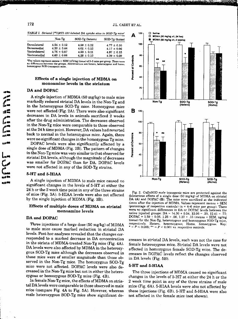
,,,__ A single injection of MDMA (50 mg/kg) to male mice o _
[[f_'_ markedly reduced striatal DA levels in the Non-Tg and Non-Tg SOO-Tg SOO-Tgm in the heterozygous SOD-Tg mice. Homozygous mice (Hetero) (Homo)Iit:_ were not affected (Fig. 2A). There were also significant B ,_ol_ decreases in DA levels in animals sacrificed 2 weeks /
_ after the drug administration. The decreases observed _oo-
1_2 in the Non-Tg mice were comparable to those observedat the 24 h time point. However, DA values had reverted80-
._,. back to normal in the heterozygous mice. Again, therewere no significant changes in the homozygous Tg mice. :o
,_m DOPAC levels were also significantly affected by a o so-osingle dose of MDMA (Fig. 2B). The pattern of changes
L in the Non-Tg mice was very similar to that observed for _ 4o-striatal DA levels, although the magnitude of decreases :_
was smaller for DOPAC than for DA. DOPAC levelswere not affected in any of the SOD-Tg strains.
2O
{' 5-HT and 5-HIAA_lmm o
Non-Tg SOD-Tg SOD-Tgiiiiiiii A single injection of MDMA to male mice caused no (Hetero) (Homo)
significant changes in the levels of 5-HT at either the24 h or the 2 week time point in any of the three strains
Fig. 2. CuZnSOD male transgenic mice are protected against theof mice (Fig. 3A). 5-HIAA levels were also not affected deleterious effects of a single dose (50 mg/kg) of MDMA on striatal
by the single injection of MDMA (Fig. 3B). DA (A) and DOPAC (B). The mice were sacrificed at the indicatedtimes after the injection of MDMA. Values represent means + SEM
Effects of multiple doses of MDMA on striatal (percentage of respective controls) (n = 4--6 mice per group). There
monoamine levels were no significant differences in DA or DOPAC levels between thesaline injected groups: DA = 14.30 + 0.54, 12.06 + .20, 12.41 + .77;
DA and DOPAC DOPAC = 1.59 + 0.06, 1.26 + .06, 1.27 + .15 (means + SEM, ng/mgtissue) for the Non-Tg, heterozygous and homozygous SOD-Tg mice,
Three injections ora large dose (50 mg/kg) of MDMA respectively. Hetero, heterozygous; Homo, homozygous. Key'to male mice cause marked reduction in striatal DA * = P < 0.005; ** = P < 0.001 vs. respective controls.
levels. Post-hoc analyses revealed that the changes cor-
responded to a marked decrease in DA concentration creases in striatal DA levels, such was not the case forin the striata ofMDMA-treated Non-Tg mice (Fig. 4A). female heterozygous mice. Striatal DA levels were notDA levels were also affected by MDMA in the heterozy- affected in homozygous female SOD-Tg mice. The de-gous SOD-Tg mice although the decreases observed in creases in DOPAC levels reflect the changes observedthese mice were of smaller magnitude than those ob- in DA levels (Fig. 5B).served in the Non-Tg mice. The homozygous SOD-Tgmice were not affected. DOPAC levels were also de- 5-HT and 5-HIAA
creased in the Non-Tg mice but not in either the hetero- The three injections of MDMA caused no significantzygous or homozygous SOD-Tg mice (Fig. 4B). changes in the levels of 5-HT at either the 24 h or the
In female Non-Tg mice, the effects of MDMA on stria- 2 week time point in any of the three strains of male
tal DA levels were comparable to those observed in male mice (Fig. 6A). 5-HIAA levels were also not affected bymice (compare Fig. 4A to Fig. 5A). However, whereas these injections (Fig. 6B). 5-HT and 5-HIAA were alsomale heterozygous SOD-Tg mice show significant de- not affected in the female mice (not shown).
Fig. 3. A single injection of MDMA (50 mg/Kg) did not affect striatal Fig. 4. CuZnSod male transgenic mice are protected against the5-HT (A) and 5-HIAA (B) in Non-Tg, heterozygous (Hetero), and homo- deleterious effects of three injections of MDMA (50 rog/kg given 24 h ) ,
zygous (Homo) male SOD-Tg mice sacrificed at the indicated times apart) on striatal DA (A) and DOPAC (B). The mice were sacrificedafter the injection of the drug. Values represent means + SEM (per- two weeks after the last injection. Values represent means + SEM icentage of respective controls) (n = 4-6 mice per group). There were (percentage of respective controls) (n = 4-6 mice per group). Thereno significant differences in 5-HT or 5-HIAA levels between the saline were no significant differences in DA or DOPAC levels between theinjected groups: 5-HT = 0.62 + 0.09, 0.71 + 0.02, 0.64 + 0.03; 5- saline injected groups: DA = 13.09 + 0.78, 13.07 + 0.77, 13.10 + 0.59;HIAA = 0.42 + 0.05; 0.38 + 0.02, 0.44 + 0.02 (means + SEM, ng/mg DOPAC = 1.59 + 0.03, 1.34 + 0.05, 1.34 + 0.08 (means + SEM, ng/ i
tissue) for the Non-Tg, heterozygous, and homozygous SOD-Tg mg tissue) for Non-Tg, heterozygous and homozygous SOD-Tg, respec-mice, respectively, tively. Key: * = P < 0.05; ** = P < 0.0001 vs. respective controls. ?
/ t:
This is the first demonstration that SOD-Tg mice that
DISCUSSION have high levels of CuZnSOD in the brain are protectedOur results show, as previously reported (Logan against the effects of MDMA on the striatal dopaminer- t
et al., 1988; O'Cal]aghan and Miller, 1994), that MDMA gic system. These results are in accord with our recentaffects the DA but not the 5-HT system of mice. This demonstration that the toxic effects of methamphet- t_is in contrast to what has been observed in other mare- amine (METH) on the DA system are attenuated in u
reals (Battaglia et al., 1987; Johnson et al., 1988; Logan CuZnSOD-Tg mice (Cadet et al., 1994). These observa-et al., 1988; O'Hearn et al., 1988; Stone et al., 1986). tions suggest that the neurotoxicity of the amphet-These results suggest that there are significant species amines and of their substituted analogs may indeed bydifferences in both the subacute and chronic effects of media ted, i n_a_,bythe_verproduction oY/h-eE/ipei'°X: (::amphetamine analogs. This is consistent with .out re- -idKa_nion. This could result from-/he formation of 6-cent observations that methamphetamine affects the hydroxydopamine (6-OHDA) as suggested by Seiden ._:DA system of mice more than the 5-HT system (Cadet and Vosmer (1984). Because the neurotoxic effects of 6-et al., 1995). OHDA is associated with the production of the superox- -
J
174 J.L. CADET ET AL.
A ,20.os.... A 140.Ds..,.I_J MDMA (50 rog/kg x3, 2 weeks) [] MDMA (50 rog/kg x3, 2 weeks)
120'100-
100'
m so- mo o;o _ so.o
c _ 60'
o
_/. 40- _.40
20- 20,
_lmm
i_l:_l I 0 Non-Tg SOD-Tg SOD-Tg Non-Tg SOD-Tg SOD-Tg
Fig. 5. CuZn SOD female transgenicmice are protected against the Fig. 6. Effects o£ three injections of MDMA (50 rog/kg given 24 hdeleterious effects of MDMA on striatal DA (A) and DOPAC (B). The apart) on striatal 5-HT (A) and 5-HIAA (B) in Non-Tg heterozygousmice were sacrificed 2 weeks after the last injection. Values represent (Hereto), and homozygous (Homo) male SOD-Tg mice sacrificed atmeans + SEM (percentage of respective controls) (n = 4-6 mice per the indicated times after the injection of the drug. Values representgroup). There were no significant differences in DA or DOPAC levels means + SEM (percentage of respective controls) (n = 4--6 mice perbetween the saline injected groups: DA = 13.04 _- 0.20, 14.30 -+0.37, group). There were no significant differences in 5-HT or 5-H1AAlevels13.88 +_0.48; DOPAC = 1.58 +-0.06, 1.47 *- 0.05, 1.56 + 0.05 between the saline injected groups: 5-HT = 0.56 + 0.05, 0.71 + 0.07,(means + SEM, ng/mg tissue) for Non-Tg, heterozygous, and homozy- 0.78 + 0.05; 5-HIAA = 0.49 + 0.03, 0.56 + 0.07, 0.52 + 0.04gous SOD-Tg, respectively. Key: * = P < 0.005 vs. respective controls. (means + SEM, ng/mg tissue); for Non-Tg, heterozygous, and homozy-
gous SOD-Tg, respectively.
ide radical (Cohen and Heikkila, 1974), the intracellular choIamines, metabolic pathways involving the forma-accumulation of 6-OHDA would result in the over- tion ofcatecholamines from MDMA must also be takenwhelming of the scavenging systems within striata] do- into consideration. For example, it has been reportedpaminergic terminals. Thus, the protective effects ob- that MDMA is converted to dihydroxymethamphet-served in SOD-Tg mice may be due to rapid breakdown amine (Hiratmatsu et al., 1990). This reaction is cyto-of the superoxide radicals secondary to the high levels chrome P-450-dependent and results in a metaboliteof enzyme in these mice (Epstein et al., 1987). This (probably a quinone) that is capable of forming an ad-proposal is supported by the recent report that metham- duct with thio] compounds (Hiratmatsu et al., 1990).phetamine does cause intracellular accumulation of re- This oxidation reaction was inhibited by SOD (Hirat-active oxygen species in vitro (Cubells et al., 1994) and matsu et al., 1190). If left unchecked, these reactionsthat MDMA increases striatal DA release after a single could lead to toxic damage in monoaminergic neuronsadministration of the drug (Nash et al., 1991). in the fashion similar to that suggested for other cate-
In addition to the increased production of superoxide cholamines (Graham, 1978). Thus, these toxic reactions,radicals via the metabolic breakdown of released cate- if they occur in vivo, may be inhibited in the presence
/
MDMA, FREE RADICALS, AND TOXICITY 175
of high levels of endogenous SOD found in the SOD-Tg 3,4-methylenedioxyamphetamine destroy serotonin terminals in ratbrain: Quantification of neurodegeneration by measurement of
mice, as observed in the in vitro situation in the pres- [SH]paroxetine-labeled serotonin uptake sites. J. Pharmacol. Exp.
ence of exogenous SOD (Hiratmatsu et al., 1990). Ther., 242:911-916.The observation that homozygous SOD-Tg mice show Boveris A., and Chance B. (1973) The mitochondrial generation ofhydrogen peroxide: General properties and effect of hyperbaric oxy-
complete protection whereas there was some DA deple- gen. Biochem. J., 134:707-716.
tion in the heterozygous SOD-Tg mice when DA levels Brodkin J., Malyala A., and Nash J.F. (1993) Effect of acute mono-amine depletion on 3,4-methylenedioxymethamphetamine-induced
were measured 24 h after drug administration suggests neurotoxicity.Pharmacol. Biochem.Behav.,45:647-653.that there might be a dose-response phenomenon so Cadet J.L. (1988) A unifying hypothesis of movement and madness:Free radical involvement of free radicals in disorders of the isoden-
that very high levels of SOD are needed to counteract dritic core. Med. Hypotheses, 27:87-94.the effects of the oxidative stress that might be observed Cadet J.L., Ladenheim B., Baum I., Carlson E., and Epstein C. (1994)
after MDMA injections. These results also suggest that CuZn-superoxide dismutase (CuZnSOD) transgenic mice show re-sistance to the lethal effects of methylenedioxyamphetamine (MDA)
only stressful events might help to uncover differences and of methylenedioxymethamphetamine (MDMA). Brain Res.,
between the Non-Tg, heterozygous as well as homozy- 655:259-262.Cadet J.L., Sheng P., Ali S., Rothman R., Carlson E., and Epstein C.J.
gous SOD-Tg mice since there were no differences in (1994) Attenuation of methamphetamine-induced neurotoxicity inthe number of striatal [_25I]RTI-121-labeled DA uptake copper/zinc superoxide dismutase transgenic mice.J. Neurochem.,
i sites between mice that had not gotten any drug injec-62:38O-383.
Chan P.H., Yang G.Y., Chen S.F., Carlson E., and Epstein C.J. (1991)
_i tions (see Table I). This is important since the DA sys- Cold-induced brain edema and infarction are reduced in transgenic
tem is known to play an important role in the biochemi- mice overexpressing CuZn-superoxide dismutase. Ann. Neurol.,29:482-486.
cai effects of the amphetamines (Marek et al., 1990; CohenG.,andHeikkilaR.E.(1974)Thegenerationofhydrogenperox-ide, superoxide radical and hydroxyl radical by hydroxydopamine,
Schmidt et al., 1991; Stone et al., 1988). dialuric acid, and related cytotoxic agents. J. Biol. Chem.,
Finally, it is also of interest that female Non-Tg mice 249:2447-2452.
ii showless ofa decrement in striatal DAlevels thanmale Commins D.L., Vosmer G., Virus R.M., Woolverton W.L., SchusterC.R., and Seiden L.S. (1987) Biochemical and histological evidence
Non-Tg mice after the administration of three injections that methylenedioxymethamphetamine (MDMA) is toxic to neurons
_ of MDMA (compare Fig. 4 to Fig. 5). Moreover, male in rat brain. J. Pharmacol. Exp. Ther., 241:338-345.
i_ heterozygous mice were somewhat more affected than Cubells J.F., Rayport S., Rajendran G., and Sulzer D. (1994) Metham-phetamine neurotoxicity involves vacuolation of endocytic organ~female heterozygous mice after receiving multiple doses elles and dopamine-dependent intracellular oxidative stress. J. Neu-
of MDMA (compare Fig. 4 to Fig. 5). Thus both Non- rosci., 14:2260-2271.Epstein C.J., Avraham K.B., Lovett M., Smith S., Elroy-Stein, O.,
Tg and SOD-Tg female mice show a similar pattern in Rotman G., Bry C., and Groner Y (1987) Transgenic mice with in-
their response to MDMA. These results are consistent creased Cu/Zn-superoxidedismutase activity:Animalmodelofdos-age effects in Down syndrome. Proc. Natl. Acad. Sci. U.S.A.,
with the gender effects on lethality that we had ob- 84:8044--8048.
served previously (Cadet et al., 1994) and provide fur- Epstein C.J., Chan P.H., and Cadet J.L. (1994) CuZn-superoxide dis-
ther support for the notion of an endocrine-oxidant state mutase transgenic mice in the study of the role Of oxidative stress'_1 in neurodegenerative disorders. In: Neurodegenerative Diseases.interaction as previously suggested (Cadet et al., 1994). London, Jolles G. and Stutzmann J.M., eds. Academic Press, New
_: This may be related to the differences in prevalence of York,Chapter 14,pp. 239-252.Freeman B.A., and Crape J.D. (1982) Biology &disease: Free RadicalsParkinson's disease between men and women (Mayeux and tissue injury. Lab. Invest., 47:412-426.
et al., 1992). Further studies will need to evaluate the FridovichI. (1986)Biologicaleffectsof the superoxide radical. Arch.Biochem, Biophys., 247:1-11.
relationship between gender and oxidative stress in Graham D.C., Tiffany S.M., Bell W.R., and Gutnecht W.F. (1978) Auto-
mammalian systems. In any case, the present study oxidation versus covalent finding of quinones as the mechanism of
does support the view that free radicals are involved toxicity of dopamine, 6-hydroxydopamine, and related compoundstowards c1300 neuroblastoma cells in vitro. Molec. Pharmacol.,in neurotoxin-induced effects on the mammalian dopa- 14:644-653.
minergic systems (Cadet, 1988; Cadet et al., 1994; Cu- Halliwell B. (1992) Reactive oxygen species and the central nervoussystem. J. Neurochem., 59:1609-1623.
bells et al., 1994; Olanow, 1993). Hardman H.F., Haavik C.O., and Seevers M.H. (1973) Relationshipof the structure of mescaline and seven analogs to toxicity and
ACKNOWLEDGMENTS behavior in five species of laboratory animals. Toxicol. Appl. Phar-macol., 25:299-309
The authors thank Dr. Michael J. Kuhar for his con- Hiratmatsu M., Kumagai Y., Unger S.E., and Cho A.K. (1990) Metabo-
tinuous support and the staff of our Animal Care Facil- lism ofmethylenedioxymethamphetamine: Formation ofdihydroxy-methamphetamine and a quinone identified as its glutathione ad-
i at the Intramural Research Programs of NIH/NIDA duct. J. Pharmacol. Exp. Ther., 254:521-527.ity
for their impeccable care of the animals. This work was Insel T.T., Battaglia G., Johannessen J.N., Marra S., and De SouzaE.B. (1989) 3,4-methylenedioxymethamphetamine ("Ecstasy") selec-partially supported by NIH Grant HD 24605 and AG- tively destroys brain serotonin terminals in rhesus monkeys. J.08938 and a grant from the Lucille P. Markey Charita- Pharmacol Exp. Ther., 249:713-720.
Janssen Y.M.W., van Houten B., Borm P.J.A., and Mossman B.T. (1993)ble Trust to the UCSF Program in Biological Sciences. Cell and tissue responses to oxidativedamage. Lab. Investigation,
69:261-274.REFERENCES Johnson M., Letter A.A., Merchant K., Hanson G.R., and Gibb J.W.
(1988) Effects of 3,4-methylenedioxymethamphetamine on central
Ames B.N., Shinenaga M.K., and Hagen T. (1993) Oxidants, antioxi- serotonergic, dopaminergic and nigral neurotensin systems of thedants, and the degenerative diseases of aging. Proc. Natl. Acad. Sci. rat. J. Pharmacol. Exp. Ther., 244:977-982.U.S.A. 90:7915-7922. Kedziora J., and Bartosz G. (1988) Down's syndrome: A pathology
Battaglia G., Yeh S.Y., O'Hearn E., Molliver M.E., Kuhar M.J., and involving the lack of balance of reactive oxygen species. Free Rad.De Souza E.B. (1987) 3,4-methylenedioxymethamphetamine and Biol. Med. 4:317-330.
176 J.L. CADET ET AL.
Kinouchi H., Epstein C.J., Mizui Ti, Carlson E., Chen S., and Chan A., Donaldson D., Goto J., O'Regan J.P., Deng H.K., Gusella J.S.,P.H. (1991) Attenuation of focal cerebral ischemia in transgenic mice Horvitz H.R., and Brown R.H. (1993) Mutations in Cu/Zn superoxideoverexpressing CuZn-superoxide dismutase. Proc. Natl. Acad. Sci. dismutase gene are associated with familial amyotrophic lateralU.S.A., 88:11158--11162. sclerosis. Nature 362:59--62.
Logan B.J., Laverty R., Sanderson W.D., and Yee Y.B. (1988) Differ- Schmidt C.J. (1987) Neurotexicity of the psychedelic amphetamine,ences between rats and mice in MDMA (methylenedioxymethylam- methylenedioxymethamphetamine. J. Pharmacol. Exp. Then,phetamine) neurotexicity. Eur. J. Pharmacol., 152:227-234. 240:1-7.
Marek G.J., Vosmer G., and Seiden L.S. (1990) Dopamine uptake Schmidt C.J., Taylor V.L., Abbate G.M., and Nieduzak T.R. (1991) 5-inhibitors block long-term neurotoxic effects of methamphetamine HT2 antagonists stereoselectively prevent the neurotoxicity of 3,4-upon dopaminergic neurons. Brain Res., 513:274-279. methylenedioxymethamphetamine by blocking the acute stimula-
Mayeux R., Denaro J., Hemenegildo N., Marder K., Tang M.X., Cote tion ofdopamine synthesis: Reversal by L-DOPA. J. Pharmacol. Exp.L.J., and Stern Y. (1992) A population-based investigation of Parkin- Ther., 256:230-235.
son's disease with and without dementia. Arch. Neurol., 49:492-497. Seiden L.S., and Vosmer G. (1984) Formation of 6-hydroxydopamineNash J.F., and Nichols D.E. (1991) Microdialysis studies on 3,4-methy- in caudate nucleus of rat brain after a single large dose of metham-
lenedioxyamphetamine and structurally related analogues. Eur. J. phetamine. Pharmacol. Biochem. Behav., 21:29-31.Pharmacol., 200:53-58. Stone D.M., Hanson G.R., and Gibb J.W. (1987) Differences in the
O'Callaghan J.P., and Miller D.B. (1994) Neurotoxicity profiles of sub- serotonergic of methylenedioxymethamphetamine in mice and rats.stituted amphetamines in the C57BL/6J mouse. J. Pharmacol. Exp. Neuropharmacology, 26:1657-1661.Ther., 270:741-751. Stone D.M., Johnson M., Hanson G.R., and Gibb J.W. (1988) Role of
,_ O'Hearn E., Battaglia G., De Souza E.B., Kuhar M.J., and Molliver endogenous dopamine in the central serotonergic deficits induced
M.E. (1988) Methylenedioxyamphetamine and methylenedioxy- by 3,4-methylenedioxymethamphetamine. J. Pharmacol. Exp. Ther.f_ methamphetamine (MDMA) cause selective ablation ofserotenergic 247:79--87.
[_ axon terminals in forebrain: Immunocytochemical evidence for neu- Stone I).M., Stahl D.C, Hanson G.R., and Gibb J.W. (1986) The effectsm rotoxicity. J. Neuros., 8:2788-2903. of 3,4-methylenedioxymethamphetamine (MDMA) and 3,4-methy-Olanow C.W. (1993) A radical hypothesis for neurodegeneration, lenedioxyamphetamine (MI)A) on monoaminergic systems in the
I1[,_ TINS, 16:439-444. rat brain. Eur. J. Pharmacol., 128:41-48.Przedborski S., Kostic V., Jackson-Lewis V., Naini A.B., Simonetti S., Touati D. (1988) Molecular genetics of superoxide dismutases. Free
_dm Fahn S., Epstein C., and Cadet J.L. (1992) Transgenic mice with Radic Biol. Med., 5:393-402._m increased Cu/Zn-superoxide dismutase activity are resistant to N- Zaczek R., and Coyle J.T. (1982) Rapid and simple method for measur-
methyl-4-phenyl- 1,2,3,6-tetrahydropyridine-induced neurotexicity, ing biogenic amine and metabolites in brain homogenates by HPLC-
I_ J. Neuresci., 12:1659-1667. electrochemical detection. J. NeuralTransm. 53:1-5.