Page 1
1
Copyright © 2009 Pearson Education, Inc.
CAMPBELL
BIOLOGYReece • Urry • Cain • Wasserman • Minorsky • Jackson
© 2014 Pearson Education, Inc.
TENTH
EDITION
CAMPBELL
BIOLOGYReece • Urry • Cain • Wasserman • Minorsky • Jackson
© 2014 Pearson Education, Inc.
TENTH
EDITION
16 DNA: The Molecular Basis of Inheritance
Lecture Presentation by
Dr Burns
NVC Biol 120
DNA
Deoxyribonucleic acid – DNA
The blueprint to making proteins!!!
Chromosomes located inside the nucleus
contains long coiled strands of DNA
DNA’s Discovery
Watson and Crick
Rosalind Franklin →
The Players
Crick: Ph.D. student at Cambridge in England working on X-ray Crystallography of the protein hemoglobin
Watson: Young American scientist visiting the lab to do some work on a protein
Both were interested in unraveling the secret of DNA’s structure – it was not what they were supposed to be working on
Wilkins: Working on DNA structure, crystallized
DNA fibers
Franklin: Working at the same university as
Wilkins, just down the hall. Did the X-ray
Crystallography on Wilkins DNA fibers
Linus Pauling: discovered the three dimensional
structure of proteins know as alpha helixes
Chargaff: Discovered that A=T and G=C
Adenine levels always equal thymine levels,
Guanine levels always equal cytosine
Page 2
2
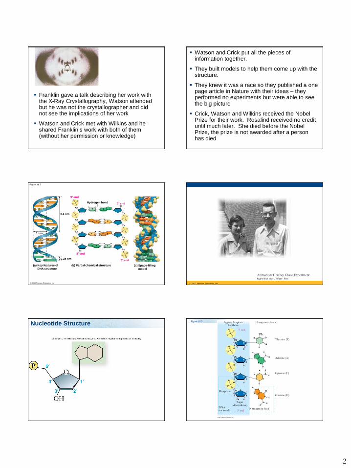
Franklin gave a talk describing her work with the X-Ray Crystallography, Watson attended but he was not the crystallographer and did not see the implications of her work
Watson and Crick met with Wilkins and he shared Franklin’s work with both of them (without her permission or knowledge)
Watson and Crick put all the pieces of information together.
They built models to help them come up with the structure.
They knew it was a race so they published a one page article in Nature with their ideas – they performed no experiments but were able to see the big picture
Crick, Watson and Wilkins received the Nobel Prize for their work. Rosalind received no credit until much later. She died before the Nobel Prize, the prize is not awarded after a person has died
© 2014 Pearson Education, Inc.
Figure 16.7
(a) Key features of
DNA structure
(b) Partial chemical structure
0.34 nm
3′ end
5′ endT
T
T
A
A
A
C
C
C
G
G
G
AT1 nm
TA
C G
CG
AT3.4 nm
CG
CG
C G
C G
3′ end
5′ end
Hydrogen bond
T A
G C
A T
C G
(c) Space-filling
model
© 2011 Pearson Education, Inc.
Animation: Hershey-Chase ExperimentRight-click slide / select “Play”
Fig. 14.4
Nucleotide StructureFigure 16.5 Sugar–phosphate
backboneNitrogenous bases
Thymine (T)
Adenine (A)
Cytosine (C)
Guanine (G)
Nitrogenous base
Phosphate
DNA nucleotide
Sugar(deoxyribose)
3 end
5 end
Page 3
3
DNA Structure
Nucleotides that build DNA have:
One phosphate (ATP has three)
One sugar = deoxyribose
One base.
The nucleotides vary in the type of base – there
are four different bases in DNA: Adenine (A),
Thymine (T), Guanine (G), Cytosine (C)
There is a 5’ end and a 3’ end
Fig. 14.3-1
Page 4
4
© 2011 Pearson Education, Inc.
Animation: DNA and RNA Structure Right-
click slide / select “Play”
Bonds
The sugars and phosphates link together by covalent bonds to form the rail on the outside = phosphodiester linkage.
The sugars are covalently bound to a base
The complementary bases are attracted to each other by hydrogen bonds
Double Helix
Two strands bonded together by hydrogen
bonds between the bases = weak bonds
Each strand has nucleotides bonded together
covalently by the phosphate and the sugar
Base pairs are two nucleotides, one on each
complementary strand of a DNA molecule
Base Pairs
The bases pair up in a specific manner:
Adenine (A) pairs with Thymine (T)
Guanine (G) pairs with Cytosine (C)
Purines: Adenine and Guanine
Pyrimidines: Thymine and Cytosine
Page 5
5
Figure 16.8
Sugar
Sugar
Sugar
Sugar
Adenine (A) Thymine (T)
Guanine (G) Cytosine (C)
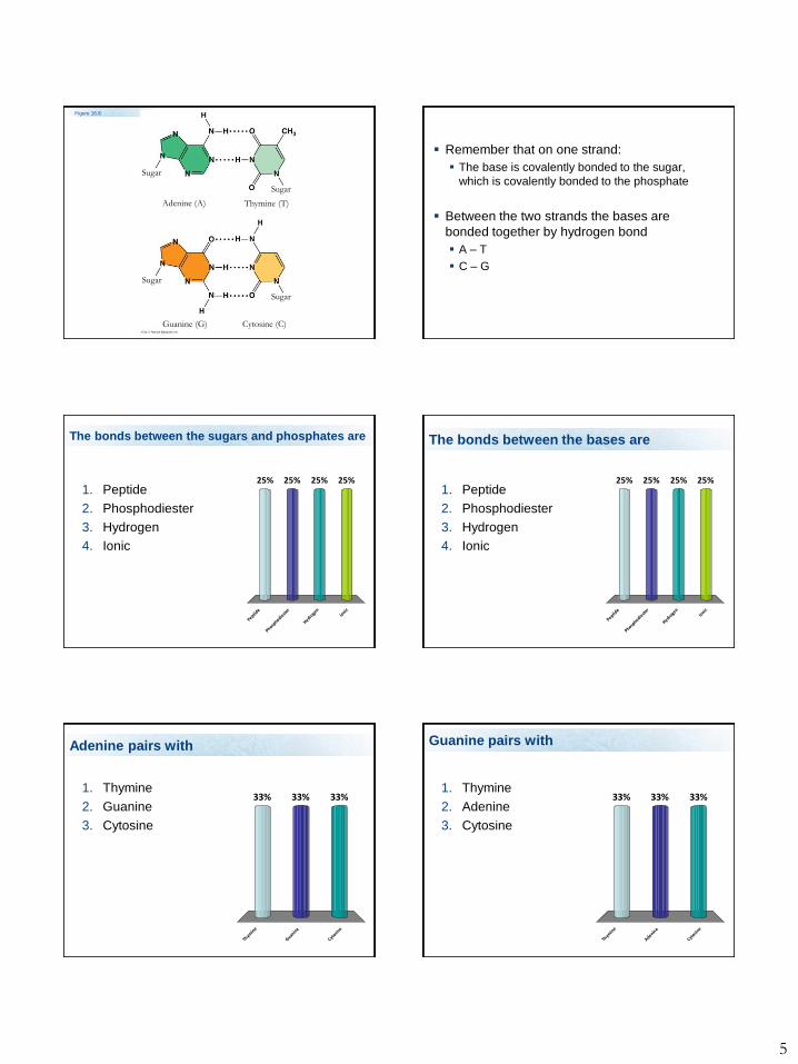
Remember that on one strand:
The base is covalently bonded to the sugar,
which is covalently bonded to the phosphate
Between the two strands the bases are
bonded together by hydrogen bond
A – T
C – G
The bonds between the sugars and phosphates are
1. Peptide
2. Phosphodiester
3. Hydrogen
4. Ionic
Peptide
Phosphodie
ster
Hydroge
n
Ionic
25% 25%25%25%
The bonds between the bases are
1. Peptide
2. Phosphodiester
3. Hydrogen
4. Ionic
Peptide
Phosphodie
ster
Hydroge
n
Ionic
25% 25%25%25%
Adenine pairs with
1. Thymine
2. Guanine
3. Cytosine
Thym
ine
Guanin
e
Cytosin
e
33%33%33%
Guanine pairs with
1. Thymine
2. Adenine
3. Cytosine
Thym
ine
Adenine
Cytosin
e
33%33%33%
Page 6
6
The bases are bound to
1. Sugars
2. Phosphates
Suga
rs
Phosphate
s
50%50%
The bases are bound to the sugar by this kind of bond
1. Covalent
2. Phosphodiester
3. Hydrogen
4. Ionic
Covale
nt
Phosphodie
ster
Hydroge
n
Ionic
25% 25%25%25%
The sugar in DNA is
1. Ribose
2. Deoxyribose
3. Glucose
4. Cellulose
Ribose
Deoxyrib
ose
Gluco
se
Cellulo
se
25% 25%25%25%
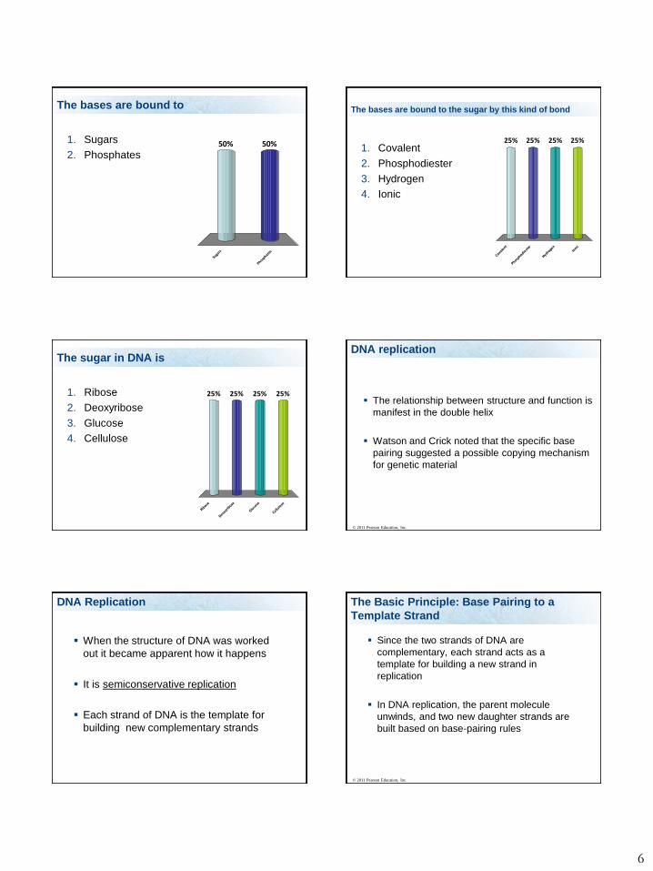
DNA replication
The relationship between structure and function is
manifest in the double helix
Watson and Crick noted that the specific base
pairing suggested a possible copying mechanism
for genetic material
© 2011 Pearson Education, Inc.
DNA Replication
When the structure of DNA was worked
out it became apparent how it happens
It is semiconservative replication
Each strand of DNA is the template for
building new complementary strands
The Basic Principle: Base Pairing to a
Template Strand
Since the two strands of DNA are
complementary, each strand acts as a
template for building a new strand in
replication
In DNA replication, the parent molecule
unwinds, and two new daughter strands are
built based on base-pairing rules
© 2011 Pearson Education, Inc.
Page 7
7
© 2011 Pearson Education, Inc.
Animation: DNA Replication OverviewRight-click slide / select “Play”
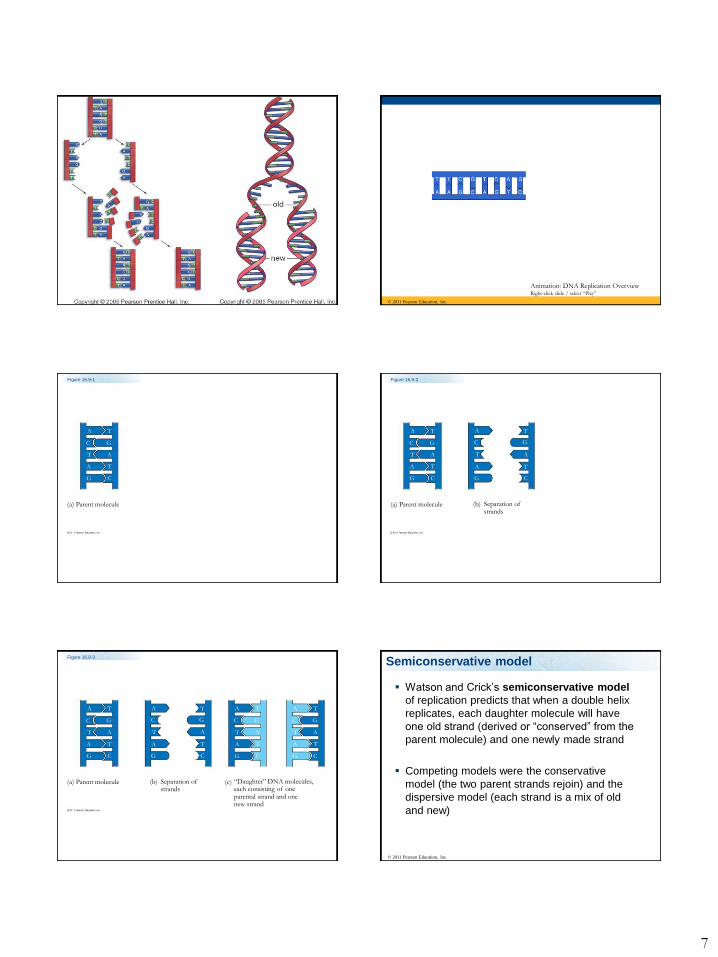
Figure 16.9-1
(a) Parent molecule
A
A
A
T
T
T
C
C
G
G
Figure 16.9-2
(a) Parent molecule (b) Separation ofstrands
A
A
A
A
A
A
T
T
T
T
T
T
C
C
C
C
G
G
G
G
Figure 16.9-3
(a) Parent molecule (b) Separation ofstrands
(c) “Daughter” DNA molecules,each consisting of oneparental strand and onenew strand
A
A
A
A
A
A
A
A
A
A
A
A
T
T
T
T
T
T
T
T
T
T
T
T
C
C
C
C
C
C
C
C
G
G
G
G
G
G
G
G
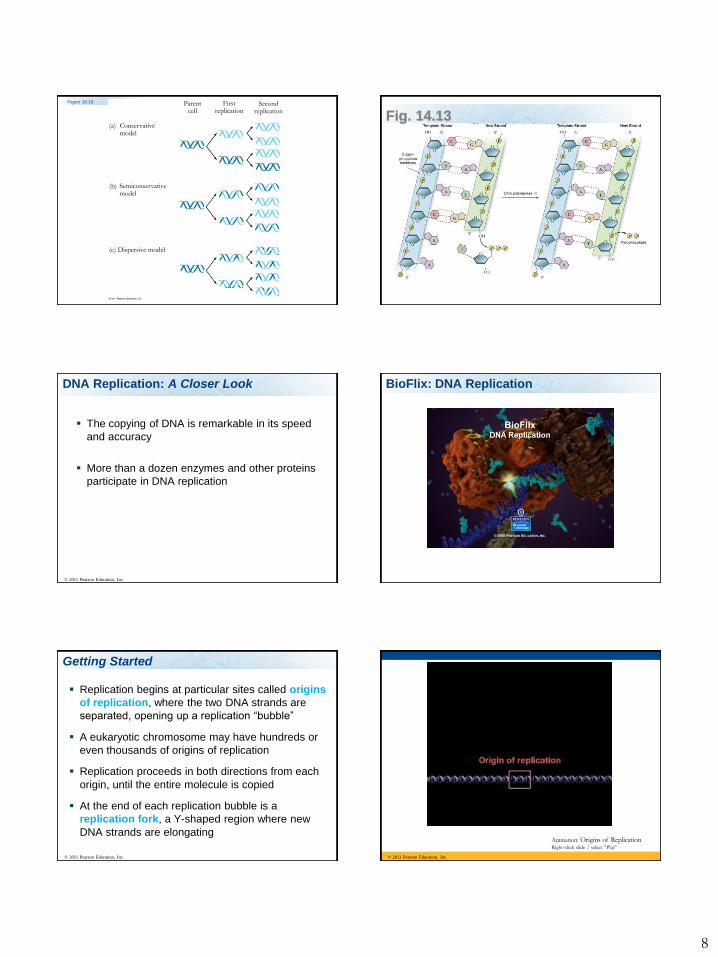
Semiconservative model
Watson and Crick’s semiconservative model
of replication predicts that when a double helix
replicates, each daughter molecule will have
one old strand (derived or “conserved” from the
parent molecule) and one newly made strand
Competing models were the conservative
model (the two parent strands rejoin) and the
dispersive model (each strand is a mix of old
and new)
© 2011 Pearson Education, Inc.
Page 8
8
Figure 16.10
(a) Conservativemodel
(b) Semiconservativemodel
(c) Dispersive model
Parentcell
Firstreplication
Secondreplication
Fig. 14.13
DNA Replication: A Closer Look
The copying of DNA is remarkable in its speed
and accuracy
More than a dozen enzymes and other proteins
participate in DNA replication
© 2011 Pearson Education, Inc.
BioFlix: DNA Replication
Getting Started
Replication begins at particular sites called origins
of replication, where the two DNA strands are
separated, opening up a replication “bubble”
A eukaryotic chromosome may have hundreds or
even thousands of origins of replication
Replication proceeds in both directions from each
origin, until the entire molecule is copied
At the end of each replication bubble is a
replication fork, a Y-shaped region where new
DNA strands are elongating
© 2011 Pearson Education, Inc. © 2011 Pearson Education, Inc.
Animation: Origins of ReplicationRight-click slide / select “Play”
Page 9
9
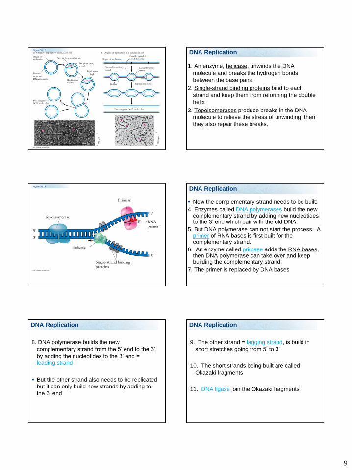
Figure 16.12(a) Origin of replication in an E. coli cell (b) Origins of replication in a eukaryotic cell
Origin ofreplication
Parental (template) strand
Double-strandedDNA molecule
Daughter (new)strand
Replicationfork
Replicationbubble
Two daughterDNA molecules
Origin of replication
Double-strandedDNA molecule
Parental (template)strand
Daughter (new)strand
Bubble Replication fork
Two daughter DNA molecules
0.5
m
0.2
5
m
DNA Replication
1. An enzyme, helicase, unwinds the DNA
molecule and breaks the hydrogen bonds
between the base pairs
2. Single-strand binding proteins bind to each
strand and keep them from reforming the double
helix
3. Topoisomerases produce breaks in the DNA
molecule to relieve the stress of unwinding, then
they also repair these breaks.
DNA Replication
Figure 16.13
Topoisomerase
Primase
RNAprimer
Helicase
Single-strand bindingproteins
5
3
5
53
3
DNA Replication
Now the complementary strand needs to be built:
4. Enzymes called DNA polymerases build the new complementary strand by adding new nucleotides to the 3’ end which pair with the old DNA.
5. But DNA polymerase can not start the process. A primer of RNA bases is first built for the complementary strand.
6. An enzyme called primase adds the RNA bases, then DNA polymerase can take over and keep building the complementary strand.
7. The primer is replaced by DNA bases
DNA Replication
8. DNA polymerase builds the new
complementary strand from the 5’ end to the 3’,
by adding the nucleotides to the 3’ end =
leading strand
But the other strand also needs to be replicated
but it can only build new strands by adding to
the 3’ end
DNA Replication DNA Replication
9. The other strand = lagging strand, is build in
short stretches going from 5’ to 3’
10. The short strands being built are called
Okazaki fragments
11. DNA ligase join the Okazaki fragments
DNA Replication
Page 10
10
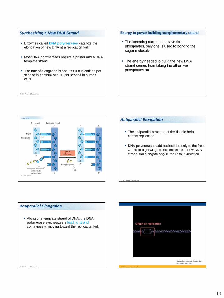
Synthesizing a New DNA Strand
Enzymes called DNA polymerases catalyze the
elongation of new DNA at a replication fork
Most DNA polymerases require a primer and a DNA
template strand
The rate of elongation is about 500 nucleotides per
second in bacteria and 50 per second in human
cells
© 2011 Pearson Education, Inc.
Energy to power building complementary strand
The incoming nucleotides have three
phosphates, only one is used to bond to the
sugar molecule
The energy needed to build the new DNA
strand comes from taking the other two
phosphates off.
Figure 16.14
New strand Template strand
Sugar
Phosphate Base
Nucleosidetriphosphate
DNApolymerase
Pyrophosphate
5
5
5
5
3
3
3
3
OH
OHP P i
2 P i
A
A
A
A
T T
T
C
C
C
C
C
C
G
G
G
G
Antiparallel Elongation
The antiparallel structure of the double helix
affects replication
DNA polymerases add nucleotides only to the free
3end of a growing strand; therefore, a new DNA
strand can elongate only in the 5to 3direction
© 2011 Pearson Education, Inc.
Along one template strand of DNA, the DNA
polymerase synthesizes a leading strand
continuously, moving toward the replication fork
© 2011 Pearson Education, Inc.
Antiparallel Elongation
© 2011 Pearson Education, Inc.
Animation: Leading Strand Right-
click slide / select “Play”
Page 11
11
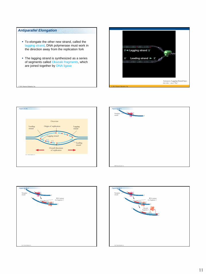
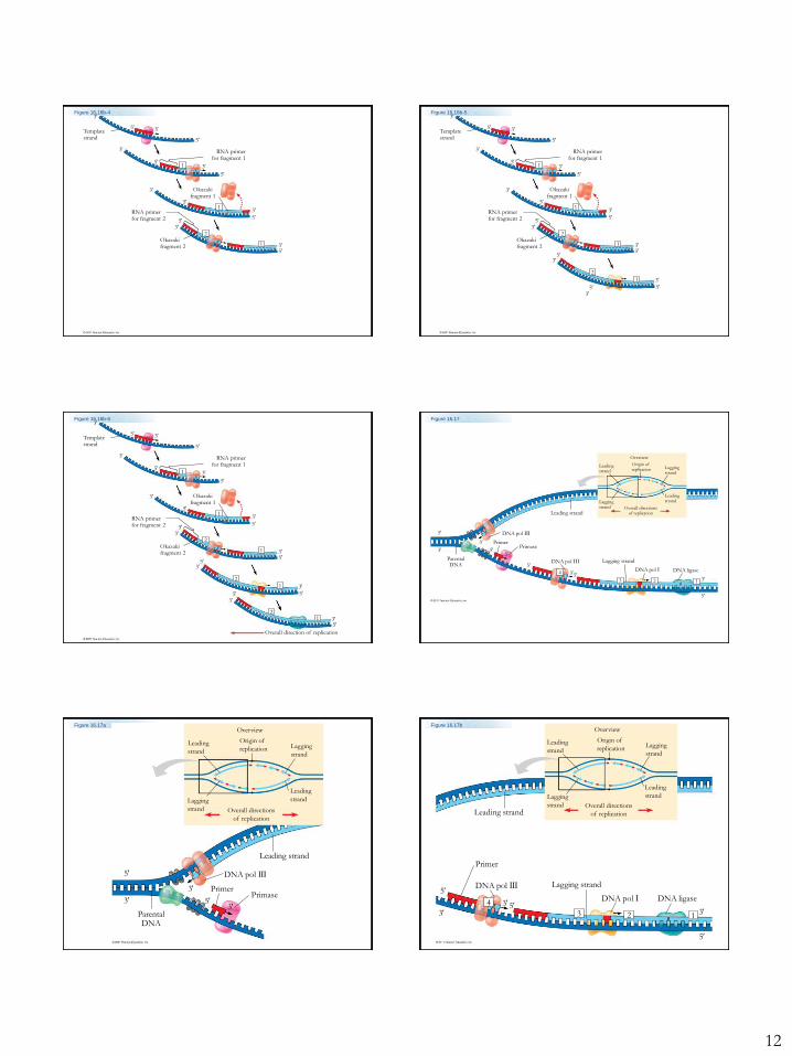
Antiparallel Elongation
To elongate the other new strand, called the
lagging strand, DNA polymerase must work in
the direction away from the replication fork
The lagging strand is synthesized as a series
of segments called Okazaki fragments, which
are joined together by DNA ligase
© 2011 Pearson Education, Inc. © 2011 Pearson Education, Inc.
Animation: Lagging Strand Right-
click slide / select “Play”
Figure 16.16a
Origin of replication
Overview
Leadingstrand
Leadingstrand
Laggingstrand
Lagging strand
Overall directionsof replication
12
Figure 16.16b-1
Templatestrand
3
35
5
Figure 16.16b-2
Templatestrand
RNA primerfor fragment 1
3
3
3
3
5
5
5
5
1
Figure 16.16b-3
Templatestrand
RNA primerfor fragment 1
Okazakifragment 1
3
3
3
3
3
3
5
5
5
5
5
5
1
1
Page 12
12
Figure 16.16b-4
Templatestrand
RNA primerfor fragment 1
Okazakifragment 1
RNA primerfor fragment 2
Okazakifragment 2
3
3
3
3
3
3
3
3
5
5
5
5
5
55
5
2
1
1
1
Figure 16.16b-5
Templatestrand
RNA primerfor fragment 1
Okazakifragment 1
RNA primerfor fragment 2
Okazakifragment 2
3
3
3
3
3
3
3
3
3
3
3
5
5
5
5
5
55
55
55
2
2
1
1
1
1
Figure 16.16b-6
Templatestrand
RNA primerfor fragment 1
Okazakifragment 1
RNA primerfor fragment 2
Okazakifragment 2
Overall direction of replication
3
3
3
3
3
3
3
3
3
3
3
3
5
5
5
5
5
55
55
55
5
2
2
21
1
1
1
1
Figure 16.17
Overview
Leadingstrand
Origin of replication Lagging
strand
LeadingstrandLagging
strand Overall directionsof replicationLeading strand
DNA pol III
DNA pol III Lagging strand
DNA pol I DNA ligase
PrimerPrimase
ParentalDNA
5
5
5
5
5
3
3
3
333 2 1
4
Figure 16.17aOverview
Leading
strand
Origin of
replication Lagging
strand
Leading
strandLagging
strand Overall directions
of replication
Leading strand
DNA pol III
PrimerPrimase
ParentalDNA
5
53
3
3
Overview
Leading
strand
Origin of
replicationLagging
strand
Leading
strandLagging
strand Overall directions
of replicationLeading strand
Primer
DNA pol III
DNA pol I
Lagging strand
DNA ligase5
5
5
33
3 3
4
2 1
Figure 16.17b
Page 13
13
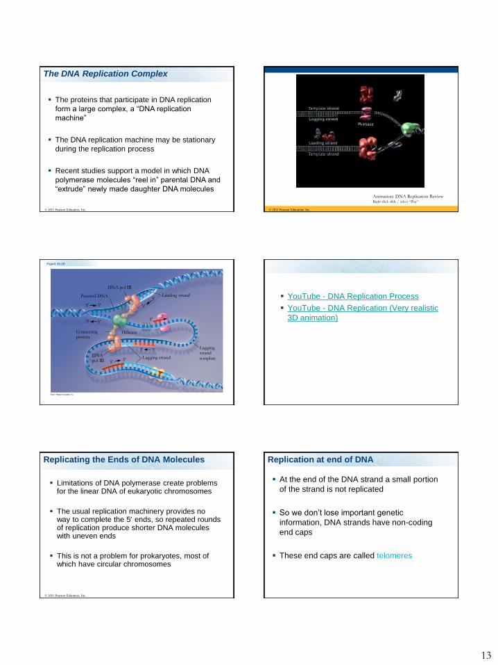
The DNA Replication Complex
The proteins that participate in DNA replication
form a large complex, a “DNA replication
machine”
The DNA replication machine may be stationary
during the replication process
Recent studies support a model in which DNA
polymerase molecules “reel in” parental DNA and
“extrude” newly made daughter DNA molecules
© 2011 Pearson Education, Inc. © 2011 Pearson Education, Inc.
Animation: DNA Replication ReviewRight-click slide / select “Play”
Figure 16.18
Parental DNA
DNA pol III
Leading strand
Connectingprotein
Helicase
Lagging strandDNA pol III
Laggingstrandtemplate
5
5
5
5
5
5
3 3
33
3
3
YouTube - DNA Replication Process
YouTube - DNA Replication (Very realistic
3D animation)
Replicating the Ends of DNA Molecules
Limitations of DNA polymerase create problems for the linear DNA of eukaryotic chromosomes
The usual replication machinery provides no way to complete the 5 ends, so repeated rounds of replication produce shorter DNA molecules with uneven ends
This is not a problem for prokaryotes, most of which have circular chromosomes
© 2011 Pearson Education, Inc.
Replication at end of DNA
At the end of the DNA strand a small portion
of the strand is not replicated
So we don’t lose important genetic
information, DNA strands have non-coding
end caps
These end caps are called telomeres
Page 14
14
Figure 16.20
Ends of parentalDNA strands
Leading strand
Lagging strand
Last fragment Next-to-last fragment
Lagging strand RNA primer
Parental strandRemoval of primers andreplacement with DNAwhere a 3 end is available
Second roundof replication
Further roundsof replication
New leading strand
New lagging strand
Shorter and shorter daughter molecules
3
3
3
3
3
5
5
5
5
5Figure 16.20a
Ends of parentalDNA strands
Leading strand
Lagging strand
Last fragment Next-to-last fragment
Lagging strand RNA primer
Parental strandRemoval of primers andreplacement with DNAwhere a 3 end is available
3
3
3
5
5
5
Figure 16.20b
Second roundof replication
Further roundsof replication
New leading strand
New lagging strand
Shorter and shorter daughter molecules
3
3
3
5
5
5
Telomeres
Eukaryotic chromosomal DNA molecules have
special nucleotide sequences at their ends
called telomeres
They postpone the erosion of genes near the
ends of DNA molecules
It has been proposed that the shortening of
telomeres is connected to aging
© 2011 Pearson Education, Inc.
Figure 16.21
1 m
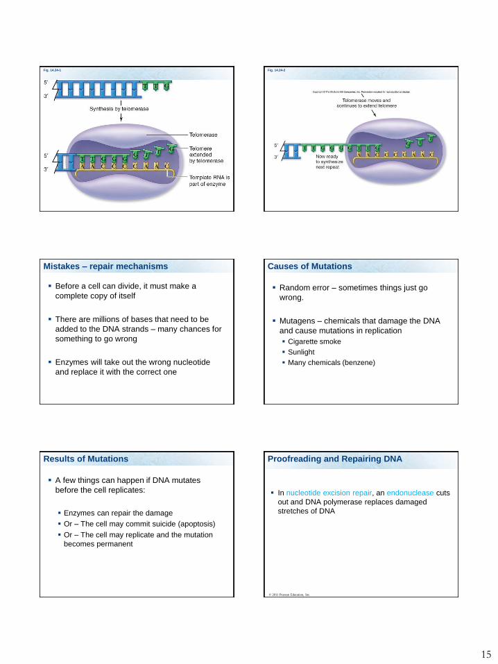
Replication at end of DNA
Telomerase build the telomeres.
Embryos have high telomerase activity, as
you age you lose this activity.
Cancer cells have telomerase activity
Page 15
15
Fig. 14.24-1 Fig. 14.24-2
Mistakes – repair mechanisms
Before a cell can divide, it must make a
complete copy of itself
There are millions of bases that need to be
added to the DNA strands – many chances for
something to go wrong
Enzymes will take out the wrong nucleotide
and replace it with the correct one
Causes of Mutations
Random error – sometimes things just go
wrong.
Mutagens – chemicals that damage the DNA
and cause mutations in replication
Cigarette smoke
Sunlight
Many chemicals (benzene)
Results of Mutations
A few things can happen if DNA mutates
before the cell replicates:
Enzymes can repair the damage
Or – The cell may commit suicide (apoptosis)
Or – The cell may replicate and the mutation
becomes permanent
Proofreading and Repairing DNA
In nucleotide excision repair, an endonuclease cuts
out and DNA polymerase replaces damaged
stretches of DNA
© 2011 Pearson Education, Inc.
Page 16
16
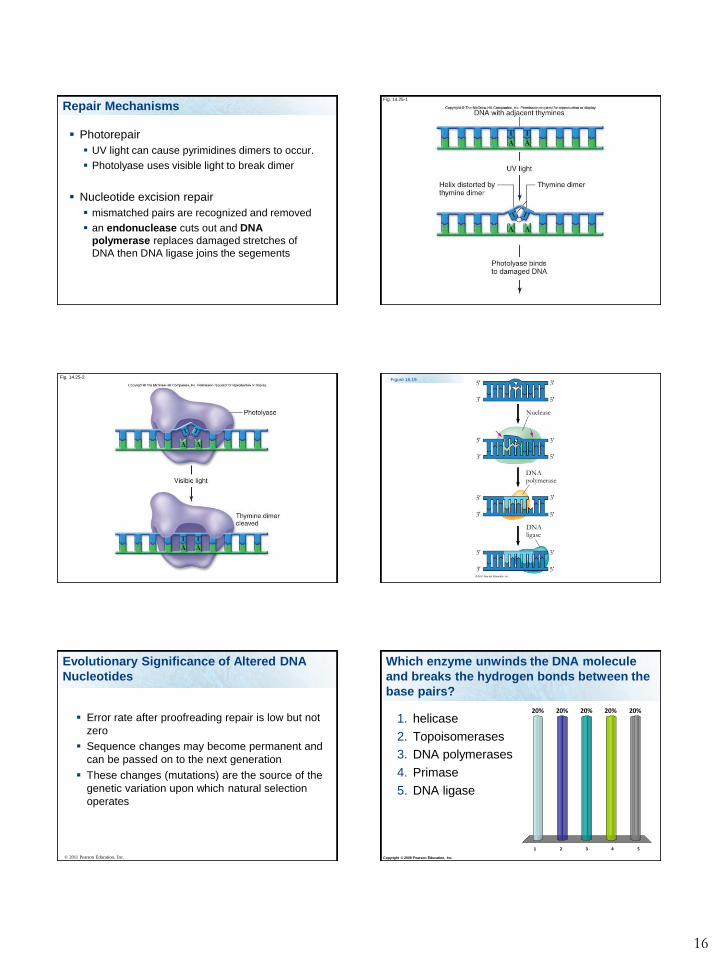
Repair Mechanisms
Photorepair
UV light can cause pyrimidines dimers to occur.
Photolyase uses visible light to break dimer
Nucleotide excision repair
mismatched pairs are recognized and removed
an endonuclease cuts out and DNA
polymerase replaces damaged stretches of
DNA then DNA ligase joins the segements
Fig. 14.25-1
Fig. 14.25-2Figure 16.19
Nuclease
DNA polymerase
DNA ligase
5
5
5
5
5
5
5
5
3
3
3
3
3
3
3
3
Evolutionary Significance of Altered DNA
Nucleotides
Error rate after proofreading repair is low but not
zero
Sequence changes may become permanent and
can be passed on to the next generation
These changes (mutations) are the source of the
genetic variation upon which natural selection
operates
© 2011 Pearson Education, Inc. Copyright © 2009 Pearson Education, Inc.
Which enzyme unwinds the DNA molecule
and breaks the hydrogen bonds between the
base pairs?
1 2 3 4 5
20% 20% 20%20%20%
1. helicase
2. Topoisomerases
3. DNA polymerases
4. Primase
5. DNA ligase
Page 17
17
Copyright © 2009 Pearson Education, Inc.
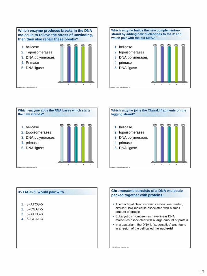
Which enzyme produces breaks in the DNA
molecule to relieve the stress of unwinding,
then they also repair these breaks?
1 2 3 4 5
20% 20% 20%20%20%
1. helicase
2. Topoisomerases
3. DNA polymerases
4. Primase
5. DNA ligase
Copyright © 2009 Pearson Education, Inc.
Which enzyme builds the new complementary
strand by adding new nucleotides to the 3’ end
which pair with the old DNA?
1 2 3 4 5
20% 20% 20%20%20%
1. helicase
2. topoisomerases
3. DNA polymerases
4. primase
5. DNA ligase
Copyright © 2009 Pearson Education, Inc.
Which enzyme adds the RNA bases which starts
the new strands?
1 2 3 4 5
20% 20% 20%20%20%
1. helicase
2. topoisomerases
3. DNA polymerases
4. primase
5. DNA ligase
Copyright © 2009 Pearson Education, Inc.
Which enzyme joins the Okazaki fragments on the
lagging strand?
1 2 3 4 5
20% 20% 20%20%20%
1. helicase
2. topoisomerases
3. DNA polymerases
4. primase
5. DNA ligase
3’-TAGC-5’ would pair with
1. 3’-ATCG-5’
2. 3’-CGAT-5’
3. 5’-ATCG-3’
4. 5’-CGAT-3’
Chromosome consists of a DNA molecule
packed together with proteins
The bacterial chromosome is a double-stranded,
circular DNA molecule associated with a small
amount of protein
Eukaryotic chromosomes have linear DNA
molecules associated with a large amount of protein
In a bacterium, the DNA is “supercoiled” and found
in a region of the cell called the nucleoid
© 2011 Pearson Education, Inc.
Page 18
18
Chromatin, a complex of DNA and protein,
is found in the nucleus of eukaryotic cells
Chromosomes fit into the nucleus through
an elaborate, multilevel system of packing
© 2011 Pearson Education, Inc.
Chromosome consists of a DNA molecule
packed together with proteins
© 2011 Pearson Education, Inc.
Animation: DNA PackingRight-click slide / select “Play”
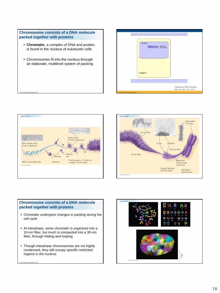
Figure 16.22a
DNA double helix(2 nm in diameter)
DNA, the double helix
Nucleosome(10 nm in diameter)
Histones
Histones
Histonetail
H1
Nucleosomes, or “beads ona string” (10-nm fiber)
Figure 16.22b
30-nm fiber
30-nm fiber
Loops Scaffold
300-nm fiber
Chromatid(700 nm)
Replicatedchromosome(1,400 nm)
Looped domains(300-nm fiber) Metaphase
chromosome
Chromatin undergoes changes in packing during the
cell cycle
At interphase, some chromatin is organized into a
10-nm fiber, but much is compacted into a 30-nm
fiber, through folding and looping
Though interphase chromosomes are not highly
condensed, they still occupy specific restricted
regions in the nucleus
© 2011 Pearson Education, Inc.
Chromosome consists of a DNA molecule
packed together with proteins
Figure 16.23
5
m
Page 19
19
Most chromatin is loosely packed in the nucleus
during interphase and condenses prior to mitosis
Loosely packed chromatin is called euchromatin
During interphase a few regions of chromatin
(centromeres and telomeres) are highly
condensed into heterochromatin
Dense packing of the heterochromatin makes it
difficult for the cell to express genetic information
coded in these regions
© 2011 Pearson Education, Inc.
Chromosome consists of a DNA molecule
packed together with proteins
DNA wrapping around proteins
Important Concepts
Know the vocabulary in this lecture
Structure of DNA – and their nucleotides
The four bases, and which are paired together
Be able to recognize the four base structures
Know which bases are purines and Pyrimidines
Type of bonds/linkages
Be able to draw DNA for me (you can use S
and P for sugar and phosphate, ATCG for
bases, 5’ and 3’)
Important Concepts
Be able to describe how is DNA replicated
Semiconservative replication
Steps
Complementary pairing
Direction of building the complementary pair
The role of helicase, Single-strand binding proteins
Topoisomerases, DNA polymerases, DNA ligase
Understand how the leading strand is build vs how
the lagging strand is built, know what Okazaki
fragments are,
Important Concepts
Know what telomers and telomerases are
What supplies the energy to be used to build the
new strand
Be able to identify correctly paired bases and
incorrectly paired bases
Know the repair mechanisms for DNA