THE JOURNAL OF BIOLOGICAL CHEMISTRY 0 1985 by The American Society of Biological Chemists, Inc. Val. 260, No. 27, Issue of November 25, pp. 14873-14878,1985 Printed in U.S.A. Camptothecin Induces Protein-linked DNA Breaks via Mammalian DNA Topoisomerase I* (Received for publication, April 8,1985) Yaw-Huei Hsiangl, Robert Hertzbergj, Sidney Hechtj, and Leroy F. LiuS From the $Department of Biological Chemistry, Johns Hopkins School of Medicine, Baltimore, Maryland 21205 and §Smith Kline and French Laboratories, Philadelphia, Pennsylvania 19101 Camptothecin, a cytotoxic drug, is a strong inhibitor ofnucleicacid synthesis inmammalian cells anda potent inducer of strand breaks in chromosomal DNA. Neither the equilibrium dialysis northeunwinding measurement indicates any interaction between camp- tothecinandpurifiedDNA.However,camptothecin induces extensive single strand DNA breaks in reac- tions containing purified mammalian DNA topoisom- erase I. DNA breakage in vitro is immediate and reversible. Analyses of camptothecin-induced DNA breaks show that topoisomerase I is covalently linked to the 3’ end of the broken DNA. In addition, campto- thecininhibitsthe catalytic activity of mammalian DNA topoisomerase I. We propose that camptothecin blocks the rejoining step of the breakage-reunion re- action of mammalian DNA topoisomerase I. This block- age results in the accumulation of a cleavable complex which resembles the transient intermediate proposed for eukaryotic DNA topoisomerase I. The inhibition of nucleic acid synthesis and the induction of DNA strand breaks observed in vivo may be related to the forma- tion of this drug-induced cleavable complex. Campothecin (Fig. 1) is a cytotoxic alkaloid isolated from Camptotheca acuminata (family Nyssaceae), a tree indigenous to China (reviewed in Ref. 1). Camptothecin has strong anti- tumor activity against a wide range of experimental tumors (2). It also inhibits both DNA and RNA synthesis in mam- malian cells. The inhibition of RNA synthesis results in shortened RNA chains and is rapidly reversible upon drug removal (3). The inhibition of DNA synthesis, on the other hand, is only partially reversible upon drug removal (4, 5). Camptothecin is a much stronger inhibitor of DNA synthesis than RNA synthesis in human lymphocytes stimulated by phytohemagglutinin (2). However, in studies using either purified DNA polymerase or RNA polymerase, the inhibitory effect of camptothecin was not observed (4). Another promi- nent effect of camptothecin is the tapid and reversible frag- mentation of cellular DNA in cultured mammalian cells (6). Studies of camptothecin analogs have suggested a correlation between suppression of tumor growth and the ability to cause fragmentation of DNA (reviewed in Ref. 1). Interestingly, camptothecin by itself does not cleave purified DNA. The cellular target(s) of camptothecin which is responsible for DNA fragmentation and inhibition of nucleic acid synthesis is still unknown. * This work has been supported by National Institutes of Health Grants GM-27731 and CA-96632. The costs of publication of this article were defrayed in part by the payment of page charges. This article must therefore be hereby marked “advertisement” in accord- ance with 18 U.S.C. Section 1734 solelyto indicate this fact. 0 FIG. 1. Chemical structure of camptothecin (lactone form). There has been increasing evidence that mammalian DNA topoisomerase I1 is a common target for a number of antineo- plastic agents (7-11). The acridine derivative, m-AMSA,’ and the epipodophyllotoxin, VM-26, are two representative anti- tumor drugs that affect the breakage-reunion reaction of mammalian DNA topoisomerase 11. Both m-AMSA and VM- 26 induce reversible DNA fragmentation and protein-DNA “cross-links” in cultured cells (10, 12, 13). In vitro and in vivo studies have suggested that these drugs block the rejoining reaction of mammalian DNA topoisomerase I1 by stabilizing a cleavable complex (7-10). Treatment of the drug-induced cleavable complex with protein denaturants results in DNA strand breaks and the covalent linkage of a topoisomerase subunit to each 5’ phosphoryl end of the broken DNA (7-10). Removal of the drugs results in an apparent repair of DNA strand breaks presumably via the continuation of the normal rejoining reaction (7-10). Preliminary studies of the effect of camptothecin onLE10 cells and sV40 infected monkey cells have shown that camptothecin induces protein-linked, single- stranded DNA breaks on cellular and SV40 DNA.’ To deter- mine if topoisomerase I1 is the target of camptothecin, we carried out in vitro studies using purified DNA topoisomer- ases. Surprisingly, camptothecin showed no effect on mam- malian DNA topoisomerase I1 but exhibited strong inhibitory effects on mammalian DNA topoisomerase I. EXPERIMENTAL PROCEDURES Enzymes, Nucleic Acids, and Antitumor Drugs-DNA topoisom- erase I was purified to homogeneity from HeLa cells and calf thymus glands using published procedures (14). DNA topoisomerase I1 from calf thymus was purified to homogeneity as described previously (15). Plasmid pBR322 dimer was purified by phenol deproteinization of cleared lysates followedby CsCl/ethidium isopycnic centrifugation and gel filtration on an A-50m column. m-AMSA (NSC 249992) was The abbreviations used are: m-AMSA, 4’-(9-acridinylamino)- methanesulfon-m-anisidide; SDS, sodium dodecyl sulfate; VM-26, 4’-demethylepipodophyllotoxin thenylidene-P-D-glucoside; bp, base pairs. Y.-H. Hsiang, R. Hertzberg, S. Hecht, and L. F. Liu, unpublished results. 14873
Transcript
THE JOURNAL OF BIOLOGICAL CHEMISTRY 0 1985 by The American Society of Biological Chemists, Inc.
Val. 260, No. 27, Issue of November 25, pp. 14873-14878,1985 Printed in U.S.A.
Camptothecin Induces Protein-linked DNA Breaks via Mammalian DNA Topoisomerase I*
(Received for publication, April 8,1985)
Yaw-Huei Hsiangl, Robert Hertzbergj, Sidney Hechtj, and Leroy F. LiuS From the $Department of Biological Chemistry, Johns Hopkins School of Medicine, Baltimore, Maryland 21205 and §Smith Kline and French Laboratories, Philadelphia, Pennsylvania 19101
Camptothecin, a cytotoxic drug, is a strong inhibitor of nucleic acid synthesis in mammalian cells and a potent inducer of strand breaks in chromosomal DNA. Neither the equilibrium dialysis nor the unwinding measurement indicates any interaction between camp- tothecin and purified DNA. However, camptothecin induces extensive single strand DNA breaks in reac- tions containing purified mammalian DNA topoisom- erase I. DNA breakage in vitro is immediate and reversible. Analyses of camptothecin-induced DNA breaks show that topoisomerase I is covalently linked to the 3’ end of the broken DNA. In addition, campto- thecin inhibits the catalytic activity of mammalian DNA topoisomerase I. We propose that camptothecin blocks the rejoining step of the breakage-reunion re- action of mammalian DNA topoisomerase I. This block- age results in the accumulation of a cleavable complex which resembles the transient intermediate proposed for eukaryotic DNA topoisomerase I. The inhibition of nucleic acid synthesis and the induction of DNA strand breaks observed in vivo may be related to the forma- tion of this drug-induced cleavable complex.
Campothecin (Fig. 1) is a cytotoxic alkaloid isolated from Camptotheca acuminata (family Nyssaceae), a tree indigenous to China (reviewed in Ref. 1). Camptothecin has strong anti- tumor activity against a wide range of experimental tumors (2). It also inhibits both DNA and RNA synthesis in mam- malian cells. The inhibition of RNA synthesis results in shortened RNA chains and is rapidly reversible upon drug removal (3). The inhibition of DNA synthesis, on the other hand, is only partially reversible upon drug removal (4, 5). Camptothecin is a much stronger inhibitor of DNA synthesis than RNA synthesis in human lymphocytes stimulated by phytohemagglutinin (2). However, in studies using either purified DNA polymerase or RNA polymerase, the inhibitory effect of camptothecin was not observed (4). Another promi- nent effect of camptothecin is the tapid and reversible frag- mentation of cellular DNA in cultured mammalian cells (6). Studies of camptothecin analogs have suggested a correlation between suppression of tumor growth and the ability to cause fragmentation of DNA (reviewed in Ref. 1). Interestingly, camptothecin by itself does not cleave purified DNA. The cellular target(s) of camptothecin which is responsible for DNA fragmentation and inhibition of nucleic acid synthesis is still unknown.
* This work has been supported by National Institutes of Health Grants GM-27731 and CA-96632. The costs of publication of this article were defrayed in part by the payment of page charges. This article must therefore be hereby marked “advertisement” in accord- ance with 18 U.S.C. Section 1734 solely to indicate this fact.
0 FIG. 1. Chemical structure of camptothecin (lactone form).
There has been increasing evidence that mammalian DNA topoisomerase I1 is a common target for a number of antineo- plastic agents (7-11). The acridine derivative, m-AMSA,’ and the epipodophyllotoxin, VM-26, are two representative anti- tumor drugs that affect the breakage-reunion reaction of mammalian DNA topoisomerase 11. Both m-AMSA and VM- 26 induce reversible DNA fragmentation and protein-DNA “cross-links” in cultured cells (10, 12, 13). In vitro and in vivo studies have suggested that these drugs block the rejoining reaction of mammalian DNA topoisomerase I1 by stabilizing a cleavable complex (7-10). Treatment of the drug-induced cleavable complex with protein denaturants results in DNA strand breaks and the covalent linkage of a topoisomerase subunit to each 5’ phosphoryl end of the broken DNA (7-10). Removal of the drugs results in an apparent repair of DNA strand breaks presumably via the continuation of the normal rejoining reaction (7-10). Preliminary studies of the effect of camptothecin on LE10 cells and sV40 infected monkey cells have shown that camptothecin induces protein-linked, single- stranded DNA breaks on cellular and SV40 DNA.’ To deter- mine if topoisomerase I1 is the target of camptothecin, we carried out in vitro studies using purified DNA topoisomer- ases. Surprisingly, camptothecin showed no effect on mam- malian DNA topoisomerase I1 but exhibited strong inhibitory effects on mammalian DNA topoisomerase I.
EXPERIMENTAL PROCEDURES
Enzymes, Nucleic Acids, and Antitumor Drugs-DNA topoisom- erase I was purified to homogeneity from HeLa cells and calf thymus glands using published procedures (14). DNA topoisomerase I1 from calf thymus was purified to homogeneity as described previously (15). Plasmid pBR322 dimer was purified by phenol deproteinization of cleared lysates followed by CsCl/ethidium isopycnic centrifugation and gel filtration on an A-50m column. m-AMSA (NSC 249992) was
The abbreviations used are: m-AMSA, 4’-(9-acridinylamino)- methanesulfon-m-anisidide; SDS, sodium dodecyl sulfate; VM-26, 4’-demethylepipodophyllotoxin thenylidene-P-D-glucoside; bp, base pairs.
Y.-H. Hsiang, R. Hertzberg, S. Hecht, and L. F. Liu, unpublished results.
14873
14874 An Inhibitor of DNA Topoisomerase I obtained from the Drug Synthesis and Chemistry Branch, Division of Cancer Treatment, National Cancer Institute. VM-26 was a gift from Rristol-Myers Co. The lactone form of camptothecin (NSC 94600), which is about IO-fold more potent than sodium camptothecin (NSC 100880) (I6) , was either obtained from National Cancer Insti- tute or prepared by acidification of the sodium camptothecin (17). Briefly, 52 mg (0.13 mmol) of sodium camptothecin was dissolved in 1 0 ml of water. 10 ml of 0.01 N HCI was then added with stirring. A fluffy yellow precipitate formed as the pH dropped below 3. Camp- tothecin lactone was isolated by filtration a s a pale green solid and was dried in uacuo to yield 30 mg (64%). All experiments were carried out using camptothecin lactone. Drugs were dissolved in dimethyl sulfoxide a t 10 mM concentration and kept frozen (-20 "C) in ali- qu0t.s.
Preparation of End-labeled pRR322 DNA-The procedure for end labeling of pRR822 DNA has been descrihed previously (18). Briefly, pRR322 dimer was digested with EcoRI and dephosphorylated with alkaline phosphatase (18). Linearized pBR322 DNA was then either laheled a t its 3' ends with the large fragment of Escherichia coli DNA polymerase I and [w3*P]dATP or labeled a t its 5' ends with T4 polynucleotide kinase and [y-"P]ATP (18). Unincorporated triphos- phates were removed hy two cycles of ethanol precipitation in the presence of 2.5 M ammonium acetate. For gel mapping experiments, the labeled DNA was further digested with Hindlll restriction endo- nuclease to remove a small fragment (31 bp) containing one labeled end.
Topoisomerase Actiuity Assays-Topoisomerase I relaxation assays were done as described previously (14). Topoisomerase I1 catalytic activity was monitored by using the P4 unknotting assay (19).
Topoisomeruse Cleavage Assays-Reaction mixtures (20 pl each) containing 40 mM Tris, pH 7.5, 100 mM KCI, 10 mM M&I,, 0.5 mM dithiothreitol, 0.5 mM EDTA, 30 pg/ml bovine serum albumin, 50 ng of '"P-labeled DNA (or 20 pg/ml of unlabeled dimeric pRR322 DNA), 20 ng of calf thymus DNA topoisomerase I or I1 (or 70 ng of enzyme if unlabeled pBR322 DNA is used), and drugs were incubated a t 37 "C for 30 ,min. The reactions were terminated by the addition of 2 pl of 5% SDS. Unless indicated, reaction mixtures were treated with 150 pg/ml proteinase K for another h a t 37 "C.
Gel Blectrophoresis-For mapping douhle strand breaks on DNA, samples were electrophoresed using a 1% agarose gel in TBE buffer (0.089 M Tris-borate, 0.002 M EDTA, pH 8.0) (20). For mapping single strand breaks, DNA samples (22 pl final volume) were dena- tured with 10 pl of 0.45 N NaOH, 30 mM EDTA. 15% sucrose, and 0.25 mg/ml bromocresol green. DNA samples were then analyzed using either a 1% agarose gel in TRE buffer or a 1% alkaline agarose gel in 30 mM NaOH and 1 mM EDTA.
Unwinding Measurements-Plasmid pBR322 DNA was linearized with EcoRI restriction endonuclease and then ligated with T4 DNA ligase in the presence of different drugs. The ligation reaction (20 pl each) was done under conditions identical to those described for topoisomerase cleavage assays (see above) except that ATP (1 mM) was added. The temperature of the ligation reactions was carefully controlled and the reactions were terminated by the addition of 5 pl of a prewarmed stop solution (5% sarkosyl, 25% sucrose, 50 mM EDTA, and 0.05 mg/ml bromphenol blue). Gel electrophoresis was performed in the cold room (4 "C) using a 1.0% agarose gel and TBE electrophoresis buffer supplemented with 5 mM MgCl2.
DNA Binding Studies by Equilibrium Dialysis-One ml of soni- cated calf thymus DNA (2.0 mM nucleotide) in 10 mM Tris-HCI, pH 7.4, 50 mM NaCl or in 20 mM potassium phosphate (pH 7.2) was placed in one well of a two-sided equilibrium dialysis cell. In the other well was placed 1 ml of camptothecin (2-50 p ~ ) in buffer. After shaking a t room temperature for 18 h, the fluorescence spectrum of each well was recorded and compared.
RESULTS
Camptothecin Does Not Affect Mammalian DNA Topoisom- erase I I in Vitro-To investigate whether mammalian DNA topoisomerase I1 is a target of camptothecin, purified calf thymus DNA topoisomerase I1 was used in an in oitro DNA cleavage assay (9). While both m-AMSA (Fig. 2, lanes H-L) and VM-26 (Fig. 2, lanes M-Q) induced DNA double strand breaks in this assay, camptothecin failed to induce any de- tectable cleavage above background (Fig. 2, lanes C-C). To test whether camptothecin can induce topoisomerase 11-me- diated single strand breaks, the following two experiments
A B C D E F G H I J K L M N O P Q " -
- 0 - -*t:
FIG. 2. Camptothecin does not affect topoisomerase 11-me- diated DNA cleavage. Topoisomerase Il-mediated DNA cleavage was done as described under "Experimental Procedures." Imne A , DNA control, no enzyme, no drug. l a n e R. no drug. lanes C-G. 0.2, 1.0, 5.0, 25, and 125 p~ of camptothecin. respectively. lanes H-I.. same as lanes C-G, respectively, except that m-AMSA was used instead of camptothecin. Lunes M-Q, same as lanes ('-(; except that VM-26 was used instead of camptothecin.
A B C D E F C H I J K L M N O P Q " P 4 circle "4 linear
P4 knots
FIG. 3. Camptothecin does not inhihit the Rtrand-pansing act ivi ty of mammalian DNA topoinomerase 11. The P4 unknot- ting assay was performed as descrltml under "Experimental Proce- dures." Lane A, control 1'4 knotted DNA. Imne H , no drug. lanes C- C, 0.2, 1.0, 5.0, 25, and 125 p M of camptothecin, respectively. Imrws H-L, 0.2, 1.0, 5.0, 25, and 125 p~ of rn-AMSA, respectively. lanes M-Q, 0.2, 1.0, 5.0, 25, and 125 pM of VM-26. respectively.
were done. First, the same samples were analyzed by alkaline gel electrophoresis to reveal single strand breaks and no increase in drug-induced cleavage was found (data not shown). Second, the same samples were also analyzed by the K-SDS precipitation procedure which was designed to assay protein- linked DNA (18). Using this assay, both m-AMSA- and VM- 26-treated samples showed dose-dependent increases in to- poisomerase 11-linked DNA, while camptothecin did not (data not shown). The possibility that camptothecin might inhibit the catalytic activity of topoisomerase I1 was also investigated using the P4 unknotting assay (19). Again, no inhibition by camptothecin was observed (Fig. 3, lanes C-G) while strong inhibition by both m-AMSA and VM-26 was quite obvious (Fig. 3, lanes H-L and M-Q, respectively).
Camptothecin Nicks DNA in the Presence of Purified Calf Thymus DNA Topoisomerase I-It is known that many an- titumor drugs which induce protein-linked DNA breaks are DNA intercalators (reviewed in Ref. 9). To test whether camptothecin intercalates into DNA, an unwinding measure- ment was performed using linearized pBR322 DNA and T4 DNA ligase. In this assay, m-AMSA, which is a weak inter- calator (7), was used as a positive control (Fig. 4, lanes H - L ) . VM-26, which neither intercalates into DNA nor binds to DNA (10, l l ) , was used as a negative control (Fig. 4, lanes M-Q). Similar to VM-26, camptothecin did not unwind DNA to any detectable extent at concentrations up to 125 p~ (Fig.
An Inhibitor of DNA Topoisomeraw I 14875
FIG. 4. ( ’ a m p t o t h r c i n docs not u n w i n d DNA. The unwinding measurement W Z I S clone :IS clcw-ritwcl under “Experimental Proce- dures.” IAnr A , I)NA rontrol. no enzyme, n o drug. Lanr H , no drug. IAnrs (‘4;. 0.2, 1.0, 5.0, 25, and 125 p~ camptothecin, respectively, Imnrs H-I, , 0.2, 1.0,5.0,25. and 125 p~ m-AMSA, respectively. I m w s M-(2, 0.2. 1.0. 5.0, 25, and 125 g~ VM-126, respectively. Imnes H , S . and T. no enavme hut contained 125 U M of camutothecin. rn-AMSA, and VM-26, respectively.
A B C D E F G
I I
1 1 1
FIG. 5. Nicking of plasmid I)NA hy camptothecin in the presence of calf thymus DNA topoisomerase I. Reaction condi- t ions were t he w m e as descritwd for the topoisomerase cleavage assay under “Experimental Procedures.” Calf thymus DNA topoisomerase I (70 ng) and pHR322 DNA dimer (20 pglml) were used. IAnr A , contml pRR922 DNA. Imnr R, no drug. I.anPs C-G, 0.2, 1.0, 5.0, 25, and 125 p M camptothecin, respectively.
4, lanes C-C). The possibility that camptothecin might bind to DNA was also tested by equilibrium dialysis. The intense fluorescence of camptothecin (370 nm excitation and 448 nm emission), which was unaffected by 2 mM calf thymus DNA (data not shown) was used to monitor the drug concentration on both sides of the dialysis cell. In all cases (from 1 to 50 pM of camptothecin), the fluorescence spectra and intensities were identical on both the DNA side and the camptothecin side of the dialysis cell. At these DNA and drug concentra- tions, the binding equation of McGhee and von Hippel (21) predicts that 32% of the camptothecin would be bound to the DNA if the binding affinity was 500 and the site size was 2 bp. This would result in an easily detectable increase in fluorescence intensity on the DNA side of the cell. Since no difference was observed, we can conclude that the drug has little or no affinity for calf thymus DNA. The experiments were repeated using both Tris and phosphate buffers and in the presence of 10 mM MgCI,. In addition, single-stranded DNA (heat-denatured and quickly chilled) was tested for camptothecin binding by this method with similar results.
Since DNA topoisomerase I is also known to break and rejoin DNA (22), the possibility that DNA topoisomerase I may mediate camptothecin-induced DNA damage was also investigated. Purified calf thymus DNA topoisomerase I was used in an in uitro reaction containing closed circular pRR.722 DNA and camptothecin. With increasing concentrations of camptothecin, closed circular pRR322 DNA (form I) was converted to nicked circular DNA (form 11) (Fig. 5, lanes C-
G). This apparent nicking activitv of camptothecin required DNA topoisomerase I since no nicking was observed in its absence (data not shown).
The nicking activity of camptothecin in the presence of calf thymus DNA topoisomerase I was further studied using end- labeled pRR.322 DNA (see “Experimental Procedures”). To reveal single strand breaks, the reaction products were dena- tured with alkali prior to gel electrophoresis in neutral THE buffer. In the presence of calf thymus DNA topoisomerase I and camptothecin, end-labeled pRR322 DNA was fragmented to smaller pieces which migrated faster in the gel (Fig. 6, lancs C-G). On the other hand, neither rn-AMSA nor VM-26 i n - duced any single strand breaks in the presence of calf thymus DNA topoisomerase I (Fig. 6, lancs H-I, and M-Q, respec- tively). Furthermore, camptothecin alone did not induce any single strand breaks (Fig. 6, lnnc I O . It thus appears that DNA topoisomerase I is specifically required for camptothe- cin-induced DNA damage. If one topoisomerase I molecule was bound to each nick on DNA, we calculated that approxi- mately 10% of DNA topoisomerase I molecules in the reaction mixture are trapped on DNA in the presence of 1 p~ of camptothecin.
Nrurrsihility of Camptothccin-induccd I )NA IhmaLv-Two interesting features of these single strand breaks were noted. ( a ) The induction of single strand breaks hy camptothecin and topoisomerase I was immediate. The amounts of single strand breaks reached a plateau level wit.hin 30 s after mixing (Fig. 7, lanes D-K) . The extent of DNA cleavage depended on the drug concentration (Fig. 6) rather than the time course of incubation (Fig. 7, lanes 11°K). ( h ) The formation of the single strand breaks is reversible. Rv lowering the temperature of the reaction to 0 “C after the first incubation but prior to the addition of SDS, the extent of cleavage was slightly reduced in a time-dependent fashion (Fig. 7 , lancs I , -N). In another experiment, the addition of high salt (0.5 M NaCl) to a preincubated reaction dramaticallv reduced the single strand breaks in a time-dependent fashion (Fig. 8, lanrs H - H ) .
Topoisomeraw I Is C’oualcntly I,intwd t o t t w :l’-E,‘nds of Rrokrn D N A Strands-It has been shown that calf thymus DNA topoisomerase I can he trapped on DNA by protein denaturant treatment (22). The trapped topoisomerase I is covalently linked to the .?’-phosphoryl ends of the broken DNA strands (22). I t has been proposed that this trapped topoisomerase I-DNA complex may he related to the putative transient intermediate of the topoisomerase I relaxation re-
A B C D E F C H I I K I M N O P Q R S T 4333 bp
-31 bp
FIG. 6. Camptothecin inducm site-specific DNA cleavage in the presence of topoisomerase 1. : I ’ encl-lnt)eled pHi<:122 I)NA and calf thymus DKA topoisomerase I were used in the topoisomerase cleavage assav as denrrihed under “Experimental I’rnredures.” D N A samples were denatured prior to elertrophoresis in neutral ‘THF: buffer. Law A, DNA control, no drug. n o enzyme. Imnr H. no drug. lmnrn C-G, 0.2, 1.0. 5.0. 25. and I25 p M carnptothecin. respectively. Imnes H - L . 0.2, 1.0.5.0.25. and 125 JIM rn-AMSA. respectively. 1mnr.T M-Q, 0.2. 1.0, 5.0, 25, and 125 p~ VM-26, respectively.
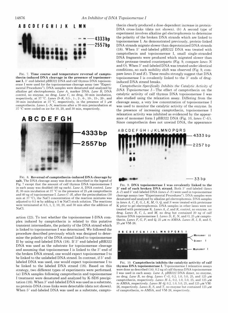
14876 An Inhibitor of DNA Topoisomerase I
A B C D E F C H I J K L M N
FIG. 7. Time course and temperature reversal of campto- thecin-induced DNA cleavage in the presence of topoisomer- ase I. 3' end-labeled pRR322 DNA and calf thymus DNA topoisom- erase I were used for the topoisomerase cleavage assay (see "Experi- mental Procedures"). DNA samples were denatured and analyzed by alkaline gel electrophoresis. Imne A, marker DNA. Imnc H , DNA control, no enzyme, no drug. Imne C, no drug, 30-min incubation, respectively, a t 37 "C. Lanes D-K, 0 5 , 1-, 2-. 6-, IO-, 1 5 , 20-, and 30-min incubation a t 37 "C, respectively, in the presence of 5 p~ camptothecin. Lanes L-N, reactions after a 30-min preincubation a t 37 "C were cooled on ice for 10, 20, and 30 min, respectively.
A B C D E F G H
thecin clearly produced a dose-dependent increase in protein- DNA cross-links (data not shown). ( b ) A second type of experiment involves alkaline gel electrophoresis to determine the polarity of the broken DNA strands which are linked to topoisomerase I. As demonstrated previously, protein-linked DNA strands migrate slower than deproteinized DNA strands (18). When 5' end-labeled pRR.722 DNA was treated with camptothecin and topoisomerase I, small single-stranded DNA fragments were produced which migrated slower than their protease-treated counterparts (Fig. 9, compare fanes N and 0). When 3' end-labeled DNA was treated under identical conditions, no such mobility shift was observed (Fig. 9, com- pare lanes D and E ) . These results strongly suggest that DNA topoisomerase I is covalently linked to the 3' ends of drug- induced DNA strand breaks.
Camptothecin Specifically Inhibits the Catal-vtic Activity of DNA Topoisomerase I-The effect of camptothecin on the catalytic activity of calf thymus DNA topoisomerase I was also studied using the relaxation assay. Differing from the cleavage assay, a very low concentration of topoisomerase I was used to monitor the catalytic activity of the enzyme. In the presence of increasing camptothecin, topoisomerase I relaxation activity was inhibited as evidenced by the appear- ance of monomer form I pRR.722 DNA (Fig. 10. fanes ( ' 4 ; ) . Since camptothecin does not unwind DNA, the appearance
-4333 bp A B C D E F G H I J K L M N O P Q R S 4333 b p
-2257 b p t o
FIG. 8. Reversal of camptothecin-induced DNA cleavage by salt. The DNA cleavage assay was done as descrihed in the legend of Fig. 7 except that the amount of calf thymus DNA topoisomerase I in each assay was douhled (40 ng each). Lane A , DNA control. Lane H . 3O-min incuhation at. 37 "C in the presence of 25 pM camptothecin and 40 ng of topoisomerase 1. 1mne.s C'-H, after the preincubation (30 min at 37 "C), the NaCl concentration of the reaction mixtures was adjusted to 0.5 M hy adding a 5 M NaCl stock solution. The reactions were terminated at 0.5, 1, 2, 10. 20, and 30 min after the addition of NaCI.
action (22). To test whether the topoisomerase I-DNA com- plex induced by camptothecin is related to this putative transient intermediate, the polarity of the DNA strand which
" -33 bp FIG. 9. DNA topoisomerase I was covalently linked to the
3' end of each broken DNA strand. Both 3' end-lnlwled (lanrs A - I ) and 5' end-laheled DNA ([anes J - S ) were used in topoisomerase cleavage assays (see "Experimental Procedures"). 1)NA samples were denatured and analyzed hy alkaline gel electrophoresis. DNA samples in lanes A, C, E , G . I , h', M , 0. Q , and S were treated with proteinase K prior to gel electrophoresis. DNA samples in other lanes were not treated with proteinase K. Imnas A, J . and K. control. no enzyme. no drug. Lanes R , C, I+ and M , no drug hut contained 2n ng of calf thymus DNA topoisomerase I. Imnes I), E,*. N . and 0 , 25 p~ campto- thecin. Lanes F, C , P, and (2, 25 p~ rn-AMSA. 1,unrs I { . 1. N , and S. 25 pM VM-26.
is linked to topoisomerase I was determined. We followed the procedure described previously which was designed to deter- mine the polarity of the DNA strand linked to topoisomerase I1 by using end-labeled DNA (18). If 3' end-labeled pBR322 Dimer I DNA was used as the substrate for topoisomerase cleavage and assuming that topoisomerase I is linked to the 3'-end of Monomer I the broken DNA strand, one would expect topoisomerase I to be linked to the unlabeled DNA strand. In contrast, if 5' end-
A B C D E F C H I J K L M N O P O R S T Monomer I I
An Inhibitor of DNA Topoisomerase I 14877
of monomer form I pBR322 DNA is a strong indication of drug inhibition of enzyme activity. The same experiment was also repeated using m-AMSA and VM-26 to test their possible effect on topoisomerase I relaxation activity. With increasing m-AMSA, form I pBR322 DNA also accumulated (Fig. 10, lanes H-L). Since m-AMSA is known to unwind DNA upon intercalation, the increasing form I pBR322 DNA may be due to bound m-AMSA rather than inhibition of enzyme activity. Indeed, when relaxed pBR322 DNA was used as a substrate, the form I DNA also increased, suggesting that topoisomerase I activity was not inhibited (data not shown). VM-26, which is another topoisomerase I1 inhibitor, did not inhibit topo- isomerase I relaxation activity (Fig. 9, lunes M-Q). These results indicate that DNA topoisomerase I is specifically inhibited by camptothecin but not by topoisomerase I1 inhib- itors such as m-AMSA and VM-26 (7-11).
DISCUSSION
It has been demonstrated that a number of antitumor drugs affect the breakage-reunion reaction of mammalian DNA topoisomerase I1 by stabilizing a cleavable complex which upon treatment with protein-denaturants produces protein- linked DNA breaks (7-10). Our preliminary studies have shown that camptothecin similarly produces protein-linked DNA breaks both in L1210 cells and SV40-infected monkey cells. In the latter case, the protein-linked breaks have been demonstrated to be mostly single strand breaks? Our present studies in vitro using purified DNA topoisomerases have clearly established that camptothecin affects only DNA to- poisomerase I but not DNA topoisomerase 11. Since DNA topoisomerase I is known to introduce transient protein- linked single strand breaks, the lack of double strand breaks on SV40 DNA following camptothecin treatment of SV40- infected monkey cells is thus explained. Although our present studies suggest that topoisomerase I is a target of camptothe- cin, additional biochemical and genetic studies are necessary to support this proposition.
The effect of camptothecin on purified DNA topoisomerase I is specific and pronounced. Even at 0.5 PM, camptothecin induces extensive nicking of DNA in the presence of DNA topoisomerase I. The rapid induction and the reversibility of camptothecin-induced DNA damage is unusual. It is possible that camptothecin may interact with DNA topoisomerase I or a topoisomerase I-DNA complex in a noncovalent manner. Based on our present knowledge of the enzyme mechanism, a simple working model for the drug action is shown schemat- ically in Fig. 11. We assume that there are at least two enzyme intermediates, the noncleavable complex (Fig. 1lA) and the cleavable complex (Fig. l lB) , a t rapid equilibrium. The cleav- able complex (Fig. 11B) may be related to the putative tran-
A B
W W I SDS I SDS
+
FIG. 11. A possible model for camptothecin-induced DNA damage.
sient intermediate in the normal enzyme reaction. Campto- thecin may affect this equilibrium in such a way that the equilibrium concentration of drug-altered cleavable complex is greatly increased. Exposure of the cleavable complex to protein-denaturants (e.g. SDS or alkali) results in DNA single strand breaks and the covalent linkage of topoisomerase I to the 3'-phosphoryl end of the breaks through a tyrosine phos- phate linkage (22). This drug-altered cleavable complex may be responsible for the inhibition of the relaxation activity of topoisomerase I. The cytotoxic effect and the stimulation of sister-chromatid exchanges and chromosome aberrations may all be due to the accumulation of the drug-altered cleavable complex (23).
The biological function(s) of mammalian DNA topoisom- erase I has not been established. Its swivel-like enzymatic activity suggests a possible function in DNA replication and RNA transcription (22). In vitro studies have shown that topoisomerase I is required for the elongation phase of ade- novirus DNA replication (24). Studies in chicken embryos and Drosophila polytene chromosomes have also suggested that topoisomerase I may be involved in transcription (25, 26). Interference with the swivel mechanism of DNA topoi- somerase I by camptothecin may thus lead to the inhibition of both DNA synthesis and RNA synthesis. Indeed, campto- thecin has been shown to inhibit the replication of two DNA viruses, adenovirus and vaccinia virus, that replicate in the nucleus and cytoplasm of HeLa cells, respectively (1). The effect of camptothecin on cell cycle traverse has also been analyzed in synchronized cultures of Chinese hamster cells (27). Camptothecin did not block the initiation of DNA syn- thesis but prevented cells from progressing to mitosis (27). Such an effect of camptothecin can be explained if topoisom- erase I functions as a swivel for the elongation phase of DNA synthesis. The effect of camptothecin on transcription is also quite interesting. It has been shown that camptothecin inhib- its the synthesis of ribosomal RNA in HeLa cells to a greater extent than 4-5 S RNA (4). The synthesis of precursor ribosomal RNA in the nucleolus is also more sensitive 'to camptothecin than that of hnRNA (3). This differential effect of camptothecin on ribosomal RNA transcription can be explained by the recent findings that topoisomerase I is enriched in the nucleolu~.~ Whether the observed inhibition of nucleic acid synthesis and fragmentation of cellular DNA are due to drug interference with the topoisomerase swivel function in vivo remains to be determined. The establishment of camptothecin as a specific inhibitor of topoisomerase I may provide a powerful tool to probe the function(s) of this im- portant nuclear enzyme. Understanding the mechanism of action of camptothecin may also be important in the estab- lishment of topoisomerase I as a useful therapeutic target for cancer chemotherapy.
Acknowledgments-We thank Dr. Annette Bodley and Teri Gold- sand for critical reading of the manuscript.
REFERENCES
1. Horwitz, S. B. (1975) in Antibiotics, Vol. 111, Mechanism of Action of Antimicrobial and Antitumor Agents (Corcoran, J. W., and Hahn, F. E., e&) pp. 48-57, Springer-Verlag, New York
2. Gallo, R. C., Whang-Peng, J., and Adamson, R. H. (1971) J. Natl. Cancer Inst. 46,789-795
3. Abelson, H. T., and Penman, S. (1972) Nature New Biol. 237,
4. Horwitz, S. B., Chang, C., and Grollman, A. P. (1971) Mol. 144-146
Pharmacol. 7,632-644
B. D. Halligan and L. F. Liu, unpublished results.
14878 A n Inhibitor of D N A Topoisomerase I 5 . Kessel, D., Bosmann, H. B., and Lohr, K. (1972) Biochim. Bio-
6. Horwitz, M. S., and Horwitz, S. B. (1971) Biochem. Biophys. Res. Commun. 45, 723-727
7. Nelson, E. M., Tewey, K. M., and Liu, L. F. (1984) Proc. Natl. Acad. Sci. U. S. A. 81, 1361-1365
8. Tewey, K. M., Chen, G. L., Nelson, E. M., and Liu, L. F. (1984) J. Biol. Chem. 259,9182-9187
9. Tewey, K. M., Rowe, T. C., Chen, G. L., Yang, L., Halligan, B. D., and Liu, L. F. (1984) Science 226,466-468
10. Chen, G. L., Yang, L., Rowe, T. C., Halligan, B. D., Tewey, K. M., and Liu, L. F. (1984) J. Biol. Chem. 259, 13560-13566
11. Ross, W., Rowe, T., Glisson, B., Yalowich, J., and Liu,, L. (1984) Cancer Res. 44,5837-5860
12. Zwelling, L. A., Michaels, S., Erickson, L. C., Ungerleider, R. S., Nichols, M., and Kohn, K. W. (1981) Biochemistry 20, 6553- 6563
13. Wozniak, A. J., and Ross, W. E. (1983) Cancer Res. 43, 120-124 14. Liu, L. F., and Miller, K. G. (1981) Proc. Natl. Acad. Sci. U. S. A.
15. Halligan, B. D., Edwards, K. A., and Liu, L. F. (1985) J. Biol.
phys. Acta 269,210-216
76,3487-3491
Chem. 260,2475-2482
16. Wani, M. C.. Rommam. P. E., Lindlev. J. T.. and Wall. M. E. (1980) J. Ahd. Chem. 23, 554-560
“ I
17. Wall. M. E.. Wani, M. C.. Cooke, C. E.. Palmer. K. H.. McPhail. A. T., and Sim, 6. A. (1966) J.‘Am. Chem. Soc. 88,3888-3890
18. Liu, L. F., Rowe, T. C., Yang, L., Tewey, K. M., and Chen, G. L. (1983) J. Bid. Chem. 258, 15365-15370
19. Liu, L. F., Davis, J. L., and Calendar, R. (1981) Nucleic Acid Res.
20. Maniatis, T., Fritsch, E. F., and Sambrook, J. (1982) in Molecular Cloning, pp. 149-185, Cold Spring Harbor Laboratory, Cold Spring Harbor, NY
21. McGhee, J. D., and von Hippel, P. H. (1974) J. Mol. Biol. 86,
22. Gellert, M. (1981) Annu. Reu. Biochem. 50, 879-910 23. Huang, C. C., Han, C. S., Yue, X. F., Shen, C. M., Wang, S. W.,
Wu, F. G., and Xu, B. (1983) J. Natl. Cancer Inst. 71,841-846 24. Nagata, K., Guggenheimer, R. A., and Hurwitz, J. (1983) Proc.
Natl. Acad. Sci. U. S. A. 80, 4266-4270 25. Weisbrod, S. (1982) Nucleic Acids Res. 10, 2017-2042 26. Fleischmann, G., Pflugfelder, G., Steiner, E. K., Javaherian, K.,
Acad. Sci. U. S. A. 81, 6958-6962 Howard, G. C., Wang, J. C., and Elgin, S. R. (1984) Proc. Natl.