Journal of Clinical Pathology, 1979, 32, 986-989 Campylobacter colitis C. P. WILLOUGHBY, J. PIRIS, AND S. C. TRUELOVE From the Nuffield Department of Clinical Medicine and the Department of Morbid Anatomy, Radcliffe Infirmary, Oxford, UK SUMMARY A patient is described in whom a campylobacter enteritis closely resembled ulcerative colitis on clinical, sigmoidoscopic, and histological grounds. Selective stool culture techniques may be necessary to differentiate campylobacter colitis from ulcerative colitis proper. Campylobacter jejuni is an important cause of infectious diarrhoea in man (Skirrow, 1977). We have recently seen a patient who had clinical, sig- moidoscopic, and histological features which initially suggested a diagnosis of ulcerative colitis, but in whom stool culture revealed the presence of heat- tolerant campylobacteria. Her illness resolved rapidly with appropriate antibiotic treatment and has not recurred during one year of follow-up. Case report A 26-year-old woman was admitted to hospital with a 48-hour history of colicky lower abdominal pain relieved by defaecation. The discomfort was asso- ciated with about 20 watery stools per day, which contained mucus and blood. There were no other relevant symptoms. She was afebrile, and the only abnormal physical sign was mild, diffuse abdominal tenderness, which was most marked in the left iliac fossa. Sigmoidoscopy showed moderate inflammation of the mucosa without contact bleeding or ulceration. The appearances closely resembled those of mild ulcerative colitis, and a rectal biopsy specimen was taken. A full blood count, sedimentation rate, and plasma biochemistry were normal. No pathogens were isolated on routine stool culture, but specimens were also inoculated on a campylobacter-selective medium (Columbia agar (Oxoid) containing 7 % lysed horse blood, van- comycin 10 jug/ml, polymyxin B 2x5 IU/ml, and trimethoprim 5 pig/ml) and incubated microaero- philically at 400C. Examination of the plates after 18 hours and 42 hours revealed the presence of typical flat, non-haemolytic mucoid colonies; Gram- staining showed vibrio forms. The organisms were Received for publication 12 March 1979 oxidase- and catalase-positive, showed a character- istic 'darting' mobility under phase-contrast micro- scopy, and were sensitive to erythromycin. No serological investigations were performed. The patient improved a little on symptomatic treatment but was still having frequent loose motions five days later. Sigmoidoscopically, the rectal mucosa still looked inflamed, and a further biopsy specimen was taken. In view of the stool culture report, she was given erythromycin, 500 mg twice daily for three weeks. Her illness rapidly resolved and, at the end of the antibiotic course, she was asymptomatic, stool cultures were negative, and the sigmoidoscopic appearances had returned to normal. She has remained well, on no treatment, over the succeeding year. PATHOLOGY The appearances of the initial biopsy specimen are shown in Figure 1. The surface epithelium is flat- tened and depleted of goblet cells. The crypt walls are infiltrated by polymorphonuclear leucocytes, and numerous crypt abscesses are present. There is a mixed inflammatory infiltrate in the lamina propria comprising polymorphs, lymphocytes, and plasma cells. The changes were reported to be consistent with moderately active ulcerative colitis. Gram- staining and silver nitrate impregnation failed to demonstrate vibrios in the tissue sections. The rectal biopsy taken five days later shows reappearance of a tall columnar surface epithelium with mild goblet cell depletion. There are no crypt abscesses. The acute inflammatory component of the lamina propria infiltrate has virtually disappeared, but there is still an excess of chronic inflammatory cells, particularly plasma cells (Fig. 2). The final biopsy specimen, taken a month after the start of the illness, is shown in Figure 3. The rectal mucosa has returned entirely to normal. 986 on 22 March 2019 by guest. Protected by copyright. http://jcp.bmj.com/ J Clin Pathol: first published as 10.1136/jcp.32.10.986 on 1 October 1979. Downloaded from
Transcript
Journal of Clinical Pathology, 1979, 32, 986-989
Campylobacter colitisC. P. WILLOUGHBY, J. PIRIS, AND S. C. TRUELOVEFrom the Nuffield Department of Clinical Medicine and the Department ofMorbid Anatomy,Radcliffe Infirmary, Oxford, UK
SUMMARY A patient is described in whom a campylobacter enteritis closely resembled ulcerativecolitis on clinical, sigmoidoscopic, and histological grounds. Selective stool culture techniques maybe necessary to differentiate campylobacter colitis from ulcerative colitis proper.
Campylobacter jejuni is an important cause ofinfectious diarrhoea in man (Skirrow, 1977). Wehave recently seen a patient who had clinical, sig-moidoscopic, and histological features which initiallysuggested a diagnosis of ulcerative colitis, but inwhom stool culture revealed the presence of heat-tolerant campylobacteria. Her illness resolvedrapidly with appropriate antibiotic treatment andhas not recurred during one year of follow-up.
Case report
A 26-year-old woman was admitted to hospital witha 48-hour history of colicky lower abdominal painrelieved by defaecation. The discomfort was asso-ciated with about 20 watery stools per day, whichcontained mucus and blood. There were no otherrelevant symptoms. She was afebrile, and the onlyabnormal physical sign was mild, diffuse abdominaltenderness, which was most marked in the left iliacfossa.
Sigmoidoscopy showed moderate inflammationof the mucosa without contact bleeding or ulceration.The appearances closely resembled those of mildulcerative colitis, and a rectal biopsy specimen wastaken. A full blood count, sedimentation rate, andplasma biochemistry were normal.No pathogens were isolated on routine stool
culture, but specimens were also inoculated on acampylobacter-selective medium (Columbia agar(Oxoid) containing 7% lysed horse blood, van-comycin 10 jug/ml, polymyxin B 2x5 IU/ml, andtrimethoprim 5 pig/ml) and incubated microaero-philically at 400C. Examination of the plates after18 hours and 42 hours revealed the presence oftypical flat, non-haemolytic mucoid colonies; Gram-staining showed vibrio forms. The organisms were
Received for publication 12 March 1979
oxidase- and catalase-positive, showed a character-istic 'darting' mobility under phase-contrast micro-scopy, and were sensitive to erythromycin. Noserological investigations were performed.The patient improved a little on symptomatic
treatment but was still having frequent loose motionsfive days later. Sigmoidoscopically, the rectalmucosa still looked inflamed, and a further biopsyspecimen was taken. In view of the stool culturereport, she was given erythromycin, 500 mg twicedaily for three weeks. Her illness rapidly resolvedand, at the end of the antibiotic course, she wasasymptomatic, stool cultures were negative, and thesigmoidoscopic appearances had returned to normal.She has remained well, on no treatment, over thesucceeding year.
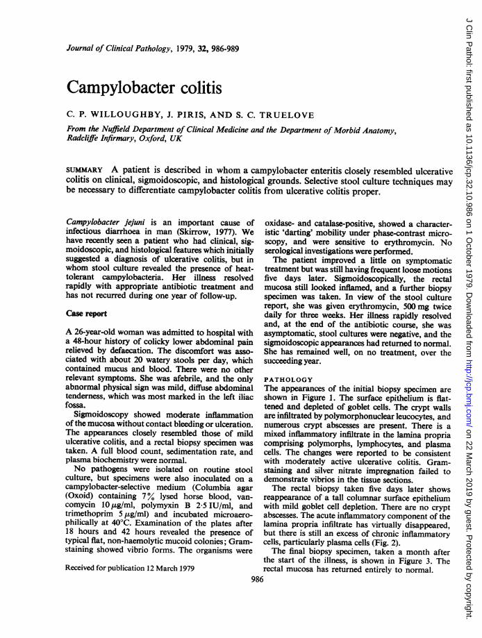
PATHOLOGYThe appearances of the initial biopsy specimen areshown in Figure 1. The surface epithelium is flat-tened and depleted of goblet cells. The crypt wallsare infiltrated by polymorphonuclear leucocytes, andnumerous crypt abscesses are present. There is amixed inflammatory infiltrate in the lamina propriacomprising polymorphs, lymphocytes, and plasmacells. The changes were reported to be consistentwith moderately active ulcerative colitis. Gram-staining and silver nitrate impregnation failed todemonstrate vibrios in the tissue sections.The rectal biopsy taken five days later shows
reappearance of a tall columnar surface epitheliumwith mild goblet cell depletion. There are no cryptabscesses. The acute inflammatory component of thelamina propria infiltrate has virtually disappeared,but there is still an excess of chronic inflammatorycells, particularly plasma cells (Fig. 2).The final biopsy specimen, taken a month after
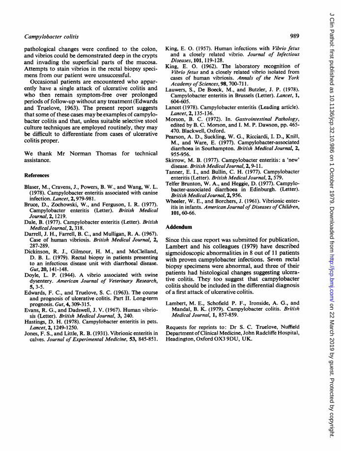
the start of the illness, is shown in Figure 3. Therectal mucosa has returned entirely to normal.
986
on 22 March 2019 by guest. P
rotected by copyright.http://jcp.bm
j.com/
J Clin P
athol: first published as 10.1136/jcp.32.10.986 on 1 October 1979. D
*Er~* *. ,^% <F . >Fig. 3 Histological appearances of the final biopsy specimen.
Discussion
Campylobacteria account for 4-2 to 13-9% of allcases of infectious diarrhoea in the United Kingdom(Bruce et al, 1977; Dale, 1977; Pearson et al., 1977;Skirrow, 1977; Tanner and Bullin, 1977; TelferBrunton and Heggie, 1977). During an attack thepatient's stools frequently contain blood, pus, andmucus. Abdominal pain, fever, and vomiting areother common features of the illness. Sources ofinfection include poultry (Bruce et al., 1977;Skirrow, 1977), dogs and cats (Wheeler and Borchers,1961; Blaser et al., 1978; Hastings, 1978), cattle(King, 1957), and possibly contaminated watersupplies (Pearson et al., 1977).The jejunum and ileum are the usual sites of the
gastrointestinal lesion (Lancet, 1978). A similarvibrionic enteritis in calves involving the uppersmall intestine has been described by Jones andLittle (1931). Campylobacteria have been isolatedfrom intestinal secretions aspirated from the jejunumof infected children (Lauwers et al., 1978). Skirrow(1977) mentions a patient who was subjected tolaparotomy because of a suspected bowel perfora-tion; most of the ileum was found to be inflamedand oedematous. Two fatal cases of the disease havebeen described in which postmortem examinationshowed a haemorrhagic jejuno-ileitis (King, 1962;Evans and Dadswell, 1967); in neither of these
patients were colonic lesions found. In anothersevere case, there was no sigmoidoscopic evidenceof colitis during the acute illness, and a later post-mortem report does not mention any colonicabnormality (Darrell et al., 1967).Our patient had clinical and sigmoidoscopic
features which suggested an attack of ulcerativecolitis. The appearances of the first biopsy specimenwere consistent with this diagnosis, although Morson(1972) has pointed out that similar histologicalchanges may be found in other inflammatory dis-eases affecting the colon. The histology of rectalbiopsy specimens from patients with infectivediarrhoea has recently been reported by Dickinsonet al. (1979). The majority of such biopsies showeda mild to moderate inflammatory infiltrate in thelamina propria. In addition, in about one-third ofcases there were epithelial cell abnormalities, focalerosions, and small numbers of poorly formed cryptabscesses; the changes do not seem to have been assevere as those in the first biopsy specimen from ourpatient. Histological features strongly suggestive ofchronic inflammatory bowel disease were found inonly two of their patients, one ofwhom had salmonel-losis; repeat biopsies a month after the acute illnessshowed normal mucosa.
Vibrios morphologically similar to Campylobacterjejuni have been reported to produce a form ofswine dysentery (Doyle, 1944). In this condition, the
988
on 22 March 2019 by guest. P
rotected by copyright.http://jcp.bm
j.com/
J Clin P
athol: first published as 10.1136/jcp.32.10.986 on 1 October 1979. D
pathological changes were confined to the colon,and vibrios could be demonstrated deep in the cryptsand invading the superficial parts of the mucosa.Attempts to stain vibrios in the rectal biopsy speci-mens from our patient were unsuccessful.
Occasional patients are encountered who appar-ently have a single attack of ulcerative colitis andwho then remain symptom-free over prolongedperiods of follow-up without any treatment (Edwardsand Truelove, 1963). The present report suggeststhat some of these cases may be examples of campylo-bacter colitis and that, unless suitable selective stoolculture techniques are employed routinely, they maybe difficult to differentiate from cases of ulcerativecolitis proper.
We thank Mr Norman Thomas for technicalassistance.
References
Blaser, M., Cravens, J., Powers, B. W., and Wang, W. L.(1978). Campylobacter enteritis associated with canineinfection. Lancet, 2, 979-981.
Bruce, D., Zochowski, W., and Ferguson, I. R. (1977).Campylobacter enteritis (Letter). British MedicalJournal, 2, 1219.
Dale, B. (1977). Campylobacter enteritis (Letter). BritishMedicalJournal, 2, 318.
Darrell, J. H., Farrell, B. C., and Mulligan, R. A. (1967).Case of human vibriosis. British Medical Journal, 2,287-289.
Dickinson, R. J., Gilmour, H. M., and McClelland,D. B. L. (1979). Rectal biopsy in patients presentingto an infectious disease unit with diarrhoeal disease.Gut, 20,141-148.
Doyle, L. P. (1944). A vibrio associated with swinedysentery. American Journal of Veterinary Research,5, 3-5.
Edwards, F. C., and Truelove, S. C. (1963). The courseand prognosis of ulcerative colitis. Part II. Long-termprognosis. Gut, 4, 309-315.
Evans, R. G., and Dadswell, J. V. (1967). Human vibrio-sis (Letter). British Medical Journal, 3, 240.
Hastings, D. H. (1978). Campylobacter enteritis in pets.Lancet, 2, 1249-1250.
Jones, F. S., and Little, R. B. (1931). Vibrionic enteritis incalves. Journal of Experimental Medicine, 53, 845-851.
King, E. 0. (1957). Human infections with Vibrio fetusand a closely related vibrio. Journal of InfectiousDiseases, 101, 119-128.
King, E. 0. (1962). The laboratory recognition ofVibrio fetus and a closely related vibrio isolated fromcases of human vibriosis. Annals of the New YorkAcademy ofSciences, 98, 700-711.
Lauwers, S., De Boeck, M., and Butzler, J. P. (1978).Campylobacter enteritis in Brussels (Letter). Lancet, 1,604-605.
Morson, B. C. (1972). In. Gastrointestinal Pathology,edited by B. C. Morson, and I. M. P. Dawson, pp. 465-470. Blackwell, Oxford.
Pearson, A. D., Suckling, W. G., Ricciardi, I. D., Knill,M., and Ware, E. (1977). Campylobacter-associateddiarrhoea in Southampton. British Medical Journal, 2,955-956.
Skirrow, M. B. (1977). Campylobacter enteritis: a 'new'disease. British MedicalJournal, 2, 9-11.
Tanner, E. I., and Bullin, C. H. (1977). Campylobacterenteritis (Letter). British Medical Journal, 2, 579.
Telfer Brunton, W. A., and Heggie, D. (1977). Campylo-bacter-associated diarrhoea in Edinburgh. (Letter).British MedicalJournal, 2,956.
Wheeler, W. E., and Borchers, J. (1961). Vibrionic enter-itis in infants. American Journal ofDiseases of Children,101,60-66.
Addendum
Since this case report was submitted for publication,Lambert and his colleagues (1979) have describedsigmoidoscopic abnormalities in 8 out of 11 patientswith proven campylobacter infections. Seven rectalbiopsy specimens were abnormal, aud three of theirpatients had histological changes suggesting ulcera-tive colitis. They too suggest that campylobactercolitis should be included in the differential diagnosisof a first attack of ulcerative colitis.
Lambert, M. E., Schofield P. F., Ironside, A. G., andMandal, B. K. (1979). Campylobacter colitis. BritishMedical Journal, 1, 857-859.
Requests for reprints to: Dr S. C. Truelove, NuffieldDepartment ofClinical Medicine, John Radcliffe Hospital,Headington, Oxford OX3 9DU, UK.
on 22 March 2019 by guest. P
rotected by copyright.http://jcp.bm
j.com/
J Clin P
athol: first published as 10.1136/jcp.32.10.986 on 1 October 1979. D