1 Cancer progression as a learning process Aseel Shomar 1,2 , Omri Barak 3,2 and Naama Brenner 1,2 1 Dept. of Chemical Engineering, 2 Network Biology Research Lab, 3 Rappaport Faculty of Medicine Technion – Israel Institute of Technology, Haifa, 32000 Israel Summary Drug resistance and metastasis - the major complications in cancer - both entail adaptation of cancer cells to stress, whether a drug or a lethal new environment. Intriguingly, these adaptive processes share similar features that cannot be explained by a pure Darwinian scheme, including dormancy, increased heterogeneity, and stress-induced plasticity. Here, we propose that learning theory offers a framework to explain these features and may shed light on these two intricate processes. In this framework, learning is performed at the single cell level, by stress-driven exploratory trial-and-error. Such a process is not contingent on pre- existing pathways but on a random search for a state that diminishes the stress. We review underlying mechanisms that may support this search, and show by using a learning model that such exploratory adaptation is feasible in a high dimensional system as the cell. At the population level, we view the tissue as a network of exploring agents that communicate and restrain cancer formation in health. In this view, disease results from the breakdown of homeostasis between cellular exploratory drive and tissue homeostasis. Introduction Cancer progression is traditionally viewed as the outcome of the accumulation of random genetic mutations and the selection of cells harboring mutations that confer them a growth advantage under certain conditions (Garraway and Lander, 2013; Nowel, P.C., 1976; Vogelstein et al., 2013). This Darwinian view is intuitively appealing and provided a powerful framework for cancer research since it reduced cancer to the molecular level of the DNA. Indeed, studies, that identified genes in which mutations are causally related to cancer, emerged at a brisk tempo, especially after the Cancer Genome Atlas and the International Cancer Genome Consortium were launched by the end of the 2000s (Martincorena and Campbell, 2015; The International Cancer Genome Consortium, 2010; Weinstein et al., 2013; Zhang et al., 2011). This reductionist approach motivated developing drugs that target single molecular abnormalities or cancer pathways (Zugazagoitia et al., 2016). Although these drugs have achieved good clinical results, they only increased the overall survival by an average of nearly
Transcript
1
Cancer progression as a learning process
Aseel Shomar1,2, Omri Barak3,2 and Naama Brenner1,2
1Dept. of Chemical Engineering, 2Network Biology Research Lab, 3Rappaport Faculty of Medicine Technion – Israel Institute of Technology, Haifa, 32000 Israel
Summary
Drug resistance and metastasis - the major complications in cancer - both entail adaptation
of cancer cells to stress, whether a drug or a lethal new environment. Intriguingly, these
adaptive processes share similar features that cannot be explained by a pure Darwinian
scheme, including dormancy, increased heterogeneity, and stress-induced plasticity. Here, we
propose that learning theory offers a framework to explain these features and may shed light
on these two intricate processes. In this framework, learning is performed at the single cell
level, by stress-driven exploratory trial-and-error. Such a process is not contingent on pre-
existing pathways but on a random search for a state that diminishes the stress. We review
underlying mechanisms that may support this search, and show by using a learning model
that such exploratory adaptation is feasible in a high dimensional system as the cell. At the
population level, we view the tissue as a network of exploring agents that communicate and
restrain cancer formation in health. In this view, disease results from the breakdown of
homeostasis between cellular exploratory drive and tissue homeostasis.
Introduction
Cancer progression is traditionally viewed as the outcome of the accumulation of random
genetic mutations and the selection of cells harboring mutations that confer them a growth
advantage under certain conditions (Garraway and Lander, 2013; Nowel, P.C., 1976;
Vogelstein et al., 2013). This Darwinian view is intuitively appealing and provided a powerful
framework for cancer research since it reduced cancer to the molecular level of the DNA.
Indeed, studies, that identified genes in which mutations are causally related to cancer,
emerged at a brisk tempo, especially after the Cancer Genome Atlas and the International
Cancer Genome Consortium were launched by the end of the 2000s (Martincorena and
Campbell, 2015; The International Cancer Genome Consortium, 2010; Weinstein et al., 2013;
Zhang et al., 2011).
This reductionist approach motivated developing drugs that target single molecular
abnormalities or cancer pathways (Zugazagoitia et al., 2016). Although these drugs have
achieved good clinical results, they only increased the overall survival by an average of nearly
2
3 months (Salas-Vega et al., 2017). A major reason for this is that malignant cells manage to
adapt to the drug by bypassing its target, rendering the tumor resistant. In fact, resistance is
the norm for all the drugs that target specific molecules (Vasan et al., 2019).
Another factor contributing to poor clinical outcome is metastasis – the process of migration
and colonization of distant organs by cells from a primary tumor. Metastasis is the major
culprit of cancer-associated mortality accounting for almost 90% of deaths (Cheung and
Ewald, 2016; Lambert et al., 2017). These two avenues of disease progression and mortality
– resistance and metastasis – are distinct biological processes. However, they seem to be
remarkably analogous in several fundamental properties. In particular, accumulating
evidence suggests that both drug resistance and colonization of cancer cells in secondary
organs exhibit aspects that do not exclusively follow a simple Darwinian scheme of mutations
and selection (Welch and Hurst, 2019). Despite many years of research, a clear genetic
signature of mutations that are associated with metastasis has not been found (Lambert et
al., 2017; Vogelstein et al., 2013). Such a signature was found in some cancers at the initiation
stage and was therefore expected and sought also in the context of metastasis. Resistance, in
parallel, can arise from purely non-genetic mechanisms (Bell and Gilan, 2020; Marine et al.,
2020; Pisco and Huang, 2015).
A cytotoxic treatment or a hostile environment in a secondary organ are both sources of stress
to which cancer cells should adapt in order to survive. Plethora of findings showed that such
a stress can actively induce an adaptive response and lead to drug resistance and the
formation of metastasis (Pisco and Huang, 2015; Welch and Hurst, 2019). Such adaptation is
supported by global epigenetic changes that enhance the plasticity of the cells. Various terms
have appeared in the literature to describe these phenomena: ‘alternative pathways’,
‘acquired’, ’adaptive’ or ‘induced’ resistance (Kim et al., 2018; Maynard et al., 2020; Pisco and
Huang, 2015; Shaffer et al., 2017; Stewart et al., 2020). Nevertheless, a comprehensive theory
and deeper understanding of these terms is still lacking; such an understanding is crucial to
pave the way to new therapeutic avenues.
In this Perspective, we propose that learning theory offers a framework that may shed some
light on the two complex processes of drug resistance and metastasis. Learning is the
fundamental ability of a system to modify itself with relation to its environment and thus
acquire novel functionality. Although most commonly attributed to the brain, we support the
view that learning is not an exclusive function of neural networks, but rather a general
property of plastic high-dimensional systems coupled to their environment (Baluška and
Levin, 2016). Experiments on yeast cells have revealed that cellular networks can provide a
substrate for learning and adaptation in the face of unforeseen challenges to which no pre-
3
programmed response is available (Braun, 2015). This experimental paradigm was based on
artificially rewiring the genetic regulation network of yeast cells. Exposing these cells to an
environment where expression of the rewired gene is essential while its natural regulation is
compromised, confronts them with regulatory challenging and stressful conditions for which
no pre-programmed response is available. Without dedicated sensory information or pre-
existing regulatory pathways, the yeast cells adapt through a trial-and-error process that can
be described as a primitive form of learning by the gene regulatory network (Schreier et al.,
2017).
Here, we propose that a similar type of adaptation is implicated in metastasis and resistance,
where cancer cells aim to achieve stress reduction. Stressful situations, such as drugs and
lethal environments, induce plasticity that can initiate an exploratory process by which new
states of the cellular networks – regulatory genetic network, metabolic and signaling
networks – can be sampled. As a more adaptive and less stressful state is reached, the drive
for additional sampling of new states decreases. The novelty of our approach is to highlight a
mode of adaptation which is not contingent on pre-programmed pathways but rather on the
ability of the cells to explore and contrive new adaptive states, and to establish the deep
connection of such adaptation with a learning process.
We combine these insights from learning theory with the tissue population level of
organization. While each individual cell can be viewed as a learning system in itself and might
explore to find an adaptive state, it is also part of a coupled cell population in a tissue
environment and interacts with neighboring cells. Thus, its propensity to explore might be
constrained at the population-level by a collective force that drives the cells to a local
synchronous state.
To make our framework more concrete, we address two corollary questions: First, can one
identify empirical signatures of exploratory dynamics in the processes of resistance and
metastasis? We will delve into corroborative findings that highlight prominent features of
exploration and tissue constraints in cancer progression. Second, how is exploration feasible
as an effective converging mechanism in a complex high dimensional system as the cell? We
will discuss the feasibility of such a mechanism based on recent modeling work done in our
lab.
4
Figure 1. The fingerprints of exploratory adaptation in resistance and metastasis. (a) Dormancy: stressed cancer cells often enter a non-proliferative state that might be crucial for them to contrive adaptive states. (b) Induced heterogeneity: despite the selective pressure of the treatment or the secondary organ, resistant cells and metastases exhibit high heterogeneity. This is concordant with exploratory adaptation as it yields multiple solutions to the same problem. (c) Stress-induced stemness: The transition to a stem cell state is enhanced when cancer cells are exposed to stress such as a new environment. This transition provides cells with high plasticity that enables them to search for a new adaptive state. Created with BioRender.com.
5
The fingerprints of exploratory adaptation in resistance and metastasis
Long and variable timescales in Dormancy
Dormancy1 is a prominent feature of both resistance and metastasis, uncharacteristic of a
pure Darwinian scheme. Cancer cells that arrive at distant organs can remain in a quiescent
non-proliferative state for a highly variable time period that extends up to decades (Fig. 1a)
(Chaffer and Weinberg, 2011; Gupta and Massagué, 2006; Lambert et al., 2017). The clinical
implication of this phenomenon is that dormant cells can cause a metastatic relapse even
years after radical dissection of the primary tumor.
Similarly, in response to cytotoxic treatments, small subpopulations of cells can survive by
initially entering a drug-tolerant state that displays little to no proliferation (Bell and Gilan,
2020; De Angelis et al., 2019; Marine et al., 2020; Ramirez et al., 2016; Sharma et al., 2010).
These cells are genetically indistinguishable from the bulk tumor population, and they regain
drug sensitivity after its withdrawal. Crucially, a fraction of these cells can gain the ability to
proliferate in drug after a long-term treatment (weeks to months). Thus, the quiescent
subpopulation provides an adaptive reservoir for the acquisition of resistance mechanisms
(Bell and Gilan, 2020).
In the absence of overt proliferation, genetic changes are less likely to occur and to be
selected for (Giancotti, 2013). This adds to the arguments suggesting that adaptation to drugs
or new environment may not occur through the selection of accumulating resistant
genotypes. What exactly happens during the dormancy period is not well understood; it has
been suggested that stress signaling is involved, as well as the emergence of stem-cell-like
properties, which will be discussed in more detail below (Giancotti, 2013). One possibility is
that dormancy time is utilized for an active search process in a large space of possible
phenotypic configurations towards an epigenetic adaptive state. Once the cells manage to
contrive an adaptive phenotype, they exit this state and reactivate growth. The yeast rewiring
experiments provide an opportunity to glimpse into the single cell dynamics during this latent
phase. Woronoff et al (Woronoff et al., 2020) utilized micro droplet techniques to reveal that
different yeast cells in the latent phase consume sugar at varying rates. This rate was
correlated with their adaptation success, suggesting that the energy invested in metabolic
activity might be the cost of contriving adaptation mechanisms during this period.
1 Several types of non-proliferative cell states have been characterized in the literature, for example dormant,
quiescent, senescent; this includes the distinction between different states by identification of specific markers. From a physiological point of view, the crucial phenomenon is the appearance of long timescales of very slow, or complete absence, of cell growth and division. In our context these can be grouped into a non-proliferative phenotype.
6
In cancer cells, no such measurement is available. An indirect clue might be obtained by
observing the state of cells at the end of the latent phase. Specifically, does passage through
a dormant state force cancer cells into a single genetic/epigenetic state, or can multiple
resistant phenotypes eventually arise? Exploratory adaptation supports the latter scenario.
The nature and extent of heterogeneity will be considered next.
Single cell heterogeneity
Both resistant and metastatic cells are highly heterogeneous (Fig. 1b) (Gupta and Massagué,
2006; Klein, 2013; Lawson et al., 2018; Scott et al., 2012; Vogelstein et al., 2013). The
heterogeneity of metastatic lesions is especially intriguing in light of the fact that metastasis
is an extremely inefficient process. The bottleneck of metastasis is colonization in distant
organs where cells need to adapt to a new deadly environment – even the most congenial
environment is still very hostile for cancer cells. While 80% of tumor cells that are injected
into the circulation manage to survive and extravasate, less than 0.02% form macro-
metastases (Brabletz, 2012; Giancotti, 2013; Luzzi et al., 1998; Massagué and Obenauf, 2016).
In this process, the miniature fraction of cells that manages to survive the new pernicious
environment produces high intra-metastatic heterogeneity. This is somewhat surprising when
compared to the effect of bottlenecks in ecology, where the signature of species migration is
a dramatic temporary decrease in population heterogeneity (Amos and Harwood, 1998). Such
a decrease is expected in any scenario that is based on selection at the population level,
regardless of the mechanisms underlying the generation of variability. Thus, the
heterogeneity of cancer cells following colonization of a new tissue, consistently with the
temporal features discussed above, suggest that the process does not rely solely on the
selection of fit cells but contains an element of intracellular dynamics.
Concordantly, recent works showed a globally increased intra-tumoral heterogeneity
following treatment resistance (Jr et al., 2016; Stewart et al., 2020). This increased
heterogeneity appears to be drug-induced, since tumors treated with a control vehicle
exhibited less heterogeneity than resistant tumors. However, in contrast to the induction of
pre-existing pathways dedicated to respond to a specific signal, here cells exposed to the
same signals activate different pathways.
Taken together, these observations suggest that both metastasis and drug resistance include
a crucial element of intracellular dynamics that results in highly variable outcomes. While
both dormancy and heterogeneity can be considered indirect evidence of a highly plastic state
of cancer cells, the next section highlights a more direct observation of plasticity.
7
Stress-induced stem cell state
The concept of a stem-cell represents an extremely high degree of cellular plasticity, with very
little constraints from epigenetic barriers. Although once thought to be a starting point of a
unidirectional path in development leading to cell differentiation, it is now known that this
path is reversible (Sánchez Alvarado and Yamanaka, 2014). Non-specific induced resistance
can be implemented by a stem-like cell state, with a larger potential to acquire new
phenotypes that are not accessible to fully differentiated cells; indeed, stemness has been
tied to resistance-enhancing plasticity in many studies (Adorno-Cruz et al., 2015; Chang, 2016;
De Angelis et al., 2019; Doherty et al., 2016; Lytle et al., 2018; Pisco and Huang, 2015).
Induction of stemness can occur through a non-proliferative state (De Angelis et al., 2019).
For example, chemotherapy can initiate such a sequence of events that results in senescence-
associated stemness (Milanovic et al., 2018).
Cancer stem cells are also pivotal players in metastasis, and were suggested to possess a
tumor initiating potential and exhibit a higher degree of plasticity that enables them to adapt
to the challenges posed by the new environment (Adorno-Cruz et al., 2015; Chang, 2016;
Doherty et al., 2016; Lytle et al., 2018). In particular, stem-like plasticity properties can be
induced by external signals that are linked to the Epithelial Mesenchymal transition (EMT),
which is considered an early step in metastasis formation (Doherty et al., 2016). It is intriguing
that the same noxious environment, that is supposed to kill the cells, enables them to acquire
essential traits for survival. This dual role of the environment in metastasis is analogous to the
role of the drug in resistance; in fact, metastasis can be viewed as resistance to the new
environment. However, in contrast to drugs which target one pathway, survival in a new
environment requires myriad general and tissue-specific adaptations.
For instance, cancer cells face a higher oxidative stress in the target organs; thus, cells that
manage to produce antioxidants are more prone to survive (Piskounova et al., 2015). In
addition, many organ-specific adaptations in the lung, bones, brain and liver were identified
(Massagué and Obenauf, 2016; Obenauf and Massagué, 2015). In the brain, for example,
astrocytes produce plasminogen activator which induces the production of plasmin that leads
to cancer cell death. Thus, to survive under such conditions cancer cells need to produce
serpins that are typically produced by neurons to shield them from plasminogen activator-
mediated cell death (Valiente et al., 2014). In the liver, the survival of cancer cells is highly
associated with their ability to consume creatinine and ATP to produce phosphocreatine
which endows them with a survival advantage (Loo et al., 2015). Such fundamental
phenotypic changes require an extremely plastic cell state, such as that provided by stem cells
(see Fig. 1C).
8
Is exploratory adaptation feasible?
Taken together, the three features described in the preceding sections combine to suggest
that single cells, in the context of metastasis and resistance, can enter a highly plastic state in
order to explore intracellular configurations that may lead to adaptive behavior. Motivated
by these observations, we proceed to examine the feasibility of exploratory adaptation.
To this end, two questions should be addressed – pertaining to the parts and to the whole:
First, what are the molecular building blocks that could lead to trial-and-error learning?
Second, does it all add up? Namely, can the process of exploratory adaptation converge within
the context of single cell networks?
What molecular mechanisms support trial-and-error learning?
When drug resistance develops, even if selection of pre-adapted sub-populations is involved,
cells still need to undergo epigenetic reprogramming to acquire resistance (Hong et al., 2019;
Kim et al., 2018; Maynard et al., 2020; Shaffer et al., 2017; Sharma et al., 2010). Interestingly
the acquired resistance is neither drug specific nor pathway specific (Sharma et al., 2010), but
rather entails global broad epigenetic changes. The coupling between cellular stress and
mechanisms that can promote such changes is a central building block in our suggested
framework (Braun, 2015; Soen et al., 2015). Stress can drive temporary plasticity, in turn
driving global epigenetic reprogramming, and eventually allowing the acquisition of novel
adaptive phenotypes. A recent study has shown that tumors consistently contain a fraction
of cells in a stress-like state (Baron et al., 2020). Intriguingly, these cells are more efficient in
seeding new tumors and hold drug-resistant properties that can be induced by heat shock.
An important mechanism that can increase cellular plasticity and enable trial-and-error
exploration is chromatin remodeling. Disruption of chromatin homeostasis can lower
epigenetic landscape barriers, making large regions of the genome accessible to transcription
factor (TF) binding, and expanding the attainable space of gene expression patterns (Fig. 2a)
(Flavahan et al., 2017; Guo et al., 2019). Importantly, chromatin remodeling can be induced
by non-genetic factors and provide transient and reversible coupling between stress and
epigenetic plasticity. For instance, overexpression of the transcription factor Nfib led to
increased accessibility to distal regulatory elements that promote pro-metastatic neural gene
expression programs (Fig. 2a) (Denny et al., 2016). Chromatin remodeling was also found to
dynamically mediate resistance in a subpopulation, in response to drug application (Sharma
et al., 2010). Thus, chromatin remodeling emerges as a candidate mechanism for modulating
the level of cellular plasticity in a transient and stress-sensitive manner. In support of this
9
picture, yeast cell experiments showing exploratory adaptation following a rewiring
perturbation, exhibited variation in the efficiency of adaptation in strains with mutations
associated with chromatin remodeling (Freddolino et al., 2018).
Both stress responses and chromatin remodeling have a global effect on the cellular network.
In addition, the local elements of the network are themselves plastic. Many proteins have
alternative binding partners that in turn give rise to multiple binding patterns depending on
context, each resulting in markedly different network configuration (Fig. 2b). For TFs, which
mediate regulatory connections, this can induce a flexible network structure. Genome-wide
binding assays in yeast have shown that TF binding patterns are context-dependent (Holland
et al., 2019; Lee et al., 2002). In mammalian cells, it was demonstrated by computational
analysis of time-varying single-cell data that edges in the regulatory networks are modulated
during induced EMT (Krishnaswamy et al., 2018).
Alternative binding of transcription factors could be supported by the coexistence of multiple
conformations (James and Tawfik, 2003). Intrinsically disordered protein regions, once
thought to be a curious feature of a small number of proteins, are now acknowledged as a
general property of many proteins, and most notably of TFs (Peng et al., 2015). Such
disordered regions can give rise to a large number of folds and binding affinities (Fig 2b). While
the role of random protein domain is still under intense study, recent results from plants
implicate their connection with adaptation to stressful conditions (Liu et al., 2017). The
existence of multiple alternative functional folds of TFs can potentially provide a powerful
mechanism to confer plasticity to the genetic network and enable to explore different
configurations due to rapid fluctuations between different conformations (Wright and Dyson,
2015). Other molecular mechanisms, such as post-transcriptional modifications, have also
been suggested as an important characteristic of TFs that induces flexibility on gene
regulation (Niklas et al., 2015).
From a systems-level perspective, such plasticity represents the ability of cellular networks to
modify themselves, in analogy to neural networks in learning. The main mechanism thought
to support learning in neural networks is synaptic plasticity, the ability of connections
between neurons to remodel following signals and experience. Connection remodelling also
underlies learning in practically all artificial network algorithms. The analogy with the ability
of gene regulatory networks to modify their interactions, as supported by the above
mechanisms, is straightforward and would thus endow the network with the ability to learn.
This analogy motivated us to address the feasibility of convergence by borrowing concepts
from learning models traditionally used to study neural networks.
10
Figure 2. Molecular mechanisms supporting trial and error learning. (a) Chromatin remodeling: (top) stress induced by the secondary organ or the drug can reshape the chromatin landscape, making it more permissive and active. (bottom) This endows the cells with higher plasticity to explore alternative states by lowering the barriers of transition between them. (b) Dynamic regulatory network: (left) Regulatory networks are dynamic and transcription factors can even take contradictory roles depending on context. Arrows represent activating interactions and caps represent inhibitory interactions. (right) Intrinsically disordered proteins can confer plasticity by alternating between different conformations. Created with BioRender.com
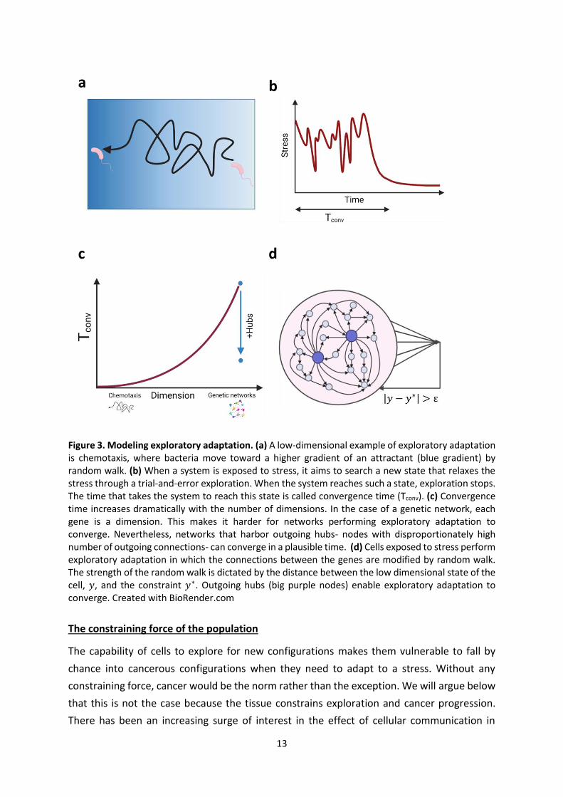
Can a high dimensional system converge under exploratory adaptation? In contrast to evolutionary trial-and-error dynamics, which takes place at the population level,
exploratory adaptation involves trial-and-error at the level of the individual. Variation is
created in a single system along time and selected by feedback from the environment. Such
dynamics have been considered in several biological contexts, such as spindle assembly and
bacterial chemotaxis (Fig. 3a-b) (Kirschner and Gerhart, 2005). However, these instances of
exploration take place in three-dimensional space, and therefore, are feasible in terms of
11
convergence success at finite time. For cancer cells to adapt by exploring gene expression,
they face a different situation of a high-dimensional system (order of thousands of degrees
of freedom) undergoing some random search in its vast configuration space (Fig. 3c). With no
sophisticated means to compute an error from some target function, the cell has only its
global stress level at its disposal to provide feedback on the exploration. Under what
conditions can such a learning scheme converge successfully? Since this is a quantitative
question, one naturally turns to mathematical modeling: a model that captures the essential
features of exploratory adaptation in high-dimensional space can shed light on the conditions
for its possibility of convergence.
We constructed such a model based on large random networks of interacting elements
(Schreier et al., 2017). This is a popular modeling approach for gene regulation when general
properties are of interest, such as number of fixed points, evolvability or canalization (Drossel,
2008; Kauffman, 1993; Li et al., 2013).
The model (Fig. 3d) describes a system of N interacting genes (N on the order of 1000), whose
state (expression level) is represented by the N-dimensional vector 𝒙 = (𝑥1, 𝑥2 … 𝑥𝑁). Their
time evolution is determined by a nonlinear dynamic rule governed by a random interaction
matrix 𝑊 , in which the element 𝑊𝑖𝑗 represents the strength of influence of gene 𝑗 on gene 𝑖.
Regulatory plasticity is represented by the ability of the interactions 𝑊𝑖𝑗 to change their
strengths over time; in exploratory dynamics, these changes will be essentially random and
their amplitude controlled by feedback. The critical ingredient of the model is to define the
feedback and close the loop.
A given cellular phenotype can be realized by different expression patterns. Mathematically,
this corresponds to a low-dimensional projection, which we take to be linear, with weights
given by arbitrary numbers 𝒃 :
𝑦(𝑡) = 𝒃 ∙ 𝒙(𝑡) (2)
The challenge is presented to the system by the constraint of maintaining the phenotype in a
range 𝑦(𝑡) ≈ 𝑦∗. This allows for multiple gene expression patterns to comply with the
constraint in different microscopic implementations. If, however, the projection deviates
outside an allowed range, a global cellular stress will emerge, 𝑆(𝑦 − 𝑦∗), causing the system
to initiate exploratory dynamics in the form of random changes in the interaction matrix 𝑊.
The amplitude of these changes is dictated by cellular stress 𝑆: it relaxes if and when it
reaches a stable state that relieves stress, 𝑆 ≈ 0 (Fig. 3b). In this way, a simple feedback loop
couples internal exploratory dynamics to the suitability of the current configuration to relieve
stress and to match the environmental demand.
12
One might imagine that such a simple algorithm will not allow convergence to an appropriate
phenotype in a reasonable time for large networks, and indeed this is the case for
homogeneous random matrices 𝑊, corresponding to identical and independent probability
of connections between any two genes. Intriguingly, the main conclusion from the modeling
work was that exploratory adaptation is sensitive to network structure; in particular, it is likely
to converge for networks with outgoing hubs - a small number of nodes with
disproportionately high connectivity (Fig. 3c). For example, in a scale-free network topology
there are hubs in the tail of the distribution that are connected to a large fraction of the
network (Schreier et al., 2017). However, scale-free networks are merely a mathematical tool
to classify topologies; we have shown that a handful of outgoing genes in an arbitrary network
are sufficient to induce the effect. In real gene networks, master regulators are well known
genes that control up to hundreds of other genes (Cai et al., 2020). Such hubs are usually
considered in the context of specific gene programs that they regulate, but our model
proposes that they can also coordinate network plasticity and drive it more easily through an
exploratory process to discover novel stable states. This new role is similar to the stabilizing
effect of an external feedback that suppresses irregular network activity (Rivkind et al., 2020).
Importantly, it is in line with recent discoveries on master regulators in the context of cancer
reprogramming.
For instance, ZEB1 is a key player in EMT, where its most well-known function is the
suppression of epithelial genes. Interestingly, under some conditions the flexible nature of
this regulator is revealed and ZEB1 can turn into a transcriptional activator in aggressive
cancer types (Lehmann et al., 2016). In fact, this is not a special property of ZEB1, but also
exists for other transcription factors (Stemmler et al., 2019). Thus, master regulators can
direct alternative expression patterns in different situations. In the absence of an external
agent that directs the hubs which program to choose, our framework of exploratory
adaptation suggests that the appropriate configuration emerges through stress-mediated
feedback.
In conclusion, the feasibility of exploratory adaptation in structured random networks, and
the finding that actual gene regulatory networks fulfill the key requirements of the model,
suggest that this mechanism can be implemented in cells adapting to stressful conditions. In
particular it can help explain the behavior of cancer progression in drug resistance and
metastasis. Intriguingly, applying this model to experimental data, Celiku et al (Celiku et al.,
2019) have shown that the adaptation of glioblastoma cells as they spread to diverse tumor
microenvironments can be at least partly attributed to exploratory dynamics. Future work
may use this framework and model to analyze experimental data in other systems.
13
Figure 3. Modeling exploratory adaptation. (a) A low-dimensional example of exploratory adaptation is chemotaxis, where bacteria move toward a higher gradient of an attractant (blue gradient) by random walk. (b) When a system is exposed to stress, it aims to search a new state that relaxes the stress through a trial-and-error exploration. When the system reaches such a state, exploration stops. The time that takes the system to reach this state is called convergence time (Tconv). (c) Convergence time increases dramatically with the number of dimensions. In the case of a genetic network, each gene is a dimension. This makes it harder for networks performing exploratory adaptation to converge. Nevertheless, networks that harbor outgoing hubs- nodes with disproportionately high number of outgoing connections- can converge in a plausible time. (d) Cells exposed to stress perform exploratory adaptation in which the connections between the genes are modified by random walk. The strength of the random walk is dictated by the distance between the low dimensional state of the cell, 𝑦, and the constraint 𝑦∗. Outgoing hubs (big purple nodes) enable exploratory adaptation to converge. Created with BioRender.com
The constraining force of the population
The capability of cells to explore for new configurations makes them vulnerable to fall by
chance into cancerous configurations when they need to adapt to a stress. Without any
constraining force, cancer would be the norm rather than the exception. We will argue below
that this is not the case because the tissue constrains exploration and cancer progression.
There has been an increasing surge of interest in the effect of cellular communication in
14
cancer and many excellent reviews gather previous findings in this area (Bissell and Hines,
2011; Capp, 2005; Soto and Sonnenschein, 2011). Here, we tie these findings to our learning
framework and emphasize the significance of cellular communication as a constraint on
exploratory adaptation.
Mutations and growth control in normal tissues
Cancer is traditionally associated with the occurrence of specific patterns of mutations that
are thought to drive transformation. It turns out that these mutations are not sufficient for
transformation. A recent comprehensive RNA sequencing analysis detected thousands of
somatic mutations across all human tissues and in almost all tested individuals, including
mutations at cancer hotspots and other cancer genes (Yizhak et al., 2019). Greater numbers
of mutations were observed in tissues that are more exposed to carcinogenic environmental
factors, such as sun-exposed skin, esophagus mucosa and lung (Yizhak et al., 2019).
Congruently, a recent analysis of sun-exposed skin tissues revealed a remarkably high burden
of somatic mutations of averaged two to six mutations per megabase per cell, similar to that
found in many cancers (Martincorena et al., 2015). The frequency of mutations that were
previously identified as "driver mutations" in these tissues was surprisingly high. For instance,
there were more NOTCH1 mutations in a sun-exposed skin biopsy than have been identified
in more than 5000 cancers sequenced by The Cancer Genome Atlas (Martincorena et al.,
2015). Remarkably, clones carrying driver genes expand in normal skin tissues; however, this
growth stops early in the expansion, giving rise to limited sized clones (Martincorena et al.,
2015). Some of the analyzed clones carried two to three driver mutations while having a
totally normal skin phenotype. Similarly, sequencing studies of normal blood cells revealed
signatures of somatic mutations broadly similar to blood cancer (Genovese et al., 2014;
Jaiswal et al., 2014). These findings raise the question: how is homeostasis maintained in
normal tissues despite the high burden of cancer driver mutations? What prevents the
appearance of cancer phenotype in these tissues?
One widely-accepted explanation is that driver mutations trigger oncogene-induced
senescence (OIS), arresting the proliferation of cells before the accumulation of additional
mutations (Bennett, 2003; Huang et al., 2017; Kaplon et al., 2014; Michaloglou et al., 2005;
Serrano et al., 1997; Wajapeyee et al., 2008). OIS is usually depicted as a cell-autonomous
stress response, namely expressing an oncogene in a cell leads to stress which induces a
growth arrest in that cell. However, a recent work by Ruiz-Vega at al. (Ruiz-Vega et al., 2020),
using a mouse model of BRAF-driven nevus formation, showed that this is not necessarily the
case. BRAF mutation is the most common driver mutation in melanoma (Davies et al., 2002),
yet it is present in 89% of benign nevi (pigmented ‘moles’) (Pollock et al., 2003). In this work,
no evidence supported the senescence of nevus cells, either compared with other skin cells
15
or other melanocytes. Moreover, nevus size distribution could not be fit by any simple cell-
autonomous model of growth arrest yet, were easily fit by models of collective feedback
between the cells. This emphasizes the significance of the tissue level of organization, in
particular cell-cell interactions, in maintaining homeostasis and constraining cancer
development.
For better or worse: micro-environment controls cell fate
Intriguingly, the effect of cellular communication is so dominant that it not only stops the
development of a cancerous phenotype, but can actually turn around cell fate. The classic
work of Beatrice Mintz and Karl Illmenesse (Mintz and Illmensee, 1975) showed that placing
teratocarcinoma (undifferentiated embryonic carcinoma cells) in a blastocyst gave rise to
perfectly normal and tumor free offspring that displayed many traits of the parental tumor
cells. Similarly, transplantation of nuclei from malignant cells in enucleated oocytes gave rise
to stem cells that were able to produce mice (Hochedlinger, 2004).
Numerous works provided additional evidence for the ability of the micro-environment to
suppress tumor growth and induce differentiation to a variety of functional tissues. In their
seminal work, the laboratories of Ole Petersen and Mina Bissel showed that breast cancer
cells revert to nearly normal phenotype when cultured in three-dimensional culture that
mimics the normal breast tissue (Howlett et al., 1994). The genome of the reverted cells was
shown to be similar to mutated and malignant cells grown in two-dimensional cultures (Rizki
et al., 2008; Weaver et al., 1995). Similarly, highly malignant melanoma cells injected into
Zebra fish embryos (Kasemeier-Kulesa et al., 2008), mammary carcinoma cells recombined
with normal mammary gland stroma (Maffini et al., 2005), and liver cancer cells injected into
normal liver (McCullough et al., 1997) are all additional examples for the ability of the
microenvironment to normalize cancer cells. More recently, it was shown that converting
invasive breast cancer cells into adipocytes by treating them with appropriate cues inhibits
cancer metastasis (Ishay-Ronen et al., 2019). Unequivocally, all these findings point to the fact
that the environment can play a crucial role in redirecting the phenotype of cancer cells and
determining whether cancer is contained or spreads. They also demonstrate the high
plasticity of cancer cells which can be exploited to adapt to dynamic changes, triggered by
external signals.
Unfortunately, the stroma (fibroblasts, vasculature, immune cells and interstitial ECM) that
suppresses the growth of tumors, can induce tumorigenesis when it undergoes detrimental
changes. Maffini et al. (Maffini et al., 2004) showed that combining a carcinogen-exposed
mammary stroma with vehicle-exposed mammary epithelium resulted in neoplasm. The
reverse combination did not. Similar results were obtained from a normal mammary cell line
16
with an irradiated stroma (Mh and Sa, 2000), and a normal prostate cell line and fibroblasts
derived from prostate cancer(Barclay et al., 2005). These findings indicate that carcinogenesis
can be the result of an abnormal interaction between the stroma and epithelial cells.
Moreover, such interaction can support tumor progression and induce resistance (Chan et al.,
2019; Shaked, 2019).
These arguments suggest a balance between the exploratory drive of individual cells under
stress, and the constraining effect of interaction with the environment in a healthy tissue. The
emerging picture suggests a view of cancer which goes beyond the single-cell and places the
disease at the level of the cell-tissue interface. In this framework cellular communication is a
double-edged sword (Bissell and Hines, 2011). It can prevent transformation of normal cells
harboring mutations and normalize cancer cells despite their mutations, but also induce
tumorigenesis when aberrant. As Smithers stated it: “Cancer is no more a disease of cells
than a traffic jam is a disease of cars. A lifetime study of the internal combustion engine would
not help anyone to understand our traffic problems”(Smithers, 1962).
Conclusion
Taken together, we propose that single cells have the capability to learn novel phenotypes by
utilizing their internal plasticity and under the direction of global cellular stress. This
proposition is based on analogies between features of cellular adaptation to stress and
learning in neural networks. We view cells in a tissue as a system of coupled explorers or
learners. The state of the entire system is determined by an interplay between the constraints
of the population and the exploratory drive of individual cells. At one end of the spectrum,
normal tissues are characterized by high tissue constraints and low exploration. Tissue
homeostasis is maintained by various mechanisms that include biochemical and mechanical
interactions between cells. At the other end of the spectrum, cancer progression is associated
with looser tissue constraints that allow a high exploratory behavior. This view shares with
the Tissue Organization Field Theory (TOFT) of cancer the premise that cells are not quiescent
by default, but rather maintain continuous internal drive, while homeostasis is enforced on
them by the tissue level of organization (Sonnenschein and Soto, 2020). It is also consistent
with the idea that aging affects the propensity for cancer development through the decrease
in effective tissue homeostasis (Capp and Thomas, 2021). Our conceptual framework may
open the door to novel directions in cancer research and therapeutic development.
Acknowledgements This work was supported in part by the Israeli Science Foundation (Grant no. 346/16, O.B.; and Grant No. 155/18, N.B.). We acknowledge the Adams Fellowship Program of the Israel Academy of Science and Humanities (AS).
17
References
Adorno-Cruz, V., Kibria, G., Liu, X., Doherty, M., Junk, D.J., Guan, D., Hubert, C., Venere, M., Mulkearns-Hubert, E., Sinyuk, M., et al. (2015). Cancer Stem Cells: Targeting the Roots of Cancer, Seeds of Metastasis, and Sources of Therapy Resistance. Cancer Res 75, 924–929.
Amos, W., and Harwood, J. (1998). Factors affecting levels of genetic diversity in natural populations. Philos Trans R Soc Lond B Biol Sci 353, 177–186.
Baluška, F., and Levin, M. (2016). On having no head: Cognition throughout biological systems. Frontiers in Psychology 7, 902.
Barclay, W.W., Woodruff, R.D., Hall, M.C., and Cramer, S.D. (2005). A System for Studying Epithelial-Stromal Interactions Reveals Distinct Inductive Abilities of Stromal Cells from Benign Prostatic Hyperplasia and Prostate Cancer. Endocrinology 146, 13–18.
Baron, M., Tagore, M., Hunter, M.V., Kim, I.S., Moncada, R., Yan, Y., Campbell, N.R., White, R.M., and Yanai, I. (2020). The Stress-Like Cancer Cell State Is a Consistent Component of Tumorigenesis. Cell Systems 11, 536-546.e7.
Bell, C.C., and Gilan, O. (2020). Principles and mechanisms of non-genetic resistance in cancer. Br J Cancer 122, 465–472.
Bennett, D.C. (2003). Human melanocyte senescence and melanoma susceptibility genes. Oncogene 22, 3063–3069.
Bissell, M.J., and Hines, W.C. (2011). Why don’t we get more cancer? A proposed role of the microenvironment in restraining cancer progression. Nat Med 17, 320–329.
Brabletz, T. (2012). To differentiate or not — routes towards metastasis. Nature Reviews Cancer 12, 425–436.
Braun, E. (2015). The unforeseen challenge: from genotype-to-phenotype in cell populations. Rep. Prog. Phys. 78, 036602.
Cai, W., Zhou, W., Han, Z., Lei, J., Zhuang, J., Zhu, P., Wu, X., and Yuan, W. (2020). Master regulator genes and their impact on major diseases. PeerJ 8, e9952.
Capp, J.-P. (2005). Stochastic gene expression, disruption of tissue averaging effects and cancer as a disease of development. Bioessays 27, 1277–1285.
Capp, J.-P., and Thomas, F. (2021). Tissue-disruption-induced cellular stochasticity and epigenetic drift: Common origins of aging and cancer? Bioessays 43, e2000140.
Celiku, O., Gilbert, M.R., and Lavi, O. (2019). Computational modeling demonstrates that glioblastoma cells can survive spatial environmental challenges through exploratory adaptation. Nature Communications 10, 5704.
Chaffer, C.L., and Weinberg, R.A. (2011). A Perspective on Cancer Cell Metastasis. Science 331, 1559–1564.
Chan, T.-S., Shaked, Y., and Tsai, K.K. (2019). Targeting the Interplay Between Cancer Fibroblasts, Mesenchymal Stem Cells, and Cancer Stem Cells in Desmoplastic Cancers. Front. Oncol. 9.
18
Chang, J.C. (2016). Cancer stem cells. Medicine (Baltimore) 95:S1(e4766).
Cheung, K.J., and Ewald, A.J. (2016). A collective route to metastasis: Seeding by tumor cell clusters. Science 352, 167–169.
Davies, H., Bignell, G.R., Cox, C., Stephens, P., Edkins, S., Clegg, S., Teague, J., Woffendin, H., Garnett, M.J., Bottomley, W., et al. (2002). Mutations of the BRAF gene in human cancer. Nature 417, 949–954.
De Angelis, M.L., Francescangeli, F., La Torre, F., and Zeuner, A. (2019). Stem Cell Plasticity and Dormancy in the Development of Cancer Therapy Resistance. Front. Oncol. 9, 626.
Denny, S.K., Yang, D., Chuang, C.-H., Brady, J.J., Lim, J.S., Grüner, B.M., Chiou, S.-H., Schep, A.N., Baral, J., Hamard, C., et al. (2016). Nfib Promotes Metastasis through a Widespread Increase in Chromatin Accessibility. Cell 166, 328–342.
Doherty, M.R., Smigiel, J.M., Junk, D.J., and Jackson, M.W. (2016). Cancer Stem Cell Plasticity Drives Therapeutic Resistance. Cancers 8, 8.
Drossel, B. (2008). Random Boolean Networks. In Reviews of Nonlinear Dynamics and Complexity, (John Wiley & Sons, Ltd), pp. 69–110.
Flavahan, W.A., Gaskell, E., and Bernstein, B.E. (2017). Epigenetic plasticity and the hallmarks of cancer. Science 357, eaal2380.
Freddolino, P.L., Yang, J., Momen-Roknabadi, A., and Tavazoie, S. (2018). Stochastic tuning of gene expression enables cellular adaptation in the absence of pre-existing regulatory circuitry. ELife 7, e31867.
Garraway, L.A., and Lander, E.S. (2013). Lessons from the cancer genome. Cell 153, 17–37.
Genovese, G., Kähler, A.K., Handsaker, R.E., Lindberg, J., Rose, S.A., Bakhoum, S.F., Chambert, K., Mick, E., Neale, B.M., Fromer, M., et al. (2014). Clonal Hematopoiesis and Blood-Cancer Risk Inferred from Blood DNA Sequence. N Engl J Med 371, 2477–2487.
Guo, M., Peng, Y., Gao, A., Du, C., and Herman, J.G. (2019). Epigenetic heterogeneity in cancer. Biomark Res 7, 23.
Gupta, G.P., and Massagué, J. (2006). Cancer Metastasis: Building a Framework. Cell 127, 679–695.
Hochedlinger, K. (2004). Reprogramming of a melanoma genome by nuclear transplantation. Genes & Development 18, 1875–1885.
Holland, P., Bergenholm, D., Börlin, C.S., Liu, G., and Nielsen, J. (2019). Predictive models of eukaryotic transcriptional regulation reveals changes in transcription factor roles and promoter usage between metabolic conditions. Nucleic Acids Research 47, 4986–5000.
Howlett, A.R., Petersen, O.W., Steeg, P.S., and Bissell, M.J. (1994). A Novel Function for the nm23-H1 Gene: Overexpression in Human Breast Carcinoma Cells Leads to the Formation of Basement Membrane and Growth Arrest. J Natl Cancer Inst 86, 1838–1844.
Huang, J.M., Chikeka, I., and Hornyak, T.J. (2017). Melanocytic Nevi and the Genetic and Epigenetic Control of Oncogene-Induced Senescence. Dermatol Clin 35, 85–93.
Ishay-Ronen, D., Diepenbruck, M., Kalathur, R.K.R., Sugiyama, N., Tiede, S., Ivanek, R., Bantug, G., Morini, M.F., Wang, J., Hess, C., et al. (2019). Gain Fat—Lose Metastasis: Converting Invasive Breast Cancer Cells into Adipocytes Inhibits Cancer Metastasis. Cancer Cell 35, 17-32.e6.
Jaiswal, S., Fontanillas, P., Flannick, J., Manning, A., Grauman, P.V., Mar, B.G., Lindsley, R.C., Mermel, C.H., Burtt, N., Chavez, A., et al. (2014). Age-Related Clonal Hematopoiesis Associated with Adverse Outcomes. N Engl J Med 371, 2488–2498.
James, L.C., and Tawfik, D.S. (2003). Conformational diversity and protein evolution – a 60-year-old hypothesis revisited. Trends in Biochemical Sciences 28, 361–368.
Jr, P.B.F., Diggins, K.E., Polikowsky, H.G., Mohan, S.R., Seegmiller, A.C., and Irish, J.M. (2016). High-Dimensional Analysis of Acute Myeloid Leukemia Reveals Phenotypic Changes in Persistent Cells during Induction Therapy. PLOS ONE 11, e0153207.
Kaplon, J., Hömig‐Hölzel, C., Gao, L., Meissl, K., Verdegaal, E.M.E., Burg, S.H. van der, Doorn, R. van, and Peeper, D.S. (2014). Near-genomewide RNAi screening for regulators of BRAFV600E-induced senescence identifies RASEF, a gene epigenetically silenced in melanoma. Pigment Cell & Melanoma Research 27, 640–652.
Kasemeier-Kulesa, J.C., Teddy, J.M., Postovit, L.-M., Seftor, E.A., Seftor, R.E.B., Hendrix, M.J.C., and Kulesa, P.M. (2008). Reprogramming multipotent tumor cells with the embryonic neural crest microenvironment. Dev Dyn 237, 2657–2666.
Kauffman, S.A. (1993). The Origins of Order: Self-organization and Selection in Evolution (Oxford University Press).
Kim, C., Gao, R., Sei, E., Brandt, R., Hartman, J., Hatschek, T., Crosetto, N., Foukakis, T., and Navin, N.E. (2018). Chemoresistance Evolution in Triple-Negative Breast Cancer Delineated by Single-Cell Sequencing. Cell 173, 879-893.e13.
Kirschner, M.W., and Gerhart, J.C. (2005). The Plausibility of Life: Resolving Darwin’s Dilemma (Yale University Press).
Klein, C.A. (2013). Selection and adaptation during metastatic cancer progression. Nature 501, 365–372.
Krishnaswamy, S., Zivanovic, N., Sharma, R., Pe’er, D., and Bodenmiller, B. (2018). Learning time-varying information flow from single-cell epithelial to mesenchymal transition data. PLoS ONE 13, e0203389.
Lambert, A.W., Pattabiraman, D.R., and Weinberg, R.A. (2017). Emerging Biological Principles of Metastasis. Cell 168, 670–691.
Lawson, D.A., Kessenbrock, K., Davis, R.T., Pervolarakis, N., and Werb, Z. (2018). Tumour heterogeneity and metastasis at single-cell resolution. Nat Cell Biol 20, 1349–1360.
20
Lee, T.I., Rinaldi, N.J., Robert, F., Odom, D.T., Bar-Joseph, Z., Gerber, G.K., Hannett, N.M., Harbison, C.T., Thompson, C.M., Simon, I., et al. (2002). Transcriptional Regulatory Networks in Saccharomyces cerevisiae. Science 298, 799–804.
Lehmann, W., Mossmann, D., Kleemann, J., Mock, K., Meisinger, C., Brummer, T., Herr, R., Brabletz, S., Stemmler, M.P., and Brabletz, T. (2016). ZEB1 turns into a transcriptional activator by interacting with YAP1 in aggressive cancer types. Nature Communications 7, 10498.
Li, Z., Bianco, S., Zhang, Z., and Tang, C. (2013). Generic properties of random gene regulatory networks. Quant Biol 1, 253–260.
Liu, Y., Wu, J., Sun, N., Tu, C., Shi, X., Cheng, H., Liu, S., Li, S., Wang, Y., Zheng, Y., et al. (2017). Intrinsically Disordered Proteins as Important Players during Desiccation Stress of Soybean Radicles. J Proteome Res 16, 2393–2409.
Loo, J.M., Scherl, A., Nguyen, A., Man, F.Y., Weinberg, E., Zeng, Z., Saltz, L., Paty, P.B., and Tavazoie, S.F. (2015). Extracellular Metabolic Energetics Can Promote Cancer Progression. Cell 160, 393–406.
Luzzi, K.J., MacDonald, I.C., Schmidt, E.E., Kerkvliet, N., Morris, V.L., Chambers, A.F., and Groom, A.C. (1998). Multistep Nature of Metastatic Inefficiency. The American Journal of Pathology 153, 865–873.
Lytle, N.K., Barber, A.G., and Reya, T. (2018). Stem cell fate in cancer growth, progression and therapy resistance. Nat Rev Cancer 18, 669–680.
Maffini, M.V., Soto, A.M., Calabro, J.M., Ucci, A.A., and Sonnenschein, C. (2004). The stroma as a crucial target in rat mammary gland carcinogenesis. Journal of Cell Science 117, 1495–1502.
Maffini, M.V., Calabro, J.M., Soto, A.M., and Sonnenschein, C. (2005). Stromal Regulation of Neoplastic Development. Am J Pathol 167, 1405–1410.
Marine, J.-C., Dawson, S.-J., and Dawson, M.A. (2020). Non-genetic mechanisms of therapeutic resistance in cancer. Nat Rev Cancer 20, 743–756.
Martincorena, I., and Campbell, P.J. (2015). Somatic mutation in cancer and normal cells. Science 349, 1483–1489.
Martincorena, I., Roshan, A., Gerstung, M., Ellis, P., Loo, P.V., McLaren, S., Wedge, D.C., Fullam, A., Alexandrov, L.B., Tubio, J.M., et al. (2015). High burden and pervasive positive selection of somatic mutations in normal human skin. Science 348, 880–886.
Massagué, J., and Obenauf, A.C. (2016). Metastatic colonization by circulating tumour cells. Nature 529, 298–306.
Maynard, A., McCoach, C.E., Rotow, J.K., Harris, L., Haderk, F., Kerr, D.L., Yu, E.A., Schenk, E.L., Tan, W., Zee, A., et al. (2020). Therapy-Induced Evolution of Human Lung Cancer Revealed by Single-Cell RNA Sequencing. Cell 182, 1232-1251.e22.
McCullough, K.D., Coleman, W.B., Smith, G.J., and Grisham, J.W. (1997). Transformed Rat Liver Epithelial Cells into the Liver’. Cancer Research 57, 1807–1813.
Mh, B.-H., and Sa, R. (2000). Irradiated mammary gland stroma promotes the expression of tumorigenic potential by unirradiated epithelial cells. Cancer Res 60, 1254–1260.
21
Michaloglou, C., Vredeveld, L.C.W., Soengas, M.S., Denoyelle, C., Kuilman, T., van der Horst, C.M.A.M., Majoor, D.M., Shay, J.W., Mooi, W.J., and Peeper, D.S. (2005). BRAF E600 -associated senescence-like cell cycle arrest of human naevi. Nature 436, 720–724.
Milanovic, M., Fan, D.N.Y., Belenki, D., Däbritz, J.H.M., Zhao, Z., Yu, Y., Dörr, J.R., Dimitrova, L., Lenze, D., Monteiro Barbosa, I.A., et al. (2018). Senescence-associated reprogramming promotes cancer stemness. Nature 553, 96–100.
Mintz, B., and Illmensee, K. (1975). Normal genetically mosaic mice produced from malignant teratocarcinoma cells. Proc. Natl. Acad. Sci. U.S.A. 72, 3585–3589.
Niklas, K.J., Bondos, S.E., Dunker, A.K., and Newman, S.A. (2015). Rethinking gene regulatory networks in light of alternative splicing, intrinsically disordered protein domains, and post-translational modifications. Front. Cell Dev. Biol. 3, 8.
NOWEL, PC. (1976). The clonal evolution of tumor cell populations. Acquired genetic lability permits stepwise selection of variant sublines and underlies tumor progression. Science 194, 223–228.
Obenauf, A.C., and Massagué, J. (2015). Surviving at a Distance: Organ-Specific Metastasis. Trends in Cancer 1, 76–91.
Peng, Z., Yan, J., Fan, X., Mizianty, M.J., Xue, B., Wang, K., Hu, G., Uversky, V.N., and Kurgan, L. (2015). Exceptionally abundant exceptions: comprehensive characterization of intrinsic disorder in all domains of life. Cell. Mol. Life Sci. 72, 137–151.
Pisco, A.O., and Huang, S. (2015). Non-genetic cancer cell plasticity and therapy-induced stemness in tumour relapse: ‘What does not kill me strengthens me.’ British Journal of Cancer 112, 1725–1732.
Piskounova, E., Agathocleous, M., Murphy, M.M., Hu, Z., Huddlestun, S.E., Zhao, Z., Leitch, A.M., Johnson, T.M., DeBerardinis, R.J., and Morrison, S.J. (2015). Oxidative stress inhibits distant metastasis by human melanoma cells. Nature 527, 186–191.
Pollock, P.M., Harper, U.L., Hansen, K.S., Yudt, L.M., Stark, M., Robbins, C.M., Moses, T.Y., Hostetter, G., Wagner, U., Kakareka, J., et al. (2003). High frequency of BRAF mutations in nevi. Nature Genetics 33, 19–20.
Ramirez, M., Rajaram, S., Steininger, R.J., Osipchuk, D., Roth, M.A., Morinishi, L.S., Evans, L., Ji, W., Hsu, C.-H., Thurley, K., et al. (2016). Diverse drug-resistance mechanisms can emerge from drug-tolerant cancer persister cells. Nat Commun 7, 10690.
Rivkind, A., Schreier, H., Brenner, N., and Barak, O. (2020). Scale free topology as an effective feedback system. PLOS Computational Biology 16, e1007825.
Rizki, A., Weaver, V.M., Lee, S.-Y., Rozenberg, G.I., Chin, K., Myers, C.A., Bascom, J.L., Mott, J.D., Semeiks, J.R., Grate, L.R., et al. (2008). A human breast cell model of preinvasive to invasive transition. Cancer Res 68, 1378–1387.
Ruiz-Vega, R., Chen, C.-F., Razzak, E., Vasudeva, P., Krasieva, T.B., Shiu, J., Caldwell, M.G., Yan, H., Lowengrub, J., Ganesan, A.K., et al. (2020). Dynamics of nevus development implicate cell cooperation in the growth arrest of transformed melanocytes. ELife 9, e61026.
Salas-Vega, S., Iliopoulos, O., and Mossialos, E. (2017). Assessment of Overall Survival, Quality of Life, and Safety Benefits Associated With New Cancer Medicines. JAMA Oncol 3, 382–390.
22
Sánchez Alvarado, A., and Yamanaka, S. (2014). Rethinking differentiation: stem cells, regeneration, and plasticity. Cell 157, 110–119.
Schreier, H.I., Soen, Y., and Brenner, N. (2017). Exploratory adaptation in large random networks. Nature Communications 8, 14826.
Scott, J., Kuhn, P., and Anderson, A.R.A. (2012). Unifying metastasis — integrating intravasation, circulation and end-organ colonization. Nature Reviews Cancer 12, 445–446.
Serrano, M., Lin, A.W., McCurrach, M.E., Beach, D., and Lowe, S.W. (1997). Oncogenic ras Provokes Premature Cell Senescence Associated with Accumulation of p53 and p16INK4a. Cell 88, 593–602.
Shaffer, S.M., Dunagin, M.C., Torborg, S.R., Torre, E.A., Emert, B., Krepler, C., Beqiri, M., Sproesser, K., Brafford, P.A., Xiao, M., et al. (2017). Rare cell variability and drug-induced reprogramming as a mode of cancer drug resistance. Nature 546, 431–435.
Shaked, Y. (2019). The pro-tumorigenic host response to cancer therapies. Nat Rev Cancer 19, 667–685.
Sharma, S.V., Lee, D.Y., Li, B., Quinlan, M.P., Takahashi, F., Maheswaran, S., McDermott, U., Azizian, N., Zou, L., Fischbach, M.A., et al. (2010). A chromatin-mediated reversible drug tolerant state in cancer cell subpopulations. Cell 141, 69–80.
Smithers, D.W. (1962). CANCER AN ATTACK ON CYTOLOGISM. The Lancet 279, 493–499.
Soen, Y., Knafo, M., and Elgart, M. (2015). A principle of organization which facilitates broad Lamarckian-like adaptations by improvisation. Biology Direct 10, 68.
Sonnenschein, C., and Soto, A.M. (2020). Over a century of cancer research: Inconvenient truths and promising leads. PLoS Biol 18, e3000670.
Soto, A.M., and Sonnenschein, C. (2011). The tissue organization field theory of cancer: A testable replacement for the somatic mutation theory. Bioessays 33, 332–340.
Stemmler, M.P., Eccles, R.L., Brabletz, S., and Brabletz, T. (2019). Non-redundant functions of EMT transcription factors. Nature Cell Biology 21, 102–112.
Stewart, C.A., Gay, C.M., Xi, Y., Sivajothi, S., Sivakamasundari, V., Fujimoto, J., Bolisetty, M., Hartsfield, P.M., Balasubramaniyan, V., Chalishazar, M.D., et al. (2020). Single-cell analyses reveal increased intratumoral heterogeneity after the onset of therapy resistance in small-cell lung cancer. Nat Cancer 1, 423–436.
The International Cancer Genome Consortium (2010). International network of cancer genome projects. Nature 464, 993–998.
Valiente, M., Obenauf, A.C., Jin, X., Chen, Q., Zhang, X.H.-F., Lee, D.J., Chaft, J.E., Kris, M.G., Huse, J.T., Brogi, E., et al. (2014). Serpins Promote Cancer Cell Survival and Vascular Co-Option in Brain Metastasis. Cell 156, 1002–1016.
Vasan, N., Baselga, J., and Hyman, D.M. (2019). A view on drug resistance in cancer. Nature 575, 299–309.
23
Vogelstein, B., Papadopoulos, N., Velculescu, V.E., Zhou, S., Diaz, L.A., and Kinzler, K.W. (2013). Cancer Genome Landscapes. Science 339, 1546–1558.
Wajapeyee, N., Serra, R.W., Zhu, X., Mahalingam, M., and Green, M.R. (2008). Oncogenic BRAF Induces Senescence and Apoptosis through Pathways Mediated by the Secreted Protein IGFBP7. Cell 132, 363–374.
Weaver, V.M., Howlett, A.R., Langton-Webster, B., Petersen, O.W., and Bissell, M.J. (1995). The development of a functionally relevant cell culture model of progressive human breast cancer. Seminars in Cancer Biology 6, 175–184.
Weinstein, J.N., Collisson, E.A., Mills, G.B., Shaw, K.R.M., Ozenberger, B.A., Ellrott, K., Shmulevich, I., Sander, C., and Stuart, J.M. (2013). The Cancer Genome Atlas Pan-Cancer analysis project. Nature Genetics 45, 1113–1120.
Welch, D.R., and Hurst, D.R. (2019). Defining the Hallmarks of Metastasis. Cancer Res 79, 3011–3027.
Woronoff, G., Nghe, P., Baudry, J., Boitard, L., Braun, E., Griffiths, A.D., and Bibette, J. (2020). Metabolic cost of rapid adaptation of single yeast cells. Proc Natl Acad Sci USA 117, 10660–10666.
Wright, P.E., and Dyson, H.J. (2015). Intrinsically disordered proteins in cellular signalling and regulation. Nat Rev Mol Cell Biol 16, 18–29.
Yizhak, K., Aguet, F., Kim, J., Hess, J.M., Kübler, K., Grimsby, J., Frazer, R., Zhang, H., Haradhvala, N.J., Rosebrock, D., et al. (2019). RNA sequence analysis reveals macroscopic somatic clonal expansion across normal tissues. Science 364, eaaw0726.
Zhang, J., Baran, J., Cros, A., Guberman, J.M., Haider, S., Hsu, J., Liang, Y., Rivkin, E., Wang, J., Whitty, B., et al. (2011). International Cancer Genome Consortium Data Portal—a one-stop shop for cancer genomics data. Database 2011.
Zugazagoitia, J., Guedes, C., Ponce, S., Ferrer, I., Molina-Pinelo, S., and Paz-Ares, L. (2016). Current Challenges in Cancer Treatment. Clin Ther 38, 1551–1566.