1371 CHAPTER 57 CANCER REHABILITATION Andrea L. Cheville Cancer rehabilitation addresses physical impairments related to tumor effects and cancer treatment. Most func- tional problems experienced by patients with cancer also occur as a result of other disease processes, such as isch- emia, trauma, and arthritis. The fact that impairments arise in the context of cancer might alter their management little. Their successful rehabilitation, however, mandates that treatments integrate cancer-specific concerns—limited prognoses, dynamic lesions, heavy symptom burden, and treatment-related toxicities—into a humane and realistic treatment plan. Cancer is a pathologic process characterized by dysregu- lated cell growth and systemic spread. All tissue types have neoplastic potential and can become cancerous. Tissues distinguished by rapid cell turnover (e.g., gastrointestinal mucosa), hormone sensitivity (e.g., breast and prostate), and regular exposure to environmental mutagens (e.g., lung and skin) have higher rates of malignant transforma- tion. The fact that any tissue can develop cancer means that cancer rehabilitation must address all body parts and sys- tems. Despite this broad scope, the field condenses into a manageable body of expertise predominantly focused on the sequelae of cancer treatment, maladaptive host responses (e.g., paraneoplastic syndromes), and the erosive effects of cancer on bones and neural tissue. Cancer rehabilitation extends far beyond the control functional decline in patients with metastatic disease. With ever-increasing cancer survivorship, the number of patients whose disease has been eliminated or successfully tem- porized continues to grow. The National Cancer Institute estimates that more than 13 million Americans with a his- tory of cancer were alive in January 2010. These patients are eager to lead highly functional and productive lives despite the legacy of their disease. A unique and intensely chal- lenging feature of cancer rehabilitation is the need to treat patients with vastly different degrees of infirmity. Given the magnitude of current need, physiatrists can elect to treat patients who are cured of their cancers or whose cancers have progressed to being widely metastatic. This chapter is intended to provide physiatrists and other readers with an overview of the issues relevant to the rehabilitation of patients with cancer. Emphasis is placed on problems that affect the nervous and musculoskeletal systems. Epidemiology Cancer is a prevalent condition that becomes increasingly common with advancing age. Just under 1.5 million new cancers were diagnosed within the United States in 2009, and more than 560,000 people died of cancer. 119 Cancer causes one in four deaths, and is second only to heart disease as the leading cause of mortality in the United States. 119 Roughly 76% of all cancers occur in patients 55 years of age and older. 7 Men are more commonly affected by cancer (excluding basal and squamous cell cancers of the skin), with a lifetime risk in the United States of one in two. The lifetime risk in women is one in three. 7 Many cancers could be prevented through behavioral modifica- tion. One third of all cancer deaths are related to obesity, physical inactivity, and other lifestyle factors. 7 Only 5% to 10% of cancers are hereditary and directly related to aberrantly expressed or regulated genes. Demographic Disparities in Cancer Racial, economic, and gender disparities influence cancer incidence, stage at diagnosis, and mortality. Race-related disparities are prominent for some cancers and are difficult to accurately distinguish from economic disparities. Afri- can Americans have the highest mortality associated with cancers of the lung, breast, prostate, and cervix among all racial groups in the United States. 6 When African Ameri- cans are compared with whites, cancer death rates are 40% higher in males and 20% higher in females. 6 The adverse impact of low economic status on cancer outcomes is being increasingly recognized. The 5-year survival rate is more than 10% higher for individuals liv- ing in affluent census tracts. 6 The effects of economic dis- parity can significantly undermine cancer rehabilitation efforts through marginally covered or uncovered items such as compression garments, high physical and occu- pational therapy copayments, and reduced home therapy benefits. Survivorship Cancer 5-year survival rates are increasing as a result of a variety of factors, including successful early detection efforts, improved multimodality treatments, and expan- sion of the chemotherapeutics and biopharmaceuticals available to treat patients with metastatic disease. Sixty-six percent of adult cancer patients live 5 years beyond diag- nosis. 107 These numbers do not accurately reflect current trends because the statistics pertain to patients who were treated 5 years ago, and the standards of care for many cancers have changed. The prevalence of cancer survivors will increase, given the anticipated persistence of factors responsible for current survivorship trends. 107 First, the aging of the population will produce an increase in the incidence of age-related cancers such as colon, breast, and
Transcript
CHAPTER 57
CANCER REHABILITATIONAndrea L. Cheville
Cancer rehabilitation addresses physical impairments related to tumor effects and cancer treatment. Most func-tional problems experienced by patients with cancer also occur as a result of other disease processes, such as isch-emia, trauma, and arthritis. The fact that impairments arise in the context of cancer might alter their management little. Their successful rehabilitation, however, mandates that treatments integrate cancer-specific concerns—limited prognoses, dynamic lesions, heavy symptom burden, and treatment-related toxicities—into a humane and realistic treatment plan.
Cancer is a pathologic process characterized by dysregu-lated cell growth and systemic spread. All tissue types have neoplastic potential and can become cancerous. Tissues distinguished by rapid cell turnover (e.g., gastrointestinal mucosa), hormone sensitivity (e.g., breast and prostate), and regular exposure to environmental mutagens (e.g., lung and skin) have higher rates of malignant transforma-tion. The fact that any tissue can develop cancer means that cancer rehabilitation must address all body parts and sys-tems. Despite this broad scope, the field condenses into a manageable body of expertise predominantly focused on the sequelae of cancer treatment, maladaptive host responses (e.g., paraneoplastic syndromes), and the erosive effects of cancer on bones and neural tissue.
Cancer rehabilitation extends far beyond the control functional decline in patients with metastatic disease. With ever-increasing cancer survivorship, the number of patients whose disease has been eliminated or successfully tem-porized continues to grow. The National Cancer Institute estimates that more than 13 million Americans with a his-tory of cancer were alive in January 2010. These patients are eager to lead highly functional and productive lives despite the legacy of their disease. A unique and intensely chal-lenging feature of cancer rehabilitation is the need to treat patients with vastly different degrees of infirmity. Given the magnitude of current need, physiatrists can elect to treat patients who are cured of their cancers or whose cancers have progressed to being widely metastatic.
This chapter is intended to provide physiatrists and other readers with an overview of the issues relevant to the rehabilitation of patients with cancer. Emphasis is placed on problems that affect the nervous and musculoskeletal systems.
Epidemiology
Cancer is a prevalent condition that becomes increasingly common with advancing age. Just under 1.5 million new cancers were diagnosed within the United States in 2009,
1371
and more than 560,000 people died of cancer.119 Cancer causes one in four deaths, and is second only to heart disease as the leading cause of mortality in the United States.119 Roughly 76% of all cancers occur in patients 55 years of age and older.7 Men are more commonly affected by cancer (excluding basal and squamous cell cancers of the skin), with a lifetime risk in the United States of one in two. The lifetime risk in women is one in three.7 Many cancers could be prevented through behavioral modifica-tion. One third of all cancer deaths are related to obesity, physical inactivity, and other lifestyle factors.7 Only 5% to 10% of cancers are hereditary and directly related to aberrantly expressed or regulated genes.
Demographic Disparities in Cancer
Racial, economic, and gender disparities influence cancer incidence, stage at diagnosis, and mortality. Race-related disparities are prominent for some cancers and are difficult to accurately distinguish from economic disparities. Afri-can Americans have the highest mortality associated with cancers of the lung, breast, prostate, and cervix among all racial groups in the United States.6 When African Ameri-cans are compared with whites, cancer death rates are 40% higher in males and 20% higher in females.6
The adverse impact of low economic status on cancer outcomes is being increasingly recognized. The 5-year survival rate is more than 10% higher for individuals liv-ing in affluent census tracts.6 The effects of economic dis-parity can significantly undermine cancer rehabilitation efforts through marginally covered or uncovered items such as compression garments, high physical and occu-pational therapy copayments, and reduced home therapy benefits.
Survivorship
Cancer 5-year survival rates are increasing as a result of a variety of factors, including successful early detection efforts, improved multimodality treatments, and expan-sion of the chemotherapeutics and biopharmaceuticals available to treat patients with metastatic disease. Sixty-six percent of adult cancer patients live 5 years beyond diag-nosis.107 These numbers do not accurately reflect current trends because the statistics pertain to patients who were treated 5 years ago, and the standards of care for many cancers have changed. The prevalence of cancer survivors will increase, given the anticipated persistence of factors responsible for current survivorship trends.107 First, the aging of the population will produce an increase in the incidence of age-related cancers such as colon, breast, and
1372 SECTION 4 Issues in Specific Diagnoses
prostate cancer. Second, early detection efforts are being aggressively funded and implemented. We can expect that more and more cancers will be identified at early, curable stages. Last, clinical research continues to refine strategies for delivering established and novel anticancer therapies. During the past several years we have witnessed an unprec-edented influx of targeted biopharmaceuticals into the cancer treatment arsenal. Collectively these efforts have consistently produced incremental outcome improve-ments. It is reasonable to assume that this trend will con-tinue to benefit both patients who are cured and those who are living with cancer.
Disease Considerations
Staging
The specifics of cancer staging vary by disease site, but all conform to a general format geared toward describing the spread of disease from its site of origin. The T, N, and M system is the most widely used. T depends on the charac-teristics of the primary tumor, N on the extent of regional lymph node spread, and M on the presence of distant metastases. Once TNM status has been determined, a dis-ease stage I through IV is assigned. Stage I is early, locally contained disease, whereas stage IV is advanced disease characterized by distant metastases.
Cancer can also be described as in situ, local, regional, and distant. This approach distinguishes whether cancer has remained in the layer of cells where it developed (in situ) or spread beyond the tissue layer (local). Cancer stag-ing dictates the type, duration, and aggressiveness of anti-cancer therapy. Staging also provides critical information for the appropriate design of rehabilitation interventions, and for gauging each patient’s risk of recurrence or progres-sion. A safe rule of thumb during cancer rehabilitation is to attribute new or progressive signs and symptoms to malig-nancy until proven otherwise.
Prognosis and Metastatic Spread
Cancer presents patients and clinicians with a staggering array of prognoses, differential treatment approaches, and patterns of metastatic spread. This reflects the fact that cancer is, in truth, many diseases. In planning a long-term rehabilitation approach, it is important to anticipate where cancer is likely to spread, how it will respond to treatment, what cumulative toxicities might be associated with ongo-ing therapies, and how long patients will live. This is no easy feat, given the number of different cancer types and the inconsistent natural histories of cancer subtypes arising from the same tissue. Treatment approaches are also con-tinuously evolving. Nonetheless, the effort to anticipate the course of disease is critical for the optimal delivery of cancer rehabilitation services. What follows is a synopsis of the characteristics of prevalent cancers and those that com-monly lead patients to seek rehabilitative services.
Table 57-1 presents 5-year survival statistics collected between 1996 and 2004 for different cancers.107,119 The impli-cations of regional and distant spread at the time of diagnosis vary considerably by cancer type. For example, prostate can-cer patients enjoy an excellent prognosis when their cancer is detected at the local or regional level, with virtually 100% 5-year survival. However, only 21% of lung cancer patients with regional spread are alive at 5 years. It is critical to bear in mind that survival statistics are mean values, with potentially wide confidence intervals that provide crude estimates—but are potentially imprecise when applied to individual patients.
This information informs rehabilitation goal setting, determines the level of emphasis placed on symptom-ori-ented versus disease-modifying treatments, and allows for the assessment of patients’ unduly optimistic or grim expec-tations. Table 57-1 also lists common sites of metastases for prevalent malignancies. Understanding patterns of meta-static spread can help clinicians focus the search for metasta-ses. Lung, breast, colon, and melanoma commonly spread to the brain. Regular neurologic screening examinations should therefore be incorporated into posttreatment, surveillance care. Prostate, breast, and lung cancer commonly produce
Table 57-1 Five-Year Survival Statistics for Different Cancers, 1996-2004Cancer Five-Year Survival Percentage Common Sites of Metastatic Spread
bone metastases. Musculoskeletal pain in these cancer popu-lations can be due to the primary or secondary consequences of bony disease and should trigger an appropriate evaluation.
Phases of Cancer
For rehabilitation purposes, cancer can be divided into sev-eral distinct stages. This approach calls clinical attention to certain nodal points along the disease trajectory that should trigger a revaluation of functional deficits, reinvolvement of rehabilitation services, and redefinition of functional goals. Five distinct stages of malignant disease—initial diagnosis and treatment, surveillance, recurrence, tempori-zation, and palliation—were initially outlined in a model proposed by Gerber et al.80 Phases of cancer should deter-mine rehabilitation goals with interphase transitions man-dating reassessment of goals. Attention to cancer phases ensures that significant shifts in prognosis and treatment requirements inform rehabilitative efforts.
At the time of initial cancer diagnosis, patients deemed curable are treated aggressively with a regimen of antican-cer therapies designed to eradicate their disease. A primary rehabilitation goal during initial cancer treatment is atten-uation of the acute functional impact of cancer treatments: surgery, radiation, and chemotherapy. Once primary can-cer treatments are complete, patients enter a period of sur-veillance. For most patients this is an uneasy and indefinite interval characterized by persistent vigilance for latent treat-ment toxicities and recurrent cancer. For some patients, the surveillance phase ends with cancer recurrence.
If cure is possible after recurrence, patients are aggres-sively re-treated with multimodal therapy to eliminate dis-ease. If not, they enter the temporization phase discussed below. Patients treated for recurrent cancer are rendered extremely vulnerable to lasting functional impairments because cancer treatments are often delivered to pretreated tissues and the cumulative toxicities can be devastating. Patients who present with stage IV disease or who recur enter a phase characterized by efforts to temporize the impact and progression of their cancer. Anticancer thera-pies during this phase are geared toward reducing tumor burden, metastatic spread, and the development of medi-cal comorbidities. Patients generally undergo serial che-motherapy trials, which can contribute to progressive deconditioning and disablement. As patients enter the final phase of cancer, rehabilitative goals become palliative and focus on maximizing patients’ comfort, psychologic well-being, and independence in mobility and the perfor-mance of activities of daily living (ADL).
Constitutional Symptoms
Many symptoms are common in cancer, particularly among patients with stage IV disease. A failure to ade-quately address symptoms such as fatigue, nausea, pain, anxiety, insomnia, and dyspnea can undermine rehabilita-tive efforts. The burgeoning of palliative care as a medi-cal discipline has produced an extensive literature and several excellent textbooks detailing current strategies for managing cancer-related symptoms. Interested readers are referred to the Oxford Textbook of Palliative Medicine and
Principles and Practice of Palliative Care and Supportive Oncol-ogy. Below is a brief discussion on strategies for managing cancer-related fatigue (CRF) and pain. In the author’s expe-rience, pain and fatigue present the most consistent and challenging obstacles to successful rehabilitation.
Fatigue
Fatigue is the most common symptom experienced by can-cer patients.180 The prevalence of fatigue ranges from 70% to 100%, contingent on the type and stage of cancer. It is also related to whether patients are receiving anticancer treatments.180,219 A majority of patients in active treatment rate their fatigue as ‘‘severe’’ or 7 or more on an 11-point numerical rating scale.102 Because fatigue is an inherently subjective, definitions of fatigue understandably differ. The National Comprehensive Cancer Network defines CRF as: “an unusual, persistent, subjective sense of tiredness related to cancer or cancer treatment that interferes with usual functioning.”181 Experts concur that fatigue reduces the energy, mental capacity, functional status, and psycho-logic resilience of cancer patients.181 The novel International Classification of Diseases, 10th edition criteria for CRF, listed in Box 57-1, reflect this consensus.
A discrete source of fatigue can be identified in some patients, leading to effective treatment and symptom rever-sal. More often the responsible mechanisms are multifac-torial. Box 57-2 lists possible contributing factors. Anemia has typically received the greatest amount of attention
• Diminished energy • Increasing need for rest • Limb heaviness • Diminished ability to concentrate • Decreased interest in engaging in normal activities • Sleep disorder • Inertia • Emotional lability as a result of fatigue • Perceived problems with short-term memory • Postexertional malaise exceeding several hours
BOX 57-1
International Classification of Diseases, 10th edition, Criteria for Cancer-Related Fatigue
as a source of fatigue, but this focus has shifted in recent years. Previous interest was due, in part, to the high preva-lence of fatigue among cytopenic cancer patients receiv-ing chemotherapy (38% to 86%),10,254 and to reports that the onset and severity of fatigue paralleled reductions in serum hemoglobin.34 More recent, comprehensive data demonstrate that the time course of fatigue differs from fluctuations in blood counts and that normalization of hemoglobin levels often fails to reduce fatigue. No specific decrement or increment in hemoglobin levels has been definitely associated with meaningful changes in patients’ quality of life (QOL). Of greater concern are the findings that patients receiving erythropoiesis-stimulating agents have an elevated risk of thromboembolism, that several randomized trials have demonstrated decreased survival times in cancer patients receiving erythropoiesis-stimulat-ing agents, and that two randomized trials have demon-strated poorer “locoregional” control or progression-free survival in cancer patients receiving these agents.18,212
Despite this, erythropoiesis-stimulating agents continue to be used in the treatment of anemia related to cancer treat-ment. A case has been made for initiating therapeutic doses in appropriate patients receiving anticancer treatment. The American Society of Clinical Oncology/American Society of Hematology guidelines endorse the use of 10 g/dL as the threshold hemoglobin value to recommend initiating an erythropoiesis-stimulating agent.212 Starting doses should be determined by the package insert of the specific agent. Continuing erythropoiesis-stimulating agents beyond 6 to 8 weeks in nonresponding patients does not appear to be effec-tive. Iron stores should be monitored and supplemented as required for patients treated with erythropoiesis-stimulating agents. Patients who have poor responses to epoetin therapy, intensely symptomatic anemia, hemoglobin levels less than 9 g/dL, or economic constraints to erythropoiesis-stimulat-ing agents might require red blood cell transfusion.
CRF often occurs in the absence of anemia or ongoing cancer therapy. In such cases, the differential diagnosis is based on patients’ previous cancer treatment, medi-cal comorbidities, and current medications. Compromise of the adrenal axis, thyroid gland, testes, and ovaries by chemical ablation, surgical resection, and irradiation can cause fatigue. Appropriate laboratory studies can rule out remediable disorders in patients with suggestive treatment histories. Patients reporting poor sleep might require a sleep study if the elimination of daytime napping and use of soporifics provide no benefit. Menopausal symptoms can also degrade sleep quality and warrant close scrutiny.
Deconditioning secondary to inactivity is common among cancer patients.49 If deconditioning does not initiate fatigue, it frequently aggravates fatigue arising from other sources. Mood-related factors such as anxiety and depres-sion are also prevalent among cancer patients. Thirty percent of patients develop clinical depression after a cancer diag-nosis.69 Centrally acting medications should be carefully reviewed in patients complaining of fatigue. A reduction or withdrawal trial of nonessential drugs can identify those producing fatigue.181 Medications that commonly produce fatigue include opioids, benzodiazepines, antiemetics, anti-histamines, tricyclic antidepressants, anticonvulsants (e.g., carbamazepine, gabapentin, and oxcarbazepine), thalido-mide, and α2-adrenergic agonists (e.g., tizanidine).
In the absence of a discernible etiology, CRF might be associated with elevated cytokine levels. Cytokines such as tumor necrosis factor, interleukin-1, and interleukin-6 have been implicated in CRF.87 The mechanism(s) by which elevations in circulating cytokine levels produce fatigue, however, and whether they are elaborated by host or tumor cells, remains unclear. Cytokine antagonists are not recommended at this time for the treatment of CRF.
When potentially reversible sources of fatigue (see Box 57-2) have been ruled out or definitively addressed, symptom-oriented fatigue management is indicated. The National Comprehensive Cancer Network endorses a multimodal approach that includes medications, exercise, psychologic interventions, and improved sleep hygiene as offering the greatest likelihood of success.181 The use of aerobic exercise to reduce CRF is discussed at length in the “Aerobic Conditioning” section of this chapter.
Methylphenidate has been used most extensively to treat fatigue in cancer patients. Four open-label studies in mixed cancer cohorts have demonstrated reduced fatigue with methylphenidate.23,91,105,224 A fifth open-label pilot study combining exercise and methylphenidate also reported ben-efit.232 However, results from five randomized, controlled, double-blinded studies conflict. Two studies published by Lower et al.150,151 detected reduced fatigue in patients who had completed cytotoxic chemotherapy. In three addi-tional trials in mixed brain and breast cancer populations, however, methylphenidate did not differ from placebo in reducing CRF.25,27,155 These inconsistencies could be due to different maximal doses, trial duration, and inclusion cri-teria. Currently it is reasonable to trial methylphenidate at a starting dose of 5 to 10 mg/day. Dose-limiting toxicities associated with methylphenidate include anorexia, insom-nia, anxiety, confusion, tremor, and tachycardia. Dose titra-tion continues gradually until a therapeutic response is achieved or adverse side effects preclude further dose esca-lation. Doses greater than 60 mg/day are rarely required.
Modafinil has been less extensively studied in two open-label trials with disparate study populations. Both breast cancer survivors and patients with brain tumors reported less fatigue while taking modafinil.122,177 Modafinil is gen-erally tolerated with few side effects (e.g., headache, anxi-ety, nausea). When present, these symptoms are rated as mild and resolve on discontinuation. Modafinil therapy can be initiated at 100 to 200 mg/day and titrated to a maximal dose of 400 mg/day.
Corticosteroids, L-carnitine (500 to 600 mg/day), and antidepressants have also been clinically used to man-age CRF based on anecdotal and tenuous evidence. The choice to trial these agents might hinge on the presence of other adverse symptoms and psychologic morbidity. For example, a trial of corticosteroids might be warranted in patients with limited prognoses whose fatigue coexists with pain and/or nausea. Antidepressants can be helpful for patients whose fatigue is complicated by depression, anxiety, insomnia, or anorexia.
Pain
The prevalence of cancer-related pain is 28% among patients with newly diagnosed cancer,269 50% to 70% among patients receiving antineoplastic therapy,198,199 and
1375CHAPTER 57 Cancer Rehabilitation
64% to 80% among patients with advanced disease.29,63,257 Adequate pain control is an absolute requisite for success-ful rehabilitation. Cancer patients generally experience multiple concurrent pain syndromes. Thorough evalua-tion requires assessment of all relevant pain etiologies and pathophysiologic processes. Pain control might require the integrated use of anticancer treatments, agents from mul-tiple analgesic classes, interventional techniques, topical agents, manual approaches, and modalities.
The unique disease context in which cancer pain devel-ops distinguishes it from many other pain-associated diag-noses managed by physiatrists. Considerations in cancer pain management are listed in Box 57-3 and explained below. One of the most salient features of cancer pain management is the reliance on high-dose opioid therapy. The doses required by many cancer patients can extend far beyond the conventional levels used by physiatrists. Fifteen percent of a cohort of stage IV pancreatic cancer patients required more than the daily equivalent of 5 g of paren-teral morphine.72 However, extensive international litera-ture and multiple guidelines resoundingly endorse this approach.12,64,168,187
The majority of cancer pain is due to tumor effects. For this reason, disease-modifying, anticancer therapy plays a critical role in pain management. For example, a single radiation fraction of 8 Gy offers a definitive and effective means of controlling pain associated with symptomatic and uncomplicated bone metastases.277 Cancer progres-sion frequently causes pain to worsen, and escalating anal-gesic requirements should be anticipated.72 Cancer-related depression, anxiety, and existential distress can exacerbate patients’ pain experience.246 For this reason, contributing psychiatric factors should be addressed.
The enteral administration of analgesics is frequently not feasible in cancer patients, particularly those with advanced cancers of the gastrointestinal tract and ovaries. Analgesics with transdermal, parenteral, and transmuco-sal routes of administration should be preferentially used when the enteral route cannot be used. Because of the lim-ited life expectancy and intense pain associated with far advanced cancer, the cost–benefit ratio of permanent neu-roablative procedures might be acceptable. Excellent suc-cess rates have been reported with anterolateral cordotomy (84% to 95%) and myelotomy (59% to 92%).88,272
Acute Pain
Acute pain after surgery or radiation therapy can be success-fully treated using conventional algorithms for acute post-operative pain.3 Nerves are frequently severed, compressed,
• Therapeutic reliance on high-dose opioid analgesia • Importance of disease-modifying analgesic approaches • Potential loss of enteral administration • Dynamic and rapidly progressive pain complaints • Multiple concurrent pain syndromes • Affective and organic psychopathology • Feasibility of permanent ablative procedures • Concurrent nociceptive and neuropathic pain
BOX 57-3
Considerations in Cancer Pain Management
or stretched during tumor resection, making it possible for neuropathic pain to be a major factor during the postoper-ative period. Neural compromise contributes significantly to postmastectomy and postthoracotomy pain syndromes. Adjuvant analgesics (e.g., gabapentin) should be initiated when a neurogenic contribution to the pain is suspected. As with all postoperative pain that impedes function, aggres-sive opioid-based and antiinflammatory analgesia should be considered. Acute pain control allows movement and limits immobility. This is particularly important in cancer patients who face the debilitating effects of chemotherapy or radiation therapy shortly after surgery.
To allow patients whose cancers eventually recur or progress to benefit from opioid rotation, opioid use should be confined to the “immediate-” and “sustained-” or “con-tinuous-release” formulations of a single drug. The dose threshold for switching opioids because of lack of efficacy in patients with poor prognoses should be high. In this way, patients’ exposure can be restricted to a limited num-ber of opioids, allowing them to benefit from opioid rota-tion in the late stages of disease.112,166
Acute pain can also complicate the administration of chemotherapy, hormonal therapy, and irradiation. Most of the associated pain syndromes are transient but can produce intense discomfort that warrants aggressive anal-gesia. Acute pain syndromes associated with cancer ther-apy include paclitaxel-related arthralgias and myalgias,217 bisphosphonate-related bone pain,104 radiation muco-sitis,211 steroid pseudorheumatism (after withdrawal of corticosteroids),216 intravesicular Bacillus Calmette Guerin (BCG)–induced cystitis, hepatic artery infusion pain,124 bone pain associated with colony-stimulating factor (CSF) and granulocyte macrophage CSF administration,266 and radiopharmaceutical-induced pain.
Chronic Pain
Chronic cancer-related pain can arise from visceral or neu-ral structures but is most commonly associated with bone metastases.145 Bone metastases occur in 60% to 84% of patients with solid tumors. Pain intensity does not correlate with the number, size, or location of bone metastases. Pain intensity also does not correlate with tumor type because 25% of patients with bone metastases report no pain.210 Bone pain is particularly relevant to physiatrists because recruiting muscles that act on or loading affected structures can precipitate severe pain. Too often the excellent pain con-trol achieved while patients remain in bed proves inadequate when they begin to transfer and ambulate. As mentioned above, bone pain responds well to local irradiation.277
Nonsteroidal Antiinflammatory Drugs for Bone Pain
Pharmacologic interventions reduce the intensity of bone pain. Prostaglandins have been implicated in pain associ-ated with lytic bone metastases.167 Blockade of prostaglan-din synthesis is likely the principal mechanism by which nonsteroidal antiinflammatory drugs (NSAIDs) alleviate bone pain.221 NSAIDs are considered first-line therapy for bone pain, and a trial is warranted unless contraindicated. Patients’ limited prognoses and the intensity of their suf-fering might eclipse cyclooxygenase (COX)-2 inhibitors’ worrisome cardiovascular risk profile. Although caution should be exercised, the significant potential benefits of
1376 SECTION 4 Issues in Specific Diagnoses
COX-2 inhibitors outweigh their risks in many cancer patients with thrombocytopenia and/or gastropathy. Cur-rently celecoxib is the only COX-2 inhibitor for oral use that remains available on the U.S. market.
COX nonselective inhibitors offer comparable or greater pain relief but a less desirable toxicity profile.223 Choline magnesium trisalicylate causes less inhibition of platelet aggregation than other COX nonselective inhibitors, but did not statistically outperform placebo when trialed in cancer-related bone pain.121 COX nonselective inhibitors with less desirable toxicity profiles have proven more effec-tive. Several placebo-controlled, randomized trials found that ketoprofen reduced cancer pain to a greater extent than either codeine or morphine.249 Dosing NSAIDs for bone pain is no different from using them at antiinflam-matory doses for pain of other etiologies.
Adjuvant for Bone Pain
Adjuvant and opioid analgesics can augment NSAID-related control of bone pain. A study found corticosteroids to be beneficial in relieving cancer pain,24 and extensive anecdotal experience supports their use. The toxicity pro-file of corticosteroids includes edema, bone demineraliza-tion, immunosuppression, and myopathies. This mandates that they be used transiently and rapidly tapered, except for patients in whom sustained analgesic benefit justifies the associated toxicity risk.
Use of calcitonin for bone pain is discouraged because of the weak supportive evidence and rapid tachyphy-laxis.158,167 Evidence supporting the use of parenteral bisphosphonates in the management of bone pain is more robust.54,167,267 Aminobisphosphonates appear to have greater effectiveness in reducing bone pain than nonami-nobisphosphonates (such as clodronate), and are preferred for patients with high pain scores. Effective doses include 30 to 90 mg of intravenous pamidronate every 4 weeks, 4 mg of intravenous zoledronic acid every 3 weeks, and 1600 mg of oral clodronate daily. Opioids enhance analgesia afforded by NSAIDs and can reduce the doses required for adequate pain relief.242
Opioids
As previously mentioned, opioid-based pharmacother-apy is the current standard of care for the management of moderate to severe cancer pain, irrespective of its eti-ology.12,64,168,185 Opioid use should be restricted to pure μ-receptor agonists. Many μ-receptor agonists are commer-cially available in the United States. Those most commonly used in cancer pain management include morphine, hydromorphone, oxycodone, oxymorphone, fentanyl, and methadone. Opioid analgesic requirements change over time depending on whether a patient’s cancer progresses or responds to treatment. Ongoing dose adjustment maxi-mizes pain control while reducing the incidence of side effects. The dominant paradigm for opioid administration has a well-established track record and has been reiterated by many experts in the field with few changes over the past.72,115,145
Recognizing that most patients experience constant, baseline pain punctuated by potentially severe inci-dent pain, combined use of immediate and sustained-or continuous-release opioid preparations is recommended.
Providing patients with liberal access to an immediate-release opioid formulation (generally through use of a patient-controlled analgesia pump or enteral route) allows rapid estimation of initial dose requirements. Once use has stabilized, mean daily or hourly consumption can be cal-culated, and an oral or transdermal sustained-release prep-aration can be initiated. For enteral or transdermal routes, the mean daily opioid dose is divided by the dosing inter-val of the sustained-release preparation. Use of a patient-controlled analgesia pump accelerates the dose estimation process, and an appropriate starting basal infusion rate can be estimated after only 6 to 12 hours of monitored patient-initiated dosing. Initial rates and doses provide a crude estimate of true opioid requirements. The ongoing dose titration should be driven by patients’ use of supplemental immediate-release or “rescue” opioid doses. Rescue doses are typically 10% to 15% of the total daily dose.
Several practices can increase the likelihood of a success-ful opioid trial. First, anticipate side effects (particularly constipation and nausea), and address them proactively. Second, in the absence of dose-limiting side effects, resist the urge to switch or to add additional opioids when a single μ-receptor agonist initially fails to control pain. Cur-rent recommendations urge dosing a single agent to effect or side effect and each agent should be adequately trialed. Third, remain vigilant for opioid-induced hyperalgesia and alterations in patients’ capacity to absorb, metabolize, or eliminate opioids in the face of progressive cancer.
Opioid Conversion
Significant intraindividual variations in response to differ-ent opioids have long been recognized and are now pre-sumed to result from genetically determined differences in pharmacokinetics and pharmacodynamics.75,214 An alter-native opioid should be considered when an “adequate” trial of a particular agent has failed to achieve an accept-able decrement in pain intensity or has engendered refrac-tory and untenable side effects. An adequate opioid trial in the cancer patient can entail use of high doses (e.g., >1 g/day intravenous morphine sulfate). Opioid dose con-version requires calculation of the equianalgesic dose of the novel agent (Table 57-2) and reduction by 50% for incomplete cross-tolerance. Incomplete cross-tolerance describes the property of opioids to induce analgesic tol-erance with sustained high-dose opioid exposure. Toler-ance is usually considerably lower to a novel agent. For this reason, patients often experience greater sedation and needless side effects when exposed to 100% of the equian-algesic dose. Reductions of 50% provide better estimates of the minimal effective opioid dose. If patients are being converted from methadone, reductions of 80% to 90% of the equianalgesic dose have been recommended because of methadone’s long half-life.274 Opioid conversions are based on imperfect dose equivalencies. Providing patients with liberal access to rescue doses is critical during the con-version period to avoid precipitation of pain crises.
Invasive and Intraspinal Analgesic Approaches
As mentioned previously, permanent ablation of cen-tral afferent tracts becomes tenable in the context of advanced cancer, and has been used with considerable success.36,88,255,272 More discrete neural blockade can
1377CHAPTER 57 Cancer Rehabilitation
effectively reduce pain transmitted by one or several adja-cent peripheral nerves. Intercostal, paravertebral, geni-tofemoral, ilioinguinal, and trigeminal nerve blocks can afford dramatic relief and reduce analgesic requirements. Nociceptive impulses of visceral origin can be blocked by ablation of sympathetic ganglia. Celiac plexus blockade affords excellent relief of visceral cancer pain.67 Intraspinal opioid administration can reduce dose requirements and associated side effects.243 The potential benefits, however, must be weighed against the added cost, required mainte-nance, and risk of infection. Despite efforts to demonstrate cost savings through the use of implantable intrathecal opi-oid delivery systems,95 these devices are not widely used.
Impairments in Cancer
Cancer can invade all tissue types and regions of the body, producing a wide array of functional impairments. Tumor-related deficits generally arise as a result of pain, neural compromise, loss of osseous or articular integrity, and invasion of cardiopulmonary structures. Cancer-related impairments are often dynamic, characterized by improve-ment or progression, depending on treatment responsive-ness. Altering or initiating antineoplastic therapy should always be considered a treatment option in the face of new or progressive impairments. By controlling tumor spread, many deficits can be ameliorated or stabilized.
Impairments Caused by Tumor Effects
Bone Metastases
Bone metastases are an important source of cancer-related impairment and a critical consideration in reha-bilitation.43 Surgical stabilization of acute or impending
Table 57-2 Opioid Dose ConversionOpioid (Generic)
Branded Product Route Dose
Morphine MS Contin, Avinza Oral: Tablet 30 mg
Kadian, Oromorph SR
Oral: Elixir 30 mg
Roxanol Intravenous or intramuscular
10 mg
Fentanyl Actiq Transmucosal 500 mcg
Intravenous or intramuscular
250 mcg
Duragesic Transdermal 250 mcg
Hydromorphone Dilaudid Oral: Tablet 7.5 mg
Intravenous or intramuscular
1.5 mg
Oxycodone OxyContin Oral: Tablet 20 mg
Oral: Elixir 20 mg
Methadone Dolophine Oral 20 mg
Intravenous or intramuscular
10 mg
Oxymorphone Intravenous or intramuscular
1 mg
fractures produces impairments that warrant physiatric attention. Greater challenges arise when bone metas-tases produce severe, function-limiting pain or pose an uncertain fracture risk during therapeutic exercise. Bone metastases are highly prevalent because bone is the most common site of metastatic spread, and osseous lesions complicate the most frequently occurring cancers: lung, breast, and prostate. Thyroid cancer, lymphoma, renal cell carcinoma, myeloma, and melanoma also commonly spread to bone. Between 60% and 84% of patients with solid tumors will develop bone metastases.146,210 Manage-ment of bony metastatic pain is discussed in the preceding section on chronic pain. Of greatest physiatric concern are lesions involving the spine and long bones. These struc-tures are critical for weight-bearing and mobility, and are the most prone to fracture. Bone metastases are managed with medications, radiopharmaceuticals, orthoses, radia-tion therapy, and/or surgical stabilization. The choice of intervention(s) will depend on lesion location, degree of associated pain, presence or risk for fracture, radiation responsiveness, and related neurologic compromise. The overall clinical context (e.g., prognosis, severity of medi-cal comorbidities, and operative risk) must also be taken into consideration. Most patients with unfractured bony lesions can be treated nonoperatively with systemic ther-apy and radiation.
Bisphosphonates are the primary medications used to manage bone metastases. Use of these agents relieves pain and mitigates the spread and progression of bone metasta-ses. Bisphosphonates are generally delivered parenterally. Bisphosphonates can reduce the risk of vertebral fracture (odds ratio, 0.69), nonvertebral fracture (odds ratio, 0.65), and hypercalcemia (odds ratio, 0.54).215 Bisphosphonates also significantly increase the time to first skeletal event after the initial detection of osseous metastases. Current evidence supports the empiric initiation of bisphospho-nates in patients with bone metastases. Radiopharma-ceuticals such as strontium-99 are predominantly used to manage severe, refractory pain associated with widely dis-seminated bone metastases. Drawbacks to radiopharma-ceuticals include prolonged bone marrow suppression and potentially severe pain flares after administration.
Radiation delivered to bone metastasis offers an effective means of rapidly achieving local control of pain and tumor growth. Palliative radiation was formerly delivered in 10 fractions of 300 cGy. However, single fractions of 8 Gy also effectively alleviate pain.277 At present the protocols in use range between these extremes, with the choice of dose and schedule being heavily influenced by individual patient factors and institutional culture. Radiation can be delayed after surgical stabilization. It is an important adjunctive treatment, however, because it suppresses tumor growth in areas where surgical management could have distributed microscopic emboli.
Painful osteolytic lesions are predominantly responsible for pathologic fractures. The incidence of pathologic frac-ture among all cancer types is 8%.220 Breast carcinoma is responsible for roughly 53% of these. Other solid tumors associated with pathologic fractures are kidney, lung, thy-roid cancer, and lymphoma. Sixty percent of all long bone fractures involve the femur, with 80% of these located in the proximal portion.210
1378 SECTION 4 Issues in Specific Diagnoses
Management of osseous metastases that present a risk of fracture remains a source of clinical uncertainty. Precise quantification of fracture risk has been a persistent chal-lenge in orthopedic oncology. Table 57-3 outlines Mire-ls’s proposed rating system for calculating fracture risk, whereby specific attributes are ascribed points.170 Neither this nor any other approach based on retrospective review has been adequately validated in clinical practice.
Pathologic fractures are generally managed through well-established surgical algorithms. Four main goals direct surgical management of pathologic fractures: pain relief, preservation or restoration of function, skeletal stabiliza-tion, and local tumor control.98 The general indications for surgery are life expectancy of more than 1 month with frac-ture of a weight-bearing bone, and more than 3 months for fracture of a non–weight-bearing bone. Internal fixation and prosthetic replacements with polymethylmethacrylate are the most effective ways of relieving pain and restoring function in patients with pathologic fractures.98 These pro-cedures allow immediate weight-bearing. Intraoperative resection removes residual tumor that would impede bony healing, but healing rates are low after pathologic fractures. One review of 123 patients reported a 35% incidence of fracture healing.74
Fractures of the pelvis are generally treated conserva-tively, unless pain persists after radiation or they involve the acetabulum. In the latter case, patients are generally surgi-cally reconstructed with screws or pins, and with an acetab-ular component. Vertebral fractures that are not associated with neurologic compromise are generally treated conser-vatively with radiation and bracing. Operative decompres-sion and stabilization might be indicated for persistent pain refractory to aggressive analgesic therapy. Vertebro-plasty can be considered for patients who are not at risk of tumor displacement into the spinal canal and associated myelopathy. Two large, recent randomized trials, however, have failed to demonstrate benefit in compression frac-tures related to osteoporosis.26,123 These results have raised skepticism regarding vertebroplasty’s benefit in cancer.
Brain Tumors: Primary and Metastases
Brain metastases occur in 15% to 40% of cancer patients, accounting for 200,000 new cases per year in the United States.78 They are the most common intracranial tumors.193 The incidence has increased in recent years, presumably as a result of prolonged patient survival and better early detec-tion of small tumors through superior imaging modali-ties.66 Lung cancer is the most common primary source of brain metastases. As many as 64% of patients with stage IV
Table 57-3 Proposed Rating System for Calculating Fracture Risk
Character Point Assigned
1 2 3
Anatomic location
Upper limb Lower limb Trochanter
Lesion type Blastic Blastic or lytic Lytic
Lesion size ≤1⁄3 diameter >1⁄3, <2⁄3 ≥2⁄3
Intensity of pain Mild Moderate Severe
lung cancer develop metastases.227 Breast cancer is the sec-ond most common source, followed by melanoma, with 2% to 25% and 4% to 20% of patients developing brain metastases, respectively.227 Brain metastases from colorec-tal cancers, genitourinary cancers, and sarcomas occur with considerably less frequency (1%).263 The distribution of metastases reflects cerebral blood flow, with 90% situ-ated in the supratentorial region and 10% in the posterior fossa.263 Brain metastases are multiple in approximately 50% to 75% of cases.
Presentation. Lung cancer and melanoma often produce multiple metastases, whereas breast, colon, and renal can-cer more commonly generate single lesions.263 Presenting symptoms at the time of diagnosis with brain metastasis, in order of decreasing frequency, are as follows (patients can have more than one): headache, 49%; mental dis-turbance, 32%; focal weakness, 30%; gait ataxia, 21%; seizures, 18%; speech difficulty, 12%; visual disturbance, 6%; sensory disturbance, 6%; and limb ataxia, 6%.201 Neu-rologic examination shows the following clinical signs at presentation: hemiparesis, 59%; impaired cognitive func-tion, 58%; hemisensory loss, 21%; papilledema, 20%; gait ataxia, 19%; aphasia, 18%; visual field cut, 7%; and limb ataxia, 4%.201
Treatment. Corticosteroids are the first-line treatment, with dexamethasone being the drug of choice. By virtue of their ability to reduce peritumoral edema, corticosteroids reverse local brain compression and associated deficits. Treatment generally involves whole brain radiation ther-apy with stereotactic radiosurgery or surgical resection via craniotomy.125 Adjunctive chemotherapy can be used, con-tingent on patient performance status, type of cancer, and previous exposure to antineoplastics. Although seizures occur in 25% of patients with brain metastasis, studies and a metaanalysis have failed to show that antiepileptic drugs reduce their incidence.83,84
Prognosis. Untreated patients with brain metastases have a median survival of 1 to 2 months.154 Analyses performed by the Radiation Therapy Oncology Group that span multiple trials have produced a three-tiered classification scheme that predicts survival.77 Patients with the best prog-noses (class 1), mean 7.1 months, had Karnofsky perfor-mance status (KPS) more than 70, age less than 65 years, controlled primary tumor, and no extracranial metastases. Patients with intermediate prognoses (class 2), mean 4.2 months, had KPS more than 70, with at least one of the following factors: more than 65 years of age, uncontrolled primary tumor, or systemic disease. Patients with poor prognoses (class 3), mean 2.3 months, had KPS less than 70. Although mean survival is less than 1 year for patients whose brain metastases are treated, the distribution is skewed, with some patients surviving more than 2 years with good functional preservation and QOL.
The rehabilitation needs of patients with brain metasta-ses are determined by baseline functional status, progno-ses, location and number of metastases, and antineoplastic treatment plan. The tremendous heterogeneity in the sever-ity and type of associated impairments defies the formu-lation of a uniform algorithm. Cancer patients should be
1379CHAPTER 57 Cancer Rehabilitation
assessed on an individual basis using an approach analo-gous to that applied to patients with ischemic or traumatic intracranial lesions.
Epidural Spinal Cord Compression
Malignant spinal cord compression (SCC) occurs in up to 5% of patients.42 In contrast to brain metastases, which involve the brain parenchyma, most symptomatic tumors compress the spinal cord or cauda equina from the epidu-ral space.200 Epidural lesions generally arise from vertebral metastases and rarely breach the dura.94 Invasion of the dural space accounts for only 5% of neoplastic SCC, and is due to either growth of tumor along the spinal roots or hematogenous spread to the cord.44,209 The cancers that most commonly cause SCC are those that produce vertebral metastases (e.g., breast, lung, myeloma, and prostate).17,276
Presentation. Pain is by far the most common initial (94%) and presenting (97% to 99%) symptom of malig-nant SCC.14,200 Radicular pain is present in 58% of patients at the time of diagnosis.14 Pain associated with SCC is gen-erally exacerbated when supine or by coughing, sneezing, or the Valsalva maneuver. If malignant SCC is detected when pain is the only symptom, efforts to preserve func-tion through surgical decompression or radiation therapy have high success rates.200 Unfortunately, this is rarely the case. Reports of symptom prevalence when the diagnosis of malignant SCC is eventually made are remarkably con-sistent. Weakness is present in 74% to 76% of patients, autonomic dysfunction in 52% to 57%, and sensory loss in 51% to 53%.81,200 The thoracic spine is the most com-mon site of epidural SCC, followed by the lumbosacral and cervical spine in a ratio of 4:2:1.200
Diagnosis and Treatment. Magnetic resonance imaging (MRI) is the procedure of choice to evaluate the epidural space and spinal cord.251 MRI allows rapid evaluation of the entire spine with sagittal views. Computed tomography (CT) scans are helpful if there is an absolute contraindica-tion to MRI, or if SCC is related to tumor encroachment through the foramina.
Prognosis. Tumors that cause rapid progression of neu-rologic deficits are associated with poorer functional out-comes after decompression.81 In general, patients remain ambulatory if able to walk at the time of definitive treat-ment. Motor and coordination deficits rarely resolve when present at the time of diagnosis. The recurrence rate for metastatic epidural SCC after successful treatment of the initial compression is 7% to 14%.42
Cancer Involving Cranial and Peripheral Nerves
Compromise of cranial and peripheral nerves is a com-mon source of cancer-related pain and impairment. Can-cer can affect nerves through local extension of primary tumors (e.g., brachial plexopathy associated with Pancoast tumors) or through metastatic spread.
Cranial Nerves
Cranial nerve palsies are caused by tumors that either origi-nate near the base of the skull or metastasize there. Cancer can directly invade cranial nerves or exogenously compress
them. Tumors often invade the neural foramina, which is seen in 15% to 35% of patients with nasopharyngeal car-cinoma (a highly neurotrophic cancer).264 Bone metasta-ses from lung, breast, and prostate cancers involving the base of the skull are also common sources of cranial nerve compromise.209 The incidence with which different cranial nerves are affected by cancer remains poorly quantified. One series of breast cancer patients reported a 13% inci-dence of cranial nerve dysfunction.90 The trigeminal and facial nerves were most frequently involved.
Clinical presentations vary depending on the cranial nerve being compressed. Evaluation should include MRI, which is the diagnostic test of choice.206 If patients have a bone-avid tumor (e.g., lung, breast, or prostate), a CT scan should be considered because bone destruction is more easily observed on CT scan.189 Positron emission tomography (PET) scanning, particularly in conjunction with CT scanning, can help to discretely localize tumor if extensive postradiation change or surgical alteration of the bony architecture has occurred. Acute management should include oral steroids, unless contraindicated, to preserve neurologic function until definitive treatment is delivered. Treatment generally involves chemotherapy and radiation.202
Spinal Roots
Malignant radiculopathies arise through direct hematog-enous spread to the nerve roots or dorsal root ganglia, or more commonly by invasion from the paravertebral space. When the latter occurs, tumor can grow longitudinally in the paravertebral space and concurrently invade multiple foramina to produce a polyradiculopathy.202 Most cancer-related radiculopathies initially produce dysesthetic, ach-ing, or burning pain in the affected dermatome, which can be associated with lancinations. Sympathetic hyperactivity or hypoactivity can be present.50 Involvement of the lower cervical or upper thoracic roots can produce Horner syn-drome. In patients with a history of cancer, a new case of Horner syndrome should be attributed to malignancy until proven otherwise. Patients can complain of muscle cramps in affected myotomes.244
Diagnosis and Treatment. Evaluation of spinal roots for cancerous involvement is best achieved with MRI. MRI will permit assessment of the paravertebral space, foramina, and epidural space. Electromyography allows pathophysio-logic characterization of the nerves involved and can com-plement the anatomic information provided by imaging studies. Corticosteroids should be considered to minimize peritumoral edema until disease-modifying therapy can be delivered. Radiation is effective at alleviating symptoms, but its capacity to spare neurologic function has not been adequately characterized. The role of surgical decompres-sion is generally determined on a contextual basis.
Nerve Plexuses
The brachial and lumbosacral plexi are commonly com-pressed or invaded by tumor. The frequency of neoplastic brachial plexopathy is 0.43%, and lumbosacral plexopathy 0.71%, based on retrospective case series.117,132 The most common sources of brachial plexopathy are tumors at the lung apex and regional spread of breast cancer.132 Because
1380 SECTION 4 Issues in Specific Diagnoses
cancer generally grows superiorly to invade the lower bra-chial plexus, the inferior trunk and medial cord are most commonly involved. Occasionally head and neck neo-plasms grow inferiorly to invade the upper trunk.116
Pain in the shoulder region and proximal arm occurs in 89% of patients with malignant brachial plexopathy and is the most common presenting symptom.133 The presence of pain helps to distinguish malignant from radiation-induced plexopathy. Only 18% of patients with radiation-induced plexopathy develop pain.133 Radiation plexopathies also differ in their propensity to cause progressive weakness in the C5–C6 myotomes as opposed to the lower cervi-cal levels.133 Horner syndrome occurs in 23% of cancer patients with malignant brachial plexopathies.116 The pres-ence of Horner syndrome suggests potential neuroforami-nal encroachment and SCC. Numbness and paresthesias associated with malignant plexopathies typically are per-ceived in the C8 dermatome, especially digits 4 and 5.202 Loss of hand dexterity and power can be the initial motor complaint. Weakness subsequently extends proximally to involve the finger flexors, wrist extensors and flexors, and elbow extensors.202
Malignancies responsible for lumbosacral plexopathies include colorectal carcinomas; retroperitoneal sarcomas; or metastatic tumors from breast, lymphoma, uterus, cervix, bladder, melanoma, or prostate.117 When primary intrapel-vic neoplasms are not responsible, the lumbosacral plexus is generally invaded from lymphatic and osseous metasta-ses.116 Sacral plexopathies are more common than those in the lumbar region. Lumbar and sacral plexopathies can also occur concurrently.202 Lumbosacral plexopathies are bilateral in 25% of patients, particularly when the sacral plexus is more extensively involved.116,202 Incontinence and impotence strongly suggest bilateral involvement.117 Back, buttock, and/or leg pain is present in 98% of patients with malignant lumbosacral plexopathies. Among the 60% of patients who eventually develop neurologic deficits, 86% have leg weakness and 73% sensory loss.116 Positive straight leg raise is present in more than 50% of patients.117 As many as 33% of patients complain of a “hot dry foot” resulting from involvement of sympathetic components of the plexus.50
Diagnosis and Treatment. The evaluations of a suspected brachial plexopathy should include chest radiography to assess the lung apex. MRI with gadolinium is the diag-nostic test of choice for evaluating the brachial and lum-bosacral plexi.251 Cancerous invasion of plexi can extend along adjacent connective tissue or the epineurium of nerve trunks, without producing a discrete mass.65 For this reason, MRI findings can be erroneously interpreted as postradiation change. Electromyography can distinguish plexopathies from radiculopathies by defining the distribu-tion of denervation. The presence of myokymia on needle examination is believed to be pathognomonic for radia-tion plexopathy.93
Acute treatment should include steroids for preserva-tion of neurologic function. Radiation can effectively relieve pain but is less helpful in restoring lost function.116 Chemotherapy is commonly initiated or altered when plexus involvement heralds cancer progression; however, the success of this approach remains poorly characterized.
Refractory pain requires aggressive coadministration of opi-oid and adjuvant analgesics, and potentially high cervical cordotomy or rhizotomy.113 Stellate ganglion blockade can relieve pain that is caused by sympathetically involvement.
Peripheral Nerves
Peripheral nerves are affected most often by cancer when extension of a bone metastasis produces a mononeuropa-thy.213 Rare polyneuropathy or mononeuritis multiplex resulting from myeloma, lymphoma, or leukemia has been reported.118,162 More commonly, nerves are compressed where they pass directly over an involved bone or through a bony canal.213 Common sites of nerve compression include the radial nerve at the humerus, obturator nerve at the obturator canal, ulnar nerve at the elbow and axilla, sciatic nerve in the pelvis, intercostal nerves, and peroneal nerve at the fibular head. Pain generally precedes motor and sensory loss.202
Diagnosis and Treatment. Evaluation includes plain radiographs, MRI, and electromyography. Treatment depends on the clinical context in which the mononeuropathy occurs. Radiation, surgical decompression, and chemo-therapy, individually or in combination, are common treat-ment approaches. Significant sensorimotor recovery should not be expected, irrespective of the type of antineoplastic intervention.
Paraneoplastic Syndromes
Paraneoplastic syndromes are pertinent to rehabilitation because they produce refractory neurologic deficits and severe disability.58 The incidence of paraneoplastic neuro-logic disorders (PNDs) is low, occurring in less than 1% of all cancer patients.106 PNDs can affect any level of the nervous system. Classic PNDs are listed in Table 57-4. These syndromes are produced when antibodies are made against tumors that express nervous system proteins. Dis-crete or multifocal neural degeneration produces diverse symptoms and deficits. Most PNDs are triggered during the early stages of cancer, when primary tumors and metasta-ses might be undetectable by conventional imaging tech-niques. The emergence of a PND in a patient with known cancer should trigger workup for recurrent or progressive disease. PNDs are characterized by symptoms that develop and progress rapidly in days to weeks, and then stabilize. Spontaneous improvement is rare. Diagnostic workup can include serum and cerebrospinal fluid studies, brain MRI, and PET.5,47 Screening patients’ serum or cerebrospinal fluid for antineuronal antibodies known to be associated with particular cancers can direct the search for an occult malignancy. Timely diagnosis and treatment of the tumor offer the greatest chance of success in managing PNDs.11,35 PNDs do not generally respond solely to immunothera-pies, including intravenous immunoglobulin, corticoste-roids, and immunosuppressants. However, these can be useful adjuvant treatments.
Rehabilitation of patients with PNDs is determined by the type, distribution, and severity of the associated neu-rologic deficits. Potential improvement with planned anti-neoplastic therapy should be taken into consideration. Supportive and preventive measures to protect the integrity of the skin, affected joints, and genitourinary symptoms
Retinopathy NA SCLC Recoverin-AbCV2-Ab (CRMP5-Ab‡)
Chronic gastrointestinal pseudoobstruction
NA Melanoma SCLC Rod-bipolar-cell-AbHu-Ab (ANNA1-Ab‡)CV2-Ab (CRMP5-Ab‡)
*VGCC-Ab are not really paraneoplastic antibodies because they are involved in LEMS but are not independent of the presence of a tumor.†Sox1 is a marker of small cell lung cancer (SCLC).
‡Alternative nomenclature that can be found in the literature.
are critical while awaiting stabilization of neurologic defi-cits. Communication, respiratory, and nutritional issues should be addressed in patients with bulbar involvement.
Skin Metastases
Dermal metastases occur in 5.3% of patients and are most common in breast cancer.134 Skin metastases can be a source of pain and an entry point for infectious patho-gens. Because the associated wounds seldom heal, chronic wound care is necessary and becomes an integral part of patients’ rehabilitation needs. Figure 57-1 shows a breast cancer patient with dermal metastases involving the breast and proximal arm. Dermal metastases often engender or aggravate lymphedema. Use of compression is limited only by patient tolerance. Malignant wounds should be man-aged with nonadherent, bacteriostatic, hyperabsorbent dressings (e.g., SilvaSorb or Aquacel Ag). Associated pain must be managed aggressively to minimize adverse func-tional consequences. Proactive range of motion (ROM) activities can prevent the formation of contractures in joints adjacent to malignant wounds, facilitating hygiene and autonomous self-care.
Cardiopulmonary Metastases
Lung, pleural, and pericardial metastases involving the heart and lungs can produce dramatic and abrupt reduc-tions in patients’ stamina and functional status. Virtually all cancers have the potential to spread to the lungs and pleura. At autopsy, 25% to 30% of all cancer patients have lung metastases.49 Pleural metastases occur in 12% of breast and 7% to 15% of lung cancers.135,138 Metastases to the heart and pericardium are less common, although their functional impact can be similarly devastating. A series of 4769 autopsies revealed the presence of cardiac metastases in 8.4% of cancer patients.237 Melanoma, mesothelioma,
lung tumors, and renal neoplasms had the highest preva-lence of cardiac spread. The clinical diagnosis of heart or lung metastases can be generally made by CT scans. PET scans and plain radiographs can also be helpful, depend-ing on the clinical context.
Treatment of lung, pleural, pericardial, or cardiac metas-tases varies considerably. The type and efficacy of antican-cer treatment will depend on the primary tumor, number and location of metastases, previous antineoplastic thera-pies, overall medical condition of the patient, and degree of associated symptomatic distress. Surgical metastectomy has the potential to definitively eliminate disease in cer-tain patients.260 Discrete metastases that are not resectable might be amenable to radiation therapy. Patients with
FIGURE 57-1 Skin metastases producing unhealing wounds in a breast can-cer patient.
1382 SECTION 4 Issues in Specific Diagnoses
extrathoracic metastases are commonly treated with sys-temic chemotherapy.
Malignant pleural effusions should be evacuated when patients become symptomatic. The associated dyspnea often arises from other causes as well, and reducing the effusion might fail to alleviate patients’ shortness of breath if the lung is trapped because of parenchymal or pleural dis-ease. Reaccumulation of malignant effusions can be man-aged through intermittent thoracentesis or pleurodesis, or placement of an indwelling pleural catheter.39 Chemical pleurodesis has an overall complete response rate of 64% when all sclerotic agents are considered.138 Talc appears the most effective, with a complete response rate of 91%.
The functional relevance of heart and lung metastases stems from their deleterious effect on patients’ aerobic capacity. Small reductions in cardiopulmonary reserve can devastate patients who are deconditioned or have other impairments. For this reason, all potentially treatable causes should be definitively addressed. Supplemental oxygen should be initiated as soon as dyspnea becomes function-limiting. In this way, patients can remain inde-pendent and ambulatory. If tolerated, gradual, progressive aerobic conditioning will optimize peripheral condition-ing, reducing the percentage of maximal volume of oxy-gen consumption (Vo2max) required for activities. Referral for outpatient aerobic training should be considered when cancer patients with cardiopulmonary disease are hospi-talized for other problems (e.g., neutropenic fever). These patients are prone to rapid functional decline that usually proves permanent in the absence of structured therapy.
Impairments Caused by Cancer Treatment
Combined Modality Therapy
The push toward organ preservation in primary cancer care has led to widespread use of combined modality therapy. Clinical trials have consistently shown that concurrent or sequential administration of radiation and chemotherapy reduces the extent of tissue resection required to achieve local cancer control, without compromising 5-year survival rates. The trend toward use of combined modality therapy is relevant to rehabilitation because most cancer patients receive some combination of chemotherapy, radiation therapy, and surgery contingent on the type and stage of cancer. This makes patients vulnerable to cumulative nor-mal tissue toxicities associated with each modality.
Surgery-Related Impairments
Primary impairments resulting from surgery depend on the extent, location, and type of tumor. Normal tissue is inevitably affected by surgical efforts to achieve local con-trol of cancer. The principal reasons for resecting normal tissue, with the associated risk of adverse long-term conse-quences, include accurate staging (e.g., sampling of lymph nodes, and visceral and parietal peritoneum), definitive eradication of tumor, assurance of local disease control (e.g., removal of lymph nodes that might harbor cancer cells), and harvest for reconstructive purposes.
Cancer surgery has the greatest physiatric relevance when certain tissue types are affected. These tissues include bone, nerve, muscle, lung parenchyma, and lymphatics. Normal
postoperative healing is often compromised by the previ-ous administration or coadministration of additional anti-cancer treatment(s) (e.g., radiation and chemotherapy).
The list of established surgical approaches to eradicate tumor is vast, and readers are referred to Surgical Oncol-ogy: Contemporary Principles and Practice for more precise and extensive procedure-specific discussions. Operations that commonly warrant the immediate, postoperative attention of a physical medicine specialist include neck dissection for oropharyngeal carcinomas (spinal accessory nerve palsy), limb salvage or amputation for osteosarcoma (impairments vary by site), resection of truncal or limb myosarcoma (weakness, gait dysfunction, biomechanical imbalance), and pneumonectomy or lobectomy for lung neoplasms (aerobic insufficiency). Procedures such as nephrectomy, colectomy, mastectomy, and oophorectomy can involve the resection of muscles, nerves, and/or ves-sels to achieve clean margins resulting in acute functional loss. Muscles can also be transposed for coverage of bony prominences or to substitute for resected muscles. Review of patients’ surgical reports is essential to accurately iden-tify all potential sources of impairment.
Neurosurgical resection of central and peripheral ner-vous system malignancies mandates physiatric evalua-tion, irrespective of the presence of gross deficits, given the potentially devastating effects of subtle impairments and the high likelihood of future recurrence and progression.
Secondary Impairments
Secondary surgery-associated impairments often emerge well after the responsible operations and can present as familiar musculoskeletal problems (e.g., tendinopathies and arthropathies). Patients’ compensatory attempts to negotiate impairments during mobility and ADL per-formance can produce maladaptive movement patterns that might, in turn, engender secondary pain sources and impairments. A common example is myofascial dysfunc-tion of the scapular retractors and middle trapezius and rhomboid muscles as a result of pectoralis major and minor tightness after mastectomy or chest wall radiation and breast implant insertion. Secondary impairments are fortunately readily reversible through timely and compre-hensive physiatric evaluation and treatment.
Donor Site Morbidity
Donor site morbidity associated with surgical tissue har-vest for reconstructive purposes produces significant impairments less often than might be anticipated. Muscle, skin, bone, and fat are used to achieve adequate coverage of surgical defects and to optimize cosmesis. Radial fore-arm and fibular flaps are commonly harvested to eliminate defects produced by mandibular resection. Both are typi-cally well tolerated and seldom produce functional deficits. Impairments associated with the harvest of myocutaneous flaps vary by extent and site, and are no different in cancer than in other rehabilitation cohorts. Partial transposition of the pectoralis major muscle from its insertion on the humerus has been used to repair soft tissue defects involv-ing the anterolateral neck. This procedure can destabilize the shoulder in the absence of therapeutic intervention.
By virtue of the high incidence of breast cancer, sig-nificant donor site morbidity is most prevalent with
1383CHAPTER 57 Cancer Rehabilitation
autogenous tissue transposition for breast reconstruction. Transverse rectus abdominis muscle (TRAM), gluteus max-imus, and latissimus myocutaneous flaps are used, with the former being more common. With a relatively low com-plication rate (25.3%) and potentially excellent cosmesis (Figure 57-2), the TRAM flap procedure is an increasingly common choice, given the potential to create a natural-looking breast with normal ptosis and an inframammary fold. More patients are electing to undergo immediate breast reconstruction to reduce the risk associated with repeat operations and the psychologic distress engendered by mastectomy.
The TRAM procedure involves the transposition of muscle and adipose tissue to match preoperative breast appearance (Figure 57-3). Other advantages of the TRAM procedure include relatively hidden scars and a satisfactory donor site resulting in a flat abdomen.188 The TRAM flap can be divided into the pedicled or free flap procedures. These procedures differ in that the pedicled, or conven-tional, procedure uses the epigastric vessels supplying the rectus muscle to perfuse the subumbilical fat. Subumbili-cal adipose tissue is tunneled under the abdominal skin to repair the defect created by mastectomy. The inferior end of the contralateral rectus abdominis muscle is tunneled with the fat (see Figure 57-3). In contrast, the free flap pro-cedure involves the creation of anastomoses with vessels in the chest, such as the thoracodorsal or internal mammary arteries. Although the free flap procedure requires increased operative time, it is associated with decreased incidence of partial flap loss resulting from fat necrosis.9
Despite declining perioperative complication rates, the adverse musculoskeletal sequelae of TRAM flap breast reconstruction can be significant.175 Fat necrosis within the reconstructed breast can significantly undermine cos-mesis.38 Donor site complications include abdominal wall bulge (2.9% to 3.8%), abdominal hernia (2.6% to 2.9%), and dehiscence (3.8%).40,136 Patients experience abdomi-nal weakness and reduced exertional tolerance, particu-larly those undergoing bilateral procedures.171 Because the TRAM procedure produces a defect in the abdominal wall, patients have difficulty stabilizing the trunk while transfer-ring from supine and seated positions. Partial denervation
FIGURE 57-2 Excellent cosmesis achieved with bilateral transverse rectus abdominis flap breast reconstructions.
of the abdominal wall also leads to deficits in propriocep-tion and truncal balance. Weakness of the abdominal wall can lead to exaggerated lumbar lordosis and an increased incidence of back pain. An algorithm for treatment of patients post–TRAM flap reconstruction is presented in the “Rehabilitation of Specific Cancer Populations” section of this chapter.
Radiation Therapy–Related Impairments
Radiation therapy has become an integral part of combined modality and organ preservation therapy for many cancers. Approximately 50% of cancer patients undergo radiation therapy during the course of their disease. Although highly effective in eliminating radiosensitive tumors, controlling regional disease, and palliating symptomatic metastases, radiation therapy also injures normal tissue. The tolerance of normal tissues surrounding tumors is the most important radiation dose-limiting consideration.96 Radiation injury is multiphasic, characterized by discrete acute and late phases mediated by distinct pathophysiologic processes. Acute injury is predominantly caused by inflammation and the death of rapidly proliferating cell types. Cell death occurs through the induction of apoptosis and free radical-medi-ated DNA damage. Patients can develop desquamation of the dermis and mucous membranes, visceral inflammation (e.g., colitis, cystitis, and enteritis), and muscle hypertonic-ity, among other symptoms. Biologic response modifiers released from injured tumor cells are thought to mediate systemic radiation effects such as fatigue and malaise.143 The time course of acute radiation effects on normal tissue varies significantly by tissue type and radiation dose. Most patients return to their preradiation baseline by the second month after treatment. The distribution is highly skewed, however, and some patients remain symptomatic as many as 12 months after treatment.
Transposedrectusmuscle
Superiorepigastricartery
Rectusfascia
Posteriorlevel of rectussheath
Lineaarcuata
Inferiorepigastric
artery
FIGURE 57-3 The transverse rectus abdominis flap procedure uses the supe-rior epigastric artery to supply blood to the subumbilical fat, which is used to reconstruct the mastectomized breast. Both the fat and inferior end of the rectus muscle are tunneled under the abdominal wall into the defect caused by mastectomy.
1384 SECTION 4 Issues in Specific Diagnoses
The deleterious effects of late radiation injury are being subjected to increasing scrutiny. Adverse late effects can be attributed to tissue necrosis and fibrosis. The mechanisms underlying these end processes are being actively investi-gated. Microvascular injury predisposes to thrombus for-mation and produces a hypoxic interstitial environment.68 Hypoxia is believed to favor the generation of free radi-cal species that produce further damage, and ultimately a self-perpetuating cycle of tissue injury and fibrosis.268 In addition to compression from fibrosis, neural and micro-vascular injury can occur from occlusion of the vasonervo-rum, vasorum, and lymphorum with resultant infarction. Dysregulated transforming growth factor β (TGF-β)–mediated fibroblast activation has also been implicated, as has aberrant signaling of TGF-β–related pathways.197
The adverse late effects of radiation therapy depend on the extent and location of the radiation field. Identifying the tattoos placed during radiation therapy simulation can help delineate the extent of the irradiated tissue. This is particularly helpful when clinical records are unavailable. Table 57-5 lists conditions caused by delayed radiation toxicity by system. Late radiation effects most relevant to rehabilitation medicine include those involving connec-tive tissue, muscles, and nerves. Fibrosis occurs to some degree in all muscles and connective tissue within a radia-tion portal. In the absence of ongoing ROM, patients can develop contractures. Because free radical-mediated and TGF-β–mediated late radiation injury is an ongoing and potentially self-perpetuating process, ranging of affected muscles and fascia should continue indefinitely.
The most devastating neural effects of radiation ther-apy include myelopathy, plexopathy, and encephalopathy. Delayed radiation myelopathy produces symptoms 12 to 50 months after radiation therapy, and progresses over weeks
Table 57-5 Conditions Caused by Delayed Radiation Toxicity
or months to paraparesis or quadriparesis.62,228 Symptoms can worsen or stabilize, producing deficits ranging from mild to complete motor weakness. Although radiation hyperfractionation has reduced the incidence of myelopa-thy, it has been reported to affect 5% of patients who sur-vive 18 months after receiving 5000 cGy (1 Gy = 100 rads, 1 cGy = 1 rad) to the mediastinum for lung cancer.271 Risk factors include radiation therapy fraction size greater than 180 cGy and older age.218 The presenting symptom is usu-ally a Brown-Séquard syndrome, which begins distally and ascends to reach the irradiated level of the cord.203 MRI is use-ful in distinguishing radiation from malignant myelopathy. Hyperbaric oxygen might offer benefit if initiated soon after the onset of weakness; however, this remains controversial.
Radiation plexopathies occur in 1.8% to 4.9% of treated patients.196,207 Risk is dose-related and seems to increase with radiation therapy exposure greater than 5000 cGy.126 Concurrent administration of chemotherapy increases the risk.185 Radiation therapy-induced brachial plexopathies develop between 3 months and 14 years (median, 1.5 years) after therapy.132 Lumbosacral plexopathies develop between 1 month and 31 years (generally 1 to 5 years) after radiation therapy.258 Characteristics of radiation therapy plexopathies that distinguish them from malignant plexopathies include lower incidence of pain (18%), pain that develops after weak-ness, and the presence of myokymia on electromyography.
Delayed radiation therapy encephalopathy resulting from necrosis of brain parenchyma occurs in 3% to 5% of patients receiving more than 5000 cGy, and in 5% to 15% of patients receiving 6000 cGy.157 Symptoms generally develop 2 years after completion of radiation therapy. The clinical presentation often resembles that of the primary malignancy, raising the question of local recurrence. PET scanning is of greater usefulness in distinguishing tumor from radiation necrosis than either MRI or CT because radiation necrosis is hypometabolic.21,57
Cerebral atrophy occurs more commonly than radiation therapy necrosis, being present invariably after whole brain radiation therapy of 3000 cGy in 10 fractions.203 Virtually all patients complain of memory loss, which can be sufficiently severe to compromise vocational viability.37 Memory loss progresses in 10% to 20% of patients to involve other cog-nitive domains, potentially leading to dementia.52 Patients can also develop gait abnormalities and urinary urgency.52
Medical treatment of radiation therapy-associated neu-ral compromise can include short-term steroids, antico-agulation, and/or hyperbaric oxygen therapy.82,208 Focal radiation necrosis of brain parenchyma can require surgical resection. Increasing use of bevacizumab to reverse radia-tion fibrosis is based on anecdotal successes and a tenuous but growing evidence base.147,261 Pentoxifylline (800 mg/day) coadministered with tocopherol attenuates radiation fibrosis.55,56 The benefits of pentoxifylline have yet to be assessed in radiation therapy-related neural compromise.
Chemotherapy
Chemotherapy is a mainstay of anticancer therapy. Che-motherapy drugs are used for different purposes and with varying efficacy in the management of cancer. Chemother-apy is used for four general purposes: 1. As induction therapy for advanced disease 2. As an adjunct to the treatment of localized tumor
1385CHAPTER 57 Cancer Rehabilitation
3. As the primary treatment of localized cancer (often to reduce tumor size in preparation for surgery)
4. By direct installation into sanctuaries or site-directed perfusion of specific body regions affected by the cancer
Induction chemotherapy is administered to patients with advanced disease for which no other treatment exists. Adju-vant chemotherapy is administered after local control is achieved through surgery or radiation when no obvious tumor is present to eliminate undetectable micrometas-tases and reduce the risk of recurrence. Neoadjuvant ther-apy can be used before surgery to reduce tumor size and thereby minimize the degree of anatomic disruption. Che-motherapy is increasingly being used serially to temporize the spread of incurable, stage IV cancer.
A staggering array of chemotherapeutic agents, or anti-neoplastics, is currently used in oncologic practice. Anti-neoplastic drugs can be mechanistically grouped into a manageable number of subclasses for the nononcologist, which include alkylating agents, platins and their analogs, antimetabolites, topoisomerase interactive agents, antimi-crotubule agents, differentiation agents, and miscellaneous agents. Table 57-6 lists antineoplastics by class.
The type, dose, and duration of chemotherapy vary across different cancer types and stages, but common over-arching strategies apply. To exploit complementary mecha-nisms of action, achieve synergy, and reduce normal tissue toxicity, different chemotherapeutic agents are generally coadministered or sequentially administered. Standardized combined chemotherapy regimens have given rise to a host of acronyms. Common examples include CHOP (cyclo-phosphamide, doxorubicin, vincristine, and prednisone), ICE (ifosfamide, carboplatin, and etoposide), and MOPP (mechlorethamine, vincristine [Oncovin], procarbazine, and prednisone); there are many others. It is currently rare for a single chemotherapeutic agent to be administered as monotherapy.
Antineoplastics are distinguished by their capacity to pref-erentially injure rapidly dividing cancer cells while sparing normal cells, but all are associated with significant potential for normal tissue toxicity. The chemotherapeutic toxicities that most commonly produce functional impairments are peripheral neuropathy, cognitive dysfunction, cardiomy-opathy, and pulmonary fibrosis. Fortunately, with proactive screening, the incidence of significant cardiopulmonary
Table 57-6 Classes of Antineoplastic DrugsClass Mechanism(s) Commonly Used Agents* Toxicities
Antitumor Formation of covalent bonds of alkyl groups to DNA to form reactive intermediates that attack nucleophilic sites; the DNA can no longer function as a template
toxicity has been substantially reduced. Bleomycin pro-duces pulmonary fibrosis in 10% of patients.192 The risk of doxorubicin-associated cardiac toxicity directly parallels increases in cumulative dose. With cumulative doses of 550, 600, and 700 mg/m2, the incidence is 7%, 15%, and 30%, respectively.101 Cardiomyopathy becomes a real concern in stage IV breast cancer patients who resume doxorubicin treatment after having received it in the context of primary adjuvant therapy. Trastuzumab produces cardiac toxicity in 3% to 5% of patients receiving monotherapy and in 28% of patients who concurrently receive anthracyclines.128
Chemotherapeutic neuropathy is a prevalent and func-tionally morbid complication of cancer treatment. The vinca alkaloids, cisplatin, ixabepilone, the taxanes, and thalidomide are among the most important drugs induc-ing peripheral neurotoxicity.238 These drugs are widely used for various malignancies, such as ovarian and breast cancer, and hematologic cancers. Chemotherapeutic neu-ropathy is related to cumulative dose or dose intensity.30 Patients who already have neuropathic symptoms resulting from diabetes mellitus, hereditary neuropathies, or earlier treatment with neurotoxic chemotherapy are believed to be at higher risk.
All platin compounds (e.g., cisplatin, carboplatin, and oxaliplatin) have the potential to produce sensory neuropa-thy. Cisplatin is a more frequent source of neurotoxicity than the latter two compounds. Symptoms often occur after com-pletion of treatment.148,236 Large sensory fibers are prefer-entially affected, leading to proprioceptive deficits. Pinprick and temperature sensation, as well as motor function, are relatively spared.30 Lower-limb muscle stretch reflexes often disappear. Autonomic nerves remain unaffected. Nerve con-duction studies show decreased sensory nerve action poten-tials and prolonged sensory distal latencies, whereas nerve conduction velocities are minimally impaired.148,236
Peripheral neuropathy related to vinca alkaloid treat-ment is observed most commonly with vincristine. Pares-thesias in the distal extremities are the initial symptoms, and loss of lower-limb muscle stretch reflexes is the initial sign. Weakness of the wrist and digital extensors can occur. Autonomic neuropathy is common and might lead to paralytic ileus, orthostatic hypotension, and impotence.89 Vibration sense generally remains intact.30 Nerve con-duction studies show decreased distal motor and sensory nerve action potentials, with only slight reduction in nerve conduction velocities, indicating an axonal rather than a demyelinating mechanism of injury.20
Taxanes have become first-line therapy in the treatment of primary breast, ovarian, and lung cancers. Docetaxel is a more frequent and severe source of neuropathy. Signs and symptoms that characterize taxane neuropa-thy include paresthesias, loss of muscle stretch reflexes, and diminished vibration sense.205 Patients can develop mild proximal muscle weakness that resolves spontane-ously.73 Autonomic neuropathy occurs uncommonly.120 Nerve conduction studies show reduction of sensory nerve action potentials in patients treated with taxanes.222 Reduced motor nerve action potentials and diminished sensory and motor nerve conduction velocity have been reported.222
Novel targeted biopharmaceuticals are increasingly dis-placing established treatment standards. These include
monoclonal antibodies to epidermal growth factor recep-tors (pertuzumab), small molecule tyrosine kinase inhibi-tors that targeted the various epidermal growth factor receptors (gefitinib, erlotinib), monoclonal antibodies directed at the vascular endothelial growth factor (bevaci-zumab), and the small tyrosine kinase inhibitors that target the vascular endothelial growth factor receptor.28 The risk profiles of many of these agents remain inadequately char-acterized, particularly when administered to elderly and infirm patients who differ considerably from the cohorts studied in trials. Thromboembolic events are of concern in patients receiving therapies targeting the vascular endothe-lial growth factor receptor.85
Rehabilitation Approaches
General StrategiesRehabilitation of Bone Metastases
Strategies to rehabilitate patients with bone metastases and pathologic fractures remain largely theoretic because of a lack of empiric data. An overarching mandate is the need to coordinate therapeutic efforts with oncologic orthopedists, radiation oncologists, and medical oncolo-gists. Bone metastases occur in complex, highly individual, and dynamic settings. Developing an integrated cross-disciplinary, long-term management plan offers patients the best chance of preserved comfort and function. Phys-iatric approaches can be grouped into the use of orthoses, assistive devices, therapeutic exercise, and environmental modification. All essentially deweight or immobilize com-promised bones. Orthoses can be fabricated to stabilize bones in positions that limit potentially damaging forces. A common example is the use of thoracolumbosacral or spinal extension orthoses, such as cruciform anterior spinal hyperextension or Jewett braces. These orthoses limit spinal flexion, thereby reducing loads on the anterior vertebrae to protect against compression fractures. Orthoses can also be used to protect and deweight sites of fracture or impend-ing fracture. Thermoplast arm troughs allow patients with humeral metastases to immobilize the affected limb and reduce damaging forces. Extreme caution must be used in patients with diffuse bone metastases while redistributing weight and loading patterns. Careful radiologic evaluation of the bones to be loaded reduces the likelihood of compli-cations. Bone metastases are rarely discrete. It can be chal-lenging in widespread osseous disease to find sufficiently intact bone to unload weight-bearing structures.
Assistive devices and instruction in compensatory strate-gies might similarly unload compromised bones. Canes, crutches, and walkers are frequently used to minimize fracture risk. Identical caveats regarding the need to evalu-ate skeletal structures that will receive additional load via the assistive devices apply. Patients should be instructed to minimize forces by performing activities close to the body, which limits torque on long bones.
Although theoretically appealing, evidence is lacking for the usefulness of therapeutic exercise in the prevention of pathologic fractures. Regardless, patients at risk for verte-bral fractures routinely tolerate exercise programs designed to strengthen the abdominal and spinal extensor muscles
1387CHAPTER 57 Cancer Rehabilitation
and to enhance their awareness of body positioning. A comprehensive exercise program should include pos-tural and balance training, as well as truncal strengthen-ing. Simple environmental modifications can significantly reduce patients’ fracture risk. Throw rugs and other hazards that increase fall risk should be removed. Railings can be added to stairwells and bathrooms as appropriate. Patients’ prognoses should obviously be considered in the zeal and expense with which such modifications are implemented.
Exercise
Aerobic Conditioning and Resistive Exercise. Trials of aerobic conditioning in cancer populations have been predominantly conducted to determine whether exercise attenuates treatment-associated fatigue and enhances QOL. Breast cancer patients receiving adjuvant chemotherapy have comprised the majority of study cohorts, although Dimeo et al.59-61 contributed significantly to the literature with studies of aerobic conditioning immediately after bone marrow transplantation.
Studies in breast and other cancer populations currently under or after cancer treatments have consistently noted improved symptom burden: fatigue,172,173,229-231 insom-nia,172 nausea,275 and emotional distress.172,173 Trials have varied considerably in the intensity, frequency, and duration of aerobic training, the targeted interval in can-cer treatment (e.g., active, posttreatment), as well as in the level of investigator supervision.226,239 Self-paced exercise regimens have reliably achieved modest improvements in 12-minute walk time.172,173,229-231 Use of more rigorous, structured programs (more than three exercise sessions per week at 60% to 90% of maximal heart rate) increases relative lean body mass275 and Vo2max.97,111,153,184 Proto-cols involving less intense exercise, for example, five times per week at 50% to 60% of Vo2max, have not consistently achieved statistically significant improvements in oxidative capacity (Vo2max).233 This suggests a potential exertional threshold below which physiologic benefits are limited, but this remains speculative. The literature suggests that at virtually all points along the cancer trajectory, patients benefit from incremental aerobic exercise, and that exer-cise intensities as high as 90% of maximal heart rate three times weekly can be safely tolerated. The common sense caveat regarding the need for program oversight and indi-vidualization by clinicians knowledgeable in both reha-bilitation approaches and patients’ disease and treatment specifics applies.
Aerobic conditioning reduces symptom burden and mitigates the physiologic impact of high-dose chemother-apy delivered in the context of bone marrow transplan-tation as well. Performance of cardiovascular cycling at 50% of heart rate reserve reduced participants’ decline in physical performance (e.g., walking distance and speed), physiologic parameters, neutropenia and thrombocytope-nia, and psychologic distress relative to those of control subjects.59,61 Training on a treadmill after high-dose che-motherapy administration at an intensity set to increase blood lactate concentrations to 3 mmol/L produced simi-lar improvements in mean blood lactate concentrations,60 and training distances improved more than 100%.60
Investigations of the impact of aerobic exercise on immunologic parameters in cancer patients have produced
mixed results. Short-term (2-week) aerobic training in stomach cancer patients using arm and cycle ergometers at 60% of maximal heart rate caused a mean 27.9% increase in natural killer (NK) cell activity.180 Cardiovascular training at 60% of maximal heart rate during a 7-month interven-tion in breast cancer survivors similarly improved NK cell activity without increasing NK cell numbers.194,195 A mixed aerobic (75% of heart rate maximum) and resistance train-ing program failed to alter NK cell activity among breast cancer survivors.182 This study was inadequately powered, however, with a sample size of only six per group. The lim-ited literature available suggests that exercise can modulate immunologic parameters. Defining the magnitude, dura-tion, and reproducibility of the exercise effect requires further investigation, as does the clinical relevance of alter-ations in immunologic parameters.
Initially programs combining resistance training with aerobic conditioning yielded inconsistent improvement in overall QOL, with some studies failing to note change1,45,233 and others showing improvement.131,234 Recent, random-ized, and adequately powered trials, however, have con-sistently demonstrated marked improvements in fatigue, physical functioning, and mental health.2,76,99,101,169 No study has reported compromised QOL associated with participation in exercise programs, irrespective of their intensity. Of note, integrated physical training approaches appear to be superior to psychocognitive approaches in enhancing physical well-being and QOL.159
Limited trials have evaluated the impact of resistance training in cancer populations.48 Definitive improvement was reported with resistance training among prostate can-cer patients receiving androgen deprivation therapy,234 as well as in breast4,225 and head and neck cancer survivors.163 A single trial that compared resistance and aerobic training found both to be effective, but the former afforded longer-term improvements.235 Several studies suggest that resis-tance training might be an effective means to reduce bone loss in postmenopausal breast cancer survivors.265,270 The exercise interventions were well tolerated without adverse effects in both resistance trials.
The number of trials evaluating exercise interventions in cancer populations has burgeoned in recent years. Tri-als have consistently demonstrated that exercise is safe, but not always effective, contingent on study endpoints. A comprehensive summary is well beyond the scope of this chapter. Interested readers are referred to multiple excellent and recently published systematic reviews.*
Rehabilitation of Cardiopulmonary Dysfunction. Exertional intolerance resulting from cardiopulmonary factors occurs commonly among cancer patients. Surgical pneumonectomy or lobectomy, the current standard of care for management of local and regional lung cancer, abruptly reduces aerobic capacity. Radiation of the thorax produces fibrosis of lung parenchyma, visceral pleura, and pericar-dium. Review of patients’ radiation treatment records can be invaluable in gauging their risk of cardiopulmonary fibrosis. Many patients requiring treatment for intratho-racic tumor have smoking histories and some degree of
premorbid subclinical chronic obstructive pulmonary or reactive airway disease.253 As a consequence, resection or irradiation of lung tissue can result in far greater dyspnea and functional compromise than anticipated. Chemother-apy and intrathoracic metastases can also produce cardio-pulmonary dysfunction.
Rehabilitation of cardiopulmonary dysfunction in can-cer patients uses protocols well established in cardiac and pulmonary rehabilitation (see Chapters 33 and 34). Incre-mental aerobic conditioning with supplemental oxygen as needed usually produces a reduction in exertional intol-erance. Similar to both cardiac and pulmonary rehabilita-tion, aerobic conditioning has limited beneficial impact on heart and lung physiology. Improvements in stamina and perceived exertion are due to muscle-training effects.
Flexibility Exercises. Activities to enhance ROM are criti-cal for rehabilitation of postsurgical and postradiation soft tissue contractures. The rationale for active and pas-sive stretching is empiric. Anecdotal evidence has noted that stretching prevents, mitigates, and reverses radiation-induced contractures. Interventions to enhance flexibility are integral to the rehabilitation of other conditions asso-ciated with progressive fibrosis such as burns. Flexibility activities should be optimally tailored to each patient’s unique requirements. This involves determination of the radiation port and identification of all irradiated muscles. For example, tangent beams for conventional breast irradi-ation encompass the pectoralis major and minor muscles. Contingent on the orientation of the posterior tangent, the serratus anterior and latissimus muscles can also be affected.
Protocols for the prevention or treatment of radia-tion contractures have not been published or empirically assessed. Patients are generally provided with a series of active-assisted ROM activities that target all affected mus-cle groups, with emphasis placed on restricted planes of motion and instructions to hold each stretch for three to five deep breaths. Stretching should be performed at least twice per day during the first year after treatment. If soft tis-sue restrictions progress despite adequate compliance, the duration, frequency, and degree of active assistance should be increased. As with any restriction in soft tissue excursion, patients should be examined for secondary myofascial dys-function, tightness in muscles outside the radiation field, and biomechanical imbalance. A single report describes the successful treatment of refractory radiation-induced contractures with botulinum toxin injections.248
Patients who receive radiation for intrapelvic cancers (e.g., bladder, prostate, colorectal, cervical, or uterine malignancies) often develop restricted flexibility of the muscles acting on the hip joint. Because they gradually adapt their gait and movement patterns to accommodate decreased muscle excursion, problems can arise latently with resulting sacroiliac or lumbar pain. Full reversal and prevention of recurrence requires that all flexibility deficits be identified and addressed.
Comprehensive Inpatient Rehabilitation
The appropriateness and potential benefits of compre-hensive inpatient rehabilitation must be assessed on a case-by-case basis. Cancer patients’ candidacy is generally
deemed appropriate when their deficits conform to a neurologic or musculoskeletal syndrome familiar in the inpatient rehabilitation setting, that is, hemiparesis, para-plegia, or amputation. Several studies have reported equal functional independence measure (FIM) efficacies when patients with malignant SCC are compared with patients with similar but traumatically and ischemically induced impairments. Patients with malignant SCC achieve less functional improvement but, because of shorter lengths of stay, have comparable FIM efficiencies relative to patients with traumatic spinal cord injury.161 Home discharge rates are equal, 84% in a retrospective case series,160 or higher among patients with malignant SCC.
Retrospective case series of patients transferred to rehabilitation after treatment for primary brain tumors and intracranial metastases describe substantial gains in cognitive ADL and mobility domains.110,156 The func-tional gains achieved by brain tumor patients are similar to those of patients with acute stroke108 and traumatic brain injury.109,183 Patients with brain tumors are consis-tently discharged to the community more than 80% of the time109 and have significantly shorter lengths of stay.108,182 Studies have differed on the impact of concurrent radia-tion therapy. Some describe greater FIM mobility efficien-cies with radiation, whereas others report the opposite.183
A recent comparison of patients admitted for inpatient rehabilitation with wide-ranging cancer-related impair-ments noted no significant differences in FIM efficiencies or length of stay relative to noncancer patients. This sug-gests that inpatient admissions should be considered for cancer patients whose debilities arise from impairments other that intracranial or epidural metastases.256 That said, roughly 31% of cancer patients admitted for acute inpa-tient rehabilitation undergo unplanned transfer back to acute care units. The predictors for transfer are low albu-min, elevated creatinine, and a requirement for tube feed-ing or a Foley catheter.86
Lymphedema Management
Lymphedema is a chronic and currently incurable condi-tion that frequently complicates cancer therapy. After resec-tion or irradiation of lymph nodes and vessels, lymphatic congestion can develop in any region of the body drained by the affected structures. If congestion becomes suffi-ciently severe, swelling can result from accumulation of protein-rich fluid.273 Far from being a treatment-refractory and inexorably progressive condition, lymphedema is now amenable to highly effective and widely available therapy. Complete (or complex) decongestive therapy (CDT) repre-sents the current international standard of care for lymph-edema management.13 This was formalized in a white paper published by the International Society of Lymphol-ogy in 2001.13 CDT is an intensive integration of manual approaches and is able to achieve and maintain substan-tial volume reduction for the majority of lymphedema patients. Surgical, dietary, and pharmacologic approaches offer equivocal benefit at best but can be considered when appropriate manual and compression therapy fail to ade-quately reduce lymphedema.252
CDT is a two-phase, multimodal system that incorpo-rates manual lymphatic drainage (MLD), short-stretch compressive bandaging, skin care, therapeutic exercise,
1389CHAPTER 57 Cancer Rehabilitation
and elastic compression garments. The initial phase, some-times designated with a Roman numeral I or described by the term reductive, has as its primary goal decreasing lymphedema volume.70 During daily phase I CDT ses-sions, patients receive approximately 45 minutes of MLD, followed by the application of compression bandages and performance of remedial exercises. Compressive ban-dages are left in place 21 to 24 hr/day. The efficacy of treat-ment delivered at this intensity has been demonstrated in numerous case series.71,130,176 Figure 57-4 shows pre- and post-CDT images in a patient with bilateral stage 3 lymph-edema. After maximal volume reduction, patients are grad-ually transitioned to a long-term maintenance program (phase II). In this phase, compressive garments are used during the day, with application of compressive bandages overnight. Patients perform remedial exercises daily while bandaged and receive MLD as needed.
Compression forms the basis of virtually all successful lymphedema therapy. During both CDT phases I (day and night) and II (night-time only), compression is achieved through the use of short-stretch bandages. Short-stretch bandages have a high working pressure by virtue of con-tractions in the underlying muscles.190,191,245 The bandages exert low pressure while the muscles are resting. A distal to proximal compression gradient is achieved by applying more layers of bandages distally, rather than varying the amount of tension used to apply the bandages. Compres-sion garments are added to patients’ phase II regimens for daytime compression. Compression garments achieve the following: • Improve lymphatic flow and reduce accumulated
protein • Improve venous return • Properly shape and reduce the size of the limb • Maintain skin integrity • Protect the limb from potential trauma31
MLD or “lymphatic massage” is a highly specialized technique designed to enhance the sequestration and transport of lymph. Specific stroke duration, orientation, pressure, and sequence characterize MLD. MLD stimulates the intrinsic contractility of the lymph vessels, leading to
increased sequestration and transformation of macromol-ecules in the interstitium.33 Through gentle and rhyth-mic skin distention, congested lymph is directed through residual lymph vessels into intact lymph node beds. MLD permits shifting of congested lymph to lymphotomes (anatomic regions drained by a specific lymph node bed with preserved drainage, as illustrated in Figure 57-5). The massage is light and superficial, limited to finger or hand pressures of around 30 to 45 mm Hg. MLD treatments are initiated proximally in lymphostatic regions adjacent to functioning lymphotomes. Lymph is constantly directed toward functional lymphotomes and lymph node beds with strategic hand movement. Treatments gradually prog-ress distally to terminate in the regions farthest removed from intact lymphatics.
Remedial lymphedema exercises refer to repetitive movements designed to encourage rhythmic, serial muscle contractions in lymphedematous territories. Remedial exer-cises are always performed with external compression, most commonly compressive garments or bandages. Remedial exercises repeatedly compress the lymph vessels through sequential muscle contraction and relaxation, thereby triggering smooth muscle contraction in lymph vessel walls.186 An internal pumping mechanism is established that encourages congested lymph to flow along the com-pression gradient created with bandages or garments.141,142 Progressive strength training, when supervised and gradu-ally progressed, was shown to reduce lymphedema flares in a large, randomized controlled trial. Based on this find-ing, strength training should be integrated into the routine management of breast cancer–related lymphedema.225
Skin care is stressed in manual approaches to lymph-edema. The goals of skin care include controlling skin col-onization with bacteria and fungi, eliminating overgrowth in skin crevices, and hydrating the skin to eliminate micro-fissuring. Daily cleansing with mineral oil-based soap will remove debris and bacteria while moisturizing the skin.32
The current shortage of adequately trained and expe-rienced lymphedema therapists in the United States is a consistent impediment to successful treatment. The Lymphology Association of North America (LANA) has
A B
FIGURE 57-4 Lower extremity lymphedema before (A) and after (B) complete decongestive therapy, which afforded dramatic volume reduction.
1390 SECTION 4 Issues in Specific Diagnoses
developed a certification examination to identify therapists with the requisite knowledge and manual training. A list of certified therapists is available through the LANA website (http://www.cltlana.org). The National Lymphedema Net-work (http://www.lymphnet.org) offers an extensive list of lymphedema-related resources. Patients with lymphedema complicated by aggressive recurrent cancer, dermal metas-tases, chemotherapeutic neuropathies, or pain will require specialized care generally available only at tertiary medical and comprehensive cancer centers.
Augmentative and Compensatory Strategies
Cancer-related impairments often render necessary daily activities challenging or impossible. The adaptation of con-ventional rehabilitation programs for the development of alternative and compensatory strategies allows patients to remain functionally independent. Use of assistive devices for mobility and ADL performance might be necessary. Environmental modification and augmentative commu-nication devices should be explored in appropriate cases.
1 1
1
22
2
3 3
4 4
FIGURE 57-5 Manual lymphatic drainage sequence in the treatment of right lower limb lymphedema resulting from inguinal lymph node dissection: 1, stim-ulate intact lymph node beds where the stagnant lymph will be directed; 2, clear the pathways that will be used to redirect stagnant lymph into functioning lymphotomes; 3, direct stagnant lymph proximally along the cleared pathways, working backward into the congested territory; 4, complete treatment with proximal redirection of lymph from the most distal portions of the lymph-edematous territory.
Pacing strategies become essential for fatigued patients receiving intensive anticancer therapy, or for those with advanced disease. The appropriateness and cost–benefit ratio of interventions must be determined on a case-by-case basis.
Rehabilitation of Specific Cancer Populations
Breast Cancer
Functional impairments unique to breast cancer patients develop after surgical procedures for tumor removal and breast reconstruction. These procedures include modi-fied radical mastectomy (MRM), lumpectomy, sentinel lymph node biopsy (SLNB), axillary lymph node dissec-tion (ALND), and autogenous tissue transposition for reconstruction.
Persistent deficits in shoulder ROM occur in as many as 35% of patients after ALND140 (Table 57-7). Even after SNLB, 16% of patients have self-reported limitations.144 Lotze et al.149 reported that vigorous shoulder mobilization in the immediate postoperative period led to an increase in seroma formation. The timeline for shoulder mobiliza-tion presented in Table 57-8 adequately restores shoulder mobility without increasing the incidence of postsurgical complications but has not been empirically evaluated. MRM and ALND are performed as same-day procedures at some institutions. In such cases, a gradual, supervised, and progressive ROM program is not possible. In such cases patients are often provided with illustrated exercise sheets covering “wall walking,” forward flexion assisted by the unaffected arm, shoulder rolls, etc. A growing litera-ture suggests that physical therapy after surgery for breast cancer offers a number of compelling benefits, including reduced pain, shoulder limitations, and lymphedema, as well as enhanced psychologic well-being.15,19,139,262 This evidence is arguably strong enough to mandate the inclu-sion of physical therapy as standard care in postsurgical breast cancer management.
For patients who have undergone immediate breast reconstruction, particularly the TRAM flap procedure, shoulder mobilization should be reviewed with the plas-tic surgeon unless an institutional algorithm has been formulated.
Axillary web syndrome (Figure 57-6) refers to the pres-ence of taut, palpable cords originating in the axilla and extending distally along the anterior surface of the arm, often below the elbow.178 What precise tissues comprise the cords remains a source of speculation. In a limited case series resected cords were pathologically evaluated and contained either lymphatic vessels or veins and surround-ing connective tissue.178 The clinical relevance of axillary web syndrome arises from its potential for painful restric-tions in shoulder ROM. In severe cases, the cords tether the humerus, preventing full shoulder flexion or abduc-tion. Pain generally responds to NSAIDs, but opioid anal-gesics might be necessary during passive and active assisted ROM if the pain is severe. Therapy involves incremental ROM activities, topical heat, manipulation to soften and potentially “pop” the cords, as well as provision with a home exercise program. Heat should be used briefly, if at all, given the risk of lymphedema and the almost universal
presence of intercostal brachial neuropathy in the axilla and upper arm.
The surgical community has increasingly recognized the need for rehabilitation after TRAM flap breast reconstruc-tion. The procedure denervates and disrupts the integrity of the abdominal wall, producing significant deficits in truncal stability, particularly during functional transfers. The goals of post-TRAM rehabilitation are to prevent sub-dermal fibrosis and adhesions, restore truncal alignment, minimize stress on the lumbar spine, optimize propriocep-tive acuity in residual abdominal muscles, and encourage normal muscle recruitment patterns. The algorithm for post-TRAM flap rehabilitation (Table 57-9) was well toler-ated and eliminated lasting impairment in a cohort of 52 patients.247 No patients developed donor site herniation or wound dehiscence.
Head and Neck Cancer
Combined modality therapy for head and neck cancer has afforded improved cure rates and reduced normal tissue compromise. The type and sequence of therapies used to treat head and neck cancer vary by the location of the primary tumor, the extent of cervical lymph node involvement, and the pathologic characteristics of the tumor. Treatment approaches increasingly reflect a trend toward organ preservation. For example, the emphasis on “normal” tissue preservation has led to the substitution of supracrichoid partial laryngectomy for total laryngec-tomy, and of a functional neck dissection for radical neck dissection.
Treatment of head and neck cancer continues to produce some of the most challenging impairments within the scope of cancer rehabilitation. Many of the impairments directly undermine patients’ ability to socialize because of facial dysmorphism, loss of spontaneous or intelligible speech, and the inability to eat normally. Common rehabilitation problems include spinal accessory nerve palsy, radiation-induced xerostomia, soft tissue contracture of the neck and anterior chest wall soft tissues, dysphagia, dysphonia, and myofascial dysfunction. Impairments evolve over the course of head and neck cancer treatment and recovery. Reha-bilitative interventions must be adjusted accordingly. This chapter addresses the common problems that occur uni-quely in the context of head and neck cancer: spinal accessory nerve palsy, cervical soft tissue contracture, and dysphonia.
Spinal Accessory Nerve Palsy
The recognition that comparable cure rates can be achieved with more conservative surgical resection has spurred the shift from radical to functional neck dissections. The for-mer procedure removes the sternocleidomastoid muscle, the spinal accessory nerve, and the external jugular vein. The nerve to the levator scapulae muscle was also fre-quently resected, producing severe ipsilateral shoulder dysfunction. Functional neck dissections, however, pre-serve all structures that can be safely left intact, producing dramatically lower rates of postoperative shoulder morbid-ity. Many head and neck cancer patients now emerge from surgery with largely spared spinal accessory nerve function. The integrity of the spinal accessory nerve can be easily assessed by side-to-side comparison of resisted end-range forward flexion of the shoulder. Some degree of weakness
can be elicited in most patients on the side of the neck dissection.
The severity and distribution of trapezius weakness sec-ondary to spinal accessory nerve palsy (Figure 57-7) are subject to great individual variability. The upper, middle, and lower trapezius muscles can be innervated solely by the spinal accessory nerve or receive partial or total inner-vation from the cervical plexus.22 When the spinal acces-sory nerve was routinely sacrificed during radical neck dissections, some patients developed little to no shoulder compromise, suggesting that innervation was predomi-nantly derived from the cervical plexus. Baseline anatomic variability is compounded by inconsistency in the type and degree of intraoperative nerve injury. The spinal acces-sory nerve can be entirely spared or subject to neurapraxic, axonotmetic, or neurotmetic insult, all with different rates and degrees of recovery. Electrocautery of blood vessels can undermine blood supply to the vasonervorum, producing ischemic nerve injury.
The timing and intensity of rehabilitation should be guided by patients’ prognosis for recovery. Frequent reeval-uation is essential. Spinal accessory nerve reinnervation can continue over 12 months after surgery. Important ele-ments of spinal accessory nerve rehabilitation include: • Prevention of frozen shoulder through active ROM and
active-assisted ROM • Prevention of anterior chest wall flexibility deficits • Strengthening of alternate scapular elevators and
retractors • Instruction in compensatory techniques for activities
requiring sustained shoulder abduction and forward flexion
• Neuromuscular retraining • Preservation of trapezius muscle tone through electrical
stimulation if reinnervation is anticipated • Postural modification • Instruction in shoulder support to allow recovery of the
levator scapulaeRehabilitative efforts should by constantly informed by
the rate of reinnervation. Patients with complete, persistent spinal accessory nerve palsy can be fitted with an ortho-sis. To date, none of the braces designed to substitute for absent or weak trapezius muscles have enjoyed widespread success. The relevant literature does not extend beyond limited case reports. For patients plagued by levator scap-ulae fatigue and spasms, a “shelf” orthosis (Figure 57-8) designed to encircle the waist, and to provide a ledge on which patients can rest their affected arms when not in use, reduces symptoms.
Cervical Contracture
Progressive fibrosis of the anterior and lateral cervical soft tissue can be highly problematic for head and neck cancer patients, particularly those who receive external beam radiation. A general approach to radiation-induced fibrosis was outlined previously in this chapter. Because of the high radiation doses delivered to some head and neck cancer patients, proactive ROM in all planes of neck motion should be initiated as soon as safely possible. Cer-vical ROM can continue throughout radiation therapy in the absence of significant skin breakdown. Ranging activi-ties should ideally begin immediately after surgery and
1392 SECTION 4 Issues in Specific Diagnoses
Elapsed Time After Breast Cancer Surgery
9-12 mo 24 mo >2 yr
ALND SLNB ALND SLNB ALND SLNB
21.0 degrees 5.5 degrees
6.3 degrees 3.1 degrees
1.9 degrees 2.5 degrees
14.0% 6.4 degrees
8.0%
11.3% 3.5%
34% 16.0%
8.0%
21.0% 0.0%
35%
156.6 degrees AB
143.8 degrees AB 158.9 degrees AB
Table 57-7 Deficits in Shoulder Range of Motion After Axillary Surgery for Breast CancerOutcome Measure Author*
Elapsed Time After Breast Cancer Surgery6 wk 3 mo 6 mo
Mean difference in AB relative to untreated shoulder
Hack et al.
ROM <160 degrees AB Ernst et al.
ROM <20 degrees nor-mal value ≥1 plane
Langer et al.
Self-reported limit ROM Leidenius et al.
Warmuth et al.
Veronesi et al. 27.0% 0.0%
ROM < normative values any plane
Lauridsen et al.
Mean ROM (normal = 180 degrees)
Rietman et al.
Gossenlink et al. FF 126 degrees MRM 150 degrees BCT
Peintinger et al.
Gosselink R, Rouffaer L, Vanhelden P, et al: Recovery of upper limb function after axillary dissection, J Surg Oncol 83:204-211, 2003.Hack TF, Cohen L, Katz J, et al: Physical and psychological morbidity after axillary lymph node dissection for breast cancer, J Clin Oncol 17:143-149, 1999.Langer I, Guller U, Berclaz G, et al: Morbidity of sentinel lymph node biopsy (SLN) alone versus SLN and completion axillary lymph node dissection after breast cancer surgery: a prospective Swiss multicenter study on 659 patients, Ann Surg 245:452-461, 2007.Lauridsen MC, Overgaard M, Overgaard J, et al: Shoulder disability and late symptoms following surgery for early breast cancer, Acta Oncol 47:569-575, 2008.Leidenius M, Leivonen M, Vironen J, et al: The consequences of long-time arm morbidity in node-negative breast cancer patients with sentinel node biopsy or axillary clearance, J Surg Oncol 92:23-31, 2005.Mansel RE, Fallowfield L, Kissin M, et al: Randomized multicenter trial of sentinel node biopsy versus standard axillary treatment in operable breast cancer: the ALMANAC Trial, J Natl Cancer Inst 98:599-609, 2006.Peintinger F, Reitsamer R, Stranzl H, et al: Comparison of quality of life and arm complaints after axillary lymph node dissection vs sentinel lymph node biopsy in breast cancer patients, Br J Cancer 89:648-652, 2003.Rietman JS, Dijkstra PU, Geertzen JH, et al: Short-term morbidity of the upper limb after sentinel lymph node biopsy or axillary lymph node dissection for Stage I or II breast carcinoma, Cancer 98:690-696, 2003.Rietman JS, Dijkstra PU, Geertzen JH, et al: Treatment-related upper limb morbidity 1 year after sentinel lymph node biopsy or axillary lymph node dissection for stage I or II breast cancer, Ann Surg Oncol 11:1018-1024, 2004.Rietman JS, Geertzen JH, Hoekstra HJ, et al: Long term treatment related upper limb morbidity and quality of life after sentinel lymph node biopsy for stage I or II breast cancer, Eur J Surg Oncol 32:148-152, 2006.Veronesi U, Paganelli G, Viale G, et al: A randomized comparison of sentinel-node biopsy with routine axillary dissection in breast cancer, N Engl J Med 349:546-553, 2003.Warmuth MA, Bowen G, Prosnitz LR, et al: Complications of axillary lymph node dissection for carcinoma of the breast: a report based on a patient survey, Cancer 83:1362-1368, 1998.ALND, Axillary lymph node dissection; MRM, modified radical mastectomy; ROM, range of motion: SLNB, sentinel lymph node biopsy.*Ernst MF, Voogd AC, Balder W, et al: Early and late morbidity associated with axillary levels I-III dissection in breast cancer, J Surg Oncol 79:151-155; discussion 156, 2002.
before radiation. The delicate balance between flexibility and postsurgical wound healing must be respected. Sur-geons should be consulted regarding the length of the postoperative interval before ROM activity can begin. For an uncomplicated radical or functional neck dissection, 3 days is generally considered safe. Reconstruction with tis-sue transposition requires a longer recovery period. ROM activities should be delayed until all drains are removed to avoid seroma formation.
Irradiated patients should perform ROM activities twice daily during the first 2 years after cancer treatment and daily thereafter. As previously mentioned, radiation-induced fibrosis can be indefinitely progressive. Figure 57-9 dem-onstrates the head-forward posture and thoracic kyphosis characteristic of head and neck cancer patients with severe anterior cervical soft tissue fibrosis. For optimal results,
patients should be taught to provide additional stretch
during end-range lateral bending or rotation by exerting additional pressure with the contralateral hand. Stretches should be held for five deep breaths and repeated between 5 and 10 times per session. Isometric strengthening of the cervical extensors and postural modification with visual cuing are beneficial.
Manual fibrous release techniques are indicated when ROM is restricted by robust soft tissue fibrosis or tether-ing of the skin to subdermal tissues. Patients can be taught self-massage to augment the efficacy of ROM activities. Compression of severely fibrotic areas breaks down estab-lished scar tissue and inhibits re-formation. Compression garments, either off the shelf or customized, are a conve-nient means of applying compression. Custom-cut foam pieces that are strategically inserted can achieve greater focal pressure on stubborn areas. Constant vigilance must be maintained to ensure that friable, irradiated skin is not
1393CHAPTER 57 Cancer Rehabilitation
Table 57-7 Deficits in Shoulder Range of Motion After Axillary Surgery for Breast CancerOutcome Measure Author*
Elapsed Time After Breast Cancer Surgery6 wk 3 mo 6 mo
Mean difference in AB relative to untreated shoulder
Hack et al.
ROM <160 degrees AB Ernst et al.
ROM <20 degrees nor-mal value ≥1 plane
Langer et al.
Self-reported limit ROM Leidenius et al.
Warmuth et al.
Veronesi et al. 27.0% 0.0%
ROM < normative values any plane
Lauridsen et al.
Mean ROM (normal = 180 degrees)
Rietman et al.
Gossenlink et al. FF 126 degrees MRM 150 degrees BCT
Peintinger et al.
Gosselink R, Rouffaer L, Vanhelden P, et al: Recovery of upper limb function after axillary dissection, J Surg Oncol 83:204-211, 2003.Hack TF, Cohen L, Katz J, et al: Physical and psychological morbidity after axillary lymph node dissection for breast cancer, J Clin Oncol 17:143-149, 1999.Langer I, Guller U, Berclaz G, et al: Morbidity of sentinel lymph node biopsy (SLN) alone versus SLN and completion axillary lymph node dissection after breast cancer surgery: a prospective Swiss multicenter study on 659 patients, Ann Surg 245:452-461, 2007.Lauridsen MC, Overgaard M, Overgaard J, et al: Shoulder disability and late symptoms following surgery for early breast cancer, Acta Oncol 47:569-575, 2008.Leidenius M, Leivonen M, Vironen J, et al: The consequences of long-time arm morbidity in node-negative breast cancer patients with sentinel node biopsy or axillary clearance, J Surg Oncol 92:23-31, 2005.Mansel RE, Fallowfield L, Kissin M, et al: Randomized multicenter trial of sentinel node biopsy versus standard axillary treatment in operable breast cancer: the ALMANAC Trial, J Natl Cancer Inst 98:599-609, 2006.Peintinger F, Reitsamer R, Stranzl H, et al: Comparison of quality of life and arm complaints after axillary lymph node dissection vs sentinel lymph node biopsy in breast cancer patients, Br J Cancer 89:648-652, 2003.Rietman JS, Dijkstra PU, Geertzen JH, et al: Short-term morbidity of the upper limb after sentinel lymph node biopsy or axillary lymph node dissection for Stage I or II breast carcinoma, Cancer 98:690-696, 2003.Rietman JS, Dijkstra PU, Geertzen JH, et al: Treatment-related upper limb morbidity 1 year after sentinel lymph node biopsy or axillary lymph node dissection for stage I or II breast cancer, Ann Surg Oncol 11:1018-1024, 2004.Rietman JS, Geertzen JH, Hoekstra HJ, et al: Long term treatment related upper limb morbidity and quality of life after sentinel lymph node biopsy for stage I or II breast cancer, Eur J Surg Oncol 32:148-152, 2006.Veronesi U, Paganelli G, Viale G, et al: A randomized comparison of sentinel-node biopsy with routine axillary dissection in breast cancer, N Engl J Med 349:546-553, 2003.Warmuth MA, Bowen G, Prosnitz LR, et al: Complications of axillary lymph node dissection for carcinoma of the breast: a report based on a patient survey, Cancer 83:1362-1368, 1998.ALND, Axillary lymph node dissection; MRM, modified radical mastectomy; ROM, range of motion: SLNB, sentinel lymph node biopsy.*Ernst MF, Voogd AC, Balder W, et al: Early and late morbidity associated with axillary levels I-III dissection in breast cancer, J Surg Oncol 79:151-155; discussion 156, 2002.
Elapsed Time After Breast Cancer Surgery
9-12 mo 24 mo >2 yr
ALND SLNB ALND SLNB ALND SLNB
21.0 degrees 5.5 degrees
6.3 degrees 3.1 degrees
1.9 degrees 2.5 degrees
14.0% 6.4 degrees
8.0%
11.3% 3.5%
34% 16.0%
8.0%
21.0% 0.0%
35%
156.6 degrees AB
143.8 degrees AB 158.9 degrees AB
compromised. Botulinum toxin injection can be trialed in refractory cases.248
Aphonia and Dysphonia
Impaired vocal communication occurs in the majority of head and neck cancer patients at some point during treat-ment. Many conditions other than total laryngectomy can compromise phonation. These include radiation-induced laryngeal or pharyngeal swelling and fibrosis, tracheos-tomy, partial or total glossectomy, reduced oral excursion secondary to trismus, copious secretions, and neurogenic pharyngeal or laryngeal paralysis. Some patients are ren-dered acutely voiceless after surgery. Such acute loss occurs most dramatically after total laryngectomy but is also fre-quent after tracheostomy and glossectomy. Gradual com-promise of vocal precision, endurance, and volume is more common with organ preservation therapies. Irrespective of the acuity of onset, loss of spontaneous, intelligible speech can be profoundly isolating. It renders patients dependent in communication and can be vocationally devastating.
Various approaches to restore communication can be used depending on the anticipated duration, severity, and nature of the deficit. The most common compensatory strategies used by acutely voiceless adults include mouth-ing words, gestures, writing, and head nods.8,92 Patients rendered chronically aphonic by total laryngectomy can communicate through esophageal speech, tracheoesopha-geal speech, or use of an electrolarynx. The frequency with which these options are offered to and accepted by patients varies considerably across physician practices, institutions, and geographic regions.165 The following frequencies for different types of alaryngeal speech have been reported:
esophageal speech, 1% to 32%; tracheoesophageal speech, 20% to 45%; electrolarynx, 0% to 50%; and nonvocal, 17% to 26% (see Chapter 3).79,103,165,241
Both esophageal and tracheoesophageal speech use the oropharynx, lips, and tongue to produce intelligible sound. Esophageal speech is time consuming and difficult to learn. Among a cohort of laryngectomized esophageal speakers, a subjectively “good enough” result was achieved by 41% in less than 6 months, by an additional 20% in 6 to 12 months, and by a further 10% in more than 12 months.278 Despite the challenges of mastery, tracheoesophageal puncture, or the TEP procedure, represents an increasingly common approach to voice restoration. The TEP proce-dure creates a stoma between the trachea and esophagus. A one-way valved prosthesis is inserted in the stoma, allow-ing pulmonary air to enter the esophageal reservoir when patients manually occlude their tracheostomies. Although guttural, the resultant speech can be exceedingly intelli-gible, with natural-sounding inflection. Compared with other types of alaryngeal speech, TEP speakers most closely approximate the frequency and intensity of the normal voice.104 In two surveys, TEP speech received higher satis-faction ratings than alternative methods, particularly with telephone use.41,278
For patients who elect not to undergo TEP, speech is most often accomplished through use of an electrolarynx. Many head and neck cancer patients who fail to achieve intel-ligible esophageal speech eventually opt for an artificial larynx. The currently marketed devices vary with respect to placement of the transducer. Some sense vibrations within the oral cavity, whereas others are placed on the submen-tal or buccal skin. Training is essential to the achievement
1394 SECTION 4 Issues in Specific Diagnoses
of acceptable voice quality. Various transducers should be trialed because patients’ preferences vary. A significant downside of electrolaryngeal speech is its mechanical and monotonic quality.
Additional Concerns
Head and neck cancer patients, by virtue of their treatment and premorbid risk profile, are prone to the development of osteoradionecrosis, dental caries, and recurrent substance abuse. Comprehensive rehabilitation involves proactive screening for these conditions. Because of the high radia-tion doses delivered in head and neck cancer treatment, 5% to 15% of these patients develop osteoradionecrosis, an extremely painful condition caused by radiation-induced bone death. The mandible is most often affected. Patients complain of relentless jaw pain aggravated by chewing and vocalization. Associated pain should be aggressively treated with combined opioids and NSAIDs. Referral for hyperbaric oxygen treatments should be considered.
Precautions in Cancer Rehabilitation
Modalities
A climate of exaggerated caution too often limits cancer rehabilitation. Specific therapeutic precautions reflect a fear of injuring patients, or worse, spreading their cancer. Although it is important to appreciate that cancer patients
Table 57-8 Time Course of Shoulder MobilizationPostoperative Day
Flexion Abduction Internal or External Rotation
1-3 40-45 degrees 40-45 degrees To tolerance
4-6 45-90 degrees 45 degrees To tolerance
7 onward To tolerance To tolerance To tolerance
FIGURE 57-6 Axillary web syndrome manifest by thick, fibrous cords tether-ing the arm.
are predisposed to a host of adverse complications (e.g., hemorrhage and disease recurrence), it is equally impor-tant to recognize that a causal relationship has not been established between such complications and physiatric interventions. Inactivity causes far greater long-term diffi-culty for the majority of cancer patients. Most precautions are not supported by empiric data, and they frequently reinforce ambivalence toward structured, incremental physical activity.
Table 57-9 Post-TRAM Procedure Rehabilitation Program
Weeks Activities
0-3 Patient education
Lymphedema precautions
Body mechanics
Back safety
(Driving permitted 3-4 weeks)
3-5 Active upper extremity range of motion (to tolerance)
Supine forward flexion with wand
Supine external rotation with wand
Standing abduction, wall walking
Postural body mechanics
Shoulder retraction—active with mirror cues
Upright standing
Head up or chin tucks
Manual techniques (as needed)
Manual lymphatic drainage
Scar mobilization (if healed)
Gentle myofascial release if restrictions are notable
Walking program if needed
6-7 Postural body mechanics
Shoulder retraction—active with mirror cues
Pectoral stretch (corner stretch)
If good alignment with retraction, may initiate
Resistive Thera-Band at yellow level for shoulder
retraction
Upright standing posture—posterior pelvic tilt in supine
8-12 Stabilization or strengthening exercises
Prone lying
Isometric pelvic or lumbar stabilization (in supine)
Lumbar extensor strengthening or stabilization
Abdominal or oblique stabilization or strengthening
Physioball activities
Aerobic exercise
Biking
Treadmill
Manual techniques
Manual scar mobilization
Myofascial release
1395CHAPTER 57 Cancer Rehabilitation
A B
FIGURE 57-7 A, Resting posture of a head and neck cancer patient with complete spinal accessory nerve palsy. B, Active shoulder abduction is limited to 90 degrees on the affected side because of middle trapezius muscle weakness.
Warnings against treating cancer patients with deep heat and massage are ubiquitous in the rehabilitation literature. Precautions regarding heating modalities are based on the concern that heat will dilate local blood vessels and increase metabolic activity in tumor cells, thereby hastening local or systemic spread. Similarly, massage is presumed to poten-tiate metastasis by encouraging blood and lymph flow or by dislodging tumor cells. This line of purely theoretic rea-soning is simplistic and at odds with several facts. First, exercise does more to promote blood and lymph flow than localized heating modalities. Evidence suggests that exer-cise has a protective effect against the recurrence of breast and colon cancer. Second, thousands of cancer patients
FIGURE 57-8 “Shelf” orthosis fabricated to encircle the torso of patients with complete spinal accessory nerve palsies and to provide a ledge on which they can rest their affected extremities when not in use.
have received MLD to deliberately stimulate lymph flow to decongest their lymphedema. Many of these patients have had known cancer at the treatment site. So far, no associa-tion has been established between lymphedema or its treat-ment and cancer progression. Lastly, complex genetic and biochemical events have been implicated by a vast body of basic science research as being requisite for tumor cells to develop metastatic potential. Tumor cells must acquire the ability to penetrate basement membranes, adhere to endo-thelial cells, elude the body’s formidable internal defenses, and stimulate angiogenesis, among many other genetically determined attributes. Relative to these complex changes, being manually dislodged from a tumor mass or tran-siently exposed to increased blood flow probably has little, if any, impact on tumor cells.
Questioning the current rigid precautions against the use of heating modalities in cancer patients might be moot. Deep heat is rarely of clinical usefulness, or
FIGURE 57-9 Head-forward posture and exaggerated thoracic kyphosis, associated with radiation-induced soft tissue contracture, in a head and neck cancer patient.
1396 SECTION 4 Issues in Specific Diagnoses
therapeutic goals can be realized by alternative means. If the clinical context warrants a trial of ultrasound or related modalities, however, the option should not be reflexively abandoned because of unsubstantiated warnings. In the author’s experience, patients with widespread tumor have benefited from the discrete use of ultrasound in areas of dense radiation-induced fibrosis and postsurgical scarring. Massage has the potential to greatly benefit cancer patients through its antispasmodic, fibrolytic, and counterstimula-tory effects. Additionally, MLD is an integral part of lymph-edema management. Aside from vigorous massage in the immediate vicinity of established tumor, massage is likely to be of far greater benefit than harm.
Cytopenias
Leukopenia and thrombocytopenia commonly occur after the administration of chemotherapy. The dura-tion and severity of cytopenias have been considerably reduced through the introduction of CSFs that acceler-ate bone marrow recovery. In cancer patients receiving initial chemotherapy who are not pretreated with CSFs, leukopenia and thrombocytopenia can be detected on the ninth or tenth day after chemotherapy administration. Nadir blood counts generally occur between days 14 and 18, with recovery beginning by day 21. The time course of bone marrow recovery dictates the widely used 3- to 4-week chemotherapy cycle, with new cycles being initi-ated 21 to 28 days after administration of the previous chemotherapy dose.
Inconsistent guidelines limit physical activity in the face of chemotherapy-induced cytopenias. Existing precautions are arbitrary and lack empiric testing. None have been shown to limit adverse events. Leukopenia is of less con-cern than thrombocytopenia, given the associated risk of intracranial hemorrhage or uncontrolled bleeding after a fall. Among National Cancer Institute–designated compre-hensive cancer centers, cutoff platelet counts below which physical therapy is contraindicated range from 25,000 to no lower limit. No differences between institutions in the incidence of spontaneous hemorrhage have been reported. Patients undergoing allogeneic and autogeneic bone mar-row transplants typically spend 7 to 21 days with plate-let counts of 5000 to 12,000. During this interval, most patients perform all ADL independently, ambulate, transfer, and lift more than 10 lb repeatedly without hemorrhage. When spontaneous bleeding does occur, it is typically not associated with physical activity. Given the routinely well-tolerated levels of physical activity in severely thrombocy-topenic patients, reconsideration of current precautions is warranted. Overzealous imposition of restrictions on phys-ical therapy and exercise in this population can contrib-ute to rapid deconditioning, bone demineralization, and contractures.
Conclusion
Cancer rehabilitation is a varied and challenging field of increasing public health importance. An accruing evidence base suggests that conventional rehabilitative interventions succeed in preserving and restoring the functional status
of cancer patients. A marked lack of hypothesis-driven research continues to limit the field, as does a lack of expe-rienced and interested clinicians. It is hoped that these deficits will be remedied given the projections for steadily increasing cancer survivorship.
REFERENCES
1. Abramsen L, Midtgaard J, Rorth M, et al: Feasibility, physical capac-ity, and health benefits of a multidimensional exercise program for cancer patients undergoing chemotherapy, Support Care Cancer 11:707-716, 2003.
2. Adamsen L, Quist M, Andersen C, et al: Effect of a multimodal high intensity exercise intervention in cancer patients undergoing chemo-therapy: randomised controlled trial, BMJ 339:b3410, 2009.
3. Agency for Health Care Policy and Research: Acute pain management: operative or medical procedures and trauma, Washington, DC, 1992, U.S. Departmente of Health and Human Services.
4. Ahmed RL, Thomas W, Yee D, et al: Randomized controlled trial of weight training and lymphedema in breast cancer survivors, J Clin Oncol 24:2765-2772, 2006.
5. Alamowitch S, Graus F, Uchuya M, et al: Limbic encephalitis and small cell lung cancer. Clinical and immunological features, Brain 120(Pt 6):923-928, 1997.
6. Altekruse SF, Kosary CL, Krapcho M, et al., editors: SEER Cancer Sta-tistics Review, 1975-2007, National Cancer Institute. Bethesda, MD, http://seer.canser.gov/csr/1975_2007/. Based on November 2009 SEER data submission, posted to the SEER web site, 2010. Accessed October 1, 2010.
7. American Cancer Society: Cancer facts & figures. Available at: http://www.cancer.org/acs/groups/content/@epidemiologysurveilance/documents/document/acspc-026238.pdf. Accessed October 1, 2010.
8. Ashworth PM: Staff-patient communication in coronary care units, J Adv Nurs 9:35-42, 1984.
9. Baldwin BJ, Schusterman MA, Miller MJ, et al: Bilateral breast reconstruction: conventional versus free TRAM, Plast Reconstr Surg 93:1410-1416, 1994:discussion 1417.
10. Barrett-Lee PJ, Bailey NP, O’Brien ME, et al: Large-scale UK audit of blood transfusion requirements and anaemia in patients receiving cytotoxic chemotherapy, Br J Cancer 82:93-97, 2000.
11. Bataller L, Graus F, Saiz A, et al: Clinical outcome in adult onset idiopathic or paraneoplastic opsoclonus-myoclonus, Brain 124:437-443, 2001.
12. Benedetti C, Brock C, Cleeland C, et al: NCCN Practice Guidelines for Cancer Pain, Oncology (Williston Park) 14:135-150, 2000.
13. Bernas MJ, Witte CL, Witte MH: The diagnosis and treatment of peripheral lymphedema: draft revision of the 1995 Consensus Document of the International Society of Lymphology Execu-tive Committee for discussion at the September 3-7, 2001, XVIII International Congress of Lymphology in Genoa, Italy, Lymphology 34:84-91, 2001.
14. Bernat JL, Greenberg ER, Barrett J: Suspected epidural compression of the spinal cord and cauda equina by metastatic carcinoma. Clini-cal diagnosis and survival, Cancer 51:1953-1957, 1983.
15. Beurskens CH, van Uden CJ, Strobbe LJ, et al: The efficacy of phys-iotherapy upon shoulder function following axillary dissection in breast cancer, a randomized controlled study, BMC Cancer 7:166, 2007.
16. Bicego D, Brown K, Ruddick M, et al: Effects of exercise on quality of life in women living with breast cancer: a systematic review, Breast J 15:45-51, 2009.
17. Boccardo M, Ruelle A, Mariotti E, et al: Spinal carcinomatous metas-tases: retrospective study of 67 surgically treated cases, J Neurooncol 3:251-257, 1985.
18. Bohlius J, Schmidlin K, Brillant C, et al: Erythropoietin or Darbe-poetin for patients with cancer–meta-analysis based on individual patient data, Cochrane Database Syst Rev, 2009:CD007303.
19. Box RC, Reul-Hirche HM, Bullock-Saxton JE, et al: Shoulder move-ment after breast cancer surgery: results of a randomised controlled study of postoperative physiotherapy, Breast Cancer Res Treat 75:35-50, 2002.
20. Bradley WG, Lassman LP, Pearce GW, et al: The neuromyopathy of vincristine in man: clinical, electrophysiological and pathological studies, J Neurol Sci 10:107-131, 1970.
21. Brennan KM, Roos MS, Budinger TF, et al: A study of radiation necrosis and edema in the canine brain using positron emission tomography and magnetic resonance imaging, Radiat Res 134:43-53, 1993.
22. Brown H: Anatomy of the spinal accessory nerve plexus: relevance to head and neck cancer and atherosclerosis, Exp Biol Med (Maywood) 227:570-578, 2002.
23. Bruera E, Driver L, Barnes EA, et al: Patient-controlled methylpheni-date for the management of fatigue in patients with advanced can-cer: a preliminary report, J Clin Oncol 21:4439-4443, 2003.
24. Bruera E, Roca E, Cedaro L, et al: Action of oral methylprednisolone in terminal cancer patients: a prospective randomized double-blind study, Cancer Treat Rep 69:751-754, 1985.
25. Bruera E, Valero V, Driver L, et al: Patient-controlled methylpheni-date for cancer fatigue: a double-blind, randomized, placebo-con-trolled trial, J Clin Oncol 24:2073-2078, 2006.
26. Buchbinder R, Osborne RH, Ebeling PR, et al: A randomized trial of vertebroplasty for painful osteoporotic vertebral fractures, N Engl J Med 361:557-568, 2009.
27. Butler JM Jr, Case LD, Atkins J, et al: A phase III, double-blind, placebo-controlled prospective randomized clinical trial of d-threo-methylphenidate HCl in brain tumor patients receiving radiation therapy, Int J Radiat Oncol Biol Phys 69:1496-1501, 2007.
28. Campos SM, Ghosh S: A current review of targeted therapeutics for ovarian cancer, J Oncol 149362:2010, 2010.
29. Caraceni A, Portenoy RK: An international survey of cancer pain characteristics and syndromes. IASP Task Force on Cancer Pain. Inter-national Association for the Study of Pain, Pain 82:263-274, 1999.
30. Carla C, Verstappen J, Heimans K, et al: Neurotoxin complications of chemotherapy in patients with cancer, Drugs 63:1549-1563, 2003.
31. Casley-Smith JR: Modern treatment of lymphedema. 1. Complex physical therapy: the first 200 Australian limbs, Australas J Dermatol 33:61-68, 1992.
32. Casley-Smith: JR, Boris M, Weindorf S, Lasinski B: Treatment for lymphedema of the arm–the Casley-Smith method: a noninvasive method produces continued reduction, Cancer 83:2843-2860, 1998.
33. Casley-Smith JR, Casley-Smith JR: The pathophysiology of lymph-edema and the action of benzo-pyrones in reducing it, Lymphology 21:190-194, 1988.
34. Cella D: Factors influencing quality of life in cancer patients: anemia and fatigue, Semin Oncol 25:43-46, 1998.
35. Chalk CH, Murray NM, Newsom-Davis J, O’Neill JH, Spiro SG: Response of the Lambert-Eaton myasthenic syndrome to treatment of associated small-cell lung carcinoma, Neurology 40:1552-1556, 1990.
36. Chambers WA: Nerve blocks in palliative care, Br J Anaesth 101:95-100, 2008.
37. Chang EL, Wefel JS, Hess KR, et al: Neurocognition in patients with brain metastases treated with radiosurgery or radiosurgery plus whole-brain irradiation: a randomised controlled trial, Lancet Oncol 10:1037-1044, 2009.
38. Cheema B, Gaul CA, Lane K, et al: Progressive resistance training in breast cancer: a systematic review of clinical trials, Breast Cancer Res Treat 109:9-26, 2008.
40. Chun YS, Sinha I, Turko A, et al. Outcomes and patient satisfaction following breast reconstruction with bilateral pedicled TRAM flaps in 105 consecutive patients, Plast Reconstr Surg 125:1-9
41. Clements K, Rassekh C, Seikaly H, et al: Communications after lar-yngectomy: an assessment of patient satisfaction, Otolaryngol Head Neck Surg 123:493-496, 1997.
43. Coleman RE: Clinical features of metastatic bone disease and risk of skeletal morbidity, Clin Cancer Res 12:6243s-6249s, 2006.
44. Costigan DA, Winkelman MD: Intramedullary spinal cord metasta-sis: a clinicopathological study of 13 cases, J Neurosurg 62:227-233, 1985.
45. Courneya KS, Friedenreich CM, Quinney HA, et al: A randomized trial of exercise and quality of life in colorectal cancer survivors, Eur J Cancer Care (Engl) 12:347-357, 2003.
46. Cramp F, Daniel J: Exercise for the management of cancer-related fatigue in adults, Cochrane Database Syst Rev CD006145, 2008.
47. Crotty E, Patz EF Jr: FDG-PET imaging in patients with paraneoplas-tic syndromes and suspected small cell lung cancer, J Thorac Imaging 16:89-93, 2001.
1397CHAPTER 57 Cancer Rehabilitation
48. Cunningham A, Morris G, Cheney C: Effects of resistance exercise on skeletal muscle in marrow transplant recipients receiving total parenteral nutrition, J Parenter Enteral Nutr 10:558-563, 1986.
49. Dahele M, Skipworth RJ, Wall L, et al: Objective physical activity and self-reported quality of life in patients receiving palliative chemo-therapy, J Pain Symptom Manage 33:676-685, 2007.
50. Dalmau J, Graus F, Marco M: ‘Hot and dry foot’ as initial manifestation of neoplastic lumbosacral plexopathy, Neurology 39:871-872, 1989.
51. Davidson RS, Nwogu CE, Brentjens MJ, et al: The surgical manage-ment of pulmonary metastasis: current concepts, Surg Oncol 10:35-42, 2001.
52. De Backer IC, Schep G, Backx FJ, et al: Resistance training in cancer survivors: a systematic review, Int J Sports Med 30:703-712, 2009.
53. De Marinis F, Eberhardt W, Harper PG, et al: Bisphosphonate use in patients with lung cancer and bone metastases: recommendations of a european expert panel, J Thorac Oncol, 2009.
54. DeAngelis LM, Delattre JY, Posner JB: Radiation-induced dementia in patients cured of brain metastases, Neurology 39:789-796, 1989.
55. Delanian S, Lefaix JL: Current management for late normal tissue injury: radiation-induced fibrosis and necrosis, Semin Radiat Oncol 17:99-107, 2007.
56. Delanian S, Porcher R, Balla-Mekias S, et al: Randomized, placebo-controlled trial of combined pentoxifylline and tocopherol for regression of superficial radiation-induced fibrosis, J Clin Oncol 21:2545-2550, 2003.
57. DiChiro G, Oldfield E, Wright D, et al: Cerebral necrosis after radio-therapy and/or intracranial chemotherapy for brain tumors: PET and neuropathic studies, Am J Roentgenol 150:189-197, 1988.
58. Didelot A, Honnorat J: Update on paraneoplastic neurological syn-dromes, Curr Opin Oncol 21:566-572, 2009.
59. Dimeo F, Fetscher S, Lange W, et al: Effects of aerobic exercise on the physical performance and incidence of treatment-related complica-tions after high-dose chemotherapy, Blood 90:3390-3394, 1997.
60. Dimeo F, Rumberger B, Keul J: Aerobic exercise as therapy for cancer fatigue, Med Sci Sports 30:475-478, 1998.
61. Dimeo FC, Stieglitz RD, Novelli-Fischer U, et al: Effects of physical activity on the fatigue and psychologic status of cancer patients dur-ing chemotherapy, Cancer 85:2273-2277, 1999.
62. Dische S, Warburton MF, Saunders MI: Radiation myelitis and sur-vival in the radiotherapy of lung cancer, Int J Radiat Oncol Biol Phys 15:75-81, 1988.
63. Donnelly S, Walsh D: The symptoms of advanced cancer, Semin Oncol 22:67-72, 1995.
64. Dy SM, Asch SM, Naeim A, et al: Evidence-based standards for can-cer pain management, J Clin Oncol 26:3879-3885, 2008.
65. Ebner I, Anderl H, Mikuz G, et al: [Plexus neuropathy: tumor infil-tration or radiation damage], Rofo 152:662-666, 1990.
66. Eichler AF, Loeffler JS: Multidisciplinary management of brain metastases, Oncologist 12:884-898, 2007.
67. Erdek MA, Halpert DE, Fernandez MG, et al: assessment of celiac plexus block and neurolysis outcomes and technique in the manage-ment of refractory visceral cancer pain, Pain Med, 2009.
68. Fajardo LF: The pathology of ionizing radiation as defined by mor-phologic patterns, Acta Oncol 44:13-22, 2005.
69. Fisch M: Treatment of depression in cancer, J Natl Cancer Inst Monogr 32:105-111, 2004.
70. Foldi E, Foldi M, Weissleder H: Conservative treatment of lymphoe-dema of the limbs, Angiology 36:171-180, 1985.
71. Foldi M, Foldi E: Komplexe physikalische enstauungstherapie des chro-nischen gliedmaben lymphnodems, Folia Angiol 29:161-168, 1981.
72. Foley KM: The treatment of cancer pain, N Engl J Med 313:84-95, 1985. 73. Freilich RJ, Balmaceda C, Seidman AD, et al: Motor neuropathy due
to docetaxel and paclitaxel, Neurology 47:115-118, 1996. 74. Gainor G, Buchert P: Fracture healing in metastatic bone disease,
Clin Orthop 178:297-302, 1983. 75. Galer BS, Coyle N, Pasternak GW, et al: Individual variability in the
response to different opioids: report of five cases, Pain 49:87-91, 1992. 76. Galvao DA, Taaffe DR, Spry N, et al: Combined resistance and
aerobic exercise program reverses muscle loss in men undergo-ing androgen suppression therapy for prostate cancer without bone metastases: a randomized controlled trial, J Clin Oncol 28:340-347.
77. Gaspar LC, Scott M, Rotman S, et al: Recursive partitioning analysis (RPA) of prognostic factors in three Radiation Therapy Oncology Group (RTOG) brain metastases trials, Int J Radiat Oncol Biol Phys 37:745-751, 1997.
79. Geraghty JA, Wenig BL, Smith BE, et al: Long-term follow-up of tracheoesophageal puncture results, Ann Otol Rhinol Laryngol 105:501-503, 1996.
80. Gerber L, Hicks J, Klaiman M, et al: Rehabilitation in the cancer patient. Cancer: principles and practice of oncology, Philadelphia, 1997, Lippincott Williams and Wilkins.
81. Gilbert RW, Kim JH, Posner JB: Epidural spinal cord compression from metastatic tumor: diagnosis and treatment, Ann Neurol 3:40-51, 1978.
82. Glantz MJ, Burger PC, Friedman AH, et al: Treatment of radiation-induced nervous system injury with heparin and warfarin, Neurology 44:2020-2027, 1994.
83. Glantz MJ, Cole BF, Forsyth PA, et al: Practice parameter: anticon-vulsant prophylaxis in patients with newly diagnosed brain tumors. Report of the Quality Standards Subcommittee of the American Academy of Neurology, Neurology 54:1886-1893, 2000.
84. Glantz MJ, Cole BF, Friedberg MH, et al: A randomized, blinded, placebo-controlled trial of divalproex sodium prophylaxis in adults with newly diagnosed brain tumors, Neurology 46:985-991, 1996.
85. Grivas AA, Trafalis DT, Athanassiou AE: Implication of bevacizumab in fatal arterial thromboembolic incidents, J BUON 14:115-117, 2009.
86. Guo Y, Persyn L, Palmer JL, et al: Incidence of and risk factors for transferring cancer patients from rehabilitation to acute care units, Am J Phys Med Rehabil 87:647-653, 2008.
87. Gutstein H: The biological basis for fatigue, Cancer 92:1678-1683, 2001.
88. Gybels J, Swet W: Neurosurgical treatment of persistent pain: physiologi-cal and pathological mechanisms of human pain, Basel, 1989, Karger.
89. Haim N, Epelbaum R, Ben-Shahar M, et al: Full dose vincristine (without 2-mg dose limit) in the treatment of lymphomas, Cancer 73:2515-2519, 1994.
90. Hall SM, Buzdar AU, Blumenschein GR: Cranial nerve palsies in metastatic breast cancer due to osseous metastasis without intracra-nial involvement, Cancer 52:180-184, 1983.
91. Hanna A, Sledge G, Mayer ML, et al: A phase II study of methylphe-nidate for the treatment of fatigue, Support Care Cancer 14:210-215, 2006.
92. Happ MB: Interpretation of nonvocal behavior and the meaning of voicelessness in critical care, Soc Sci Med 50:1247-1255, 2000.
93. Harper C, Thomas J, Cascino T, et al: Distinction between neoplastic and radiation-induced brachial plexopathy with emphasis on role of EMG, Neurology 39:502-506, 1989.
94. Harrington K: Metastatic disease of the spine, J Bone Joint Surg Am 68: 1110-1115, 1988.
95. Hassenbusch SJ: Cost modeling for alternate routes of administration of opioids for cancer pain, Oncology (Williston Park) 13:63-67, 1999.
96. Hauer-Jensen M, Fink LM, Wang J: Radiation injury and the protein C pathway, Crit Care Med 32:S325-S330, 2004.
97. Hayes S, Davies PS, Parker T, et al: Total energy expenditure and body composition changes following peripheral blood stem cell transplantation and participation in an exercise programme, Bone Marrow Transplant 31:331-338, 2003.
98. Healey JH, Brown HK: Complications of bone metastases: surgical management, Cancer 88:2940-2951, 2000.
99. Heim ME, v d Malsburg ML, Niklas A: Randomized controlled trial of a structured training program in breast cancer patients with tumor-related chronic fatigue, Onkologie 30:429-434, 2007.
100. Herrero F, San Juan AF, Fleck SJ, et al: Combined aerobic and resis-tance training in breast cancer survivors: a randomized, controlled pilot trial, Int J Sports Med 27:573-580, 2006.
101. Hershman DL, Shao T: Anthracycline cardiotoxicity after breast can-cer treatment, Oncology (Williston Park) 23:227-234, 2009.
102. Hickok JT, Roscoe JA, Morrow GR, et al: Frequency, severity, clinical course, and correlates of fatigue in 372 patients during 5 weeks of radiotherapy for cancer, Cancer 104:1772-1778, 2005.
103. Hillman RE, Walsh MJ, Wolf GT, et al: Functional outcomes fol-lowing treatment for advanced laryngeal cancer. Part I. Voice preservation in advanced laryngeal cancer. Part II. Laryngectomy rehabilitation: the state of the art in the VA System. Research Speech-Language Pathologists. Department of Veterans Affairs Laryngeal Cancer Study Group, Ann Otol Rhinol Laryngol Suppl 172:1-27, 1998.
104. Hillner B, Ingle N, Berenson J, et al: American Society of Clinical Oncology guideline in the role of bisphosphonates in breast can-cer: American Society of Clinical Oncology Bisphosphonates Expert Panel, J Clin Oncol 18:1378-1391, 2000.
105. Homsi J, Nelson KA, Sarhill N, et al: A phase II study of methyl-phenidate for depression in advanced cancer, Am J Hosp Palliat Care 18:403-407, 2001.
107. Horner MJ, Ries LAG, Krapcho M, et al. (eds). SEER cancer statistics review, 1975-2006. Available at: http://seer.cancer.gov/csr/1975_2006/. Accessed 2009.
108. Huang ME, Cifu DX, Keyser-Marcus L: Functional outcome after brain tumor and acute stroke: a comparative analysis, Arch Phys Med Rehabil 79:1386-1390, 1998.
109. Huang ME, Cifu DX, Keyser-Marcus L: Functional outcomes in patients with brain tumor after inpatient rehabilitation: compari-son with traumatic brain injury, Am J Phys Med Rehabil 79:327-335, 2000.
110. Huang ME, Wartella JE, Kreutzer JS: Functional outcomes and qual-ity of life in patients with brain tumors: a preliminary report, Arch Phys Med Rehabil 82:1540-1546, 2001.
111. Hutnick NA, Williams NI, Kraemer WJ, et al: Exercise and lympho-cyte activation following chemotherapy for breast cancer, Med Sci Sports Exerc 37:1827-1835, 2005.
112. Indelicato RA, Portenoy RK: Opioid rotation in the management of refractory cancer pain, J Clin Oncol 20:348-352, 2002.
113. Ischia S, Ischia A, Luzzani A, et al: Results up to death in the treat-ment of persistent cervico-thoracic (Pancoast) and thoracic malig-nant pain by unilateral percutaneous cervical cordotomy, Pain 21:339-355, 1985.
114. Jacobsen PB, Donovan KA, Vadaparampil ST, et al: Systematic review and meta-analysis of psychological and activity-based interventions for cancer-related fatigue, Health Psychol 26:660-667, 2007.
115. Jacox A, Carr DB, Payne R: New clinical-practice guidelines for the management of pain in patients with cancer, N Engl J Med 330:651-655, 1994.
116. Jaeckle KA: Neurological manifestations of neoplastic and radiation-induced plexopathies, Semin Neurol 24:385-393, 2004.
117. Jaeckle KA, Young DF, Foley KM: The natural history of lumbosacral plexopathy in cancer, Neurology 35:8-15, 1985.
118. Jellinger K, Radiaszkiewicz T: Involvement of the central nervous system in malignant lymphomas, Virchows Arch A Pathol Anat Histol 370:345-362, 1976.
119. Jemal A, Siegel R, Ward E, et al: Cancer statistics, 2009, CA Cancer J Clin 59:225-249, 2009.
120. Jerian SM, Sarosy GA, Link CJ Jr, et al: Incapacitating autonomic neuropathy precipitated by taxol, Gynecol Oncol 51:277-280, 1993.
121. Johnson JR, Miller AJ: The efficacy of choline magnesium trisalicy-late (CMT) in the management of metastatic bone pain: a pilot study, Palliat Med 8:129-135, 1994.
122. Kaleita TA, WD, Graham CA, et al: Pilot study of modafinil for treat-ment of neurobehavioral dysfunction and fatigue in adult patients with brain tumors, J Clin Oncol ASCO Meeting Abstract 1503, 2006.
123. Kallmes DF, Comstock BA, Heagerty PJ, et al: A randomized trial of vertebroplasty for osteoporotic spinal fractures, N Engl J Med 361:569-579, 2009.
124. Kemeny MM: Continuous hepatic artery infusion (CHAI) as treat-ment of liver metastases: are the complications worth it? Drug Saf 6:159-165, 1991.
125. Khuntia D, Brown P, Li J, et al: Whole-brain radiotherapy in the management of brain metastasis, J Clin Oncol 24:1295-1304, 2006.
126. Killer HE, Hess K: Natural history of radiation-induced brachial plexopathy compared with surgically treated patients, J Neurol 237:247-250, 1990.
127. Kirshbaum MN: A review of the benefits of whole body exercise dur-ing and after treatment for breast cancer, J Clin Nurs 16:104-121, 2007.
128. Kirsten J, Schimmel K, Richel D, et al: Cardiotoxicity of cytotoxic drugs, Cancer Treat Rev 30:181-191, 2004.
129. Knols R, Aaronson NK, Uebelhart D, et al: Physical exercise in cancer patients during and after medical treatment: a systematic review of randomized and controlled clinical trials, J Clin Oncol 23:3830-3842, 2005.
130. Ko DS, Lerner R, Klose G, et al: Effective treatment of lymphedema of the extremities, Arch Surg 133:452-458, 1998.
131. Kolden GG, Strauman TJ, Ward A, et al: A pilot study of group exer-cise training (GET) for women with primary breast cancer: feasibility and health benefits, Psychooncology 11:447-456, 2002.
132. Kori SH, Foley KM, Posner JB: Brachial plexus lesions in patients with cancer: 100 cases, Neurology 31:45-50, 1981.
133. Kori SH, Foley KM, Posner JB: Brachial plexus lesions in patients with cancer: 100 cases. Neurology 1981; 31(1):45-50.
134. Krathen RA, Orengo IF, Rosen T: Cutaneous metastasis: a meta-anal-ysis of data, South Med J 96:164-167, 2003.
135. Kreisman H, Wolkove N, Finkelstein HS, et al: Breast cancer and tho-racic metastases: review of 119 patients, Thorax 38:175-179, 1983.
136. Kroll SS, Shusterman MA, Reece GP, et al: Abdominal wall strength, bulging, and hernia after TRAM flap breast reconstruction, Plast Reconstr Surg 96:616-619, 1995.
137. Kuchinski AM, Reading M, Lash AA: Treatment-related fatigue and exercise in patients with cancer: a systematic review, Medsurg Nurs 18:174-180, 2009.
138. Kvale PA, Simoff M, Prakash UB: Lung cancer. Palliative care, Chest 123:284S-311S, 2003.
139. Lauridsen MC, Christiansen P, Hessov I: The effect of physiotherapy on shoulder function in patients surgically treated for breast cancer: a randomized study, Acta Oncol 44:449-457, 2005.
140. Lauridsen MC, Overgaard M, Overgaard J, et al: Shoulder disability and late symptoms following surgery for early breast cancer, Acta Oncol 47:569-575, 2008.
141. Leduc O, Klein P, Demaret P, et al: Dynamic pressure variation under bandages with different stiffness. In Boccalon H, editor: Vas-cular medicine, Amsterdam, 1993, Elsevier.
142. Leduc O, Peeters A, Bourgeois P: Bandages: scintigraphic demon-stration of its efficacy on colloidal protein reabsorption during muscular activity. In Nishi M, Uctino S, Yabuki S, editors: Progress in lymphology, Tokyo, 1993, Congress Book.
143. Lee BN, Dantzer R, Langley KE, et al: A cytokine-based neuroim-munologic mechanism of cancer-related symptoms, Neuroimmuno-modulation 11:279-292, 2004.
144. Leidenius M, Leivonen M, Vironen J, et al: The consequences of long-time arm morbidity in node-negative breast cancer patients with sentinel node biopsy or axillary clearance, J Surg Oncol 92:23-31, 2005.
145. Levy MH: Pharmacologic treatment of cancer pain, N Engl J Med 335:1124-1132, 1996.
146. Lipton A: Pathophysiology of bone metastases: how this knowledge may lead to therapeutic intervention, J Support Oncol 2:205-213, 2004, discussion 213-204, 216-207, 219–220.
147. Liu AK, Macy ME, Foreman NK: Bevacizumab as therapy for radi-ation necrosis in four children with pontine gliomas, Int J Radiat Oncol Biol Phys 75:1148-1154, 2009.
148. LoMonaco M, Milone M, Batocchi AP, et al: Cisplatin neuropa-thy: clinical course and neurophysiological findings, J Neurol 239:199-204, 1992.
149. Lotze MT, Duncan MA, Gerber LH, et al: Early versus delayed shoul-der motion following axillary dissection: a randomized prospective study, Ann Surg 193:288-295, 1981.
150. Lower EFS, Cooper A, et al: A phase III, randomized placebo-con-trolled trial of the safety and efficacy of d-MPH as a new treatment of fatigue and “chemobrain” in adult cancer patients, J Clin Oncol Proceedings of the 2005 ASCO Annual Meeting, 23(16 suppl):8000, 2005.
151. Lower EE, Fleishman S, Cooper A, et al: Efficacy of dexmethyl-phenidate for the treatment of fatigue after cancer chemotherapy: a randomized clinical trial, J Pain Symptom Manage 38:650-662, 2009.
152. Luctkar-Flude MF, Groll DL, Tranmer JE, et al: Fatigue and physical activity in older adults with cancer: a systematic review of the litera-ture, Cancer Nurs 30:E35-E45, 2007.
153. MacVicar MG, Winningham ML, Nickel JL: Effects of aerobic inter-val training on cancer patients’ functional capacity, Nurs Res 38:348-351, 1989.
154. Manon R, O’Neill A, Knisely J, et al: Phase II trial of radiosurgery for one to three newly diagnosed brain metastases from renal cell carci-noma, melanoma, and sarcoma: an Eastern Cooperative Oncology Group study (E 6397), J Clin Oncol 23:8870-8876, 2005.
155. Mar Fan HG, Clemons M, Xu W, et al: A randomised, placebo-con-trolled, double-blind trial of the effects of d-methylphenidate on fatigue and cognitive dysfunction in women undergoing adjuvant che-motherapy for breast cancer, Support Care Cancer 16:577-583, 2008.
156. Marciniak CM, Sliwa JA, Heinemann AW, et al: Functional outcomes of persons with brain tumors after inpatient rehabilitation, Arch Phys Med Rehabil 82:457-463, 2001.
157. Marks JE, Wong J: The risk of cerebral radionecrosis in relation to dose, time and fractionation: a follow-up study, Prog Exp Tumor Res 29:210-218, 1985.
158. Martinez-Zapata MJ, Roque M, Alonso-Coello P, et al: Calcitonin for metastatic bone pain, Cochrane Database Syst Rev. 3:CD003223, 2006.
159. May AM, Van Weert E, Korstjens I, et al: Improved physical fitness of cancer survivors: a randomised controlled trial comparing physi-cal training with physical and cognitive-behavioural training, Acta Oncol 47:825-834, 2008.
160. McKinley WO, Conti-Wyneken AR, Vokac CW, et al: Rehabilitation functional outcome of patients with neoplastic spinal cord compres-sions, Arch Phys Med Rehabil 77:892-895, 1996.
161. McKinley WO, Huang ME, Tewksbury MA: Neoplastic vs. traumatic spinal cord injury: an inpatient rehabilitation comparison, Am J Phys Med Rehabil 79:138-144, 2000.
162. McLeod, JG: Peripheral neuropathy associated with lymphomas, leukemias, and polycythemia vera. In Dyck PJ, Thomas PK, editors: Peripheral neuropathy, philadelphia, 1993, WB Saunders.
163. McNeely ML, Campbell KL, Rowe BH, et al: Effects of exercise on breast cancer patients and survivors: a systematic review and meta-analysis, CMAJ 175:34-41, 2006.
164. McNeely ML, Parliament MB, Seikaly H, et al: Effect of exercise on upper extremity pain and dysfunction in head and neck cancer sur-vivors: a randomized controlled trial, Cancer 113:214-222, 2008.
165. Mendenhall WM, Morris CG, Stringer SP, et al: Voice rehabilitation after total laryngectomy and postoperative radiation therapy, J Clin Oncol 20:2500-2505, 2002.
166. Mercadante S: Opioid rotation for cancer pain: rationale and clinical aspects, Cancer 86:1856-1866, 1999.
167. Mercadante S, Fulfaro F: Management of painful bone metastases, Curr Opin Oncol 19:308-314, 2007.
168. Miaskowski C Cleary J, Burney R, et al: Guideline for the management of Cancer pain in adults and children, Glenview, IL, 2005, American Pain Society.
169. Milne HM, Wallman KE, Gordon S, et al: Effects of a combined aero-bic and resistance exercise program in breast cancer survivors: a ran-domized controlled trial, Breast Cancer Res Treat 108:279-288, 2008.
170. Mirels H: Metastatic disease in long bones: a proposed scoring sys-tem for diagnosing impending pathologic fractures, Clin Orthop Relat Res 256-264, 1989.
171. Mizgala CL, Hartrampf CR Jr, Bennett GK: Abdominal function after pedicled TRAM flap surgery, Clin Plast Surg 21:255-272, 1994.
172. Mock V, Dow KH, Meares CJ, et al: Effects of exercise on fatigue, physical functioning, and emotional distress during radiation ther-apy for breast cancer, Oncol Nurs Forum 24:991-1000, 1997.
173. Mock V, Pickett M, Ropka ME, et al: Fatigue and quality of life outcomes of exercise during cancer treatment, Cancer Pract 9:119-127, 2001.
174. Monninkhof EM, Elias SG, Vlems FA, et al: Physical activity and breast cancer: a systematic review, Epidemiology 18:137-157, 2007.
175. Monteiro M: Physical therapy implications following the TRAM pro-cedure, Phys Ther 77:765-770, 1997.
176. Morgan RG, Casley-Smith JR, Mason MR, et al: Complex physical ther-apy for the lymphoedematous arm, J Hand Surg Br 17:437-441, 1992.
177. Morrow GR, Gilles LJ, Hickok JT, et al: The positive effect of the psycho-stimulant modafinil on fatigue from cancer that persists after treatment is completed, J Clin Oncol Proceedings of the 2005 ASCO Annual meetings, 23(16 suppl): 8012, 2005.
178. Moskovitz AH, Anderson BO, Yeung RS, et al: Axillary web syn-drome after axillary dissection, Am J Surg 181:434-439, 2001.
179. Mustian KM, Morrow GR, Carroll JK, et al: Integrative nonphar-macologic behavioral interventions for the management of cancer-related fatigue, Oncologist 12(suppl 1):52-67, 2007.
180. Na YM, Kim MY, Kim YK, et al: Exercise therapy effect on natural killer cell cytotoxic activity in stomach cancer patients after curative surgery, Arch Phys Med Rehabil 81:777-779, 2000.
181. National Comprehensive Cancer Network Clinical practice guide-lines in oncology: Cancer-related fatigue, v1.2006. Available at: http://www.nccn.org/professionals/physician_glf/PDF/fatigue.pdf. Accessed 2006.
182. Nieman DC, Cook VD, Henson DA, et al: Moderate exercise training and natural killer cell cytotoxic activity in breast cancer patients, Int J Sports Med 16:334-337, 1995.
183. O’Dell MW, Barr K, Spanier D, et al: Functional outcome of inpa-tient rehabilitation in persons with brain tumors, Arch Phys Med Rehabil 79:1530-1534, 1998.
184. Oldervoll LM, Kaasa S, Knobel H, et al: Exercise reduces fatigue in chronic fatigued Hodgkins disease survivors: results from a pilot study, Eur J Cancer 39:57-63, 2003.
185. Olsen NK, Pfeiffer P, Mondrup K, et al: Radiation-induced brachial plexus neuropathy in breast cancer patients, Acta Oncol 29:885-890, 1990.
186. Olszewski W, Engeset A: Vasomotoric function of lymphatics and lymph transport in limbs during massage and with elastic support. In Partsch H, editor: Progress in lymphology XI, Amsterdam, 1988, Elsevier.
187. Organization WH: Cancer pain relief and palliative care: report of a WHO expert committee, Geneva, 1990, World Health Organization.
188. Paige KT J, Bostwick ,Bried JT 3rd, et al: A comparison of morbidity from bilateral, unipedicled and unilateral, unipedicled TRAM flap breast reconstructions, Plast Reconstr Surg 101:1819-1827, 1998.
189. Paling MR, Black WC, Levine PA, et al: Tumor invasion of the ante-rior skull base: a comparison of MR and CT studies, J Comput Assist Tomogr 11:824-830, 1987.
190. Partsch H: Do we need firm compression stocking exerting high pressure? Vasa 13:52-57, 1984.
191. Partsch H: Verbesserte forderleistung der wadenmuskelpumpe unter kompressionstrumpfen bei varizen und venoser insuffizienz, Phlebol Proktol 7:58, 1978.
192. Pavinen L, Kikku P, Maekinen E, et al: Factors affecting the pulmo-nary toxicity of bleomycin, Acta Radiol 22:417, 1983.
193. Peacock KH, Lesser GJ: Current therapeutic approaches in patients with brain metastases, Curr Treat Options Oncol 7:479-489, 2006.
194. Peters C, Lotzerich H, Niemeier B, et al: Influence of a moderate exercise training on natural killer cytotoxicity and personality traits in cancer patients, Anticancer Res 14:1033-1036, 1994.
195. Peters C, Lotzerich H, Niemeir B, et al: Exercise, cancer and the immune response of monocytes, Anticancer Res 15:175-179, 1995.
196. Pierce SM, Recht A, Lingos TI, et al: Long-term radiation complica-tions following conservative surgery (CS) and radiation therapy (RT) in patients with early stage breast cancer, Int J Radiat Oncol Biol Phys 23:915-923, 1992.
197. Pohlers D, Brenmoehl J, Loffler I, et al: TGF-beta and fibrosis in dif-ferent organs - molecular pathway imprints, Biochim Biophys Acta 1792:746-756, 2009.
198. Portenoy R, Kornblith A, Wong G: Pain in ovarian cancer patients: prevalence, characteristics, and associated symptoms, Cancer 74:907-915, 1994.
199. Portenoy R, Miransky J, Thaler H: Pain in ambulatory patients with lung or colon cancer: prevalence, characteristics, and effect, Cancer 70:1616-1624, 1992.
201. Posner J: Management of brain metastases. Neurological complications of cancer, Philadelphia, 1992, Davis.
202. Posner JB: Cancer involving cranial and peripheral nerves. In Neuro-logical complications of cancer, Philadelphia, 1995, FA Davis.
203. Posner JB: Side effects of radiation therapy. In Neurological complica-tions of cancer, Philadelphia, 1995, FA Davis.
204. Post J, Quencer R, Green B, et al: Intramedullary spinal cord metastases, mainly of nonneurogenic origin, Am J Roentgenol 148:1015-1022, 1987.
205. Postma TJ, Vermorken JB, Liefting AJ, et al: Paclitaxel-induced neu-ropathy, Ann Oncol 6:489-494, 1995.
206. Potts D, Zimmerman R: Nuclear magnetic resonance imaging of skull base lesions, J Neurol Sci 12:327-331, 1975.
207. Powell S, Cooke J, Parsons C: Radiation-induced brachial plexus injury: follow-up of two different fractionation schedules, Radiother Oncol 18:213-220, 1990.
208. Pritchard J, Anand P, Broome J, et al: Double-blind randomized phase II study of hyperbaric oxygen in patients with radiation-induced brachial plexopathy, Radiother Oncol 58:279-286, 2001.
209. Ransom DT, Dinapoli RP, Richardson RL: Cranial nerve lesions due to base of the skull metastases in prostate carcinoma, Cancer 65:586-589, 1990.
210. Riccio AI, Wodajo FM, Malawer M: Metastatic carcinoma of the long bones, Am Fam Physician 76:1489-1494, 2007.
211. Rider CA: Oral mucositis. A complication of radiotherapy, N Y State Dent J 56:37-39, 1990.
212. Rizzo JD, Somerfield MR, Hagerty KL, et al: Use of epoetin and dar-bepoetin in patients with cancer: 2007 American Society of Clinical Oncology/American Society of Hematology clinical practice guide-line update, J Clin Oncol 26:132-149, 2008.
213. Rodgers L, Borkowski G, Albers J, et al: Obturator mononeuropathy caused by pelvic cancer: six cases, Neurology 43:1489-1492, 1993.
214. Rollason V, Samer C, Piguet V, et al: Pharmacogenetics of analge-sics: toward the individualization of prescription, Pharmacogenomics 9:905-933, 2008.
215. Ross JR, Saunders Y, Edmonds PM, et al: Systematic review of role of bisphosphonates on skeletal morbidity in metastatic cancer, BMJ 327:469, 2003.
216. Rotstein J, Good R: Steroid pseudorheumatism, Arch Intern Med 99:545-555, 1957.
217. Rowinsky EK, Chaudhry V, Cornblath DR, et al: Neurotoxicity of Taxol, J Natl Cancer Inst Monogr 107-115, 1993.
218. Ruifrok AC, Kleiboer BJ, van der Kogel AJ: Radiation tolerance and fractionation sensitivity of the developing rat cervical spinal cord, Int J Radiat Oncol Biol Phys 24:505-510, 1992.
219. Ryan JL, Carroll JK, Ryan EP, et al: Mechanisms of cancer-related fatigue, Oncologist 12(Suppl 1):22-34, 2007.
220. Saarto T, Janes R, Tenhunen M, et al: Palliative radiotherapy in the treatment of skeletal metastases, Eur J Pain 6:323-330, 2002.
221. Sabino MA, Ghilardi JR, Jongen JL, et al: Simultaneous reduction in cancer pain, bone destruction, and tumor growth by selective inhibi-tion of cyclooxygenase-2, Cancer Res 62:7343-7349, 2002.
222. Sahenk Z, Barohn R, New P, et al: Taxol neuropathy. Electrodiagnos-tic and sural nerve biopsy findings, Arch Neurol 51:726-729, 1994.
223. Saito O, Aoe T, Yamamoto T: Analgesic effects of nonsteroidal anti-inflammatory drugs, acetaminophen, and morphine in a mouse model of bone cancer pain, J Anesth 19:218-224, 2005.
224. Sarhill N, Walsh D, Nelson KA, et al: Methylphenidate for fatigue in advanced cancer: a prospective open-label pilot study, Am J Hosp Palliat Care 18:187-192, 2001.
225. Schmitz KH, Ahmed RL, Troxel A, et al: Weight lifting in women with breast-cancer-related lymphedema, N Engl J Med 361:664-673, 2009.
226. Schmitz KH, Holtzman J, Courneya KS, et al: Controlled physical activity trials in cancer survivors: a systematic review and meta-anal-ysis, Cancer Epidemiol Biomarkers Prev 14:1588-1595, 2005.
227. Schouten LJ, Rutten J, Huveneers HA, et al: Incidence of brain metas-tases in a cohort of patients with carcinoma of the breast, colon, kidney, and lung and melanoma, Cancer 94:2698-2705, 2002.
228. Schultheiss TE, Stephens LC, Peters LJ: Survival in radiation myelop-athy, Int J Radiat Oncol Biol Phys 12:1765-1769, 1986.
229. Schwartz AL: Daily fatigue patterns and effect of exercise in women with breast cancer, Cancer Pract 8:16-24, 2000.
230. Schwartz AL: Fatigue mediates the effects of exercise on quality of life, Qual Life Res 8:529-538, 1999.
231. Schwartz AL, Mori M, Gao R, et al: Exercise reduces daily fatigue in women with breast cancer receiving chemotherapy, Med Sci Sports Exerc 33:718-723, 2001.
232. Schwartz AL, Thompson JA, Masood N: Interferon-induced fatigue in patients with melanoma: a pilot study of exercise and methylphe-nidate, Oncol Nurs Forum 29:E85-E90, 2002.
233. Segal R, Evans W, Johnson D, et al: Structured exercise improves physical functioning in women with stages I and II breast cancer: results of a randomized controlled trial, J Clin Oncol 19:657-665, 2001.
234. Segal RJ, Reid RD, Courneya KS, et al: Resistance exercise in men receiving androgen deprivation therapy for prostate cancer, J Clin Oncol 21:1653-1659, 2003.
235. Segal RJ, Reid RD, Courneya KS, et al: Randomized controlled trial of resistance or aerobic exercise in men receiving radiation therapy for prostate cancer, J Clin Oncol 27:344-351, 2009.
236. Siegal T, Haim N: Cisplatin-induced peripheral neuropathy: fre-quent off-therapy deterioration, demyelinating syndromes, and muscle cramps, Cancer 66:1117-1123, 1990.
237. Silvestri F, Bussani R, Pavletic N, et al: Metastases of the heart and pericardium, G Ital Cardiol 27:1252-1255, 1997.
238. Sioka C, Kyritsis AP: Central and peripheral nervous system toxicity of common chemotherapeutic agents, Cancer Chemother Pharmacol 63:761-767, 2009.
239. Speck RM, Courneya KS, Masse LC, et al: An update of controlled physical activity trials in cancer survivors: a systematic review and meta-analysis, J Cancer Surv 4(2):87-100, 2010.
1401CHAPTER 57 Cancer Rehabilitation
240. Spence RR, Heesch KC, Brown WJ: Exercise and cancer rehabilita-tion: a systematic review, Cancer Treat Rev, 36(2):185-194 2009.
241. St Guily JL, Angelard B, el-Bez M, et al: Postlaryngectomy voice resto-ration: a prospective study in 83 patients, Arch Otolaryngol Head Neck Surg 118:252-255, 1992.
242. Stambaugh JE Jr, Drew J: The combination of ibuprofen and oxyco-done/acetaminophen in the management of chronic cancer pain, Clin Pharmacol Ther 44:665-669, 1988.
243. Stearns L, Boortz-Marx R, Du Pen S, et al: Intrathecal drug delivery for the management of cancer pain: a multidisciplinary consensus of best clinical practices, J Support Oncol 3:399-408, 2005.
244. Steiner I, Siegal T: Muscle cramps in cancer patients, Cancer 63:574-577, 1989.
245. Stemmer R, Marescaux J, Furderer C: Compression treatment of the lower extremities particularly with compression stockings, Dermatologist 31:355-365, 1980.
246. Strang P: Emotional and social aspects of cancer pain, Acta Oncol 31:323-326, 1992.
247. Stout Gergich N, Smith H: Core stabilization and rehabilitation after breast cancer treatment involving breast reconstruction, Combined sec-tions American physical Therapy Association, 2005 San Diego, CA.
248. Stubblefield MD, Levine A, Custodio CM, et al: The role of botuli-num toxin type A in the radiation fibrosis syndrome: a preliminary report, Arch Phys Med Rehabil 89:417-421, 2008.
249. Sunshine A, Olson N: Analgesic efficacy of ketoprofen in postpar-tum, general surgery, and chronic cancer pain, J Clin Pharmacol 28:S47-S54, 1998.
250. Sureka J, Cherian RA, Alexander M, et al: MRI of brachial plexopa-thies, Clin Radiol 64:208-218, 2009.
251. Sze G: Magnetic resonance imaging in the evaluation of spinal tumors, Cancer 67:1229-1241, 1991.
252. Szuba A, Rockson SG: Lymphedema: classification, diagnosis and therapy, Vasc Med 3:145-156, 1998.
253. Tanoue LT: Preoperative evaluation of the high-risk surgical patient for lung cancer resection, Semin Respir Crit Care Med 21:421-432, 2000.
254. Tas F, Eralp Y, Basaran M, et al: Anemia in oncology practice: rela-tion to diseases and their therapies, Am J Clin Oncol 25:371-379, 2002.
255. Tasker R: Percutaneous cordotomy for persistent pain. Textbook of ste-reotactic and functional neurosurgery, New York, 1998, McGraw-Hill.
256. Tay SS, Ng YS, Lim PA: Functional outcomes of cancer patients in an inpatient rehabilitation setting, Ann Acad Med Singapore 38:197-201, 2009.
257. Tay WK, Shaw RJ, Goh CR: A survey of symptoms in hospice patients in Singapore, Ann Acad Med Singapore 23:191-196, 1994.
258. Thomas J, Cascino T, Earle J: Differential diagnosis between radia-tion and tumor plexopathy of the pelvis, Neurology 35:1-7, 1985.
259. Thompson E, Sola I, Subirana M: Non-invasive interventions for improving well-being and quality of life in patients with lung can-cer: a systematic review of the evidence, Lung Cancer 50:163-176, 2005.
260. Todd TR: The surgical treatment of pulmonary metastases, Chest 112:287S-290S, 1997.
261. Torcuator R, Zuniga R, Mohan YS, et al: Initial experience with beva-cizumab treatment for biopsy confirmed cerebral radiation necrosis, J Neurooncol 94:63-68, 2009.
262. Torres Lacomba M, Yuste Sanchez MJ, Zapico Goni A, et al: Effectiveness of early physiotherapy to prevent lymphoedema after surgery for breast cancer: randomised, single blinded, clinical trial. BMJ 340:b5396.
263. Tosoni A, Ermani M, Brandes AA: The pathogenesis and treatment of brain metastases: a comprehensive review, Crit Rev Oncol Hematol 52:199-215, 2004.
264. Turgman J, Braham J, Modan B, et al: Neurological complications in patients with malignant tumors of the nasopharynx, Eur Neurol 17:149-154, 1978.
265. Twiss JJ, Waltman NL, Berg K, et al: An exercise intervention for breast cancer survivors with bone loss, J Nurs Scholarsh 41:20-27, 2009.
266. Vial T, Descotes J: Clinical toxicity of cytokines used as haemopoi-etic growth factors, Drug Saf 13:371-406, 1995.
267. von Moos R, Strasser F, Gillessen S, et al: Metastatic bone pain: treat-ment options with an emphasis on bisphosphonates, Support Care Cancer 16:1105-1115, 2008.
268. Vujaskovic Z, Anscher MS, Feng QF, et al: Radiation-induced hypoxia may perpetuate late normal tissue injury, Int J Radiat Oncol Biol Phys 50:851-855, 2001.
269. Vuorinen E: Pain as an early symptom in cancer, Clin J Pain 9:272-278, 1993.
270. Waltman NL, Twiss JJ, Ott CD, et al: The effect of weight training on bone mineral density and bone turnover in postmenopausal breast cancer survivors with bone loss: a 24-month randomized controlled trial, Osteoporos Int, 2009 21(8): 1361-1369, 2009.
271. Wara WM, Phillips TL, Sheline GE, et al: Radiation tolerance of the spinal cord, Cancer 35:1558-1562, 1975.
272. Watling CJ, Payne R, Allen RR, et al: Commissural myelotomy for intractable cancer pain: report of two cases, Clin J Pain 12(2): 151-156, 1996.
273. Weissleder H, Schuchhardt, editors: Lymphedema: diagnosis and ther-apy, ed 2, Bonn, 1997, Kagerer Kommunikation.
274. Weschules DJ, Bain KT: A systematic review of opioid conversion ratios used with methadone for the treatment of pain, Pain Med 9:595-612, 2008.
275. Winningham ML, MacVicar MG: The effect of aerobic exercise on patient reports of nausea, Oncol Nurs Forum 15:447-450, 1988.
276. Woo E, Yu YL, Ng M, et al: Spinal cord compression in multiple myeloma: who gets it? Aust N Z J Med 16:671-675, 1986.
277. Wu JS, Wong RK, Lloyd NS, et al: Radiotherapy fractionation for the palliation of uncomplicated painful bone metastases: an evidence-based practice guideline, BMC Cancer 4:71, 2004.
278. Zeine L, Larson M: Pre- and postoperative counseling for laryngec-tomees and their spouses: an update, J Commun Disorders 32:51-71, 1999.


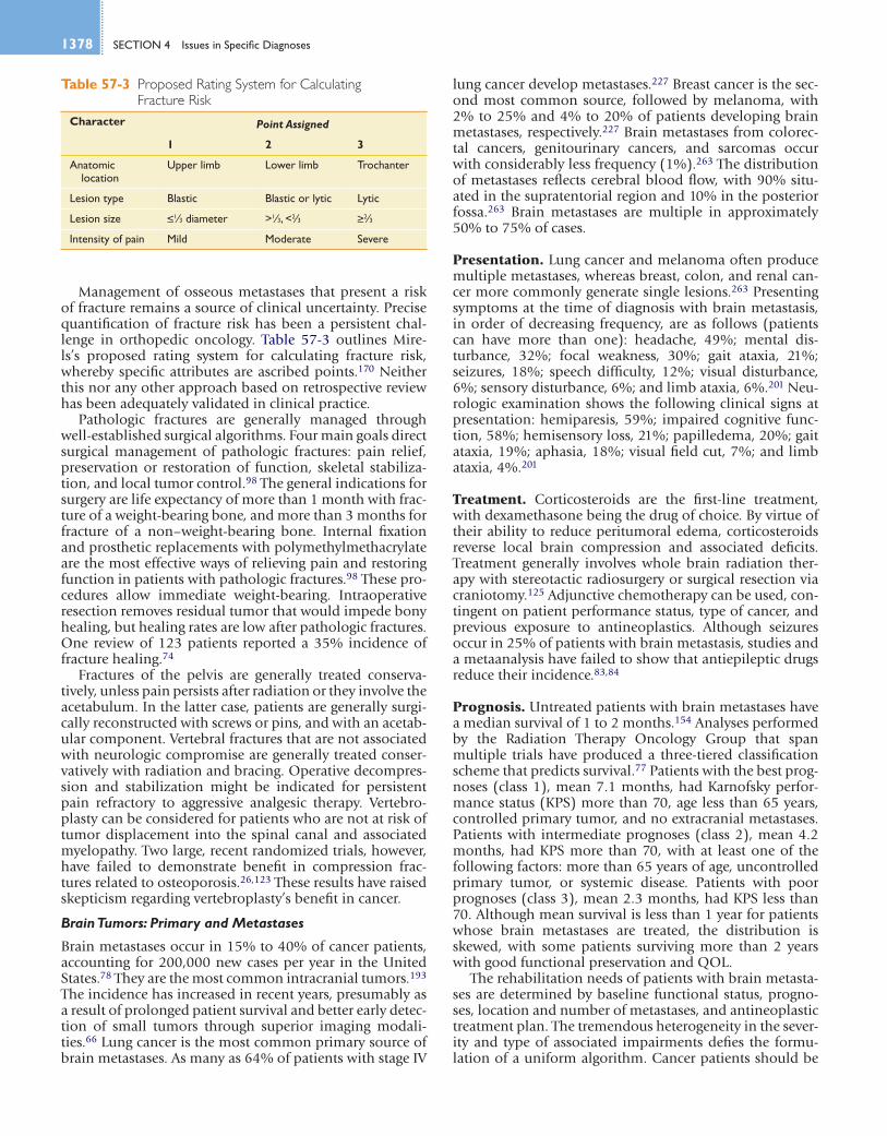


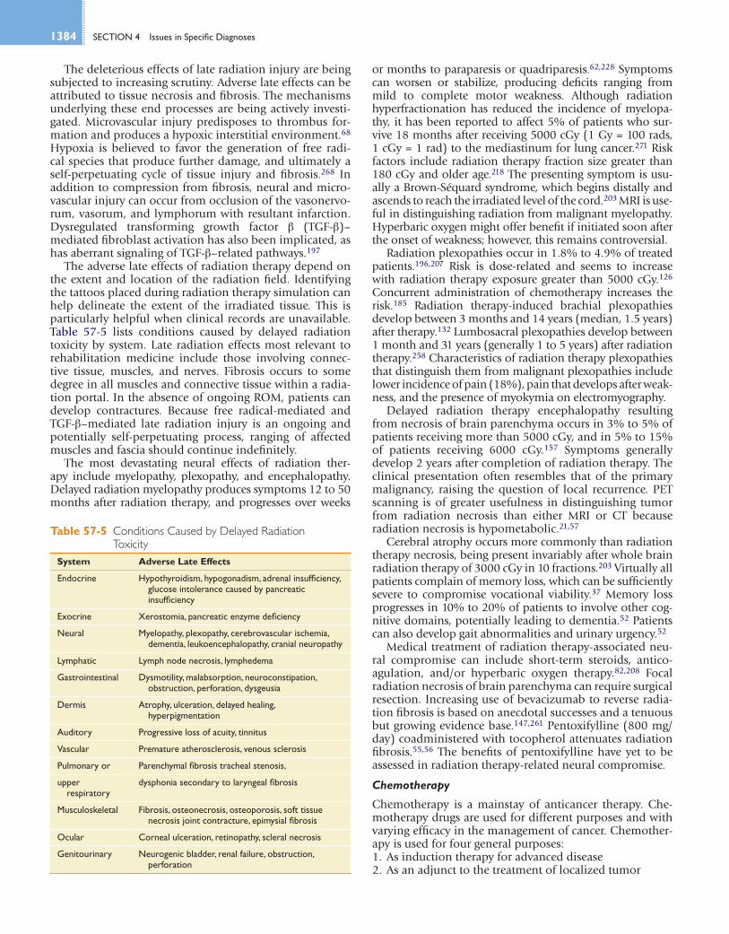
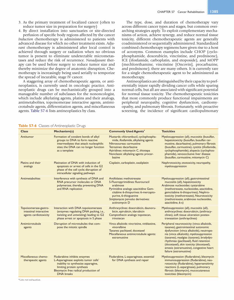


![Multidisciplinary Outpatient Cancer Rehabilitation Can ......cancer treatment. Thus, cancer is progressively seen as a chronic disease [1]. However, cancer itself and its treatment](https://static.documents.pub/doc/80x56/60ffc22fe3d7f75ebd3a887d/multidisciplinary-outpatient-cancer-rehabilitation-can-cancer-treatment.jpg)


