Page 1
This is a repository copy of Cancer Stem Cells in Osteosarcoma.
White Rose Research Online URL for this paper:http://eprints.whiterose.ac.uk/108081/
Version: Accepted Version
Article:
Heymann, D. orcid.org/0000-0001-7777-0669, Brown, H.K. and Tellez-Gabriel, M. (2017) Cancer Stem Cells in Osteosarcoma. Cancer Letters, 386. pp. 189-195. ISSN 0304-3835
https://doi.org/10.1016/j.canlet.2016.11.019
[email protected] ://eprints.whiterose.ac.uk/
Reuse
This article is distributed under the terms of the Creative Commons Attribution-NonCommercial-NoDerivs (CC BY-NC-ND) licence. This licence only allows you to download this work and share it with others as long as you credit the authors, but you can’t change the article in any way or use it commercially. More information and the full terms of the licence here: https://creativecommons.org/licenses/
Takedown
If you consider content in White Rose Research Online to be in breach of UK law, please notify us by emailing [email protected] including the URL of the record and the reason for the withdrawal request.
Page 2
1
CancerstemcellsinOsteosarcoma
HannahK.Brown1,2,MartaTellez-Gabriel
3,DominiqueHeymann
1,2,4
1. Department of Oncology and Metabolism, University of Sheffield, Medical School,
BeechHillRoad,S102RX,Sheffield,UK
2. EuropeanAssociatedLaboratory, INSERM-UniversityofSheffield,SarcomaResearch
Unit,MedicalSchool,S102RX,Sheffield,UK
3. Laboratotio Hematologia Oncologica y de Transplantes, Institut Investigacions
Biomèdiques (IBB)SantPau,Hospitalde laSantaCreui SantPau,08025Barcelona,
Spain
4. INSERM, UMR 957, Pathophysiology of Bone Resorption and Therapy of Primary
BoneTumours,EquipeLigue2012,UniversityofNantes,FacultyofMedicine,44035
Nantes,France
Runningtitle:Stemcellsandosteosarcoma
Correspondingauthors:
Prof.DominiqueHeymannandDrHannahBrown
DepartmentofOncologyandMetabolism
MedicalSchool
BeechHillRoad,S102RX,Sheffield,UK
Tel.:+44(0)1122268464
E-mail:[email protected] @sheffield.ac.uk
Page 3
2
Abstract:
Osteosarcoma is themostcommonprimarybone tumour inchildrenandadolescentsand
advanced osteosarcoma patients with evidence of metastasis share a poor prognosis.
Osteosarcoma frequently gains resistance to standard therapies highlighting the need for
improvedtreatment regimensand identificationofnovel therapeutic targets.Cancerstem
cells(CSC)representasub-typeoftumourcellsattributedtocriticalstepsincancerincluding
tumourpropagation, therapy resistance, recurrenceand in somecasesmetastasis.Recent
published work demonstrates evidence of cancer stem cell phenotypes in osteosarcoma
withlinkstodrugresistanceandtumorigenesis.Inthisreviewwewilldiscussthecommonly
used isolation techniques for cancer stem cells in osteosarcoma as well as the identified
biochemicalandmolecularmarkers.
Keywords:osteosarcoma,bonecancer,cancerstemcell,tumourheterogeneity
Page 4
3
Cancerstemcellsandtumourheterogeneity:whatdoweknowaboutosteosarcoma?
Osteosarcoma predominantly initiates in the metaphysis of the long bones with a high
prevalence in childrenandyoungadults.Theoriginofa tumour ispotentiallya singlecell
locatedinthebonemarrow,whichwilleventuallygiverisetoapolyclonal,heterogeneous
tumourmass.Analysisof tumourheterogeneityallowsus todecipher thesteps thatwere
taken from the initiating cell to the development of a heterogeneous tumour mass
comprised of an array of distinguishable sub-clones. Indeed, osteosarcoma initiates as a
monoclonaldisease,whichquicklydevelopsintoapolyclonaldiseaseandisconsideredone
of the most complex cancers in terms of molecular aberration. Deeper insight into this
diversity therefore holds great promise to identify markers associated with the most
aggressive tumour cells within a tumour mass. The vast heterogeneity found in
osteosarcomaisshowninanexomesequencingstudyinwhichmultiplepathways(14driver
genes)wereidentified(1).Theauthorssuggestthatnosingledrivergenecanbepinpointed
tobethecauseofthemajorityofinvestigatedtumoursandthatseveraloncogenicpathways
cause genetic instability in osteosarcoma development. Importantly, this high level of
heterogeneity adds increased complexity for effective treatment strategies, which is
clinicallyreflectedinrefractoryandrecurrentdisease.
Theincreasingknowledgeofthecancergenomethroughindepthanalysisusingfor
example deep sequencing has significantly added to the understanding of intra-tumour
heterogeneityandanevolutionarypatternofasubsetofcloneswithinatumourhasbeen
reported(2).Newtechnologiesnowallowustoviewheterogeneityalsoonasinglecelllevel.
This has clearly increased the tumour complexity over performing analysis on bulk tissue
showingevendeeperlevelsofintra-tumourheterogeneityinmanycancertypes(3-6).Single
Page 5
4
cell analysisonCSCs inosteosarcomahas, toourknowledge, currentlynotbeen reported
butcouldsignificantlyhelptounderstandthediversityofthesecells.
Cells-of-origininosteosarcomaandpropertiesofcancerstemcells
Inosteosarcomaseveralcell-of-originmodelshavebeenproposedincludingtransformation
ofundifferentiatedmesenchymalstemcells (MSCs)aswellasmorecommittedosteogenic
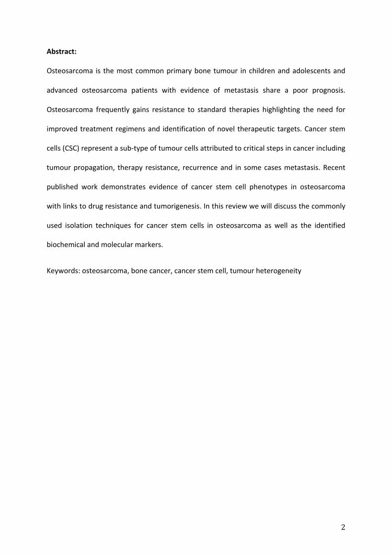
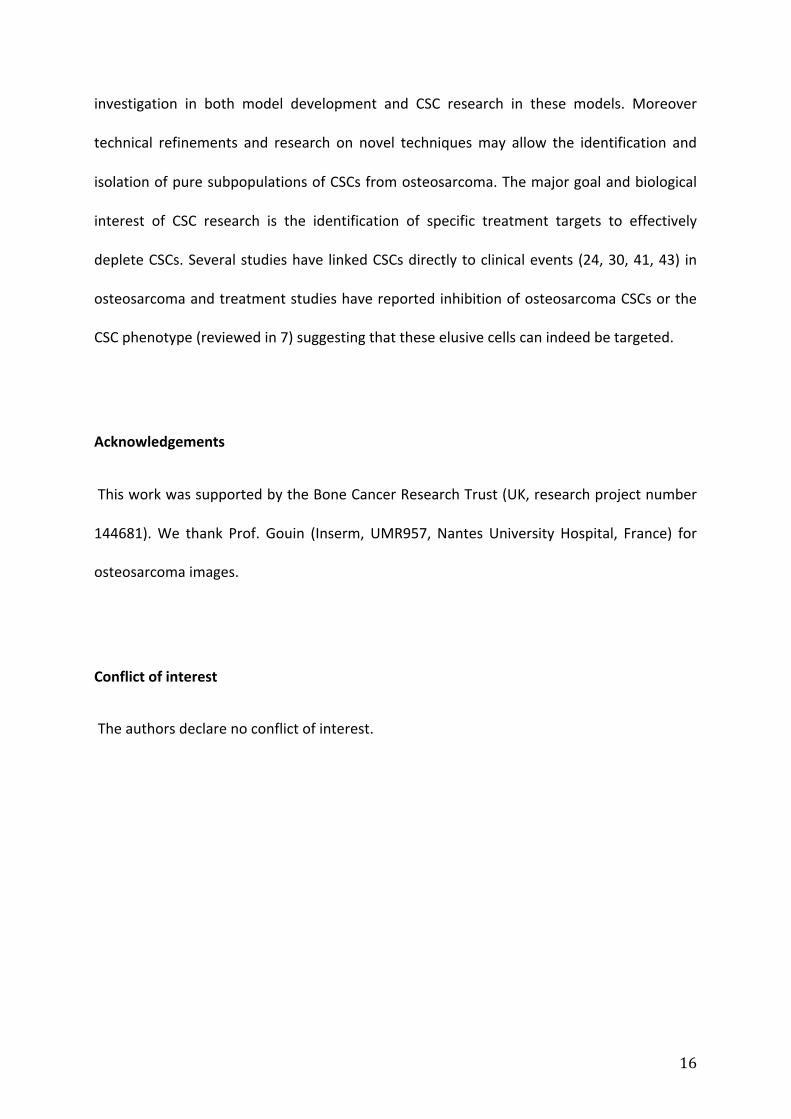
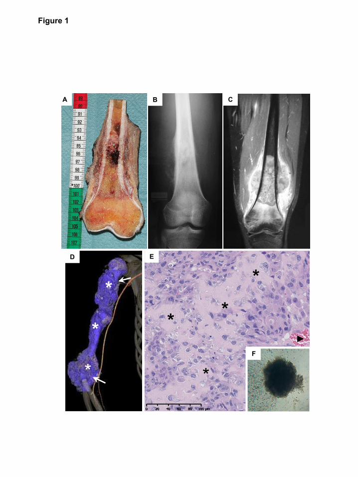
progenitorcells(7)(Figure1).Osteosarcomaisaboneformingtumourinvadingfrequently
the surrounding soft tissues as revealed by conventional imaging associating X-ray and
MagneticResonance Imaging (MRI) (Figures1A-C).Osteosarcoma isavascularized tumour
characterized by typical osteoid matrix formed by cancer cells (Figures 1D,E). Evidence
comespredominantlyfrominvivostudiesusingMSCsand/orosteoprogenitorsinwhichfor
example mutations in genes such as P53 and RB and/or aberrant Hedgehog and NOTCH
signallingwereshowntoinduceosteosarcoma(7-9).Theterminologyof‘cancerstemcells’
is still under debate with the association to stem cells remaining controversial. Indeed,
cancer stem cells and cancer initiating cells are oftenused interchangeably although they
mayindeedexhibitdifferentproperties.Inacancerstemcellmodel,tumoursarethoughtto
behierarchicallyorganisedwithasubpopulationofself-renewingcellsatthebasisoftumour
progression(10).Thesecancerstemcells(CSCs)areproposedtobeuniquesubsetsofclones
withinatumourmassattributedwithtumourpropagation,resistancetotherapyandhavein
some studiesbeenattributed to initiatemetastases. Evidenceexists inosteosarcoma that
patients may present with distant metastases decades after completion of their first
treatment (11) potentially further highlighting the tumorigenic characteristics of CSCs
although this currently remains speculative. Similar to the origin of osteosarcoma, the
Page 6
5
existing hypotheses for the origin of CSCs in general include the transformation of
undifferentiatedstemcellsormorecommittedcellstogainaberrantself-renewalproperties
(12). Remarkably, a stem cell transcription factor (Sox2) has been shown to maintain
osteosarcoma CSCs (13) through inhibition of the Hippo pathway (14). The complex self-
renewal processof normal stem cells (15) is partly regulatedby external signals from the
surrounding microenvironment, the stem cell niche. Such a specialised microenvironment
hasalsobeenproposedforCSCs,influencingCSCfunctionandsurvivalandinsomecancers
these niches (e.g. vascular, immune, bone) may even overlap with the normal stem cell
niche(16). Inaddition,thebonenicheandthebonemicroenvironmentthatosteosarcoma
cells and the putative CSCs are surrounded by apply continuous pressure thus further
influencinggenomicinstability,dormancyandpotentialnewresistantmechanisms(17).
Methodstoidentifycancerstemcells
Several methods have been developed to identify and isolate CSCs based on their self-
renewalpropertiesandhavebeendiscussed indetailelsewhere(10,18,19).Functional in
vitroassaysarefrequentlyinitiallyappliedtoenrichforCSCs.Thisisoftenfollowedbymore
descriptive assays and in vivo verification; however, the order and selection of the tests
appears to be interchangeable depending on the study. The functional in vitro test of
formationoftumourspheresundernon-adherentandserum-freeconditionsoftenservesas
the initial step toenrich forCSC-like cell populations (Figure1F). The standardmethod to
verify CSC candidates in vivo is the serial transplantation of isolated putative CSCs into
immunocompromisedmicetoassesstumorigeniccapacityatlowcellnumbers.Boththe in
vivo and in vitro functional assays have the disadvantage that truly quiescent CSCs will
Page 7
6
possiblynotbe identified. The functionalmethodsare frequentlyused in connectionwith
descriptiveassays likethemeasurementofgeneexpression levelsofstem-like factorsand
identificationbasedoncellsurfacemarkers.Inordertoassesscellpopulationsforproposed
drugresistancepropertiessidepopulationanalysis(dyeexclusionassayandALDH(aldehyde
dehydrogenase)activityarefrequentlyappliedtoenrichforCSCswiththesecharacteristics.
Importantly, all methods require careful consideration because under none of these
experimentalconditionsapureCSCpopulationordetectionofallCSCsub-populationscan
beassumed.Themethodsaremore likelytoenrichthesamplefor (specific)CSCsthrough
experimentally-induced selectionand in somecasesenvironmentalpressure. Theuseof a
number of methods including functional experiments and careful interpretation should
thereforebeappliedtoCSCresearch.
Identificationofcancerstemcellsinosteosarcoma
InthefollowingsectionthemainidentifiedmarkersandevidencefortheexistenceofCSCs
in osteosarcoma will be discussed. The studies were grouped based on their initial
enrichmentmethodandanoverviewcanbefoundintable1.
Sidepopulation(dyeexclusionassays)
OnemainfeatureofCSCsistheirpotentialtoevadetreatmentanditissuggestedthatthisis
achieved through an increase in ATP-binding cassette (ABC) multidrug efflux transporters
suchasMDR1/ABCB1andBRCP1/ABCG2andABCB5expression.Thistraithasbeenusedfor
CSC identification by measuring the ability of cells to exclude DNA-binding dyes (Hoechst
Page 8
7
33342orRhodamine123)byfluorescence-activatedcellsorting(18,20-22).Cellsexpressing
highABCtransportersexcludethedyesandarevisibleasa‘sidepopulation’duringanalysis.
Muraseetal (23)studiedthesidepopulationfraction insevenosteosarcomacell linesbut
onlyoneline(NY)wasreportedtohaveasidepopulationfractionwhilesidepopulationcells
were hardly detected in the rest of the cell lines. They also studied one bone human
malignant fibrous histiocytoma cell line (MFH2003) which was shown to have the largest
side population fraction and to exhibit increased ABCG5 expression as well as cancer-
initiating characteristics detectedby increased sphere formation and tumour formation in
vivowhencomparedtonon-sidepopulationcells.
Ahigherdetectionrateofsidepopulationcellsinosteosarcomawasshowninhuman
primarysamples(24).Downstreamanalysisofthesidepopulationcellsshowedupregulated
gene expression (ABC transporters, Oct4, Nanog), increased sphere formation and higher
multidrug resistance to doxorubicin, methotrexate and cisplatin compared to non- side
populationcells.Wheninjectedintoimmunocompromisedmice,theproposedCSCsshowed
higher tumorigenic potential although non-side population cells at higher numbers could
also form tumours. Similar experiments carried out with U2OS cells did not show a side
populationfraction.
Aldehydedehydrogenase(ALDH)
Asecond,frequentlyinvestigatedapproachthroughwhichCSCsapplytheirchemoresistance
is the expression and activity of the drug-detoxifying enzyme ALDH. A subpopulation of
ALDH1high
MG63cellswasdetectedbyHonokietal(25)whofurtherreportedALDH1high
cells
to have increased expression of stem-like genes (Nanog, Oct3/4, Stat3, Sox2), higher
Page 9
8
resistancetodoxorubicinandcisplatinandincreasedself-renewalabilityasshownbysphere
formation. Importantly,ALDHhigh
cellswerealsodetected inhumanosteosarcomasamples
andincreasedALDHactivitywasfurthercorrelatedtometastaticpotential(26).Itwasalso
shownthatinhibitionofALDHactivityusingdisulfiramresultedinreducedcellproliferation
suggesting direct targeting of CSC phenotype cells. A second study reported large ALDH-
brightcellpopulationintheOS99-1osteosarcomacelllinecomparedtolowerpercentages
in Hu09, Saos-2 and MG63 (27). Interestingly, injection of the OS99-1 cells into mice
followedbyALDHactivitymeasurementshowedalowerALDH-brightfractioncomparedto
the parental line cultured in vitro. TheALDH-bright cellswere further characterised to be
more tumorigenic in vivo and exhibit higher stem like gene expression (Nanog, Sox-2,
OCT3/4)comparedtoALDH-lowcells.
Cellsurfacemarkers
Cellsurfacemarkersarearguablythemostattractiveandsoughtafteridentificationmethod
of CSCs. A specific cell surface marker exclusively to CSCs and not normal cells, including
stemcells,woulddrasticallysimplifytheisolationandtreatmentpotentialforthesecells.
CD133(prominin)
Tirinoetal(28)showedthatCD133,amembraneglycoprotein,maybeamarkerofCSCsin
osteosarcoma.CD133+cellswere identified inthreeosteosarcomacell lines(Saos2,MG63,
U2OS). The CD133+ cellswere further characterised to bemore proliferative, overexpress
OCT3/4andABCG2,haveasmallsidepopulationfractionandformedspheresinserum-free
Page 10
9
conditionswhileCD133-cellsdidnot.Interestingly,theauthorsdetectedintracellularCD133
staininginCD133+andCD133
-cellsandreportedthatmRNAlevelsofCD133wereidentical
betweenthetwopopulations.ThegroupwasunabletogrowCD133+orCD133
-tumours in
vivo. In a followup study, CD133+ cellswere shown tobepresent in twohumanprimary
osteosarcomas and exhibiting stem-like gene expression (e.g. OCT3/4, Nanog), sphere
formationandsidepopulationfractions(29).Inaddition,cellsisolatedfromCD133+derived
spheres were shown to form large tumours in vivo compared to the adherent cells.
Interestingly,inbothstudiesitwasshownthatCD133+cellscouldgiverisetoCD133
-cellsin
culturesuggestingaphenotypicswitch.
TheexpressionofCD133onFFPEsamplesofhumanosteosarcomawasshownbyHe
et al (30) who detected the marker on 46 out of 70 analysed samples and positively
correlated CD133 expression with lung metastases and decreased overall survival. In
addition,characterisationofMG63cellsshowedthatCD133+subsetsweremoremigratory,
invasive and overexpressed Oct4, Nanog and CXCR4. CD133+ cells in patient samples of
osteosarcomaandaCD133+sub-populationinSaos2cellswerealsoreportedbyLietal(31).
A comprehensiveassessmentofCD133expressionandassociatedCSCphenotypeson cell
lines(Saos2,U2OS,MG63,HOS,MNNG/HOS,143B)andprimarytumourswasfollowedbyin
depthanalysisofmiRNAexpressionprofilingwhich identifiedmiR-133a inconnectionwith
CD133expression(32).ThemicroRNAwasfurthershowntoregulatetumorigenicpotential
since silencing in combinationwith chemotherapeutic treatment significantly reduced the
aggressivetumourtypeand lungmetastases invivo. Interestingly,CD133expressioncould
be induced by chemotherapy treatment and was associated with enhanced miR-133a
expression suggesting a CSC induction through therapeutic challenge. Overall, CD133
appearsaspotential therapeutic target inosteosarcomaandpharmacologic inductionofa
Page 11
10
switch from theCD133+ toCD133-phenotype through siRNA couldbe anew therapeutic
approach.
CD117(c-kit),Stro-1
A study assessing the feasibility of using MSC surface markers to enrich for CSCs in
osteosarcomawasdonebyAdhikarietal(33),whoconvincinglyshowedthatCD117+/Stro-1
+
double-positivecellswerepresent inmouseandhumanosteosarcomacell linesaswellas
primarycultures.Intriguingly,thedouble-positivefractionshowedhighersphereformation
ability,increasedresistancetodoxorubicintreatmentandoverexpressedCXCR4andABCG2.
Furthermore,double-positivecellsmorereadilyformedtumoursatalowercellnumberand
itwasshownthatthisCSCfractionhadincreasedmetastaticpotentialcomparedtodouble
negative cells. This study was one of the first to comprehensively assess the molecular
constitutionoftheidentifiedCSCsandtofurthermoreshowfunctionalCSCcharacteristicsin
anumberofexperiments.
CD271
CD271 is a neural crest low-affinity nerve growth factor receptor and a marker of bone
marrowmesenchymalstemcells.Astudy investigating thestem likephenotype inCD271+
cellswasperformedby Tianet al (34). CD271+ cellswere foundon FFPE tissueof human
osteosarcoma samples and the cell lines MNNG/HOS, U2OS and Saos2 also contained
CD271+ subpopulations. The authors went on to show that CD271
+ cells had upregulated
stemcellgeneexpression, resistancetochemotherapyandweremorereadilydetected in
spheres.WhenCD271+andCD271
-cellswereseparatedtheCD271
+fractionshowedhigher
Page 12
11
tumorigenic potential in the sphere formation assay and when injected into
immunocompromisedmice.
Sphereformationassays
Tissuestemcellshavebeen identifiedthroughtheabilityofcells to formspheresandthis
method has also been used to assess CSC presence. Sarcospheres (spheres growing from
sarcomacells)havebeengrownundersimilarexperimentalsetupstothosepublishedfor
neuralstemcellsincludingnon-adherent,serum-freeconditionsandthesupplementationof
mediawithgrowthfactors(N2,epidermalgrowthfactoretc.).
ThefirsttodescribeosteosarcomastemcellsusingsphereformationwasGibbsetal
in2005(35).Thiswasfollowedbyseveralothergroups,whichfurthershowedthatsphere
forming cells were tumorigenic in immunodeficient mice and that these cells were more
drug resistant suggesting a CSC phenotype (36). A detailed study on the sphere forming
fractionofMNNG/HOS cellswasdonebyMartins-Neveset al (37)who showed that cells
isolated from spheres had mesenchymal stem cell properties including upregulated gene
expressionofstemnessgenesOct3/4,Nanog,andABCtransporters.Theywentontoshow
that sphere cellsweremore resistant to chemotherapyand radiationand that these cells
exhibitedhigher cancer-initiating properties in vivowhen compared to parental cells. It is
noteworthythatthemainCSCselectionmethodwassphereformationsuggestingthatthe
experimental conditions of this assay induced CSC-like characteristics. Indeed, the same
group later showed that the selection or enrichmentmethod (i.e., sphere forming ability,
sidepopulationanalysisorALDHactivity)appliedtoeachcelllineofalargerpanelwaspre-
selectingCSCswithspecificanddissimilarcharacteristicsinsinglecelllines(38).Importantly,
Page 13
12
these data suggest that CSCs with specific molecular and functional characteristics are
enricheddependingonthemethodsandthusheterogeneousCSCsub-populationsmayexist
sidebysideinoneandthesamecancercellline.Toourknowledgethisstudyisoneofvery
fewapplyingseveral initialCSCenrichmentmethodsonthesamesamplesthussuggesting
thatheterogeneitymaybepresentwithintheosteosarcomaCSCpopulation.
ThepotentialplasticityoftheCSCphenotypeinosteosarcomawasshownbyZhang
et al (39). The authors presented thatMNNG/HOS, Saos2 andMG63 cells readily formed
spheresandthattreatmentwithTGFβ1orapplicationofhypoxicconditions(bothabundant
in the bone microenvironment) significantly increased sphere formation suggesting an
inductionofCSCsthroughenvironmentalfactors.Spherecellshadincreasedstemlikefactor
andABCtransporterexpressionlevels,increasedresistancetocisplatinandadriamycinand
invivotumourformation.TheroleofTGFingeneraltumourprogressionwaswidelystudied
anditsroleinosteosarcomasuspected(40).!Indeed,therelevanceofTGFβ1topropagatea
stem like phenotype in osteosarcoma was shown when inhibition of the TGFβ1-receptor
resulted in reduced sphere formation. In vivo experiments using CSCs from spheres, CSCs
from TGFβ1-induced spheres and parental cells further confirmed the enrichment of
aggressive CSCs in the TGFβ1-induced sphere population since this group was capable of
forming the most tumours. The authors concluded that CSCs may develop de novo from
differentiated cancer cells and that they can revert back into a more differentiated state
underlining a potential of plasticity. In addition, the study elegantly highlights the
importanceofthemicroenvironment(orexperimentalconditions)forCSCs.
MicroRNAs (miRNA) regulate gene expression on a post-transcriptional level and
havebeenproposedtobeinvolvedincancerprogressionandinitiation.ThemicroRNAmiR-
Page 14
13
26a was shown to be significantly lower in sarcospheres or the ALDH positive fraction of
severalosteosarcomacell linescomparedtotheparentallines(41).Doxorubicintreatment
alsoreducedmiR-26alevelssuggestinganincreaseinstemlikecellsaftertreatmentanda
correlationbetweenlowmiR-26alevelsandCSCs.Thiswasconfirmedsinceover-expression
ofmiR-26asignificantlyreducedgeneexpressionofstemnessmarkers,resultedinfewerand
smaller spheres, formed fewer tumoursand increased thesensitivity tochemotherapeutic
challengecomparedtocontrol.AsamechanismthegroupsuggestedthatmiR-26areduces
osteosarcomamalignancyviaJagged1(notchpathway)suppression.Importantly,analysisof
miR-26a levels in patient samples linked high levels to a better prognosis. Finally, a link
between osteosarcoma CSCs and telomerase activity was reported by Yu et al after
detectionofcellswithhightelomeraseactivityundersphereformationconditions(42).
Inductionofcancerstemcellphenotypethroughchemotherapeutictreatment
Thepresenceofatherapy-resistant,tumorigenicsub-populationofcancercellsinatumour
is visible in patients with refractory disease and significantly highlights the necessity to
further understand the nature of these cells. This chemoresistant characteristic has been
exploited in a few studies investigating whether chemotherapy treated or treatment-
resistant cells could contain CSCs (43-46). Indeed, it was shown that methotrexate pre-
treatedorresistantcellsnotonlyexpressedCD117+/Stro-1
+andhadasidepopulationbut
thatthesecellsalsoformedmorespheresinvitroandweremoretumorigenicwheninjected
subcutaneously into immunodeficient mice (44). The finding that cells with CSC
characteristicscouldbeenrichedbytreatmentviaactivatedNotchsignallingpathwayswas
showninarecentstudyusingcisplatin-resistant143BandU2Oscells(46).Cisplatin-resistant
Page 15
14
cellswereenrichedforCD117+/Stro-1
+double-positivecells,formedmorespheresandwere
more tumorigenic in vivo. Importantly, the group also assessed relapsed tumours after
cisplatintreatmentinvivoandreportedanincreaseinCSCswithelevatedOct4,Sox2,CD117,
Stro-1levelsandsphereformationcapability.
TheexploitationofchemoresistantcancercelltraitswasalsousedbyMartins-Neves
etal(43)whofoundthatstem-likecellscouldbeinducedwithchemotherapytreatmentina
number of fibroblastic and osteoblastic osteosarcoma cell lines. The identified treatment-
inducedcellshadincreasedALDHhigh
populations,upregulatedABCtransportersandshowed
overexpression of stemness genes as well as Wnt/beta-catenin signalling pathways. The
stem like featurescouldbe reducedwhentheWnt/beta-cateninpathwaywas inhibited in
vitroanditwasshownthatacombinationofchemotherapyandWnt-inhibitorwasthemost
efficacious anti-tumour treatment in vivo. The findings of the induction of a stem-like
phenotypethroughchemotherapywerestrengthenedwiththecorrelationof‘poorresponse’
tochemotherapyandstem-likegeneexpressioninosteosarcomapatients.Theselectionof
an aggressiveCSCphenotypeafter cisplatin treatment couldbe suggestedby theworkof
Tsuchida et al (45). Side population cells of the HOS cell line treated or not treatedwith
cisplatinwereisolatedandwhilecisplatin-inducedsidepopulationcellsformedtumours in
vivo the side population fraction isolated from untreated cells did not. Furthermore,
cisplatin-induced activation of VEGF/Flt1 signallingwas reported to accumulate the highly
tumorigenicCSC-typecells.
CollectivelythereportsusingchemotherapeuticsshowthataCSCphenotypecanbe
induced through therapeutic challenge. It remains to be established whether different
chemotherapeuticchallengeswillgiverisetodifferentCSCsub-populations.
Page 16
15
Conclusion
Thedebateaboutacancerstemcelldefinitionisstillongoingandimportantlyitisnotclear
whether the stem cell phenotype of a cancer cell is intrinsic or plastic. Tumour cells can
switchinandoutofastemcellphenotypefurther increasingthecomplexityofthecancer
stem cell hypothesis and suggesting that environmental pressure (experimental or in
patients) may be a trigger for heterogeneity and plasticity. For example increased
environmental pressure through chemotherapy treatment has the potential to induce a
switch fromadifferentiated towardsa stem-likephenotype inosteosarcoma (43-46). This
potentialplasticityand themanydifferentexperimentalenrichmentmethodsmayhelp to
understandthevariationsofreportedCSCmarkersbetweenstudies.Importantlyitappears
thattheenrichedCSCphenotypemaydependontheappliedisolationmethod(s),however,
more studies are required to verify this. Moreover, it is currently unclear whether CSCs
inherentlyexhibitmetastasis-initiatingproperties.
The high level of heterogeneity found in osteosarcoma, including the effect of the
tumourmicroenvironmentmaybemorecomprehensivelyassessedinPDX(patientderived
xenograft)modelsandevidenceexiststhatcellswithCSCphenotypearepresentinpatient
derivedosteosarcomasamples(24,46).Theuseofpatientderivedmaterialcouldpotentially
not only reflect the high heterogeneity of this rare cancer but would also reflect the
influenceofthehumanmicroenvironmentinmoredetailcomparedtostudiesoncelllines.
Despite thesepotentialadvantages theapproachmaybe limitedby the lowavailabilityof
primarychemonaivetumours.ThepotentialexpansionofCSCsthroughthegrowthofPDX
material invivo couldpossiblyhelpovercomethis limitationand requires further indepth
Page 17
16
investigation in both model development and CSC research in these models. Moreover
technical refinements and research on novel techniques may allow the identification and
isolationofpuresubpopulationsofCSCsfromosteosarcoma.Themajorgoalandbiological
interest of CSC research is the identification of specific treatment targets to effectively
depleteCSCs.Severalstudieshave linkedCSCsdirectlytoclinicalevents (24,30,41,43) in
osteosarcomaandtreatmentstudieshavereportedinhibitionofosteosarcomaCSCsorthe
CSCphenotype(reviewedin7)suggestingthattheseelusivecellscanindeedbetargeted.
Acknowledgements
ThisworkwassupportedbytheBoneCancerResearchTrust(UK,researchprojectnumber
144681). We thank Prof. Gouin (Inserm, UMR957, Nantes University Hospital, France) for
osteosarcomaimages.
Conflictofinterest
Theauthorsdeclarenoconflictofinterest.
Page 18
17
References
1. Kovac M, Blattmann C, Ribi S, Smida J, Mueller NS, Engert F, et al. Exome
sequencingofosteosarcomarevealsmutationsignaturesreminiscentofBRCAdeficiency.
Naturecommunications.2015;6:8940.
2. Hiley C, de Bruin EC, McGranahan N, Swanton C. Deciphering intratumor
heterogeneity and temporal acquisition of driver events to refine precisionmedicine.
GenomeBiol.2014;15(8):453.
3. Kim KT, Lee HW, Lee HO, Kim SC, Seo YJ, Chung W, et al. Single-cell mRNA
sequencing identifies subclonal heterogeneity in anti-cancer drug responses of lung
adenocarcinomacells.GenomeBiol.2015;16:127.
4. Navin N, Kendall J, Troge J, Andrews P, Rodgers L, McIndoo J, et al. Tumour
evolutioninferredbysingle-cellsequencing.Nature.2011;472(7341):90-4.
5. Patel AP, Tirosh I, Trombetta JJ, Shalek AK, Gillespie SM, Wakimoto H, et al.
Single-cell RNA-seq highlights intratumoral heterogeneity in primary glioblastoma.
Science.2014;344(6190):1396-401.
6. Ryu D, Joung JG, Kim NK, Kim KT, Park WY. Deciphering intratumor
heterogeneityusingcancergenomeanalysis.HumGenet.2016;135(6):635-42.
7. Abarrategi A, Tornin J, Martinez-Cruzado L, Hamilton A, Martinez-Campos E,
Rodrigo JP, et al. Osteosarcoma: Cells-of-Origin, Cancer Stem Cells, and Targeted
Therapies.Stemcellsinternational.2016;2016:3631764.
8. TaoJ,JiangMM,JiangL,SalvoJS,ZengHC,DawsonB,etal.Notchactivationasa
driverofosteogenicsarcoma.Cancercell.2014;26(3):390-401.
9. ChanLH,WangW,YeungW,DengY,YuanP,MakKK.Hedgehogsignalinginduces
osteosarcoma development through Yap1 and H19 overexpression. Oncogene.
2014;33(40):4857-66.
10. Basu-Roy U, Basilico C, Mansukhani A. Perspectives on cancer stem cells in
osteosarcoma.Cancerletters.2013;338(1):158-67.
11. Halldorsson A, Brooks S, Montgomery S, Graham S. Lung metastasis 21 years
afterinitialdiagnosisofosteosarcoma:acasereport.JMedCaseRep.2009;3:9298.
12. ClarkeMF,DickJE,DirksPB,EavesCJ,JamiesonCH,JonesDL,etal.Cancerstem
cells--perspectivesoncurrentstatusand futuredirections:AACRWorkshoponcancer
stemcells.Cancerresearch.2006;66(19):9339-44.
13. Basu-RoyU,SeoE,RamanathapuramL,RappTB,PerryJA,OrkinSH,etal.Sox2
maintains self renewal of tumor-initiating cells in osteosarcomas. Oncogene.
2012;31(18):2270-82.
14. Basu-RoyU,BayinNS,RattanakornK,HanE,PlacantonakisDG,MansukhaniA,et
al. Sox2 antagonizes the Hippo pathway tomaintain stemness in cancer cells. Nature
communications.2015;6:6411.
15. HeS,NakadaD,MorrisonSJ.Mechanismsofstemcellself-renewal.AnnuRevCell
DevBiol.2009;25:377-406.
16. Shiozawa Y, Berry JE, Eber MR, Jung Y, Yumoto K, Cackowski FC, et al. The
marrownichecontrolsthecancerstemcellphenotypeofdisseminatedprostatecancer.
Oncotarget.2016.
17. HeymannDR,F . Bone sarcomas: pathogenesis and new therapeutic
approaches.IBMSBoneKEy.2011;8:402–14.
18. TirinoV,DesiderioV,PainoF,PapaccioG,DeRosaM.Methods forcancerstem
celldetectionandisolation.MethodsMolBiol.2012;879:513-29.
Page 19
18
19. ValentP,BonnetD,DeMariaR,LapidotT,CoplandM,MeloJV,etal.Cancerstem
cell definitions and terminology: the devil is in the details. Nature reviews Cancer.
2012;12(11):767-75.
20. FukudaK, SaikawaY,OhashiM,KumagaiK,KitajimaM,OkanoH, et al. Tumor
initiatingpotentialofsidepopulationcellsinhumangastriccancer.Internationaljournal
ofoncology.2009;34(5):1201-7.
21. Moserle L, Indraccolo S, GhisiM, Frasson C, Fortunato E, Canevari S, et al. The
sidepopulationofovariancancercellsisaprimarytargetofIFN-alphaantitumoreffects.
Cancerresearch.2008;68(14):5658-68.
22. TirinoV,DesiderioV, Paino F,DeRosaA, Papaccio F, LaNoceM, et al. Cancer
stem cells in solid tumors: an overview and new approaches for their isolation and
characterization. FASEB journal : official publication of the Federation of American
SocietiesforExperimentalBiology.2013;27(1):13-24.
23. MuraseM,KanoM,TsukaharaT,TakahashiA,TorigoeT,KawaguchiS,etal.Side
populationcellshavethecharacteristicsofcancerstem-likecells/cancer-initiatingcells
inbonesarcomas.Britishjournalofcancer.2009;101(8):1425-32.
24. YangM,YanM,ZhangR,Li J,LuoZ. Sidepopulationcells isolated fromhuman
osteosarcomaareenrichedwithtumor-initiatingcells.CancerSci.2011;102(10):1774-
81.
25. HonokiK,FujiiH,KuboA,KidoA,MoriT,TanakaY,etal.Possibleinvolvementof
stem-like populations with elevated ALDH1 in sarcomas for chemotherapeutic drug
resistance.Oncologyreports.2010;24(2):501-5.
26. GrecoN, Schott T,Mu X, Rothenberg A, Voigt C,McGoughRL, 3rd, et al. ALDH
Activity CorrelateswithMetastatic Potential in Primary Sarcomas of Bone. Journal of
cancertherapy.2014;5(4):331-8.
27. Wang L, Park P, Zhang H, La Marca F, Lin CY. Prospective identification of
tumorigenic osteosarcoma cancer stem cells in OS99-1 cells based on high aldehyde
dehydrogenaseactivity.Internationaljournalofcancer.2011;128(2):294-303.
28. Tirino V, Desiderio V, d'Aquino R, De Francesco F, Pirozzi G, Graziano A, et al.
Detection and characterization of CD133+ cancer stem cells in human solid tumours.
PloSone.2008;3(10):e3469.
29. Tirino V, Desiderio V, Paino F, De Rosa A, Papaccio F, Fazioli F, et al. Human
primary bone sarcomas contain CD133+ cancer stem cells displaying high
tumorigenicityinvivo.FASEBjournal:officialpublicationoftheFederationofAmerican
SocietiesforExperimentalBiology.2011;25(6):2022-30.
30. HeA,QiW,HuangY,FengT,ChenJ,SunY,etal.CD133expressionpredictslung
metastasis and poor prognosis in osteosarcoma patients: A clinical and experimental
study.Experimentalandtherapeuticmedicine.2012;4(3):435-41.
31. LiJ,ZhongXY,LiZY,CaiJF,ZouL,LiJM,etal.CD133expressioninosteosarcoma
andderivationofCD133(+)cells.Molecularmedicinereports.2013;7(2):577-84.
32. FujiwaraT, KatsudaT,HagiwaraK,KosakaN, YoshiokaY, TakahashiRU, et al.
Clinical relevance and therapeutic significance of microRNA-133a expression profiles
andfunctionsinmalignantosteosarcoma-initiatingcells.Stemcells.2014;32(4):959-73.
33. Adhikari AS, Agarwal N, Wood BM, Porretta C, Ruiz B, Pochampally RR, et al.
CD117 and Stro-1 identify osteosarcoma tumor-initiating cells associated with
metastasisanddrugresistance.Cancerresearch.2010;70(11):4602-12.
34. Tian J, Li X, Si M, Liu T, Li J. CD271+ osteosarcoma cells display stem-like
properties.PloSone.2014;9(6):e98549.
Page 20
19
35. GibbsCP,KukekovVG,ReithJD,TchigrinovaO,SuslovON,ScottEW,etal.Stem-
likecellsinbonesarcomas:implicationsfortumorigenesis.Neoplasia.2005;7(11):967-
76.
36. FujiiH,HonokiK,TsujiuchiT,KidoA,YoshitaniK,TakakuraY.Sphere-forming
stem-like cell populations with drug resistance in human sarcoma cell lines.
Internationaljournalofoncology.2009;34(5):1381-6.
37. Martins-NevesSR,LopesAO,doCarmoA,PaivaAA,SimoesPC,AbrunhosaAJ,et
al.Therapeuticimplicationsofanenrichedcancerstem-likecellpopulationinahuman
osteosarcomacellline.BMCcancer.2012;12:139.
38. Martins-Neves SR, CorverWE, Paiva-OliveiraDI, vandenAkkerBE, Briaire-de-
Bruijn IH,Bovee JV,etal.OsteosarcomaStemCellsHaveActiveWnt/beta-cateninand
OverexpressSOX2andKLF4.Journalofcellularphysiology.2016;231(4):876-86.
39. ZhangH,WuH, Zheng J, Yu P, Xu L, Jiang P, et al. Transforming growth factor
beta1 signal is crucial for dedifferentiation of cancer cells to cancer stem cells in
osteosarcoma.Stemcells.2013;31(3):433-46.
40. LamoraA,TalbotJ,MullardM,Brounais-LeRoyerB,RediniF,VerrecchiaF.TGF-β
Signalinginboneremodelingandosteosarcomaprogression.JClinMed.2016;5(11).
41. Lu J, SongG, TangQ, Yin J, ZouC, ZhaoZ, et al.MiR-26a inhibits stem cell-like
phenotypeandtumorgrowthofosteosarcomabytargetingJagged1.Oncogene.2016.
42. Yu L, Liu S, Zhang C, ZhangB, Simoes BM, Eyre R, et al. Enrichment of human
osteosarcoma stem cells based on hTERT transcriptional activity. Oncotarget.
2013;4(12):2326-38.
43. Martins-Neves SR, Paiva-Oliveira DI, Wijers-Koster PM, Abrunhosa AJ, Fontes-
Ribeiro C, Bovee JV, et al. Chemotherapy induces stemness in osteosarcoma cells
throughactivationofWnt/beta-cateninsignaling.Cancerletters.2016;370(2):286-95.
44. Tang QL, Liang Y, Xie XB, Yin JQ, Zou CY, Zhao ZQ, et al. Enrichment of
osteosarcomastemcellsbychemotherapy.Chinese journalofcancer.2011;30(6):426-
32.
45. Tsuchida R, Das B, Yeger H, Koren G, Shibuya M, Thorner PS, et al. Cisplatin
treatment increases survival and expansion of a highly tumorigenic side-population
fraction by upregulating VEGF/Flt1 autocrine signaling. Oncogene. 2008;27(28):3923-
34.
46. YuL,FanZ,FangS,YangJ,GaoT,SimoesBM,etal.Cisplatinselectsforstem-like
cellsinosteosarcomabyactivatingnotchsignaling.Oncotarget.2016.
47. PenfornisP,CaiDZ,HarrisMR,WalkerR,LiciniD,FernandesJD,etal.HighCD49f
expression is associatedwithosteosarcoma tumorprogression: a studyusingpatient-
derivedprimarycellcultures.Cancermedicine.2014;3(4):796-811.
Page 21
20
Figure 1: Representative imaging of osteoblastic osteosarcoma. (A) Macroscopic
viewofaresectedosteosarcomainfiltratingsurroundingsofttissue.ConventionalX-
ray (B)and(C)MagneticResonance Imaging (MRI)ofosteosarcoma. (D)Computed
tomography of an osteosarcoma in a 15-year old patient (adapted from “Bone
Cancer” 1st Edition, Ed. Heymann D., Academic Press, 2009). Tumour tissue is
composed by mineralized component detectable to X-Ray (*) and is strongly
associated with the vasculature (arrows). (E) Typical histological view showing
osteoid extracellular matrix produced by osteosarcoma cells (*), tumour tissue is
vascularized (arrowhead). (F) Sarcosphere generated from thehumanMNNG-HOS
osteosarcomacelllineundernon-adherentserum-freeconditions.
Page 22
Table1:StudiesinvestigatingCSCsinosteosarcoma
!
!
InitialCSC
identification/
enrichmentmethod
Osteosarcomacell
lines/patientsamples
CharacterisationofenrichedCSCphenotype Reference
Sidepopulation
OS2000,KIKU,NY,
Huo9,HOS,U2OSand
Saos2
Sphereformation,stemlikegeneexpression,in
vivotumorigenicity
(23)
Humanprimary,U2OS Stemlikegenes,sphereformation,drugresistance,
invivotumorigenicity
(24)
ALDH
MG63 Stemlikegeneexpression,drugresistance,sphere
formation
(25)
OS99-1Hu09,Saos-2,
MG63
Stemlikegeneexpression,invivotumorigenicity (27)
CD133
Saos2,MG63,U2OS Stemlikegeneexpression,sidepopulation,sphere
formation
(28)
Humanprimary Stemlikegenes,sidepopulation,sphere
formation,invivotumorigenicity
(29)
Humanprimary(FFPE),
MG63
Stemlikegeneexpression (30)
Humanprimary(FFPE),
Saos2
Stemlikegeneexpression (31)
Humanprimary,Saos2,
U2OS,MG63,HOS,
MNNG/HOS,143B
Sphereformation,drugresistance,invivo
tumorigenicity,stemlikegeneexpression
(32)
CD117/Stro-1
K7M2,KHOS/NP,
MNNG/HOS,318-1,
P932,BCOS
Sphereformation,drugresistance,stemlikegene
expression,invivotumorigenicityandmetastatic
potential
(33)
CD271Humanprimary(FFPE),
MNNG/HOS,U2OS,
Saos2
Stemlikegeneexpression,sphereformation,drug
resistance,invivotumorigenicity
(34)
Sphereformation
Humanprimary,MG63 Stemlikegeneexpression (35)
MG63 Drugresistance,invivotumorigenicity (36)
MNNG/HOS Stemlikegeneexpression,drugresistance,invivo
tumorigenicity
(37)
Sphereformation,
ALDH,sidepopulation
HOS,MG-63,MHM,
MNNG-HOS,SJSA-1,
L2531,L3312,OHS,
U2OS
Stemlikegeneexpression,invivotumorigenicity (38)
SphereformationMNNG/HOS,Saos2,
MG63+TGFβ/hypoxia
Invivotumorigenicity,drugresistance,stemlike
geneexpression
(39)
Sphereformation,
chemotherapeutic
treatment,ALDH
Humanprimary,U2OS,
MG63,Saos-2and
143B,linktomiR-26a
Stemlikegeneexpression,invivotumorigenicity,
drugresistance
(41)
Chemotherapeutic
treatment
U2Os CD117+/Stro-1,sidepopulation,sphereformation,
invivotumorigenicity
(44)
Cisplatin-resistant143B
andU2Os
CD117+/Stro-1
+,sphereformation,invivo
tumorigenicity,stemlikegeneexpression
(42)
HOS,MG-63,MHM,
MNNG-HOS,OHS,U2OS
ALDHhigh,stemlikegeneexpression (43)
HOS Sidepopulation,invivotumorigenicity,stemlike
geneexpression
(45)
Page 23
D E
*
*
*
*
*
*
*
F
Figure 1
A B C
Figure 1


![Prosthetic Reconstruction for Proximal Tibial Osteosarcoma ...austinpublishinggroup.com/sarcoma/download.php?file=...rate is about 0.3 per million. It often occurs in adolescents [1]](https://static.documents.pub/doc/80x56/6142375a55c1d11d1b340d2a/prosthetic-reconstruction-for-proximal-tibial-osteosarcoma-a-rate-is-about.jpg)

![Osteosarcoma of the Distal Tibia · Osteosarcoma more frequently occurs in children and adolescents, at the position of knee-joint and proximal humerus [1] (Figure 1). It is rare](https://static.documents.pub/doc/80x56/5fd415dc79ff91782318c086/osteosarcoma-of-the-distal-tibia-osteosarcoma-more-frequently-occurs-in-children.jpg)

