Canine retraction with rare earth magnets: An investigation into the validity of the constant force hypothesis
John Daskalogiannakis, DDS, MSc, a and Kenneth Roy McLachlan, PhD b
Toronto, Ontario, and Winnipeg, Manitoba, Canada
ThE; objective of this study was to test the hypothesis that a prolonged constant force provides more effective tooth movement than an impulsive force of short duration. Six human subjects were selected, the main criterion being a need for extraction of their upper first premolars. Canine retraction on these subjects was executed on one side with the application of a force rapidly declining in magnitude, produced by a vertical loop, and on the other side with the application of a relatively constant force. This type of force was achieved by a similar vertical loop which was constantly activated by three parylene-coated neodymium-iron-boron (Nd2Fe14 P) block magnets. The vertical loop on the control side was reactivated 6 weeks after the initial activation. No reactivation was necessary on the experimental side for the duration of the experiment. The rate of tooth movement on the two sides was compared over a period of 3 months, on the basis of maxillary impressions taken at frequent intervals during the course of the study. The canines retracted with a constant force moved statistically significantly more than the control canines (p < 0.05) during the experimental period. The average differences in the mean rates of tooth movement between the two sides were in the order of 2:1 in favor of the experimental side. There were no statistically significant differences in the changes of angulation (tipping) or rotation about the y axis between the two sides. The duration of force application seems to be a critical factor in regulating rate of tooth movement. Conversely, magnitude of the applied force did not appear to be of primary significance. (Am J Orthod Dentofac Orthop 1996;109:489-95.)
T h e important question of how the magni- tude and temporal distribution of the forces produced by orthodontic appliances influence the rate of tooth movement has received relatively little experimental study in human subjects. Although the recommended magnitudes of force vary, most studies '-8 conclude that optimum tooth movement can be achieved if light and continuous torces are used. Continuity of the applied force, however, is certainly not a prerequisite for tooth movement. In fact, several investigators have sug- gested that noncontinuous forces should be preferred as more physiologic. 9''° The primary argument in sup- port of such forces is that they give the periodontal ligament the chance to regenerate during the period
that they are not active. More recently, there have been studies claiming that pulsating forces of variable fre- quencies may be advantageous in inducing more physi- ologic tooth movement. 'l''a
Investigations to date have often been difficult to interpret, mainly because of the lack of standardization of the three-dimensional characteristics and the tempo- ral pattern of the forces used (e.g., continuous, inter- rupted) and the type of tooth movement achieved (e.g., translation, tipping). These facts, along with the indi- vidual variability in the response that is expected from biologic tissues, have resulted in significant divergence of opinion.
The objective of this study was to test the hypothe- sis that a higher rate of orthodontic translation could be induced by delivering a constant force to a tooth than by using a system that delivers an impulsive (i.e., interrupted) force. We hoped that by calibrating the components of the applied force and by using an accurate technique of three-dimensional assessment of tooth position, we would be successful in discerning patterns and correlations that would otherwise be over- shadowed by variation.
489
490 Daskalogiannakis and McLachIan American Journal of Orthodontics and Dentofacial Orthopedics May 1996
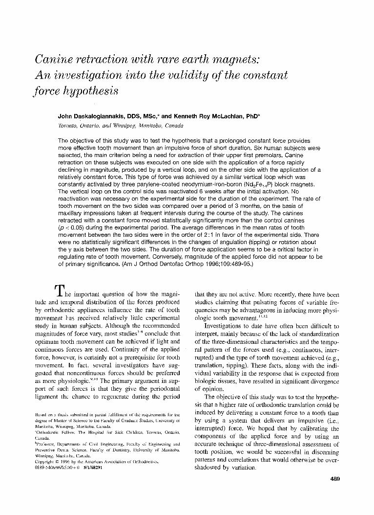
Fig. 1. Vertical loop of 0.017 x 0.025 TMA wire 0/') soldered on 0.017 x 0.025 stainless steel distal extension (T) was used for retraction of control canines. Force delivered by such mecha- nism is of impulsive nature (i,e., interrupted).
SUBJECTS AND METHODS
Six volunteer patients were selected from the screening program at the Graduate Orthodontic Clinic at the University of Manitoba on the basis of needing extraction of their maxillary first premolars. All patients received a combination Nance appliance/transpalatal arch on the maxillary first mo- lars for anchorage reinforcement. Bilateral posterior anchor- age segments (from the second molars to the second premo- lars) were established by inserting 0.017 x 0.025-inch stain- less steel wire segments (in an 0.018-inch slot size appliance) that were bent to fit the brackets passively.
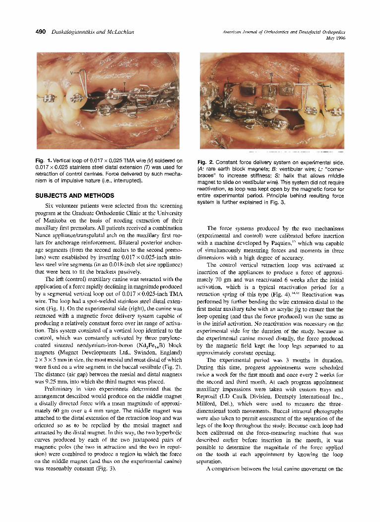
The left (control) maxillary canine was retracted with the application of a force rapidly declining in magnitude produced by a segmental vertical loop out of 0.017 x 0.025-inch TMA wire. The loop had a spot-welded stainless steel distal exten- sion (Fig. 1). On the experimental side (right), the canine was retracted with a magnetic force delivery system capable of producing a relatively constant force over its range of activa- tion. This system consisted of a vertical loop identical to the control, which was constantly activated by three parylene- coated sintered neodymium-iron-boron (Nd2Fe14B) block magnets (Magnet Developments Ltd., Swindon, England) 2 x 3 x 5 mm in size, the most mesial and most distal of which were fixed on a wire segment in the buccal vestibule (Fig. 2). The distance (air gap) between the mesial and distal magnets was 9.25 ram, into which the third magnet was placed.
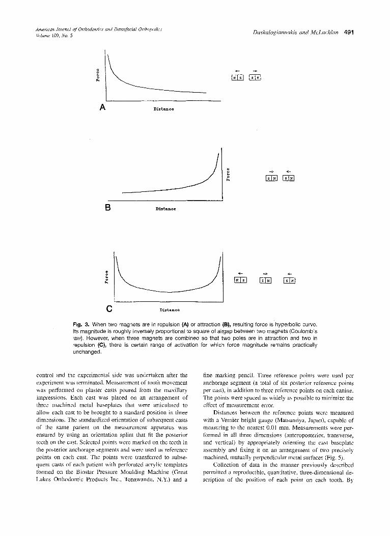
Preliminary in vitro experiments determined that the arrangement described would produce on the middle magnet a distally directed force with a mean magnitude of approxi- mately 60 gm over a 4 mm range. The middle magnet was attached to the distal extension of the retraction loop and was oriented so as to be repelled by the mesial magnet and attracted by the distal magnet. In this way, the two hyperbolic curves produced by each of the two juxtaposed pairs of magnetic poles (thetwo in attraction and the two in repul- sion) were combined to produce a region in which the force on the middle magnet (and thus on the experimental canine) was reasonably constant (Fig. 3).
Fig. 2, Constant force delivery system on experimental side. (,,4: rare earth block magnets; B: vestibular wire; L" "corner- braces" to increase stiffness; S: helix that allows middle magnet to slide on vestibular wire). This system did not require reactivation, as loop was kept open by the magnetic force for entire experimental period. Principle behind resulting force system is further explained in Fig. 3.
The force systems produced by the two mechanisms (experimental and control) were calibrated before insertion with a machine developed by Paquien, 13 which was capable of simultaneously measuring forces and moments in three dimensions with a high degree of accuracy.
The control vertical retraction loop was activated at insertion of the appliances to produce a force of approxi- mately 70 gm and was reactivated 6 weeks after the initial activation, which is a typical reactivation period for a retraction spring of this type (Fig. 4). 14'~5 Reactivation was performed by further bending the wire extension distal to the first molar auxiliary tube with an acrylic jig to ensure that the loop opening (and thus the force produced) was the same as in the initial activation. No reactivation was necessary on the experimental side for the duration of the study, because as the experimental canine moved distally, the force produced by the magnetic field kept the loop legs separated to an approximately constant opening.
The experimental period was 3 months in duration. During this time, progress appointments were scheduled twice a week for the first month and once every 2 weeks for the second and third month. At each progress appointment maxillary impressions were taken with custom trays and Reprosil (LD Caulk Division, Dentsply International Inc., Milford, Del.), which were used to measure the three- dimensional tooth movements. Buccal intraoral photographs were also taken to permit assessment of the separation of the legs of the loop throughout the study. Because each loop had been calibrated on the force-measuring machine that was described earlier before insertion in the mouth, it was possible to determine the magnitude of the force applied on the tooth at each appointment by knowing the loop separation.
A comparison between the total canine movement on the
American Journal of Orthodontics and Dentofacial Orthopedics Daskalogiannakis and McLachlan 491 Volume 109, No. 5
g
A D i e t a n c e
f B D£s~a~ce
C Distmace
Fig. 3. When two magnets are in repulsion (A) or attraction (B}, resulting force ishyperbolic curve. Its magnitude is roughly inversely proportional to square of airgap between two magnets (Coulomb's law). However, when three magnets are combined so that two poles are in attraction and two in repulsion (C}, there is certain range of activation for which force magnitude remains practically unchanged.
control and ti~e experimental side was undertaken after the experiment was terminated. Measurement of tooth movement was performed on plaster casts poured fiom the maxillary impressions. Each cast was placed on an alTangement of three machined metal baseplates that were articulated to allow each cast to be brought to a standard position in three dimensions. The standardized orientation of subsequent casts of the same patient on the measurement apparatus was ensured by using an orientation splint that fit the posterior teeth on the cast. Selected points were marked on the teeth in the posterior anchorage segments and were used as reference points on each cast. The points were transferred to subse- quent casts of each patient with perforated acrylic templates formed on the Biostar Pressure Moulding Machine (Great Lakes Orthodontic Products Inc., Tonawanda, N.Y.) and a
fine marking pencil. Three reference points were used per anchorage segment (a total of six posterior reference points per cast), in addition to three reference points on each canine. The points were spaced as widely as possible to minimize the effect of measurement era-or.
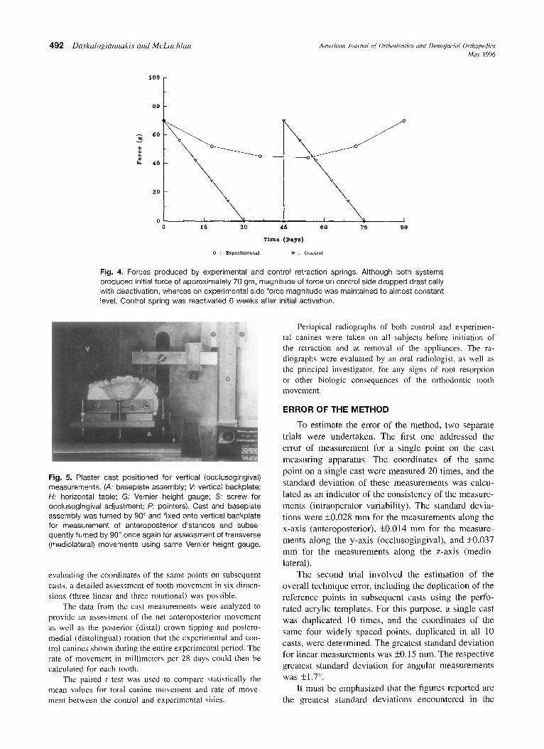
Distances between the reference points were measured with a Vernier height gauge (Matsumiya, Japan), capable of measuring to the nearest 0.01 mm. Measurements were per- formed in all three dimensions (anteroposterior, transverse, and vertical) by appropriately orienting the cast baseplate assembly and fixing it on an arrangement of two precisely machined, mutually perpendicular metal surfaces (Fig. 5).
Collection of data in the manner previously described permitted a reproducible, quantitative, three-dimensional de- scription of the position of each point on each tooth. By
492 Daskalogiannakis and McLachlan American Journal q/" Orthodontics and Dentofacial Orthopedics May 1 9 9 6
100
80
60
40
20
\ \
,¢
I I I ~ T 15 30 45
\ \
v
, I r \ ~ , I 60 75 go
T i m e (De.ys)
0 : E x p e r i m e n t a l v : C o n t r o l
Fig. 4. Forces produced by experimental and control retraction springs. Although both systems produced initial force of approximately 70 gm, magnitude of force on control side dropped drastically with deactivation, whereas on experimental side force magnitude was maintained to almost constant level. Control spring was reactivated 6 weeks after initial activation.
Fig. 5. Piaster cast positioned for vertical (occlusogingival) measurements. (,4: basepiate assembly; V." vertical backplate; H: horizontal table; G: Vernier height gauge; S- screw for occlusogingival adjustment; P: pointers). Cast and baseplate assembly was turned by 90 ° and fixed onto vertical backplate for measurement of anteroposterior distances and subse- quently turned by 90 ° once again for assessment of transverse (mediolateral) movements using same Vernier height gauge.
evaluating the coordinates of the same points on subsequent casts, a detailed assessment of tooth movement in six dimen- sions (three linear and three rotational) was possible.
The data from the cast measurements were analyzed to provide an assessment of the net anteroposterior movement as well as the posterior (distal) crown tipping and postero- medial (distolingual) rotation that the experimental and con- trol canines shown during the entire experimental period. The rate of movement in millimeters per 28 days could then be calculated for each tooth.
The paired t test was used to compare statistically the mean values for total canine movement and rate of move- ment between the control and experimental sides.
Periapical radiographs of both control and experimen- tal canines were taken on all subjects before initiation of the retraction and at removal of the appliances. The ra- diographs were evaluated by an oral radiologist, as well as the principal investigator, for any signs of root resorption or other biologic consequences of the orthodontic tooth movement.
ERROR OF THE METHOD
To estimate the error of the method, two separate
trials were undertaken. The first one addressed the
error of measurement for a single point on the cast
measuring apparatus. The coordinates of the same point on a single cast were measured 20 times, and the
standard deviation of these measurements was calcu-
lated as an indicator of the consistency of the measure-
ments (intraoperator variability). The standard devia-
tions were +0.028 mm for the measurements along the x-axis (anteroposterior), +0.014 mm for the measure- ments along the y-axis (occlusogingival), and +_0.037
mm for the measurements along the z-axis (medio- lateral).
The second trial involved the estimation of the
overall technique error, including the duplication of the reference points in subsequent casts using the perfo-
rated acrylic templates. For this purpose, a single cast was duplicated 10 times, and the coordinates of the same four widely spaced points, duplicated in all 10 casts, were determined. The greatest standard deviation for linear measurements was +0.15 mm. The respective greatest standard deviation for angular measurements
was _+1.7 ° . It must be emphasized that the figures reported are
the greatest standard deviations encountered in the
American Journal of Orthodontics and Dentofacial Orthopedics Volume 109, No. 5 Daskalogiannakis and McLach lan 49:3
error trials and represent the worst case indicators of the overall accuracy of the method.
RESULTS
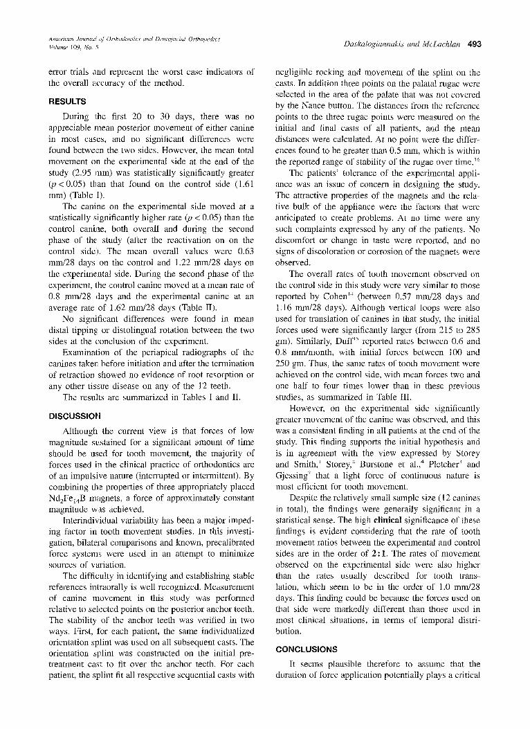
During the first 20 to 30 days, there was no appreciable mean posterior movement of either canine in most cases, and no significant differences were found between the two sides. However, the mean total movement on the experimental side at the end of the study (2.95 ram) was statistically significantly greater (p < 0.05) than that found on the control side (1.61 mm) (Table I).
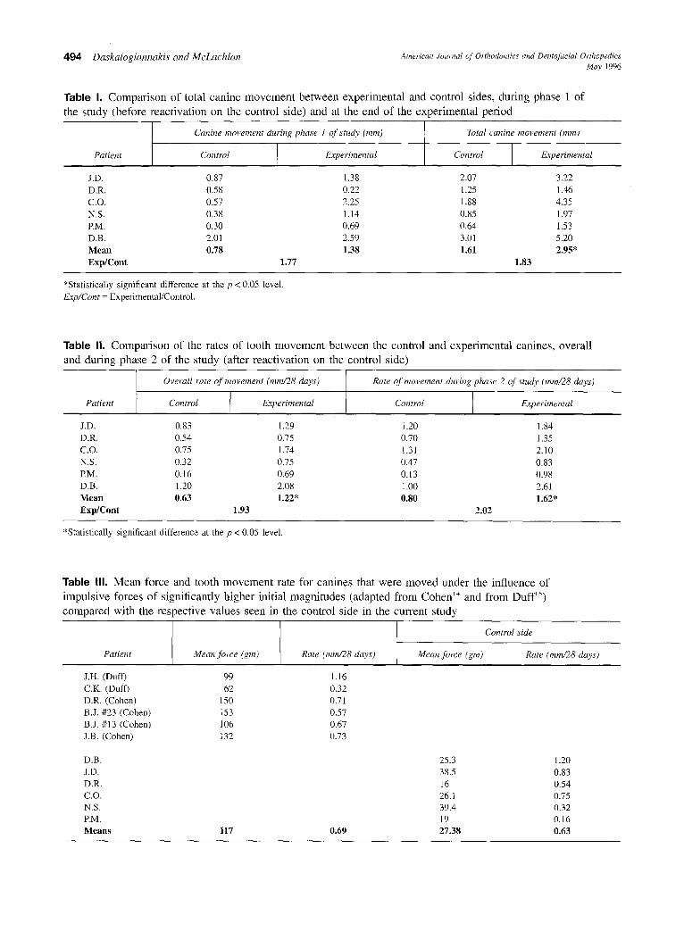
The canine on the experimental side moved at a statistically significantly higher rate (p < 0.05) than the control canine, both overall and during the second phase of the study (after the reactivation on on the control side). The mean overall values were 0.63 mrrd28 days on the control and 1.22 mm/28 days on the experimental side. During the second phase of the experiment, the control canine moved at a mean rate of 0.8 mm/28 days and the experimental canine at an average rate of 1.62 ram/28 days (Table II).
No significant differences were found in mean distal tipping: or distolingual rotation between the two sides at the conclusion of the experiment.
Examination of the periapical radiographs of the canines taken before initiation and after the termination of retraction showed no evidence of root resorption or any other tissue disease on any of the 12 teeth.
The results are summarized in Tables I and II.
DISCUSSIOH
Although the current view is that forces of low magnitude sustained for a significant amount of time should be used for tooth movement, the majority of forces used in the clinical practice of orthodontics are of an impulsive nature (interrupted or intermittent). By combining the properties of three appropriately placed Nd2Fe14B magnets, a force of approximately constant magnitude was achieved.
Interindividual variability has been a major imped- ing factor in tooth movement studies. In this investi- gation, bilateral comparisons and known, precalibrated force systems were used in an attempt to minimize sources of variation.
The difficulty in identifying and establishing stable references intraorally is well recognized. Measurement of canine movement in this study was performed relative to selected points on the posterior anchor teeth. The stability of the anchor teeth was verified in two ways. First, for each patient, the same individualized orientation splint was used on all subsequent casts. The orientation splint was constructed on the initial pre- treatment cast to fit over the anchor teeth. For each patient, the splint fit all respective sequential casts with
negligible rocking and movement of the splint on the casts. In addition three points on the palatal rugae were selected in the area of the palate that was not covered by the Nance button. The distances from the reference points to the three rugae points were measured on the initial and final casts of all patients, and the mean distances were calculated. At no point were the differ- ences found to be greater than 0.5 ram, which is within the reported range of stability of the rugae over time. ~6
The patients' tolerance of the experimental appli- ance was an issue of concern in designing the study. The attractive properties of the magnets and the rela- tive bulk of the appliance were the factors that were anticipated to create problems. At no time were any such complaints expressed by any of the patients. No discomfort or change in taste were reported, and no signs of discoloration or corrosion of the magnets were observed.
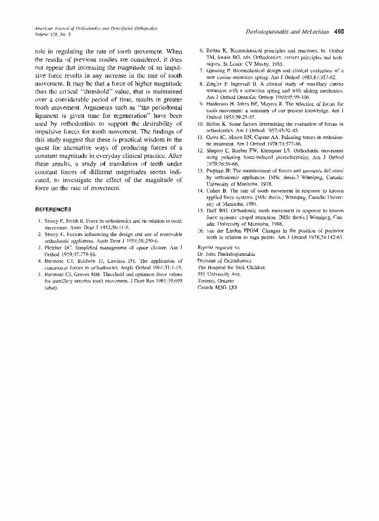
The overall rates of tooth movement observed on the control side in this study were very similar to those reported by Cohen '4 (between 0.57 mm/28 days and 1.16 mm/28 days). Although vertical loops were also used for translation of canines in that study, the initial forces used were significantly larger (from 215 to 285 gm). Similarly, DufP ~ reported rates between 0.6 and 0.8 mm/month, with initial forces between 100 and 250 gm. Thus, the same rates of tooth movement were achieved on the control side, with mean forces two and one half to four times lower than in these previous studies, as summarized in Table III.
However, on the experimental side significantly greater movement of the canine was observed, and this was a consistent finding in all patients at the end of the study. This finding supports the initial hypothesis and is in agreement with the view expressed by Storey and Smith, ~ Storey, 2 Burstone et al.,4 Pletcher 3 and Gjessing v that a light force of continuous nature is most efficient for tooth movement.
Despite the relatively small sample size (12 canines in total), the findings were generally significant in a statistical sense. The high clinical significance of these findings is evident considering that the rate of tooth movement ratios between the experimental and control sides are in the order of 2"1. The rates of movement observed on the experimental side were also higher than the rates usually described for tooth trans- lation, which seem to be in the order of 1.0 ram/28 days. This finding could be because the forces used on that side were markedly different than those used in most clinical situations, in terms of temporal distri- bution.
CONCLUSIONS
It seems plausible therefore to assume that the duration of force application potentially plays a critical
4 9 4 Daskalogiannakis and McLachlan American Journal of Orthodontics and Dentoj~tcial Orthopedics May 1996
Table I. Comparison of total canine movement between experimental and control sides, during phase 1 of the study (before reactivation on the control side) and at the end of the experimental period
Canine movement during phase 1 of study (ram) Total canine movement (mm)
*Statistically significant difference at the p < 0.05 level. Exp/Cont = Experimental/Control.
Table II. Comparison of the rates of tooth movement between the control and experimental canines, overall and during phase 2 of the study (after reactivation on the control side)
Overall rate of movement (mm/28 days) Rate of movement during phase 2 of study (ram~28 days)
Patient Control Control I Experimental Experimental
*Statistically significant difference at the p < 0.05 level.
Table Ill. Mean force and tooth movement rate for canines that were moved under the influence of impulsive forces of significantly higher initial magnitudes (adapted from Cohen '4 and from Duff '5) compared with the respective values seen in the control side in the current study
I Control side
Patient Mean Jbrce (gin) Mean force (gm) Rate (ram~28 days)
American Journal of Orthodontics and Dentofacial Orthopedics Volume 109, No. 5 DaskaIogiannakis and McLach tan 495
role in regulat ing the rate o f tooth movemen t . W h e n
the results o f previous studies are considered, it does
not appear ~.hat increas ing the magn i tude o f an impul-
sive force results in any increase in the rate o f tooth
movemen t . It may be that a force o f h igher magni tude
than the crit ical " t h r e sho ld" value, that is main ta ined
over a cons iderable per iod o f time, results in greater
tooth movemen t . Arguments such as " the per iodonta l
l igament is g iven t ime for r egenera t ion" have been
used by orthodontis ts to support the desirabi l i ty of
impuls ive forces for tooth movemen t . The findings o f
this study suggest that there is pract ical w i s d o m in the
quest for a l ternat ive ways of producing forces of a
constant magn i tude in eve ryday cl inical practice. Af te r
these results, a study of t ranslat ion o f teeth under
constant forces o f different magni tudes seems indi-
cated, to inves t igate the effect o f the magni tude o f
force on the: rate o f m o v e m e n t ,
REFERENCES
I. Storey E, Smith R. Force in orthodontics and its relation to tooth movement. Austr Dent J 1952;56:11-8.
2. Storey E. Factors influencing the design and use of removable orthodontic appliances. Austr Dent J 1954;58:230-6.
3. Pletcher EC. Simplified management of space closure. Am J Orthod 1959;45:278-86.
4. Burstone CJ, Baldwin JJ, Lawless DT. The application of continuous forces to orthodontics. Angle Orthod 1961;31:1-15.
5. Burstone C J, Groves MH. Threshold and optimum force values for maxillary anterior tooth movement. J Dent Res 1961;39:695 (abst).
6. Reitan K. Biomechanical principles and reactions. In: Graber TM, Swain BG, eds. Orthodontics: current principles and tech- niques. St Louis: CV Mosby, 1985.
7. Gjessing R Biomechanical design and clinical evaluation of a new canine-retraction spring. Am J Orthod 1985;87:353-62.
8. Ziegler R Ingervall B. A clinical study of maxillary canine retraction with a retraction spring and with sliding mechanics. Am J Orthod Dentofac Orthop 1989;95:99-106.
9. Halderson H, Johns EE, Moyers R. The selection of forces for tooth movement: a summary of our present knowledge. Am J Orthod 1953;39:25-35.
10. Reitan K. Some factors determining the evaluation of forces in orthodontics. Am J Orthod 1957;43:32-45.
11. Oates JC, Moore RN, Caputo AA. Pulsating forces in orthodon- tic treatment. Am J Orthod 1978;74:577-86.
12. Shapiro E, Roeber FW, Ktempner LS. Orthodontic movement using pulsating force-induced piezoelectricity. Am J Orthod I979;76:59~66,
13. Paquien JP. The measurement of forces and moments delivered by orthodontic appliances. [MSc thesis.] Winnipeg, Canada: University of Manitoba, 1978.
14. Cohen B. The rate of tooth movement in response to known applied force systems. [MSc thesis.] Winnipeg, Canada: Univer- sity of Manitoba, 1991.
15. Duff WG. Orthodontic tooth movement in response to known force systems: cuspid retraction. [MSc thesis.] Winnipeg, Can- ada: University of Manitoba, 1988.
16. van der Linden FPGM. Changes in the position of posterior teeth in relation to ruga points. Am J Orthod 1978;74:142-61.
Reprint requests to: Dr. John Daskalogiannakis Division of Orthodontics The Hospital for Sick Children 555 University Ave. Toronto, Ontario Canada M5G 1X8