Seminars in Cell & Developmental Biology 23 (2012) 937– 944
Contents lists available at SciVerse ScienceDirect
Seminars in Cell & Developmental Biology
jo u rn al hom epa ge: www.elsev ier .com/ locate /semcdb
eview
apturing epidermal stemness for regenerative medicine
ann Barrandona,b,∗, Nicolas Grasseta,b, Andrea Zaffalona,b, Franc ois Gorostidia,b,téphanie Claudinota,b, Stéphanie Lathion Droz-Georgeta,b, Daisuke Nanbac, Ariane Rochata,b,∗
Department of Experimental Surgery, Lausanne University Hospital (CHUV), 1011 Lausanne, SwitzerlandLaboratory of Stem Cell Dynamics, Ecole Polytechnique Fédérale de Lausanne (EPFL), 1015 Lausanne, SwitzerlandDepartment of Cell Growth and Tumor Regulation, Proteo-Medicine Research Center, Ehime University, Shitsukawa, Toon, Ehime 791-0295, Japan
r t i c l e i n f o
rticle history:vailable online 2 October 2012
eywords:kintem cellsegenerative medicineell and gene therapy
a b s t r a c t
The skin is privileged because several skin-derived stem cells (epithelial stem cells from epidermis and itsappendages, mesenchymal stem cells from dermis and subcutis, melanocyte stem cells) can be efficientlycaptured for therapeutic use. Main indications remain the permanent coverage of extensive third degreeburns and healing of chronic cutaneous wounds, but recent advances in gene therapy technology openthe door to the treatment of disabling inherited skin diseases with genetically corrected keratinocytestem cells. Therapeutic skin stem cells that were initially cultured in research or hospital laboratoriesmust be produced according strict regulatory guidelines, which ensure patients and medical teams thatthe medicinal cell products are safe, of constant quality and manufactured according to state-of-the art
technology. Nonetheless, it does not warrant clinical efficacy and permanent engraftment of autologousstem cells remains variable. There are many challenges ahead to improve efficacy among which to keeptelomere-dependent senescence and telomere-independent senescence (clonal conversion) to a mini-mum in cell culture and to understand the cellular and molecular mechanisms implicated in engraftment.Finally, medicinal stem cells are expansive to produce and reimbursement of costs by health insurancesis a major concern in many countries.
Stem cells are instrumental for renewal, regeneration and repair,
therapeutic arsenal to treat hematopoietic diseases and a variety ofdisabling conditions [1], including inherited skin diseases [2]. Stemcells or stem cell-derived products are also used in dermatology
nd hold great expectations for disease modeling, drug discov-ry and regenerative medicine. Transplantation of bone marrowerived stem cells (hematopoietic or mesenchymal) is part of the
∗ Corresponding authors at: Department of Experimental Surgery, Lausanneniversity Hospital (CHUV), 1011 Lausanne, Switzerland.
and reconstructive surgery. Furthermore, stem cell therapy is oftenregarded as the therapy of the future for diabetes, cardiac and neu-rological diseases. But it is necessary to capture stem cells beforethey are used in regenerative medicine and it is worth emphasizingthat some cells display stem cell capabilities only when challengedby tissue repair and regeneration, stress or cell culture [3]. For
instance, the pluripotent cells of the inner cell mass only undergoa few round of divisions in blastocyst but their pluripotency canbe captured in cell culture to generate embryonic stem cells that
an indefinitely self renew under appropriate conditions [4]. Sim-larly, cardiac stem cells can be captured from normal or diseased
ammalian heart [5–7] and expanded in culture for therapy [8]. Tohe opposite, hematopoietic stem cells are directly captured fromhe donor marrow or from the blood stream after mobilization fromheir niche, and used without or little ex vivo expansion. In any case,ong-term therapeutic success is achieved only if the transplanteddult stem cells can permanently engraft, self-renew and properlyerform the function for which they are specified. Maintenancef stem cell specification in cell culture is thus critical for a suc-essful cell therapy. Yet stem cell specification can be manipulatedn culture in a process termed reprogramming [3] that consists ofemoving cells from their natural microenvironment and exposinghem to a proper combination of transcription factors [9–11], small
olecules [12] or even to a completely different microenviron-ent [13]. Reprogramming of skin cells (fibroblasts, keratinocytes)
o generate induced pluripotent stem (iPS) cell is now a commonrocedure useful for disease modeling, drug testing or to generateifferentiated cells other than skin.
Stem cells do not mean the same if you are a scientist, a physicianr a patient. Obviously, scientists think self-renewal and potency,ymmetric and asymmetric divisions, fate and niches, growth fac-ors and small molecules, molecular markers and signalization, exivo expansion and banking [14] whereas physicians think success-ul therapy of diseases [15] and patients hope for a better health.ut stem cells have acquired another dimension besides the scien-ific and medical breakthroughs as they are expected to create jobsnd wealth. Hence, stem cell research and its clinical outcome arender close scrutiny from politicians, the media, regulatory affairsnd health insurances as well as from the biotechnology and phar-aceutical industry [16]. The story on how keratinocyte stem cellsade it from bench to bedside is a perfect illustration.
. A short stem cell story
In October 1983, one of us (YB) joined the laboratory of Pro-essor Howard Green at Harvard Medical School as a post-doctoralellow. It was an exciting and amazing time in the Green laboratorys the first cultured epidermal autografts (CEAs) were prepared toreat two young brothers with extensive third degree burn woundsovering over 90% of their body [17]. The areas to repair with theultured cells were extremely large, nothing to compare with amall transplant onto the back of a mouse or a human arm as itad already been done by the laboratory [17,18]. To permanentlyestore a functional epidermal barrier with autologous culturederatinocytes transplanted on burn wounds excised to muscularascia with the ultimate hope to save the patients life was a majorhallenge both in term of biology and medicine. Many questionsere opened: can massive expansion of human keratinocytes from
small skin biopsy harvested in an unburned area be achievedn a minimal time and in an emergency context? Can patients be
aintained alive during the time of preparation of cultured cells?an cultured keratinocytes permanently engraft, form an epider-is and restore the skin barrier function? All lab members were
ware that the life of two young children was at stake and thatoward Green’s decision was based on his faith that cultures ofuman epidermal keratinocytes contained stem cells [19]. Indeed,he notion that cultured keratinocytes could be useful for cell ther-py was first stated in the conclusion of the Rheinwald and Green’seminal paper [19]. Follow-up experiments performed in the Greenaboratory had then demonstrated that cultured human keratino-ytes transplanted onto athymic mice could generate an epidermis
18] and were able to heal small burn wounds [20]. Nonetheless,
oving from bench to bedside overnight was certainly a jump inhe unknown but medical breakthroughs often happen in circum-tances in which there is no alternative than pushing the limits
lopmental Biology 23 (2012) 937– 944
to save a patient’s life. The most emotional time was at the firstdressing, usually a week after CEA transplantation (take down). Theentire laboratory eagerly waited for news from the hospital; howwere the patients doing? Did the cultures engraft? Sometimes newswere good and greeted with joy, other times there were bad andeveryone was sad with a feeling of catastrophe. Finally, the youngbrothers went out of intensive care. Several months later, HowardGreen and YB visited the children at the Schriners Burn Hospital andfound them playing with a basketball in the corridor of the hospitalward. This was an amazing moment, full of emotion. The kids wereseverely handicapped and surely not at the end of suffering, butthey were full of life with twinkles of fun in their eyes. Undoubt-edly, CEA had contributed to save the children life and from here,it was evident that the transplanted CEA had to contain stem cells.This was how adult keratinocyte stem cells were captured for thefirst time ever for therapeutic purposes. From here, several otherburn children were treated and the Green lab turned into a cell fac-tory, mixing production of CEA, academic research, media exposureand training of colleagues from all over the world. Unknowingly, theGreen lab was bringing cultured adult stem cells from bench to bed-side and experiencing translational medicine [21]. Yet, the goal ofan academic laboratory was to perform experiments and not to be acell factory. In 1986, the cell culture technology was transferred toa start-up company, Biosurface Technology Inc., located in KendallSquare in Cambridge (Massachusetts) within a stone’s throw fromMIT where Howard Green had started his research on cultured kera-tinocytes in the seventies. With this move, CEA became a productand the scientists discovered that it was another ball game ruled bythe FDA (Food and Drug Administration), the health insurances andthe amount of cash in the bank. Biosurface Technology Inc. eventu-ally became part of Genzyme (Sanofi). Thirty years later, little haschanged and the Rheinwald and Green culture system remains thegold standard and most, if not all, commercial companies providingthe service of culturing stem cells for the treatment of extensivethird degree burns still rely on it (e.g. Genzyme, Cambridge, MA,USA; Tego Science, Seoul, South Korea; J-TEC, Aichi Japan; HolostemTerapie Avanzate, Modena, Italy).
3. Capturing stemness in human skin
Considerable progress in understanding the biology of skin stemcells has been accomplished in the mouse and there are excellentrecent reviews that describe these advances [22,23]. Lineage trac-ing experiments and fonctional skin reconstitution assays in themouse have unambiguously demonstrated that the interfollicularepidermis [24–26], the upper constant region of pelage hair fol-licles [27–29] and the ducts of sweat glands of the foot pad [30]contain cells with stem cells properties. These stem cells are mul-tipotent and clonogenic [27,29], slow-cycling [31,32], or expresscell surface proteins like Lgr5 [33] and Lgr6 [34], CD34 [35], Plet1(MTS24) [36] and Lrig1 [37]. Similarly, multipotent stem cells havebeen identified in whisker follicles of the mouse and the rat byclonal analysis and serial transplantation [38,39]. Furthermore, theclassical scheme of epidermal renewal based on a hierarchy of slow-cycling stem cells and rapidly cycling transient amplifying cells [40]is a matter of discussion with opposing opinions [24–26]. But howmuch of this knowledge can be translated in human stem cell ther-apy? A pessimistic but fair answer is very little, and the clinicalsituation is pretty much the same to day that it was thirty years ago.There are several reasons for that, first stem cell markers describedin the mouse do not apply to human, second label retaining, lineagetracing or genetic manipulation experiments cannot be performed
in human for obvious ethical reasons and third clonogenic assaysremains the sole reliable read-out to capture stemness in humanepidermis or epidermal appendages [41–44]. Ultimately a humankeratinocyte is considered a stem cell if it forms a colony that can
Y. Barrandon et al. / Seminars in Cell & Developmental Biology 23 (2012) 937– 944 939
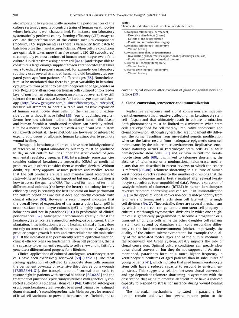
Fig. 1. Clonal analysis. A holoclone (red) generates a progeny that forms largeprogressively growing colonies and almost no terminal colonies (less than 5%). Ameroclone (yellow) forms both large progressively growing and terminal colonies.Aa
btkcEt(Ctccre
tpcci7miatedgtihtMgtstfmpsb
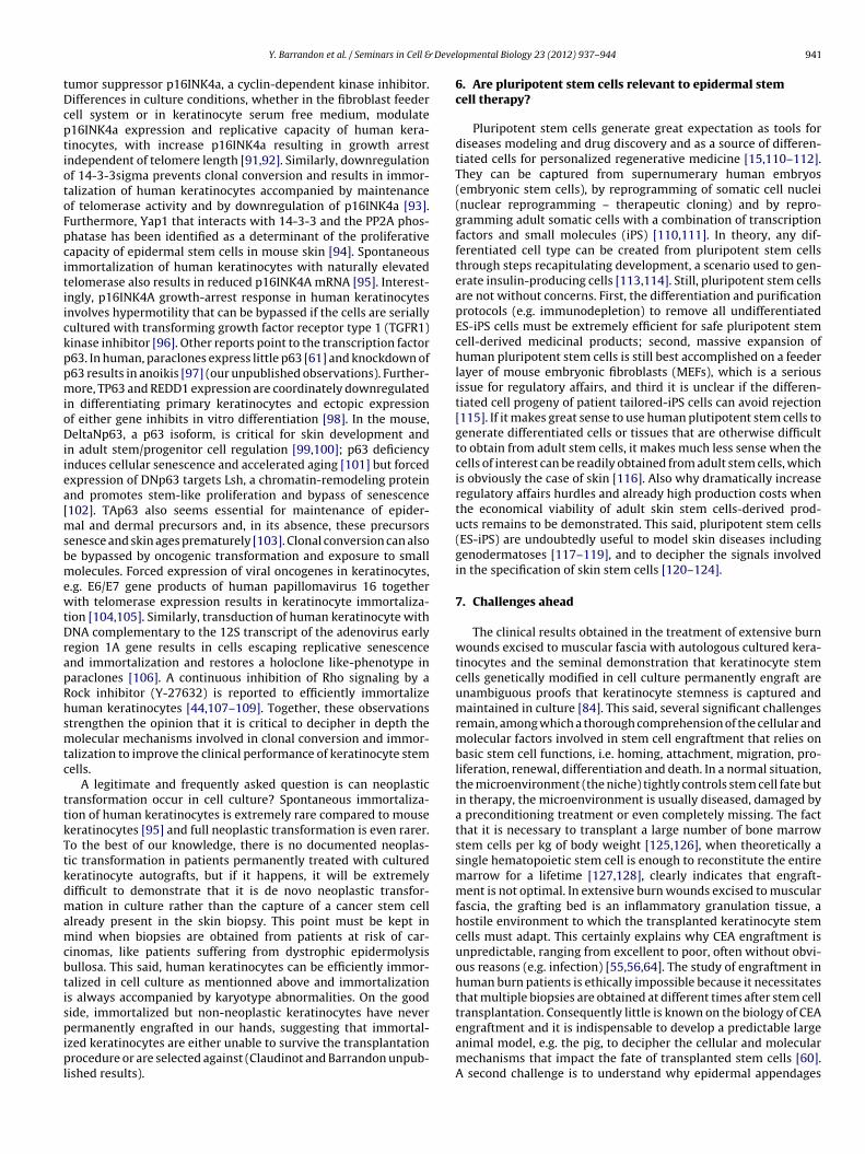
Fig. 2. Clonal conversion. Clonal conversion together with replicative senescenceaffects the lifespan of human keratinocyte stem cells in culture and results in pro-gressive loss of growth potential. A holoclone (red) with extensive growth potential
paraclone (green) generates only terminal colonies. Modified from Barrandon Ynd Green H [41].
e serially subcultured and that can generate an epidermis whenransplanted onto immunodeficient mice [41,42,45]. Are all humaneratinocyte stem cells clonogenic? Possibly not as there are non-lonogenic stem cells in rodents (our unpublished observations).xpression of cell surface proteins like CD71 (transferring recep-or) [46], CD49f (alpha 6 integrin), CD49a (alpha 1 integrin) CD49balpha 2 integrin), CD49c (alpha 3 integrin), CD104 (beta 4 integrin),D29 (beta 1 integrin) [43,47], Lrig1 [48], or ABCG2 [49] can cap-ure stemness. However none of these cell surface proteins is stemell specific and the quest for a reliable marker of human keratino-yte stem cells remains. In our opinion, cell size remains the mosteliable and efficient means to enrich in clonogenic keratinocytesither as single cells [50] or after elutriation [51,52].
Human clonogenic keratinocytes have different growth poten-ial in culture and can be classified as holoclones, meroclones andaraclones by clonal analysis [41] (Fig. 1). Ideally, single keratino-ytes are isolated under an inverted microscope and individuallyultured. Typically cloning efficiency ranges between 40 and 70%f cultured keratinocytes are selected on small size [50]. After
days of cultivation, each clone is identified under an invertedicroscope and subcultured onto indicator dishes containing an
rradiated feeder layer. After 12 days, indicator dishes are fixednd stained with Rhodamine B. The size and the number oferminal colonies containing squame-like cells expressing mark-rs of skin terminal differentiation (involucrin, filaggrin, LEKTI)efine the clonal type [41]. Holoclones generate large progressivelyrowing colonies and almost no terminal colonies. They have aremendous growth capacity up to 180 divisions and can theoret-cally generate enough CEA to cover the entire body of an adultuman [41,42,45]. It is now widely accepted that holoclones arehe phenotype of human keratinocyte stem cells in culture [53].
eroclones derive from holoclones and generate a mixture of pro-ressively growing colonies and terminal colonies. Paraclones areransient amplifying cells committed to a small number of divi-ions (from 1 to 15 divisions – 2 cells to 32,000 cells respectively)hat generate only terminal aborted colonies containing large dif-erentiated squame like-cells on indicator dishes. Paraclones are
ostly generated by meroclones [41]. Importantly the number ofaraclones in a suspension of keratinocytes freshly isolated from akin biopsy is independent of donor age and is far below the num-er of meroclones or holoclones (our unpublished results). This is
converts into a meroclone (yellow) that in turn converts into a paraclone withrestricted growth potential (green). Clonal conversion results from microenviron-mental insults and is irreversible under normal conditions.
surprising because transient amplifying cells should represent themajority of the multiplying keratinocytes in the basal layer ofepidermis according to kinetic experiments [40]. Therefore it isunlikely that paraclones are native transient amplifying cells; inour opinion they result from culture stress because the frequency ofparaclone formation rapidly increases with time in culture, whichultimately results in culture termination. The conversion of holo-clones to meroclones and paraclones is termed clonal conversionand is an irreversible phenomenon under normal circumstances[41] (Fig. 2).
4. Capturing stemness for cell therapy
The capture of keratinocyte stemness for cell therapy relieson recreating or mimicking a microenvironment that favors self-renewal and long-term expansion of stem cells, an indispensablecondition to produce a medicinal product on a large scale. So far,the best scenario to capture adult keratinocyte stem cells relieson the use of a mouse feeder layer of embryonic fibroblast, as forhuman ES or iPS cells [19]. This method remains unmatched fora rapid and massive expansion of adult keratinocyte stem cellsfor autologous therapy. The Rheinwald and Green culture sys-tem relies on the presence of a feeder layer of mouse embryonicfibroblasts (Swiss 3T3-J2 cells) [19,54] that are growth arrestedby a mitomycin C treatment or lethal gamma irradiation (60 Gy).The cultured medium is a 3:1 mix of DMEM (Dulbecco ModifiedEagle Medium) and F12 supplemented with fetal calf serum (FCS),cholera toxin and hormones including insulin, hydrocortisone andtriiodothyronine [17,55,56]. Epidermal growth factor (EGF) is alsoan important supplement to enhance colony growth [57,58]. Propermaintenance of the 3T3-J2 cells is key to long-term cultivationof human keratinocyte stem cells and this is particularly criticalfor clinical applications or if the starting biopsy is small. Withoutdoubt improper cultivation of feeder cells systematically affects thequality of a keratinocyte culture, consequently it may be impos-sible to produce enough CEA to fully treat a patient or CEA maypoorly engraft. A common mistake is to substitute the 3T3-J2 cellsby NIH 3T3 cells or to culture the 3T3-J2 cells in medium supple-mented with FCS rather than bovine serum. As a consequence, cells
are selected for rapid growth and loss of contact inhibition, whichtogether results in the irradiated feeder cells detaching within fewdays after seeding when a good feeder layer is able to supportgrowth of keratinocytes for several weeks after irradiation [58]. It is
9 Developmental Biology 23 (2012) 937– 944
acwse(batccyrpicrltabosSatcew
itecpdtshdetctohpkpnp[ctg
cst[rtrodo
Table 1Therapeutic indications of cultured keratinocyte stem cells.
Autologous cell therapy (permanent)- Extensive skin defects (burns)- Defects of the ocular surface- Plastic and reconstructive surgery
Autologous gene therapy (permanent)- Invalidating genodermatoses (junctional epidermolysis bullosa)- Production of proteins of medical interest
Allogenic cell therapy (temporary)- Wound healing
Allogenic gene therapy (temporary)
40 Y. Barrandon et al. / Seminars in Cell &
lso important to systematically monitor the performances of theulture system by means of control strains of human keratinocyteshose behavior is well characterized. For instance, our laboratory
ystematically performs colony-forming efficiency (CFE) assays tovaluate the performances of the culture medium componentsmedium, FCS, supplements) as there is variability from batch toatch despites the manufacturers’ claims. When culture conditionsre optimal, it takes more than five months (20–25 subcultures)o completely exhaust a culture of human keratinocyte, even if theulture is initiated from a single stem cell [42,45] and it is possible toonstitute a large enough supply of frozen keratinocytes that takesears to exhaust if properly managed. For example, our laboratoryoutinely uses several strains of human diploid keratinocytes pre-ared years ago from patients of different ages [58]. Nonetheless,
t must be mentioned that there is a great variability in keratino-yte growth from patient to patient independent of age, gender orace. Regulatory affairs consider human cells cultured onto a feederayer of non-human origin as xenotransplants, but even so they stillolerate the use of a mouse feeder for keratinocyte stem cell ther-py (http://www.genzyme.com/business/biosurgery/burn/epicel)ecause all attempts to obtain a rapid and massive expansionf human keratinocyte stem cells for the treatment of exten-ive burns without it have failed [59] (our unpublished results).erum free low calcium medium, irradiated human fibroblastsnd human fibroblast-conditioned medium can partially substi-ute for a mouse feeder layer but with a significant loss in stemell growth potential. These methods are however of interest toxpand autologous or allogenic human keratinocytes to promoteound healing.
Therapeutic keratinocyte stem cells have been initially culturedn research or hospital laboratories, but they must be producedo day in cell culture facilities under the strict control of gov-rnmental regulatory agencies [16]. Interestingly, some agenciesonsider cultured keratinocyte autografts (CEAs) as medicinalroducts while others consider them as medical devices. Withoutoubt, regulatory approval assures patients and medical teamshat the cell products are safe and manufactured according totate-of-the art technology. An important but unsolved question isow to assess the quality of CEA. The rate of formation of terminalifferentiated colonies (the lower the better) in a colony-formingfficiency assay is certainly the best indicator on how performanthe culture conditions are but it does not strictly correlate withlinical efficacy [60]. However, a recent report indicates thathe overall level of expression of the transcription factor p63 incular surface keratinocyte cultures (p63 is highly expressed inoloclones and not in paraclones [61]) is predictable of clinicalerformances [62]. Anticipated performances greatly differ if theeratinocyte stem cells are aimed for autologous or allogenic trans-lantation. If the indication is wound healing, clinical efficacy doesot rely on stem cell capabilities but relies on the cells’ capacity toroduce proper growth factors and extracellular matrix molecules63]. If the indication is to permanently restore epithelial function,linical efficacy relies on fundamental stem cell properties, that ishe capacity to permanently engraft, to self-renew and to faithfulyenerate a differentiated progeny for a lifetime.
Clinical applications of cultured autologous keratinocyte stemells have been extensively reviewed [59] (Table 1). The mosttriking application of cultured keratinocyte stem cells remainshe permanent coverage of extensive third degree burn wounds17,55,56,64–81], the transplantation of corneal stem cells toestore sight in patients with corneal blindness [62,82,83] and thereatment of junctional epidermolysis bullosa with genetically cor-
ected autologous epidermal stem cells [84]. Cultured autologousr allogenic keratinocytes have also been used to improve healing ofonor sites and of second degree burns, in Mohs surgery for removalf basal cell carcinoma, to prevent the recurrence of keloids, and to
- Wound healing
cover surgical wounds after excision of giant congenital nevi andtattoos [59].
5. Clonal conversion, senescence and immortalization
Replicative senescence and clonal conversion are indepen-dent phenomenon that negatively affect human keratinocyte stemcell lifespan and that ultimately result in culture termination.Both phenomenons must be limited to a minimum when stemcells are expanded for cell therapy. Replicative senescence andclonal conversion, although synergistic, are fundamentally differ-ent, the former resulting from age-related genetic modificationwhereas the latter results from inadequate epigenetic stem cellmaintenance by the culture microenvironment. Replicative senes-cence naturally occurs in keratinocyte stem cells as in adulthematopoietic stem cells [85] and ex vivo in cultured kerati-nocyte stem cells [60]. It is linked to telomere shortening, theabsence of telomerase or a nonfunctional telomerase, mecha-nisms that are described in recent reviews to which the readeris referred [86–88]. Telomere shortening in a culture of humankeratinocytes directly relates to the number of divisions that thecells have undergone and is best visualized after several kerati-nocyte subcultures (our unpublished data). Forced expression ofcatalytic subunit of telomerase (hTERT) in human keratinocytesreverses telomere shortening and can result in immortalization[89]. To the opposite, clonal conversion occurs independently fromtelomere shortening and affects stem cell fate within a singlecell division (Fig. 2). Theoretically, there are several mechanismsby which a stem cell can generate a non-stem cell progeny inculture. First through asymmetrical divisions, in which one daugh-ter cell is genetically programmed to become a progenitor or atransient amplifying cells while the other daughter cell remainsa stem cell, second by daughter stem cells responding differ-ently to the local microenvironment (niche). Importantly, thequality of the culture microenvironment, for example the qual-ity of the irradiated feeder layer and of the culture medium inthe Rheinwald and Green system, greatly impacts the rate ofclonal conversion. Optimal culture conditions can greatly slowdown clonal conversion but they do not suppress it. As afore-mentioned, paraclones form at a much higher frequency inkeratinocyte subcultures of aged patients than in subcultures ofyoung patients [41], which indicates that aged human keratinocytestem cells have a reduced capacity to respond to environmen-tal stress. This suggests a relation between clonal conversionand age-dependent telomere shortening in agreement with theobservation that aging telomerase-deficient mice have a reduced
capacity to respond to stress, for instance during wound healing[90].
The molecular mechanisms implicated in paraclone for-mation remain unknown but several reports point to the
umor suppressor p16INK4a, a cyclin-dependent kinase inhibitor.ifferences in culture conditions, whether in the fibroblast feederell system or in keratinocyte serum free medium, modulate16INK4a expression and replicative capacity of human kera-inocytes, with increase p16INK4a resulting in growth arrestndependent of telomere length [91,92]. Similarly, downregulationf 14-3-3sigma prevents clonal conversion and results in immor-alization of human keratinocytes accompanied by maintenancef telomerase activity and by downregulation of p16INK4a [93].urthermore, Yap1 that interacts with 14-3-3 and the PP2A phos-hatase has been identified as a determinant of the proliferativeapacity of epidermal stem cells in mouse skin [94]. Spontaneousmmortalization of human keratinocytes with naturally elevatedelomerase also results in reduced p16INK4A mRNA [95]. Interest-ngly, p16INK4A growth-arrest response in human keratinocytesnvolves hypermotility that can be bypassed if the cells are seriallyultured with transforming growth factor receptor type 1 (TGFR1)inase inhibitor [96]. Other reports point to the transcription factor63. In human, paraclones express little p63 [61] and knockdown of63 results in anoikis [97] (our unpublished observations). Further-ore, TP63 and REDD1 expression are coordinately downregulated
n differentiating primary keratinocytes and ectopic expressionf either gene inhibits in vitro differentiation [98]. In the mouse,eltaNp63, a p63 isoform, is critical for skin development and
n adult stem/progenitor cell regulation [99,100]; p63 deficiencynduces cellular senescence and accelerated aging [101] but forcedxpression of DNp63 targets Lsh, a chromatin-remodeling proteinnd promotes stem-like proliferation and bypass of senescence102]. TAp63 also seems essential for maintenance of epider-
al and dermal precursors and, in its absence, these precursorsenesce and skin ages prematurely [103]. Clonal conversion can alsoe bypassed by oncogenic transformation and exposure to smallolecules. Forced expression of viral oncogenes in keratinocytes,
.g. E6/E7 gene products of human papillomavirus 16 togetherith telomerase expression results in keratinocyte immortaliza-
ion [104,105]. Similarly, transduction of human keratinocyte withNA complementary to the 12S transcript of the adenovirus early
egion 1A gene results in cells escaping replicative senescencend immortalization and restores a holoclone like-phenotype inaraclones [106]. A continuous inhibition of Rho signaling by aock inhibitor (Y-27632) is reported to efficiently immortalizeuman keratinocytes [44,107–109]. Together, these observationstrengthen the opinion that it is critical to decipher in depth theolecular mechanisms involved in clonal conversion and immor-
alization to improve the clinical performance of keratinocyte stemells.
A legitimate and frequently asked question is can neoplasticransformation occur in cell culture? Spontaneous immortaliza-ion of human keratinocytes is extremely rare compared to mouseeratinocytes [95] and full neoplastic transformation is even rarer.o the best of our knowledge, there is no documented neoplas-ic transformation in patients permanently treated with culturederatinocyte autografts, but if it happens, it will be extremelyifficult to demonstrate that it is de novo neoplastic transfor-ation in culture rather than the capture of a cancer stem cell
lready present in the skin biopsy. This point must be kept inind when biopsies are obtained from patients at risk of car-
inomas, like patients suffering from dystrophic epidermolysisullosa. This said, human keratinocytes can be efficiently immor-alized in cell culture as mentionned above and immortalizations always accompanied by karyotype abnormalities. On the goodide, immortalized but non-neoplastic keratinocytes have never
ermanently engrafted in our hands, suggesting that immortal-
zed keratinocytes are either unable to survive the transplantationrocedure or are selected against (Claudinot and Barrandon unpub-
ished results).
lopmental Biology 23 (2012) 937– 944 941
6. Are pluripotent stem cells relevant to epidermal stemcell therapy?
Pluripotent stem cells generate great expectation as tools fordiseases modeling and drug discovery and as a source of differen-tiated cells for personalized regenerative medicine [15,110–112].They can be captured from supernumerary human embryos(embryonic stem cells), by reprogramming of somatic cell nuclei(nuclear reprogramming – therapeutic cloning) and by repro-gramming adult somatic cells with a combination of transcriptionfactors and small molecules (iPS) [110,111]. In theory, any dif-ferentiated cell type can be created from pluripotent stem cellsthrough steps recapitulating development, a scenario used to gen-erate insulin-producing cells [113,114]. Still, pluripotent stem cellsare not without concerns. First, the differentiation and purificationprotocols (e.g. immunodepletion) to remove all undifferentiatedES-iPS cells must be extremely efficient for safe pluripotent stemcell-derived medicinal products; second, massive expansion ofhuman pluripotent stem cells is still best accomplished on a feederlayer of mouse embryonic fibroblasts (MEFs), which is a seriousissue for regulatory affairs, and third it is unclear if the differen-tiated cell progeny of patient tailored-iPS cells can avoid rejection[115]. If it makes great sense to use human plutipotent stem cells togenerate differentiated cells or tissues that are otherwise difficultto obtain from adult stem cells, it makes much less sense when thecells of interest can be readily obtained from adult stem cells, whichis obviously the case of skin [116]. Also why dramatically increaseregulatory affairs hurdles and already high production costs whenthe economical viability of adult skin stem cells-derived prod-ucts remains to be demonstrated. This said, pluripotent stem cells(ES-iPS) are undoubtedly useful to model skin diseases includinggenodermatoses [117–119], and to decipher the signals involvedin the specification of skin stem cells [120–124].
7. Challenges ahead
The clinical results obtained in the treatment of extensive burnwounds excised to muscular fascia with autologous cultured kera-tinocytes and the seminal demonstration that keratinocyte stemcells genetically modified in cell culture permanently engraft areunambiguous proofs that keratinocyte stemness is captured andmaintained in culture [84]. This said, several significant challengesremain, among which a thorough comprehension of the cellular andmolecular factors involved in stem cell engraftment that relies onbasic stem cell functions, i.e. homing, attachment, migration, pro-liferation, renewal, differentiation and death. In a normal situation,the microenvironment (the niche) tightly controls stem cell fate butin therapy, the microenvironment is usually diseased, damaged bya preconditioning treatment or even completely missing. The factthat it is necessary to transplant a large number of bone marrowstem cells per kg of body weight [125,126], when theoretically asingle hematopoietic stem cell is enough to reconstitute the entiremarrow for a lifetime [127,128], clearly indicates that engraft-ment is not optimal. In extensive burn wounds excised to muscularfascia, the grafting bed is an inflammatory granulation tissue, ahostile environment to which the transplanted keratinocyte stemcells must adapt. This certainly explains why CEA engraftment isunpredictable, ranging from excellent to poor, often without obvi-ous reasons (e.g. infection) [55,56,64]. The study of engraftment inhuman burn patients is ethically impossible because it necessitatesthat multiple biopsies are obtained at different times after stem celltransplantation. Consequently little is known on the biology of CEA
engraftment and it is indispensable to develop a predictable largeanimal model, e.g. the pig, to decipher the cellular and molecularmechanisms that impact the fate of transplanted stem cells [60].A second challenge is to understand why epidermal appendages
9 Deve
(fwsicumcocbsaetp
A
Uaw
R
42 Y. Barrandon et al. / Seminars in Cell &
hair follicles, sebaceous and sweat glands) are not regeneratedrom the transplanted autologous human keratinocyte stem cellshen recent studies clearly demonstrate that there are multipotent
tem cells with hair- and sebaceous glands-forming capabilitiesn adult mammalian skin and that multipotency is maintained inell culture [27,38,39]. The most plausible explanation for the fail-re to regenerate skin appendages in human is again an impropericroenvironment. A third challenge is to discover cell culture
onditions that address safety concerns of regulatory affairs with-ut jeopardizing stemness and CEA efficacy. Last but not least, theosts of skin stem cell-based products, unaffordable by many, muste decreased [60]. Unfortunately, this is wishful thinking becausetem cell technologies are getting more and more sophisticatednd because the translation from bench to bedside necessitatesxpensive standard operating procedures (SOPs), good manufac-uring procedures (GMPs) and good clinical practice (GCP) to ensureatients that stem cell therapy is safe and efficient [129–131].
cknowledgements
The Ecole Polytechnique Fédérale Lausanne (EPFL), the Lausanneniversity Hospital (CHUV), the EEC (EuroSyStem and OptiStem)nd La Fondation Enfants Papillons, Switzerland supported thisork.
eferences
[1] Li HW, Sykes M. Emerging concepts in haematopoietic cell transplantation.Nature Reviews Immunology 2012;12:403–16.
[2] Wagner JE, Ishida-Yamamoto A, McGrath JA, Hordinsky M, Keene DR,Woodley DT, et al. Bone marrow transplantation for recessive dystrophic epi-dermolysis bullosa. The New England Journal of Medicine 2010;363:629–39.
[3] Smith A. A glossary for stem-cell biology. Nature 2006;441:185–9.[4] Blair K, Wray J, Smith A. The liberation of embryonic stem cells. PLoS Genetics
2011;7:e1002019.[5] Beltrami AP, Barlucchi L, Torella D, Baker M, Limana F, Chimenti S, et al. Adult
cardiac stem cells are multipotent and support myocardial regeneration. Cell2003;114:763–76.
[6] Bearzi C, Rota M, Hosoda T, Tillmanns J, Nascimbene A, De Angelis A, et al.Human cardiac stem cells. Proceedings of the National Academy of Sciencesof the United States of America 2007;104:14068–73.
[7] D’Amario D, Cabral-Da-Silva MC, Zheng H, Fiorini C, Goichberg P, Steadman E,et al. Insulin-like growth factor-1 receptor identifies a pool of human cardiacstem cells with superior therapeutic potential for myocardial regeneration.Circulation Research 2011;108:1467–81.
[8] Bolli R, Chugh AR, D’Amario D, Loughran JH, Stoddard MF, Ikram S, et al. Car-diac stem cells in patients with ischaemic cardiomyopathy (SCIPIO): initialresults of a randomised phase 1 trial. Lancet 2011;378:1847–57.
[9] Takahashi K, Yamanaka S. Induction of pluripotent stem cells frommouse embryonic and adult fibroblast cultures by defined factors. Cell2006;126:663–76.
[10] Zhou Q, Brown J, Kanarek A, Rajagopal J, Melton DA. In vivo reprogrammingof adult pancreatic exocrine cells to beta-cells. Nature 2008;455:627–32.
[11] Vierbuchen T, Ostermeier A, Pang ZP, Kokubu Y, Sudhof TC, Wernig M. Directconversion of fibroblasts to functional neurons by defined factors. Nature2010;463:1035–41.
[12] Ying QL, Wray J, Nichols J, Batlle-Morera L, Doble B, Woodgett J, et al. Theground state of embryonic stem cell self-renewal. Nature 2008;453:519–23.
[13] Bonfanti P, Claudinot S, Amici AW, Farley A, Blackburn CC, Barrandon Y.Microenvironmental reprogramming of thymic epithelial cells to skin multi-potent stem cells. Nature 2010;466:978–82.
[14] Tajbakhsh S. Stem cell: what’s in a name? Nature Reports Stem Cells. Nature2009.
[15] Daley GQ. The promise and perils of stem cell therapeutics. Cell Stem Cell2012;10:740–9.
[16] Goldring CE, Duffy PA, Benvenisty N, Andrews PW, Ben-David U, Eakins R,et al. Assessing the safety of stem cell therapeutics. Cell Stem Cell 2011;8:618–28.
[17] Gallico 3rd GG, O’Connor NE, Compton CC, Kehinde O, Green H. Permanentcoverage of large burn wounds with autologous cultured human epithelium.The New England Journal of Medicine 1984;311:448–51.
[18] Banks-Schlegel S, Green H. Involucrin synthesis and tissue assembly by kera-
tinocytes in natural and cultured human epithelia. The Journal of Cell Biology1981;90:732–7.
[19] Rheinwald JG, Green H. Serial cultivation of strains of human epidermalkeratinocytes: the formation of keratinizing colonies from single cells. Cell1975;6:331–43.
lopmental Biology 23 (2012) 937– 944
[20] O’Connor NE, Mulliken JB, Banks-Schlegel S, Kehinde O, Green H. Grafting ofburns with cultured epithelium prepared from autologous epidermal cells.Lancet 1981;1:75–8.
[21] Green H. The birth of therapy with cultured cells. BioEssays: News andReviews in Molecular, Cellular and Developmental Biology 2008;30:897–903.
[22] Blanpain C, Fuchs E. Epidermal homeostasis: a balancing act of stem cells inthe skin. Nature Reviews Molecular Cell Biology 2009;10:207–17.
[23] Tumbar T. Ontogeny and homeostasis of adult epithelial skin stem cells. StemCell Reviews and Reports 2012;8:561–76.
[24] Clayton E, Doupe DP, Klein AM, Winton DJ, Simons BD, Jones PH. A single typeof progenitor cell maintains normal epidermis. Nature 2007;446:185–9.
[25] Doupe DP, Klein AM, Simons BD, Jones PH. The ordered architecture of murineear epidermis is maintained by progenitor cells with random fate. Develop-mental Cell 2010;18:317–23.
[26] Mascre G, Dekoninck S, Drogat B, Youssef KK, Brohee S, Sotiropoulou PA, et al.Distinct contribution of stem and progenitor cells to epidermal maintenance.Nature 2012;489:257–62.
[27] Blanpain C, Lowry WE, Geoghegan A, Polak L, Fuchs E. Self-renewal, multipo-tency, and the existence of two cell populations within an epithelial stem cellniche. Cell 2004;118:635–48.
[28] Morris RJ, Liu Y, Marles L, Yang Z, Trempus C, Li S, et al. Capturing and profilingadult hair follicle stem cells. Nature Biotechnology 2004;22:411–7.
[29] Ito M, Liu Y, Yang Z, Nguyen J, Liang F, Morris RJ, et al. Stem cells in the hair folli-cle bulge contribute to wound repair but not to homeostasis of the epidermis.Nature Medicine 2005;11:1351–4.
[30] Lu CP, Polak L, Rocha AS, Pasolli HA, Chen SC, Sharma N, et al. Identification ofstem cell populations in sweat glands and ducts reveals roles in homeostasisand wound repair. Cell 2012;150:136–50.
[31] Cotsarelis G, Sun TT, Lavker RM. Label-retaining cells reside in the bulge areaof pilosebaceous unit: implications for follicular stem cells, hair cycle, andskin carcinogenesis. Cell 1990;61:1329–37.
[32] Tumbar T, Guasch G, Greco V, Blanpain C, Lowry WE, Rendl M, et al. Definingthe epithelial stem cell niche in skin. Science 2004;303:359–63.
[33] Jaks V, Barker N, Kasper M, van Es JH, Snippert HJ, Clevers H, et al.Lgr5 marks cycling, yet long-lived, hair follicle stem cells. Nature Genetics2008;40:1291–9.
[34] Snippert HJ, Haegebarth A, Kasper M, Jaks V, van Es JH, Barker N, et al. Lgr6marks stem cells in the hair follicle that generate all cell lineages of the skin.Science 2010;327:1385–9.
[35] Trempus CS, Morris RJ, Bortner CD, Cotsarelis G, Faircloth RS, Reece JM,et al. Enrichment for living murine keratinocytes from the hair follicle bulgewith the cell surface marker CD34. The Journal of Investigative Dermatology2003;120:501–11.
[36] Nijhof JG, Braun KM, Giangreco A, van Pelt C, Kawamoto H, Boyd RL,et al. The cell-surface marker MTS24 identifies a novel population of fol-licular keratinocytes with characteristics of progenitor cells. Development2006;133:3027–37.
[37] Jensen KB, Collins CA, Nascimento E, Tan DW, Frye M, Itami S, et al. Lrig1expression defines a distinct multipotent stem cell population in mammalianepidermis. Cell Stem Cell 2009;4:427–39.
[38] Oshima H, Rochat A, Kedzia C, Kobayashi K, Barrandon Y. Morphogene-sis and renewal of hair follicles from adult multipotent stem cells. Cell2001;104:233–45.
[39] Claudinot S, Nicolas M, Oshima H, Rochat A, Barrandon Y. Long-termrenewal of hair follicles from clonogenic multipotent stem cells. Proceedingsof the National Academy of Sciences of the United States of America2005;102:14677–82.
[40] Potten CS, Booth C. Keratinocyte stem cells: a commentary. The Journal ofInvestigative Dermatology 2002;119:888–99.
[41] Barrandon Y, Green H. Three clonal types of keratinocyte with different capac-ities for multiplication. Proceedings of the National Academy of Sciences ofthe United States of America 1987;84:2302–6.
[42] Rochat A, Kobayashi K, Barrandon Y. Location of stem cells of human hairfollicles by clonal analysis. Cell 1994;76:1063–73.
[43] Jones PH, Watt FM. Separation of human epidermal stem cells from transitamplifying cells on the basis of differences in integrin function and expression.Cell 1993;73:713–24.
[44] Chapman S, Liu X, Meyers C, Schlegel R, McBride AA. Human keratinocytesare efficiently immortalized by a Rho kinase inhibitor. The Journal of ClinicalInvestigation 2010;120:2619–26.
[45] Mathor MB, Ferrari G, Dellambra E, Cilli M, Mavilio F, Cancedda R, et al.Clonal analysis of stably transduced human epidermal stem cells in culture.Proceedings of the National Academy of Sciences of the United States ofAmerica 1996;93:10371–6.
[46] Schluter H, Paquet-Fifield S, Gangatirkar P, Li J, Kaur P. Functionalcharacterization of quiescent keratinocyte stem cells and their progenyreveals a hierarchical organization in human skin epidermis. Stem Cells2011;29:1256–68.
[47] Jones PH, Harper S, Watt FM. Stem cell patterning and fate in human epider-mis. Cell 1995;80:83–93.
[48] Jensen KB, Watt FM. Single-cell expression profiling of human epidermal
stem and transit-amplifying cells: Lrig1 is a regulator of stem cell quies-cence. Proceedings of the National Academy of Sciences of the United Statesof America 2006;103:11958–63.
[49] Terunuma A, Jackson KL, Kapoor V, Telford WG, Vogel JC. Side popula-tion keratinocytes resembling bone marrow side population stem cells are
Deve
Y. Barrandon et al. / Seminars in Cell &
distinct from label-retaining keratinocyte stem cells. The Journal of Inves-tigative Dermatology 2003;121:1095–103.
[50] Barrandon Y, Green H. Cell size as a determinant of the clone-forming abilityof human keratinocytes. Proceedings of the National Academy of Sciences ofthe United States of America 1985;82:5390–4.
[51] Tseng H, Green H. Association of basonuclin with ability of keratinocytes tomultiply and with absence of terminal differentiation. The Journal of CellBiology 1994;126:495–506.
[52] Parsa R, Yang A, McKeon F, Green H. Association of p63 with proliferativepotential in normal and neoplastic human keratinocytes. The Journal of Inves-tigative Dermatology 1999;113:1099–105.
[53] Blanpain C, Fuchs E. Epidermal stem cells of the skin. Annual Review of Celland Developmental Biology 2006;22:339–73.
[54] Todaro GJ, Green H. Quantitative studies of the growth of mouse embryocells in culture and their development into established lines. The Journal ofCell Biology 1963;17:299–313.
[55] Pellegrini G, Ranno R, Stracuzzi G, Bondanza S, Guerra L, Zambruno G, et al.The control of epidermal stem cells (holoclones) in the treatment of mas-sive full-thickness burns with autologous keratinocytes cultured on fibrin.Transplantation 1999;68:868–79.
[56] Ronfard V, Rives JM, Neveux Y, Carsin H, Barrandon Y. Long-term regen-eration of human epidermis on third degree burns transplanted withautologous cultured epithelium grown on a fibrin matrix. Transplantation2000;70:1588–98.
[57] Rheinwald JG, Green H. Epidermal growth factor and the multiplication ofcultured human epidermal keratinocytes. Nature 1977;265:421–4.
[58] Barrandon Y, Green H. Cell migration is essential for sustained growth ofkeratinocyte colonies: the roles of transforming growth factor-alpha and epi-dermal growth factor. Cell 1987;50:1131–7.
[59] De Luca M, Pellegrini G, Green H. Regeneration of squamous epithelia fromstem cells of cultured grafts. Regenerative Medicine 2006;1:45–57.
[60] Rochat A, Grasset N, Gorostidi F, Lathion S, Barrandon Y. Regeneration ofepidermis from adult keratinocyte stem cells. In: Atala A, Lanza R, editor.Handbook of Stem Cells. vol. 2. 2nd ed. Adult and fetal stem cells.
[61] Pellegrini G, Dellambra E, Golisano O, Martinelli E, Fantozzi I, Bon-danza S, et al. p63 identifies keratinocyte stem cells. Proceedings of theNational Academy of Sciences of the United States of America 2001;98:3156–61.
[62] Rama P, Matuska S, Paganoni G, Spinelli A, De Luca M, Pellegrini G. Lim-bal stem-cell therapy and long-term corneal regeneration. The New EnglandJournal of Medicine 2010;363:147–55.
[63] Gurtner GC, Werner S, Barrandon Y, Longaker MT. Wound repair and regen-eration. Nature 2008;453:314–21.
[64] Carsin H, Ainaud P, Le Bever H, Rives J, Lakhel A, Stephanazzi J, et al. Cul-tured epithelial autografts in extensive burn coverage of severely traumatizedpatients: a five year single-center experience with 30 patients. Burns: Journalof the International Society for Burn Injuries 2000;26:379–87.
[65] Chester DL, Balderson DS, Papini RP. A review of keratinocyte delivery to thewound bed. The Journal of Burn Care and Rehabilitation 2004;25:266–75.
[66] Cirodde A, Leclerc T, Jault P, Duhamel P, Lataillade JJ, Bargues L. Culturedepithelial autografts in massive burns: a single-center retrospective studywith 63 patients. Burns: Journal of the International Society for Burn Injuries2011;37:964–72.
[67] De Luca M, Albanese E, Bondanza S, Megna M, Ugozzoli L, Molina F, et al. Mul-ticentre experience in the treatment of burns with autologous and allogeniccultured epithelium, fresh or preserved in a frozen state. Burns: Journal of theInternational Society for Burn Injuries 1989;15:303–9.
[68] Hernon CA, Dawson RA, Freedlander E, Short R, Haddow DB, BrotherstonM, et al. Clinical experience using cultured epithelial autografts leads to analternative methodology for transferring skin cells from the laboratory to thepatient. Regenerative Medicine 2006;1:809–21.
[69] Hickerson WL, Compton C, Fletchall S, Smith LR. Cultured epidermal auto-grafts and allodermis combination for permanent burn wound coverage.Burns: Journal of the International Society for Burn Injuries 1994;20(Suppl.1:S52-5), discussion S5–6.
[70] Haith Jr LR, Patton ML, Goldman WT. Cultured epidermal autograft and thetreatment of the massive burn injury. The Journal of Burn Care and Rehabili-tation 1992;13:142–6.
[71] Kumagai N, Nishina H, Tanabe H, Hosaka T, Ishida H, Ogino Y. Clinical appli-cation of autologous cultured epithelia for the treatment of burn wounds andburn scars. Plastic and Reconstructive Surgery 1988;82:99–110.
[72] Oshima H, Inoue H, Matsuzaki K, Tanabe M, Kumagai N. Permanent restora-tion of human skin treated with cultured epithelium grafting – wound healingby stem cell based tissue engineering. Human Cell 2002;15:118–28.
[73] Paddle-Ledinek JE, Cruickshank DG, Masterton JP. Skin replacement by cul-tured keratinocyte grafts: an Australian experience. Burns: Journal of theInternational Society for Burn Injuries 1997;23:204–11.
[74] Rue 3rd LW, Cioffi WG, McManus WF, Pruitt Jr BA. Wound closure andoutcome in extensively burned patients treated with cultured autologouskeratinocytes. The Journal of Trauma 1993;34:662–7, discussion 7–8.
[75] Sood R, Balledux J, Koumanis DJ, Mir HS, Chaudhari S, Roggy D, et al. Coverage
of large pediatric wounds with cultured epithelial autografts in congenitalnevi and burns: results and technique. Journal of Burn Care and Research:Official Publication of the American Burn Association 2009;30:576–86.
[76] Sood R, Roggy D, Zieger M, Balledux J, Chaudhari S, Koumanis DJ, et al.Cultured epithelial autografts for coverage of large burn wounds in
lopmental Biology 23 (2012) 937– 944 943
eighty–eight patients: the Indiana University experience. Journal of BurnCare and Research: Official Publication of the American Burn Association2010;31:559–68.
[77] Wood FM, Kolybaba ML, Allen P. The use of cultured epithelial autograftin the treatment of major burn injuries: a critical review of the litera-ture. Burns: Journal of the International Society for Burn Injuries 2006;32:395–401.
[78] Wood FM, Kolybaba ML, Allen P. The use of cultured epithelial autograft in thetreatment of major burn wounds: eleven years of clinical experience. Burns:Journal of the International Society for Burn Injuries 2006;32:538–44.
[79] Meuli M, Burns Raghunath M. (Part 2) Tops and flops using cultured epithelialautografts in children. Pediatric Surgery International 1997;12:471–7.
[80] Munster AM, Weiner SH, Spence RJ. Cultured epidermis for the coverageof massive burn wounds. A single center experience. Annals of Surgery1990;211:676–9, discussion 9–80.
[81] Teepe RG, Kreis RW, Koebrugge EJ, Kempenaar JA, Vloemans AF, Hermans RP,et al. The use of cultured autologous epidermis in the treatment of extensiveburn wounds. The Journal of Trauma 1990;30:269–75.
[82] Pellegrini G, Traverso CE, Franzi AT, Zingirian M, Cancedda R, De Luca M. Long-term restoration of damaged corneal surfaces with autologous cultivatedcorneal epithelium. Lancet 1997;349:990–3.
[83] Rama P, Bonini S, Lambiase A, Golisano O, Paterna P, De Luca M, et al.Autologous fibrin-cultured limbal stem cells permanently restore the cornealsurface of patients with total limbal stem cell deficiency. Transplantation2001;72:1478–85.
[84] Mavilio F, Pellegrini G, Ferrari S, Di Nunzio F, Di Iorio E, Recchia A, et al. Cor-rection of junctional epidermolysis bullosa by transplantation of geneticallymodified epidermal stem cells. Nature Medicine 2006;12:1397–402.
[85] Rossi DJ, Jamieson CH, Weissman IL. Stems cells and the pathways to agingand cancer. Cell 2008;132:681–96.
[86] Blackburn EH. Telomeres and telomerase: their mechanisms of action and theeffects of altering their functions. FEBS Letters 2005;579:859–62.
[87] Azzalin CM, Lingner J. Telomeres: the silence is broken. Cell Cycle2008;7:1161–5.
[88] Armanios M, Blackburn EH. The telomere syndromes. Nature Reviews Genet-ics 2012.
[89] Dickson MA, Hahn WC, Ino Y, Ronfard V, Wu JY, Weinberg RA, et al.Human keratinocytes that express hTERT and also bypass a p16(INK4a)-enforced mechanism that limits life span become immortal yet retain normalgrowth and differentiation characteristics. Molecular and Cellular Biology2000;20:1436–47.
[90] Rudolph KL, Chang S, Lee HW, Blasco M, Gottlieb GJ, Greider C, et al.Longevity, stress response, and cancer in aging telomerase-deficient mice.Cell 1999;96:701–12.
[91] Rheinwald JG, Hahn WC, Ramsey MR, Wu JY, Guo Z, Tsao H, et al. A two-stage,p16(INK4A)- and p53-dependent keratinocyte senescence mechanism thatlimits replicative potential independent of telomere status. Molecular andCellular Biology 2002;22:5157–72.
[92] Darbro BW, Schneider GB, Klingelhutz AJ. Co-regulation of p16INK4Aand migratory genes in culture conditions that lead to premature senes-cence in human keratinocytes. The Journal of Investigative Dermatology2005;125:499–509.
[93] Dellambra E, Golisano O, Bondanza S, Siviero E, Lacal P, Molinari M, et al.Downregulation of 14-3-3sigma prevents clonal evolution and leads toimmortalization of primary human keratinocytes. The Journal of Cell Biology2000;149:1117–30.
[94] Schlegelmilch K, Mohseni M, Kirak O, Pruszak J, Rodriguez JR, Zhou D, et al.Yap1 acts downstream of alpha-catenin to control epidermal proliferation.Cell 2011;144:782–95.
[95] Rea MA, Zhou L, Qin Q, Barrandon Y, Easley KW, Gungner SF, et al. Spon-taneous immortalization of human epidermal cells with naturally elevatedtelomerase. The Journal of Investigative Dermatology 2006;126:2507–15.
[96] Natarajan E, Omobono 2nd JD, Guo Z, Hopkinson S, Lazar AJ, Brenn T, et al. Akeratinocyte hypermotility/growth-arrest response involving laminin 5 andp16INK4A activated in wound healing and senescence. The American Journalof Pathology 2006;168:1821–37.
[97] Carroll DK, Carroll JS, Leong CO, Cheng F, Brown M, Mills AA, et al. p63 regu-lates an adhesion programme and cell survival in epithelial cells. Nature CellBiology 2006;8:551–61.
[98] Ellisen LW, Ramsayer KD, Johannessen CM, Yang A, Beppu H, Minda K,et al. REDD1, a developmentally regulated transcriptional target of p63and p53, links p63 to regulation of reactive oxygen species. Molecular Cell2002;10:995–1005.
[99] Aberdam D, Mantovani R. A new p63-deficient mouse model or a fresh lookat an old one. Cell Death and Differentiation 2009;16:1073–4.
[100] Paris M, Rouleau M, Puceat M, Aberdam D. Regulation of skin aging and heartdevelopment by TAp63. Cell Death and Differentiation 2012;19:186–93.
[101] Keyes WM, Wu Y, Vogel H, Guo X, Lowe SW. Mills AA p63 deficiency activatesa program of cellular senescence and leads to accelerated aging. Genes andDevelopment 2005;19:1986–99.
[102] Keyes WM, Pecoraro M, Aranda V, Vernersson-Lindahl E, Li W, Vogel H, et al.
DeltaNp63alpha is an oncogene that targets chromatin remodeler Lsh to driveskin stem cell proliferation and tumorigenesis. Cell Stem Cell 2011;8:164–76.
[103] Su X, Paris M, Gi YJ, Tsai KY, Cho MS, Lin YL, et al. TAp63 prevents pre-mature aging by promoting adult stem cell maintenance. Cell Stem Cell2009;5:64–75.
9 Deve
44 Y. Barrandon et al. / Seminars in Cell &
[104] Kiyono T, Foster SA, Koop JI, McDougall JK, Galloway DA, Klingelhutz AJ. BothRb/p16INK4a inactivation and telomerase activity are required to immortal-ize human epithelial cells. Nature 1998;396:84–8.
[105] Fu B, Quintero J, Baker CC. Keratinocyte growth conditions modulate telome-rase expression, senescence, and immortalization by human papillomavirustype 16 E6 and E7 oncogenes. Cancer Research 2003;63:7815–24.
[106] Barrandon Y, Morgan JR, Mulligan RC, Green H. Restoration of growth poten-tial in paraclones of human keratinocytes by a viral oncogene. Proceedingsof the National Academy of Sciences of the United States of America1989;86:4102–6.
[107] McMullan R, Lax S, Robertson VH, Radford DJ, Broad S, Watt FM, et al. Kerati-nocyte differentiation is regulated by the Rho and ROCK signaling pathway.Current Biology: CB 2003;13:2185–9.
[108] Terunuma A, Limgala RP, Park CJ, Choudhary I, Vogel JC. Efficient procurementof epithelial stem cells from human tissue specimens using a Rho-associatedprotein kinase inhibitor Y-27632. Tissue Engineering Part A 2010;16:1363–8.
[109] Liu X, Ory V, Chapman S, Yuan H, Albanese C, Kallakury B, et al. ROCK inhibitorand feeder cells induce the conditional reprogramming of epithelial cells. TheAmerican Journal of Pathology 2012;180:599–607.
[110] Hanna JH, Saha K, Jaenisch R. Pluripotency and cellular reprogramming: facts,hypotheses, unresolved issues. Cell 2010;143:508–25.
[111] Yamanaka S. Induced pluripotent stem cells: past, present, and future. CellStem Cell 2012;10:678–84.
[112] Cohen DE, Melton D. Turning straw into gold: directing cell fate for regener-ative medicine. Nature Reviews Genetics 2011;12:243–52.
[113] D’Amour KA, Bang AG, Eliazer S, Kelly OG, Agulnick AD, Smart NG, et al.Production of pancreatic hormone-expressing endocrine cells from humanembryonic stem cells. Nature Biotechnology 2006;24:1392–401.
[114] Kroon E, Martinson LA, Kadoya K, Bang AG, Kelly OG, Eliazer S, et al.Pancreatic endoderm derived from human embryonic stem cells gener-ates glucose-responsive insulin-secreting cells in vivo. Nature Biotechnology2008;26:443–52.
[115] Zhao T, Zhang ZN, Rong Z, Xu Y. Immunogenicity of induced pluripotent stemcells. Nature 2011;474:212–5.
[116] Pellegrini G, De Luca M. Human embryonic stem cell-derived keratinocytes:how close to clinics. Cell Stem Cell 2010;6:8–9.
[117] Galach M, Utikal J. From skin to the treatment of diseases – the possibilities ofiPS cell research in dermatology. Experimental Dermatology 2011;20:523–8.
[118] Tolar J, Xia L, Riddle MJ, Lees CJ, Eide CR, McElmurry RT, et al. Induced pluripo-tent stem cells from individuals with recessive dystrophic epidermolysisbullosa. The Journal of Investigative Dermatology 2011;131:848–56.
lopmental Biology 23 (2012) 937– 944
[119] Itoh M, Kiuru M, Cairo MS, Christiano AM. Generation of keratinocytes fromnormal and recessive dystrophic epidermolysis bullosa-induced pluripotentstem cells. Proceedings of the National Academy of Sciences of the UnitedStates of America 2011;108:8797–802.
[120] Aberdam D, Gambaro K, Rostagno P, Aberdam E, de la Forest Divonne S,Rouleau M. Key role of p63 in BMP-4-induced epidermal commitment ofembryonic stem cells. Cell Cycle 2007;6:291–4.
[121] Coraux C, Hilmi C, Rouleau M, Spadafora A, Hinnrasky J, Ortonne JP, et al.Reconstituted skin from murine embryonic stem cells. Current Biology: CB2003;13:849–53.
[122] Guenou H, Nissan X, Larcher F, Feteira J, Lemaitre G, Saidani M, et al. Humanembryonic stem-cell derivatives for full reconstruction of the pluristratifiedepidermis: a preclinical study. Lancet 2009;374:1745–53.
[123] Iuchi S, Dabelsteen S, Easley K, Rheinwald JG, Green H. Immortalizedkeratinocyte lines derived from human embryonic stem cells. Proceedingsof the National Academy of Sciences of the United States of America2006;103:1792–7.
[124] Medawar A, Virolle T, Rostagno P, de la Forest-Divonne S, Gambaro K, RouleauM, et al. DeltaNp63 is essential for epidermal commitment of embryonic stemcells. PLoS One 2008;3:e3441.
[125] Allan DS, Keeney M, Howson-Jan K, Popma J, Weir K, Bhatia M, et al.Number of viable CD34(+) cells reinfused predicts engraftment in autolo-gous hematopoietic stem cell transplantation. Bone Marrow Transplantation2002;29:967–72.
[126] Shizuru JA, Negrin RS, Weissman IL. Hematopoietic stem and progenitor cells:clinical and preclinical regeneration of the hematolymphoid system. AnnualReview of Medicine 2005;56:509–38.
[127] Osawa M, Hanada K, Hamada H, Nakauchi H. Long-term lymphohematopoi-etic reconstitution by a single CD34-low/negative hematopoietic stem cell.Science 1996;273:242–5.
[128] Wagers AJ, Sherwood RI, Christensen JL, Weissman IL. Little evidencefor developmental plasticity of adult hematopoietic stem cells. Science2002;297:2256–9.
[129] Hyun I, Lindvall O, Ahrlund-Richter L, Cattaneo E, Cavazzana-Calvo M,Cossu G, et al. New ISSCR guidelines underscore major principles forresponsible translational stem cell research. Cell Stem Cell 2008;3:
607–9.
[130] Lysaght T, Campbell AV. Regulating autologous adult stem cells: the FDA stepsup. Cell Stem Cell 2011;9:393–6.