Vol. 170, No. 1, 1990 BIOCHEMICAL AND BIOPHYSICAL RESEARCH COMMUNICATIONS July 16, 1990 Pages 391-396 CARCINOEMBRYONIC ANTIGEN BINDING TO KUPFFER CELLS IS VIA A PEPTIDE LOCATED AT THE JUNCTION OF THE N-TERMINAL AND FIRST LOOP DOMAINS Peter Thomas and Carol A. Toth Laboratory of Cancer Biology, Department of Surgery, New England Deaconess Hospital, Harvard Medical School, BostonMA 02115 Received May 9, 1990 A 1lkD glycopeptide has been isolated by pepsin digestion of carcinoembryonic antigen (CEA) that is rapidly endocytosed by isolated rat Kupffer cells and lung alveolar macrophages. Uptake of this glycopeptide by the isolated cells can be inhibited by excess unmodified CEA. Removal of the N-linked oligosaccharide chains by N-glycanase did not alter cellular uptake but reduced the MH to approximately 5500. A seventeen amino acid N-terminal sequence locates this peptide at the junction of the N-terminal and first loop domain of CEA. It is suggested that the recognition of a peptide sequence in this area of CEA is responsible for its clearance from the circulation. 01990 Academic Press, Inc. Carcinoembryonic antigen (CRA) is a highly glycosylated protein whose measurement in serumhas beenused as an indicator of prognosis, tumor recurrence and for monitoring therapy in patients with colorectal and other carcinomas (1). CEA is composed of a single polypeptide chain of 668 amino acids, with 28 potential sites for N-linked glycosylation (2). Complex carbohydrate chains largely ofi the tetra-anntenary type comprise over 50% of the molecule (3). CEA is one of a large family of molecules that include non-specific cross reacting antigen (PICA), the biliary glycoproteins (BGPaed) and the pregnancy-specific R,- glycoproteins (4). These family members have a high level of sequence homology and belong to the much larger immunoglobulin supergene family (5). A domain model for CEA has been suggested (6,7) and comprises of a 108 residue N-terminus which is relatively free of carbohydrate, three 178 residue repeating loop domains each with two disulfide bridges and a 26 residue hydrophobic C-terminus that may be lost when the molecule is secreted (8,Y). CEA is removed from the circulation by the liver (10). Specific binding proteins :for CBA and NCA have been demonstrated on both rodent and human Kupffer DOO6-291x/90 $1.50 391 Copyright 0 1990 by Academic Press, Inc. All rights of reproduction in any form reserved.
Transcript
Vol. 170, No. 1, 1990 BIOCHEMICAL AND BIOPHYSICAL RESEARCH COMMUNICATIONS
July 16, 1990 Pages 391-396
CARCINOEMBRYONIC ANTIGEN BINDING TO KUPFFER CELLS IS VIA A PEPTIDE LOCATED AT
THE JUNCTION OF THE N-TERMINAL AND FIRST LOOP DOMAINS
Peter Thomas and Carol A. Toth
Laboratory of Cancer Biology, Department of Surgery, New England Deaconess Hospital, Harvard Medical School, BostonMA 02115
Received May 9, 1990
A 1lkD glycopeptide has been isolated by pepsin digestion of carcinoembryonic antigen (CEA) that is rapidly endocytosed by isolated rat Kupffer cells and lung alveolar macrophages. Uptake of this glycopeptide by the isolated cells can be inhibited by excess unmodified CEA. Removal of the N-linked oligosaccharide chains by N-glycanase did not alter cellular uptake but reduced the MH to approximately 5500. A seventeen amino acid N-terminal sequence locates this peptide at the junction of the N-terminal and first loop domain of CEA. It is suggested that the recognition of a peptide sequence in this area of CEA is responsible for its clearance from the circulation. 01990 Academic Press, Inc.
Carcinoembryonic antigen (CRA) is a highly glycosylated protein whose
measurement in serumhas beenused as an indicator of prognosis, tumor recurrence
and for monitoring therapy in patients with colorectal and other carcinomas (1).
CEA is composed of a single polypeptide chain of 668 amino acids, with 28
potential sites for N-linked glycosylation (2). Complex carbohydrate chains
largely ofi the tetra-anntenary type comprise over 50% of the molecule (3). CEA
is one of a large family of molecules that include non-specific cross reacting
antigen (PICA), the biliary glycoproteins (BGPaed) and the pregnancy-specific R,-
glycoproteins (4). These family members have a high level of sequence homology
and belong to the much larger immunoglobulin supergene family (5). A domain
model for CEA has been suggested (6,7) and comprises of a 108 residue N-terminus
which is relatively free of carbohydrate, three 178 residue repeating loop
domains each with two disulfide bridges and a 26 residue hydrophobic C-terminus
that may be lost when the molecule is secreted (8,Y).
CEA is removed from the circulation by the liver (10). Specific binding
proteins :for CBA and NCA have been demonstrated on both rodent and human Kupffer
DOO6-291x/90 $1.50
391 Copyright 0 1990 by Academic Press, Inc.
All rights of reproduction in any form reserved.
Vol. 170, No. 1, 1990 BIOCHEMICAL AND BIOPHYSICAL RESEARCH COMMUNICATIONS
cells (11,12). Previous studies with isolated rat Kupffer cells showed that the
area of CEA that binds to these proteins is peptide in nature (11). A more
precise definition of the binding site is required and the evidence presented
in this study localizes this binding site to the N-terminal end of the first
disulfide loop domain.
MATERIALS AND METHODS
CEA Purification
Human carcinoembryonic antigen (CEA) was obtained from hepatic metastases of colorectal cancers by perchloric acid extraction followed by chromatography on Sepharose 48 and Sephadex G200. The purity of the preparations was determined by SDS-PAGE, HPLC analysis and by immunoreactivity as previously described (11). The CEA was homogeneous by the above criteria and appeared as a single band of 180-200 kD on Western blot analysis using a polyclonal antiserum raised in goats (a gift from Dr. D. Darcy Institute of Cancer Research, London) that recognizes both CEA and non specific cross reacting antigen (NCA).
Pepsin Divests
CEA (2mg) was incubated with pepsin (0.04mg) (Sigma Chemical Co.) in 0.1 M-acetate buffer, pH 2.2 at 37'C for 16 hours. An inital separation of the digest was carried out by centrifuging through a Centricon- membrane (Amicon). The high and low molecular mass fractions were each fractionated by chromatography on an Ultropac TSK G3000SW column. Individual peaks from the chromatography were examined by SDS-polyacrylamide-gel electrophoresis.
Isolated glycopeptides were treated with N-glycanase (a gift from Dr. J. Eltin
B1 Molecular Diagnostics, CT) to remove N-linked oligosaccharide chains.
The r 51-labeled peptide was boiled for 5 minutes in 0.05% SDS/lo% DTT. The denatured glycopeptide was incubated at 37OC in 0.15M phosphate buffer pH 8.5 with O.OlM phenanthroline and 1.5% NP40 and the N-glycanase added (-50 units) in 3 aliquots over 16 hours. The reaction mixture was centrifuged through a Centricon 3 membrane and examined by SDS-PAGE.
Isolation of Rat Kuoffer Cells and Lung Alveolar Macrophases
Kupffer cells were harvested from the livers of anesthetized male Sprague-Dawley rats (250-300 g) by collagenase perfusion, differential centrifugation and final purification on a metrizamide gradient (17.5 %) as previously described (11). Greater than ninety-five percent of the resulting sinusoidal cells were viable by trypan blue dye exclusion. Sixty-five percent of the cells were identified as macrophages by nonspecific esterase and peroxidase staining and sixty-four percent of the cells phagocytosed colloidal carbon and 0.8 um latex beads. The preparations contained less than 1% hepatocytes, as counted microscopically. Alveolar macrophages were obtained by saline lavage of the lungs following perfusion of the liver and examined for purity as described for Kupffer cells. Lung lavage cells were >90% macrophages and >95% viable (13).
Uutake of Peptides bv Isolated Lung. and Liver Macronhapes
Peptides isolated from the pepsin digests were labeled with lz51 using the Chloramine T reaction (14). Labeling was approximately 5 mCi/mg of peptide in each case. Suspensions of Kupffer cells or alveolar macrophages were incubated at 37'C with the labelled peptide in buffered RPM1 1640 containing 1% bovine serum albumin. For inhibition of uptake experiments unmodified CEA or NCA was
Vol. 170, No. 1, 1990 BIOCHEMICAL AND BIOPHYSICAL RESEARCH COMMUNICATIONS
included in the reaction mixtures. The incubation mixtures were sampled at various times, and the cells separated by centrifugation through an oil phase at 11,000 rpm for 5 minutes using an Eppendorf microcentrifuge. The oil phase used for Kupffer cells was dibutylphthalate:dioctylphthalate (3:1), while for alveolar cells it was silicone oil:mineraloil (4:l) (11,13). Both glycopeptides and N-glycanase deglycosylated glycopeptides were examined in this assay.
Peptide Sequencing
Peptides were sequenced following electrophoresis on 15% polyacrylamide gels and electrophoretic transfer to PVDF membranes (Immobilon, Milipore, Bedford, MA). Gas-phase sequencing was carried out on the Coomassie blue stained bands from the PVDF membranes (15).
RESULTS AND DISCUSSION
Our inital attempts to characterize the region of the CEA molecule that
binds to the Kupffer cell suggested that it was peptide in nature (11,16). The
present study confirms the peptide nature of the interaction and locates the
binding site to an area of approximately 50 amino acids at the beginning of the
first disulfide loop domain of CEA.
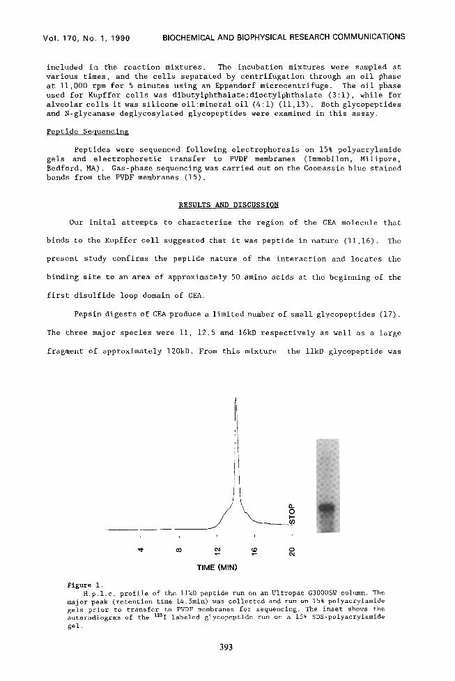
Pepsin digests of CEAproduce a limited number of smallglycopeptides (17).
The three major species were 11, 12.5 and 16kD respectively as well as a large
fragment of approximately 120kD. From this mixture the 1lkD glycopeptide was
I ! I I
t co 04 a ::
TIME (MINI
Figure 1. H.p.1.c. profile of the 1lkD peptide run on an Ultropac G3000SW column. The
major peak (retention time 14.5min) was collected and run on 15% polyacrylamide gels prior to transfer to PVDF membranes for sequencing. The inset shows the autoradiogram of the "'1 labeled glycopeptide run on a 15% SDS-polyacrylamide gel.
393
Vol. 170, No. 1, 1990 BIOCHEMICAL AND BIOPHYSICAL RESEARCH COMMUNICATIONS
isolated by HPLC and SDS-PAGE (Fig. 1). This glycopeptide bound to both rat
liver Kupffer cells and lung alveolar macrophages. Other glycopeptides isolated
from the digest including the large fragment were not capable of specific
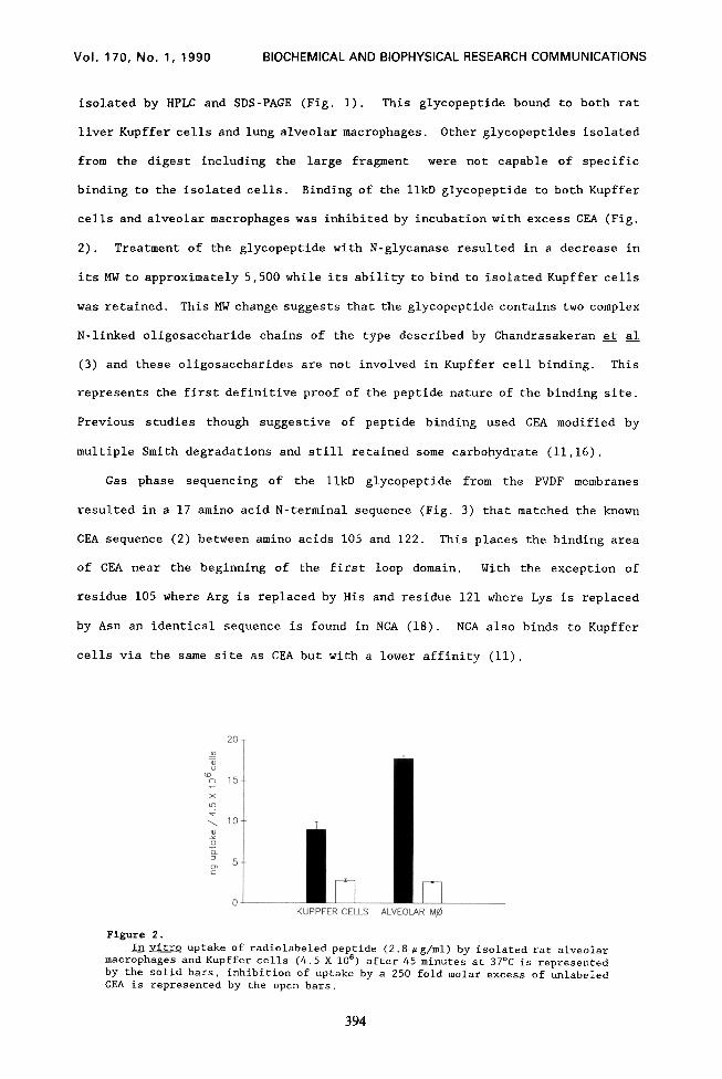
binding to the isolated cells. Binding of the 1lkD glycopeptide to both Kupffer
cells and alveolar macrophages was inhibited by incubation with excess CEA (Fig.
2). Treatment of the glycopeptide with N-glycanase resulted in a decrease in
its MW to approximately 5,500 while its ability to bind to isolated Kupffer cells
was retained. This MW change suggests that the glycopeptide contains two complex
N-linked oligosaccharide chains of the type described by Chandrasakeran et al
(3) and these oligosaccharides are not involved in Kupffer cell binding. This
represents the first definitive proof of the peptide nature of the binding site.
Previous studies though suggestive of peptide binding used CEA modified by
multiple Smith degradations and still retained some carbohydrate (11,16).
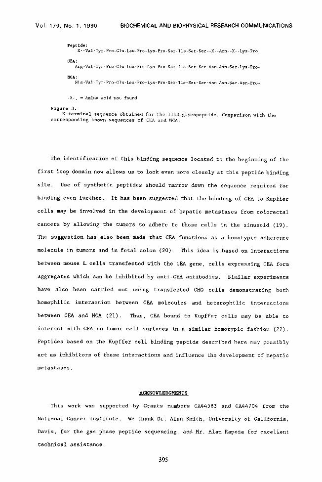
Gas phase sequencing of the 1lkD glycopeptide from the PVDF membranes
resulted in a 17 amino acid N-terminal sequence (Fig. 3) that matched the known
CEA sequence (2) between amino acids 105 and 122. This places the binding area
of CEA near the beginning of the first loop domain. With the exception of
residue 105 where Arg is replaced by His and residue 121 where Lys is replaced
by Asn an identical sequence is found in NCA (18). NCA also binds to Kupffer
cells via the same site as CEA but with a lower affinity (11).
0 KUPPFER CELLS ALVEOLAR Mj4
Figure 2.
In vitro uptake of radiolabeled peptide (2.8 rg/ml) by isolated rat alveolar macrophages and Kupffer cells (4.5 X 10⁶) after 45 minutes at 37OC is represented by the solid bars, inhibition of uptake by a 250 fold molar excess of unlabeled CEA is represented by the open bars.
394
Vol. 170, No. 1, 1990 BIOCHEMICAL AND BIOPHYSICAL RESEARCH COMMUNICATIONS
Figure 3. N-terminal sequence obtained for the 1lkD glycopeptide. Comparison with the
corresponding known sequences of CEA and NCA.
The identification of this binding sequence located to the beginning of the
first loop domain now allows us to look even more closely at this peptide binding
site. Use of synthetic peptides should narrow down the sequence required for
binding even further. It has been suggested that the binding of CEA to Kupffer
cells may be involved in the development of hepatic metastases from colorectal
cancers by allowing the tumors to adhere to these cells in the sinusoid (19).
The suggestion has also been made that CKA functions as a homotypic adherence
molecule in tumors and in fetal colon (20). This idea is based on interactions
between mouse L cells transfected with the CEA gene, cells expressing CEA form
aggregates which can be inhibited by anti-CEA antibodies. Similar experiments
have also been carried out using transfected CHO cells demonstrating both
homophilic interaction between CEA molecules and heterophilic interactions
between CEA and NCA (21). Thus, ClZA bound to Kupffer cells may be able to
interact Iwith CPA on tumor cell surfaces in a similar homotypic fashion (22).
Peptides based on the Kupffer cell binding peptide described here may possibly
act as inhibitors of these interactions and influence the development of hepatic
metastases.
B[;KNOWLEDGMENT$
This work was supported by Grants numbers CA44583 and CA44704 from the
National Cancer Institute. We thank Dr. Alan Smith, University of California,
Davis, for the gas phase peptide sequencing, and Mr. Alan Rapoza for excellent
technical assistance.
395
Vol. 170, No. 1, 1990 BIOCHEMICAL AND BIOPHYSICAL RESEARCH COMMUNICATIONS
1.
2.
3.
4. 5.
6.
7. 8.
9.
10.
11.
12.
13.
14.
15. 16.
17.
18.
19.
20.
21.
22.
REFERENCES
Zamcheck, N., Steele, G., Thomas, P. and Mayer, R. (1986) in Manual of Clinical Laboratory Immunology. (Rose, N.R., Freidman, H. and Fahey, J.L. eds.) pp. 802-809. Am. Sot. for Microbiology, Washington, DC. Oikawa, S., Imajo, S., Noguchi, T., Kosaki, G., and Nakazato, H. (1987) Biochem. Biophys. Res. Commun. 144, 634-642. Chandrasekaran, E., Davila, M., Nixon, D., Goldfarb, M. and Mendicino, J. (1983) J. Biol. Chem. 258, 7213-7222. Barnett, T. and Zimmermann, W. (1990) Tumor Biol. 11, 59-63. Paxton, R.J., Mooser, G., Pande, H., Lee, T.D. and Shively, J.E. (1987) Proc. Natl. Acad. Sci. U.S.A. 84, 920-924. Thompson, J.A., Pande, H., Paxton, R.J., Shively, L., Padma, A., Simmer, R.L., Todd, C.W., Riggs, A.D. and Shively, J.E. (1987) Proc. Natl. Acad. Sci. U.S.A. 84, 2965-2969. Thompson, J. and Zimmermann, W. (1988) 9, 63-83. Hefta, S.A., Hefta, L.J.F., Lee, T., Paxton, R.J. and Shively, J.E. (1988) Proc. Natl. Acad. Sci. U.S.A. 85, 4648-4652. Sack, T.L., Gum, J.R., Low, M.G. and Kim, Y.S. (1988) J. Clin. Invest. 82, 586-593. Thomas, P. and Hems, D.A. (1975) Biochem. Biophys. Res. Commun. 67, 1205- 1209. Toth, C.A., Thomas, P., Broitman, S.A. and Zamcheck, N. (1985) Cancer Res. 45: 392-397. Toth, C.A., Rapoza, A., Kowal, A., Steele, G.D. and Thomas, P. (1988) Biochem. Sot. Trans. 16, 1027-1028. Toth, C.A., Rapoza, A., Zamcheck, N., Steele, G.D. and Thomas, P. (1989) J. Leukocyte Biol. 45: 370-376. Greenwood, F.C., Hunter, W.M. and Glover, J.S. (1963) Biochem. J. 89, 114- 123. Matsudaria, P.T. (1988) J. Biol. Chem. 262:10035-10038. Toth, C.A., Thomas, P., Broitman, S.A. and Zamcheck, N. (1982) Biochem. J. 204, 377-381. Westwood, J.H., Bessell, E.M., Bukhari, M.A., Thomas, P. and Walker, J.M. (1974) Immunochemistry. 11, 811-818. Neumaier, M., Zimmermann, W., Shively, L., Hinoda, Y., Riggs, A.D. and Shively, J.E. (1988) J. Biol. Chem. 263, 3202-3207. Hostetter, R.B., Augustus, L.B., Mankarious, R., Chi, K., Fan, D., Toth, C Thomas, P. and Jessup, J.M. (1990) J. Natl. Cancer Inst. 82, 380-385. Bknchimol, S., Fuks, A., Jothy, S., Beauchemin, N., Shirota, K. and Stanners, C.P. (1989) Cell. 57, 327-334.
Oikawa, S., Inuzuka, C., Kuroki, M., Matsuoka, Y., Kosaki, G. and Nakazato, H. (1989) Biochem. Biophys. Res. Commun. 164, 39-45. Jessup, J.M. and Thomas, P. (1989) Cancer and Metastases Rev. 8, 263-280.