Brit. J. Ophthal.. 35, 366. CARCINOMA OF THE LACRIMAL SAC* BY N. ASHTON, D. P. CHOYCE, AND L. G. FISON Institute of Ophthalmology, and Moorfields Westmtinster and Central Eye Hospital, London BOTH benign and malignant primary tumours of the lacrimal sac are uncommon. Penman and Wolff (1938) give an analysis of 64 cases, including one of their own, and since that date further cases, nine of which are detailed in Table I, have appeared in the literature. TABLE 1 ANALYSIS OF CASES OF PRIMARY TUMOURS OF THE LACRIMAL SAC REPORTED BETWEEN 1938 AND 1950 Year Authors Sex Age Duration Pathology Treatment and Remarks M 54 6 months Mixed-cell tumour of salivary type M 66 5 years Epidermoid carcinoma Excision and radium implant. No recurrence after 10 months Exenteration of orbit and x-ray therapy 1947 Florentin and Poirot 1947 Carlevaro and Landoni 1947 Rousseau 1948 Vrabec 1949 Nichelatti 1949 Barton 1949 ToseIlli M 49 ' Naevo- carcinoma Reticulo- Generalized reticulo- sarcoma sarcomatosis F 79 ' Epithelioma Concurrent tumour of skin of temporo-malar area and lacrimal sac. Treated with radium. Good result after one year '.) '.' '.) Epithelioma F 16 4 months Sarcoma X-ray therapy, preceded by biopsy. No sign of recur- rence two years later F 35 year Basal-cell Excision and deep x rays. carcinoma Later exenteration of orbit M 55 2 years Plasmoma Excision Received for publication March 12, 1951. 366 1939 McCool 1940 Spratt on 8 July 2018 by guest. Protected by copyright. http://bjo.bmj.com/ Br J Ophthalmol: first published as 10.1136/bjo.35.6.366 on 1 June 1951. Downloaded from
Transcript
Brit. J. Ophthal.. 35, 366.
CARCINOMA OF THE LACRIMAL SAC*BY
N. ASHTON, D. P. CHOYCE, AND L. G. FISONInstitute of Ophthalmology, and Moorfields Westmtinster and
Central Eye Hospital, London
BOTH benign and malignant primary tumours of the lacrimal sacare uncommon. Penman and Wolff (1938) give an analysis of64 cases, including one of their own, and since that date furthercases, nine of which are detailed in Table I, have appeared in theliterature.
TABLE 1ANALYSIS OF CASES OF PRIMARY TUMOURS OF THE LACRIMAL SAC
REPORTED BETWEEN 1938 AND 1950
Year Authors Sex Age Duration Pathology Treatment and Remarks
M 54 6 months Mixed-celltumour ofsalivary type
M 66 5 years Epidermoidcarcinoma
Excision and radium implant.No recurrence after 10months
Penman and Wolff grouped the cases according to the histologicaldiagnosis and, although it is not very satisfactory to include in onetable pathological opinions from different sources, the nine recentcases have been added to their list and regrouped into epithelial andnon-epithelial tumours (Table II).
Epithelial neoplasms, therefore, appear to be slightly morecommon than the non-epithelial and when it is realized that thediagnoses of " carcinoma ", " papilloma ", and " epithelioma "
probably represent different stages of cylindrical-celled carcinoma, aswe shall subsequently show, it will be seen that they form about50 per cent. of all reported cases. The reasons for this opinion are
based upon a clinical and pathologicalstudy of two recently encountered caseswhich represent primary tumours ofepithelial origin but of differing degrees
-!W EL\ of malignancy.
FIG. 1.-Case 1. Drawing ofthe open sac showing a poly-poid growth hanging from theupper pole.
CASE REPORTSCase 1.-J. R., male, aged 46 years, van-driver, firstnoticed watering of the right eye. The tear duct hadpreviously been syringed elsewhere, but he had feltno fluid pass through. After a month or two hebecame aware of a lump growing at the right innercanthus, and he came to Moorfields four months later.
Examination revealed a large cystic swellingclosely resembling a mucocele of the lacrimal sac,but no mucus regurgitated on pressure. There wasmoderate epiphora. In July, 1949, a large thin-walled sac was removed. When the sac was openeda polypoid growth was found hanging from the upperpole (Fig. 1). The wound healed well but thelacrimal passages on this side are still not patent.
367
on 8 July 2018 by guest. Protected by copyright.
http://bjo.bmj.com
/B
r J Ophthalm
ol: first published as 10.1136/bjo.35.6.366 on 1 June 1951. Dow
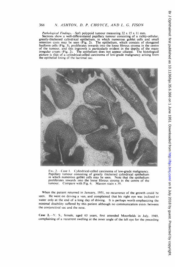
Pathological Findings.-Soft polypoid tumour measuring 32 x 15 x 1 1 mm.Sections show a well-differentiated papillary tumour consisting of a richly-cellular,
greatly-thickened cylindrical epithelium, in which numerous goblet cells and smallretention cysts may be seen (Fig. 2). The epithelium, which consists of elongatedfusiform cells (Fig. 3), proliferates inwards into the loose fibrous stroma in the centreof the tumour, and this ingrowth is particularly evident in the depths of the manyirregular crypts (Fig. 2). The epithelium does not appear ciliated. The histologicalpicture is that of a cylindrical-celled carcinoma of low-grade malignancy arising fromthe epithelial lining of the lacrimal sac.
FiG. 2.-Case 1. Cylindrical-celled carcinoma of low-grade malignancy.Papillary tumour consisting of greatly thickened cylindrical epitheliumin which numerous goblet cells may be seen. Note that the epitheliumproliferates inwards into the loose fibrous stroma in the centre of thetumour. Compare with Fig. 6. Masson stain x 39.
When the patient returned in January, 1951, no recurrence of the growth could beseen. He went on driving a van, and complained that his right eye was inclined towater only at the end of a long day of driving. It is perhaps worth emphasizing theminimal disability suffered by this patient although no communication exists betweenthe conjunctival sac and the nose.
Case 2.---Y. S., female, aged 63 years, first attended Moorfields in July, 1949.complaining of a recurrent swelling at the inner angle of the left eye for the preceding
on 8 July 2018 by guest. Protected by copyright.
http://bjo.bmj.com
/B
r J Ophthalm
ol: first published as 10.1136/bjo.35.6.366 on 1 June 1951. Dow
FiG. 3.-Case 1. High-power view showing therich cellularity of theepithelium which con-sists of elongated fusi-form cells. CompareFig. 7. Masson stainX 122.
4. ~ ~ 4 4
few weeks. There was a yellowish discharge from the inner canthus, but very littlewatering and no pain.Examination revealed a firm cystic swelling about 1 cm. square just below the inner
canthus. Pressure on the swelling made a small amount of yellow fluid regurgitatefrom the lower punctum. On syringing, the left naso-lacrimal duct was not patent.A diagnosis of left mucocele was made and a dacryocystectomy carried out under localanaesthesia. Unfortunately the specimen was lost and its histological structurecannot be recorded.The wound healed normally, but in January, 1950, the patient returned with a
further swelling in the same site. The tumour was now discharging pus into theoverlying skin, and epiphora had increased, but still there was no pain. As thedischarge and swelling persisted, a diagnosis of infected residual sac tissue was made.
In August, 1950, she underwent a further left dacryocystectomy under generalanaesthesia. Occupying the lacrimal fossa was a firm mass, indistinguishable froma lacrimal sac, and firmly adherent below and in front.
Pathological Findings.-Firm solid mass measuring 14 x 10 x 6 mm.Sections show the tumour to consist of irregular masses of hyperchromatic cylindrical
cells lying in a dense fibrous stroma lightly infiltrated with lymphocytes. Numerousgoblet cells may be seen and in many areas these have distended to form cysts ofvarying sizes containing granular mucinous material. Within the larger cysts theepithelial cells have proliferated so as to impart a glandular appearatce comparablewith that of intra-duct carcinoma (Fig. 4). Mitoses are few but' the growth isobviously malignant and it extends in all directions to the limits of the'section. In itsrelatively solid areas the neoplasm is more squamous in type (Fig. 5). The histologicalpicture is that of a highly malignant cylindrical-celled carcinoma of thoe'lacrimal sac.
In view of these findings the patient's skull was x-rayed, with negative results, andshe was referred to the ear, nose, and throat surgeon, Mr. G. H. Howells, who did notdetect any neoplasm in the naisal or para-nasal cavities.
369
adINNE~,A4%.A .,i.T
A .*
on 8 July 2018 by guest. Protected by copyright.
http://bjo.bmj.com
/B
r J Ophthalm
ol: first published as 10.1136/bjo.35.6.366 on 1 June 1951. Dow
Fi(r. 4.-Case 2. Highly malignant cylindrical-celled carcinoma oflacrimal sac. Within cysts of varying sizes the epithelial cells haveproliferated, imparting a glandular appearance comparable with thatof intra-duct carcinoma. Masson stain x 84.
FiG. 5.-Case 2. Cylindrical-celled carcinoma. In its relatively solidareas the neoplasm is more squamous in type. Note the small cysticspaces which have developed from goblet cells. Compare with Fig. 8.x 160.
on 8 July 2018 by guest. Protected by copyright.
http://bjo.bmj.com
/B
r J Ophthalm
ol: first published as 10.1136/bjo.35.6.366 on 1 June 1951. Dow
Fourteen days after operation there was already a fresh, firm swelling presentbeneath the lower part of the recent incision. In view of the patient's poor generalcondition it was considered that further surgery was to be avoided if possible and shewas sent to the eye clinic at the Royal Cancer Hospital. In December, 1950.Dr. M. Lederman kindly supplied the following note:The patient has had 5,600r in 47 days. The swelling shows considerable reduction
but has not entirely disappeared. She is being kept under observation and if thegrowth shows signs of increasing, further surgical intervention may be undertaken.
DISCUSSIONBoth from the clinical and pathological points of view these two
cases form an interesting contrast. In the first case, an apparent curefollowed a straightforward dacryocystectomy, and this benignoutcome was in accord with the histological findings of lowmalignancy. In the second case, the neoplasm was much moremalignant, and may have been provoked into further activity byincomplete removal.
Spratt (1940) has emphasized other writers' unanimous testimonythat diagnosis is not possible in the first stage of the disease, becauseepiphora is the only symptom. In the second stage of swelling a diag-nosis can be made. The presence of a round, hard mass, not reducedby pressure, especially if irrigation shows a patent duct, is indicativeof thickening of the wall of the lacrimal sac rather than of a mucocele.In the third stage, when invasion of the lids and orbit has occurred,diagnosis becomes obvious. It is only in this stage that pain hasbeen reported. The existence of Spratt's second stage may well bequestioned, because it is unlikely that syringing would be successfulin the presence of swelling and epiphora. Theoretically, one mighthave expected our first case, in which the polypoid tumour wassuspended from the fundus of the sac, to have shown a patent ducton irrigation, for the growth could have caused epiphora and yethave been pushed aside by the lacrimal cannula, but actually syringingwas never successful. Our second case should perhaps be classifiedsomewhere between Spratt's second and third stages. The fact thatthe duct was not patent could be explained by the site of the tumour,near the outlet from the sac or from the top of the naso-lacrimal ductitself, or from an oedematous obstruction due to superaddedinfection.From the pathological point of view it is of interest and importance
to note that exactly similar tumours arise from the respiratoryepithelium in the nasal and para-nasal cavities. This similarity, sofar as we can ascertain, has not yet been sufficiently emphasized, butit is important, because the nasal tumours, although uncommon, areless rare than the corresponding growths in the lacrimal sac. There-fore attention to the natural history of these nasal tumours canperhaps help to explain the malignant potentialities of those arising
371
on 8 July 2018 by guest. Protected by copyright.
http://bjo.bmj.com
/B
r J Ophthalm
ol: first published as 10.1136/bjo.35.6.366 on 1 June 1951. Dow
in the lacrimal sac, where their rarity is an obstacle to completeassessment.
This similarity had previously been noted by Muirhead (1933), whoreported a case of lacrimal-sac carcinoma:
the section shows a transitional cell carcinoma, which is very similar in appearanceto an antral tumour.
Verhoeff described the histological findings in a case reportedby Spratt (1940), as follows:
. . . in a few places in the section there are remains of normal ciliated epitheliumof mucous membrane. Such tumours may arise from accessory sinuses.
The illustrations of the histology in this article are exactly similarto those of our first case (Fig. 2). From an examination of two casesof polypoidal formation in the lacrimal sac, Tooke (1912) came tothe conclusion that polyps of the tear sac, as in the nose and accessorynasal cavities, are evidence of hyperplastic growth or actual tumourformation which may possibly be due to a pre-existing inflammation.
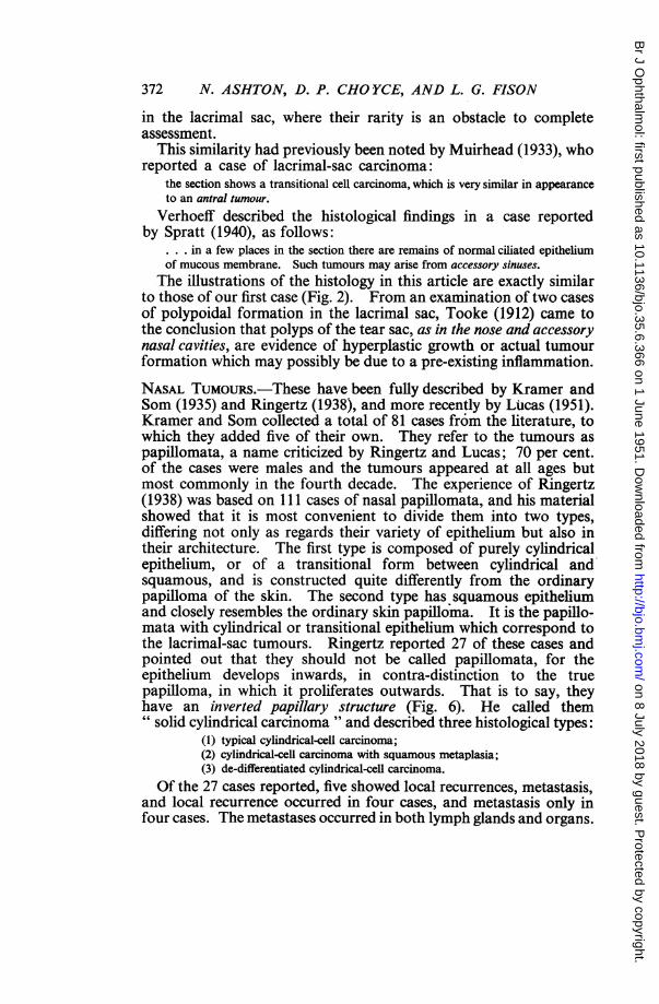
NASAL TuMouRs.-These have been fully described by Kramer andSom (1935) and Ringertz (1938), and more recently by Lucas (1951).Kramer and Som collected a total of 81 cases from the literature, towhich they added five of their own. They refer to the tumours aspapillomata, a name criticized by Ringertz and Lucas; 70 per cent.of the cases were males and the tumours appeared at all ages butmost commonly in the fourth decade. The experience of Ringertz(1938) was based on 111 cases of nasal papillomata, and his materialshowed that it is most convenient to divide them into two types,differing not only as regards their variety of epithelium but also intheir architecture. The first type is composed of purely cylindricalepithelium, or of a transitional form between cylindrical and'squamous, and is constructed quite differently from the ordinarypapilloma of the skin. The second type has squamous epitheliumand closely resembles the ordinary skin papilloma. It is the papillo-mata with cylindrical or transitional epithelium which correspond tothe lacrimal-sac tumours. Ringertz reported 27 of these cases andpointed out that they should not be called papillomata, for theepithelium develops inwards, in contra-distinction to the truepapilloma, in which it proliferates outwards. That is to say, theyhave an inverted papillary structure (Fig. 6). He called them"solid cylindrical carcinoma " and described three histological types:
Of the 27 cases reported, five showed local recurrences, metastasis,and local recurrence occurred in four cases, and metastasis only infour cases. The metastases occurred in both lymph glands and organs.
on 8 July 2018 by guest. Protected by copyright.
http://bjo.bmj.com
/B
r J Ophthalm
ol: first published as 10.1136/bjo.35.6.366 on 1 June 1951. Dow
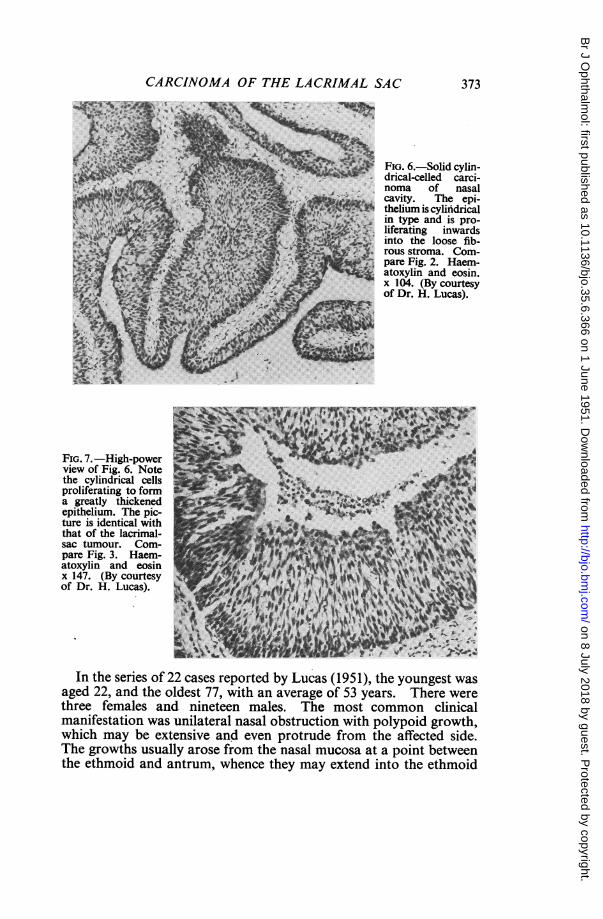
iw FIG. 6.-Solid cylin-drical-celled carci-noma of nasalcavity. The epi-
ture is identicalthelium iscyl-idricalr' li in type and is pro
liferating inwardsIn the sio2ce e r b cinto the loose fib-rous stroma. Comn-
*~pare Fig. 2. Haem-atoxylin and eosin.
_ ~~~~~~~~~~~~~~x104. (BY courtesyof Dr. H. Lucas).
FiG. 7.-High-powerview of Fig. 6. Notethe cylindrical cellsproliferating to forma greatly thickened fepithelium. The pic-ture is identical withthat of the lacrimal-sac tumour. Cond-pare Fig. 3. Haem-atoxylin and eosinSAx 147. (By courtesy p 1of Dr. H. Lucas).
In the series of 22 cases reported by Lucas (1951), the youngest wasaged 22, and the oldest 77, with an average of 53 years. There werethree females and nineteen males. The most common clinicalmanifestation was unilateral nasal obstruction with polypoid growth,which may be extensive and even protrude from the affected side.The growths usually arose from the nasal mucosa at a point betweenthe ethmoid and antrum,, whence they may extend into the ethmoid
373:-
;. h, . 1.. 1 l.7q.M'1C..X14 11
on 8 July 2018 by guest. Protected by copyright.
http://bjo.bmj.com
/B
r J Ophthalm
ol: first published as 10.1136/bjo.35.6.366 on 1 June 1951. Dow
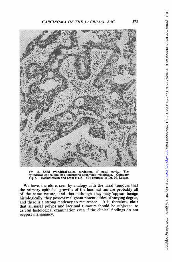
cavity or the antrum or both. According to Lucas, the lesion beginsas a widespread change in the respiratory epithelium, which undergoesmetaplasia to a cylindrical type of cell. These cells are elongated.rather deeply staining and crowded together in palisade formation.resting on a basement membrane (Fig. 7). As the cells undergode-differentiation they lose their cilia and their power to form mucousglobules. This epithelial proliferation extends down into the ductsaind mucous glainds, and the growth is continued by an infolding ofthe epitheliunm inlto it own stroma (Fig. 6), so that solid masses ofepitheliunm are gradually formed. This description conforms exactlhwvith that of Ringertz. The cylindrical epithelium may then undergosquamous metaplasia and closely resemble a squamous-cell carcinoma(Fig. 8). The third type of growth, the de-differentiated cylindrical-cell carcinoma, did not occur in Lucas' series. Lucas agreed withRingertz that the term papilloma should not be applied to thesetumours, not oinly because their structure and mode of formation isdifferent but also because such a name implies an innocent stage intheir development, whereas they are probably malignant from theonset, varying only in the degree of their malignancy and cor-responding- clinical course.
TUMOURS OF THE LACRIMAL SAC. It will already be clear- from theabove descriptions that we are here dealing with a tumour which isidentical histologically and developmentally. Nor is this surprising.for it will be remembered that in the embryo the lining of the sacand the respiratory epithelium develop along parallel lines.As Spratt (1937) pointed out, the normal lining of the lacrimal sac
is composed of cylindrical cells. Repeated irritation, due to chronicinflammation, may cause this membrane to become thickened andto form several layers of cells which change to the squamous variety.Consequently sections may show the cylindrical, transitional, orsquamous-cell type. The histology of our first case is exactlycomparable with the early lesion in the nose, as a comparison ofFig. 2 with Fig. 6 and Fig. 3 with Fig. 7 will show. The second caseis comparable with the " de-differentiated cylindrical-cell carcinoma -
of Ringertz and shows also squamous metaplasia (compare Fig. 5with Fig. 8).
It seems probable that all primary malignant epithelial tumours ofthe lacrimal sac arise in the same way, and that the different histo-logical descriptions in the literature may be attributed to its varyingdevelopmental phases. Thus " carcinoma ", " papilloma ", and" epithelioma " were probably all examples of solid cylindricalcarcinoma. In this connection the comments of Pasetti (1913) areof interest:From what we were able to learn from these few observations of epithelioma of the
lacrimal sac, all have been made up of cylindrical cells. All have exhibited markedmalignancy. Recurrence is frequent.
on 8 July 2018 by guest. Protected by copyright.
http://bjo.bmj.com
/B
r J Ophthalm
ol: first published as 10.1136/bjo.35.6.366 on 1 June 1951. Dow
FIG. 8.-Solid cylindrical-celled carcinoma of nasal cavity. Thecylindrical epithelium has undergone squamous metaplasia. CompareFig. 5. Haematoxylin and eosin x 138. (By courtesy of Dr. H. Lucas).
We have, therefore, seen by analogy with the nasal tumours thatthe primary epithelial growths of the lacrimal sac are probably allof the same nature, and that although they may appear benignhistologically, they possess malignant potentialities of varying degree,and there is a strong tendency to recurrence. It is, therefore, clearthat all nasal polyps and lacrimal tumours should be subjected tocareful histological examination even if the clinical findings do notsuggest malignancy.
375
on 8 July 2018 by guest. Protected by copyright.
http://bjo.bmj.com
/B
r J Ophthalm
ol: first published as 10.1136/bjo.35.6.366 on 1 June 1951. Dow
(1) Since the publication of the analysis of 64 reported cases ofprimary tumours of the lacrimal sac (Penman and Wolff, 1938),nine further cases have been found in the literature, and an analysisof the whole 73 cases shows that about 50 per cent. fall into the"papilloma-carcinoma " group.
(2) Two further cases of carcinoma of the lacrimal sac are reported.(3) It is pointed out that the histology of these tumours is identical
with that of solid cylindrical-cell carcinoma arising from therespiratory epithelium in the nasal and para-nasal cavities.
(4) The pathology of carcinomata of the nasal cavities and of thelacrimal sac have, therefore, been considered together, and thefollowing conclusions reached:
(a) The term " papilloma " as applied to these growths should beabandoned, since their mode of formation and structure differs from thatof a true papilloma.
(b) " Papillomata " arising from the lacrimal-sac epithelium areprobably malignant from the beginning, as in the nose, and gradually oror rapidly progress to more obviously malignant cylindrical-cell carcino-mata, a term which is, therefore, applicable to the whole group.
(5) The necessity for carrying out careful histological examinationon all lacrimal tumours, even if the clinical findings do not suggestmalignancy, is emphasized.
We are indebted to Mr. F. W. Law and to Mr. J. H. Doggart for permission topublish Cases 1 and 2 respectively. We are grateful to Dr. P. Hansell and Dr. H. Lucasfor the photomicrographs, to Dr. M. Ledermnan for his note on the radiotherapy, and toMiss J. Trotman for the drawing of the polypoid growth.
REFERENCESBARTON, D. (1949). Trans. Ophthal. Soc. U.K, 69, 523.CARiLEvARo, G., and LANDONI, P. (1947). Ann. Ottaim., 73, 468.FLoRENTN, T., and PoIRoT, J. C. (1947). Bull. Soc. Ophtal. Paris, 270.KRAMER, R., and SOM, M. L. (1935). Arch. Otolaryng., Chicago, 22, 22.LuCAs, H. (1951). Rep. Inst. Laryng. Otol. No. 1. (in the press).MCCOOL, J. L. (1939). Amer. J. Ophthal., 22, 734.MUIRHEAD, W. M. (1933). Trans. ophthal. Soc. U.K, 53, 591.NICHELATrI, P. (1949). Ann. Ottal., 75, 3.PASETrI, G. (1913). Ann. Ottal., 42, 55.PENMAN, G. G., and WOLFF, E. (1938). Lancet, 1, 1325.RINGERTZ, N. (1938). Acta Oto-laryng., Stockh., Suppl. 27.ROUSSEAU (1947). * Bull. Soc. Ophtal. Paris, p. 650.SPRATT, C. N. (1937). Arch. Ophthal., Chicago, 18, 67.
- (1940). Ibid., 24, 1237.TOOKE, F. (1912). Ibid., 41, 446.TOSELLI, C. (1949). Rass. ital. Ottalm., 18, 333.VRABEC, F. (1948). Csl. Ophthal., 4, 24.
on 8 July 2018 by guest. Protected by copyright.
http://bjo.bmj.com
/B
r J Ophthalm
ol: first published as 10.1136/bjo.35.6.366 on 1 June 1951. Dow