Cardiac Evaluation of the Newborn Donald J. Fillipps, MD a , Richard L. Bucciarelli, MD b,c, * CARDIAC EVALUATION OF THE NEWBORN The approach to the cardiac evaluation of a newborn can be challenging. As a result, many pediatricians report that they often feel uncomfortable when it comes to differ- entiating the normal from the abnormal state with regard to a newborn’s Declaration of conflict of interest: Both R.L. Bucciarelli, MD and D.J. Fillipps, MD attest that they have no conflicts of interest to declare in relation to the materials and information pro- vided in this article. a Division of General Pediatrics, College of Medicine, University of Florida, 1701 Southwest 16th Avenue, Building A, Gainesville, FL 32608, USA; b Division of Neonatology, College of Medicine, University of Florida, 2400 Archer Road, Gainesville, FL 32610, USA; c Division of Pe- diatric Cardiology, College of Medicine, University of Florida, 2400 Archer Road, Gainesville, FL 32610, USA * Corresponding author. Division of Neonatology, College of Medicine, University of Florida, 2400 Archer Road, Gainesville, FL 32610. E-mail address: [email protected]KEYWORDS Term newborn cardiovascular examination Term newborn with congenital heart disease (CHD) Common types of neonatal congenital heart disease Pulse oximetry screening Critical congenital heart disease (CCHD) KEY POINTS Although congenital heart defects can be diagnosed using fetal cardiac ultrasonography, some defects can be challenging to identify. Even with a careful complete physical examination, some infants seem normal and are discharged home undiagnosed. The persistence of fetal channels can mask the presence of critical congenital heart dis- ease, and the rather short postpartum hospital stay contributes to the diagnostic challenges. It is essential for the examiner to use all physical examination skills, including inspection, palpation, and auscultation, and to perform more than one physical assessment before discharge or shortly thereafter. The recent introduction of Pulse Oximetry Screening has been an extremely helpful adju- vant in assisting with the diagnosis of CCHD. Pediatr Clin N Am 62 (2015) 471–489 http://dx.doi.org/10.1016/j.pcl.2014.11.009 pediatric.theclinics.com 0031-3955/15/$ – see front matter Published by Elsevier Inc.
Transcript
Cardiac Evaluation of theNewborn
Donald J. Fillipps, MDa, Richard L. Bucciarelli, MDb,c,*
KEYWORDS
� Term newborn cardiovascular examination� Term newborn with congenital heart disease (CHD)� Common types of neonatal congenital heart disease � Pulse oximetry screening� Critical congenital heart disease (CCHD)
KEY POINTS
� Although congenital heart defects can be diagnosed using fetal cardiac ultrasonography,some defects can be challenging to identify.
� Even with a careful complete physical examination, some infants seem normal and aredischarged home undiagnosed.
� The persistence of fetal channels can mask the presence of critical congenital heart dis-ease, and the rather short postpartum hospital stay contributes to the diagnosticchallenges.
� It is essential for the examiner to use all physical examination skills, including inspection,palpation, and auscultation, and to perform more than one physical assessment beforedischarge or shortly thereafter.
� The recent introduction of Pulse Oximetry Screening has been an extremely helpful adju-vant in assisting with the diagnosis of CCHD.
CARDIAC EVALUATION OF THE NEWBORN
The approach to the cardiac evaluation of a newborn can be challenging. As a result,many pediatricians report that they often feel uncomfortable when it comes to differ-entiating the normal from the abnormal state with regard to a newborn’s
Declaration of conflict of interest: Both R.L. Bucciarelli, MD and D.J. Fillipps, MD attest thatthey have no conflicts of interest to declare in relation to the materials and information pro-vided in this article.a Division of General Pediatrics, College of Medicine, University of Florida, 1701 Southwest16th Avenue, Building A, Gainesville, FL 32608, USA; b Division of Neonatology, College ofMedicine, University of Florida, 2400 Archer Road, Gainesville, FL 32610, USA; c Division of Pe-diatric Cardiology, College of Medicine, University of Florida, 2400 Archer Road, Gainesville, FL32610, USA* Corresponding author. Division of Neonatology, College of Medicine, University of Florida,2400 Archer Road, Gainesville, FL 32610.E-mail address: [email protected]
Pediatr Clin N Am 62 (2015) 471–489http://dx.doi.org/10.1016/j.pcl.2014.11.009 pediatric.theclinics.com0031-3955/15/$ – see front matter Published by Elsevier Inc.
cardiovascular examination. It is the authors’ goal for this article to provide the readerwith the background knowledge that will make the cardiac evaluation of newbornsless intimidating and assist the general pediatrician in understanding, detecting, andtreating a newborn with congenital heart disease (CHD).CHD is the most common congenital disorder in newborns, occurring in approxi-
mately 8 out of 1000 live births, and is responsible for almost 30% of infant deathsrelated to birth defects. Of those children with CHD, about 1 in 4 (25%) babies bornwith a heart defect will have critical CHD (CCHD), defined as needing interventionwithin the first year of life.1–3
Although CHD can be diagnosed using fetal cardiac ultrasonography, somedefects can be challenging to identify. Similarly, even with a careful completephysical examination, some infants seem normal and are discharged home undi-agnosed. The persistence of fetal channels can mask the presence of CCHD,and the rather short postpartum hospital stay contributes to the diagnostic chal-lenges. Thus it is essential for the examiner to use all physical examinationskills, including inspection, palpation, and auscultation, and to perform morethan one physical assessment before discharge or shortly thereafter. The recentintroduction of pulse oximetry screening (POS) has been an extremely helpful adju-vant in assisting with the diagnosis of CCHD before signs of decompensationoccur.4
Initial Evaluation
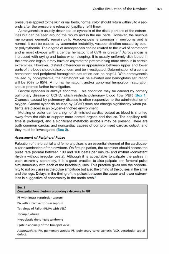
The first step in the assessment of the newborn infant’s cardiovascular system is acareful review for conditions that are associated with an increased risk of CHD(Table 1). The presence of any of these factors should raise the index of suspicion,but a complete physical examination should be performed regardless.5–7
Inspection and Palpation of the Skin and Mucous Membranes
The color of the skin and briskness of capillary refill can be indicators of the adequacyof oxygenation and cardiac output. The mucous membranes of a normal newbornshould be pink. This is usually checked by looking at the tongue and lips. When light
Table 1Common conditions associated with CHD
Maternal Perinatal
Diabetes TORCH infection
Obesity Premature delivery <37 wk
Hypertension Genetic/chromosomal disorders
Systemic lupus erythematosus VACTERL
Epilepsy Omphalocele
Influenza or flulike symptoms Congenital diaphragmatic hernia
First-trimester smoking
Maternal thyroid conditions
Maternal CHD
Maternal alcohol/medication use
Multifetal pregnancy
Abbreviations: TORCH, toxoplasmosis, other agents, rubella, cytomegalovirus, herpes simplex;VACTERL, vertebral, anal, cardiac, tracheal, esophageal, renal, and limb.
Data from Refs.1–3,5
Cardiac Evaluation of the Newborn 473
pressure is applied to the skin or nail beds, normal color should return within 3 to 4 sec-onds after the pressure is released (capillary refill time).Acrocyanosis is usually described as cyanosis of the distal portions of the extrem-
ities but can be seen around the mouth and in the nail beds. However, the mucousmembranes generally remain pink. Acrocyanosis is common in newborns and isnormal. It can be caused by vasomotor instability, vasoconstriction caused by cold,or polycythemia. The degree of acrocyanosis can be related to the level of hematocritand is most obvious with a central hematocrit of 65% or greater.7 Acrocyanosis isincreased with crying and fades when sleeping. It is usually uniformly distributed inthe arms and legs but may have an asymmetric pattern being more obvious in certainextremities. However, distinct differences in appearance between upper and lowerparts of the body should raise concern and be investigated. Determination of a centralhematocrit and peripheral hemoglobin saturation can be helpful. With acrocyanosiscaused by polycythemia, the hematocrit will be elevated and hemoglobin saturationwill be 90% to 95%. A normal hematocrit and/or abnormal hemoglobin saturationshould prompt further investigation.Central cyanosis is always abnormal. This condition may be caused by primary
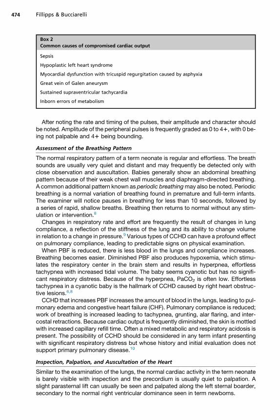
pulmonary disease or CCHD, which restricts pulmonary blood flow (PBF) (Box 1).Cyanosis caused by pulmonary disease is often responsive to the administration ofoxygen. Central cyanosis caused by CCHD does not change significantly when pa-tients are placed in an oxygen-enriched environment.Mottling or pallor can be a sign of diminished cardiac output as blood is shunted
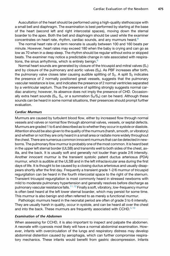
away from the skin to support more central organs and tissues. The capillary refilltime is prolonged, and a significant metabolic acidosis may be present. There areboth common cardiac and noncardiac causes of compromised cardiac output, andthey must be investigated (Box 2).
Assessment of Peripheral Pulses
Palpation of the brachial and femoral pulses is an essential element of the cardiovas-cular examination of the newborn. On first palpation, the examiner should assess thepulse rate (normal between 100 and 160 beats per minute) and rhythm (consistentrhythm without irregular beats). Although it is acceptable to palpate the pulses ineach extremity separately, it is a good practice to also palpate one femoral pulsesimultaneously with each of the brachial pulses. This practice gives one the opportu-nity to not only assess the pulse amplitude but also the timing of the pulses in the armsand the legs. Delays in the timing of the pulses between the upper and lower extrem-ities is suggestive of abnormality in the aortic arch.8
Box 1
Congenital heart lesions producing a decrease in PBF
Myocardial dysfunction with tricuspid regurgitation caused by asphyxia
Great vein of Galen aneurysm
Sustained supraventricular tachycardia
Inborn errors of metabolism
Fillipps & Bucciarelli474
After noting the rate and timing of the pulses, their amplitude and character shouldbe noted. Amplitude of the peripheral pulses is frequently graded as 0 to 41, with 0 be-ing not palpable and 41 being bounding.
Assessment of the Breathing Pattern
The normal respiratory pattern of a term neonate is regular and effortless. The breathsounds are usually very quiet and distant and may frequently be detected only withclose observation and auscultation. Babies generally show an abdominal breathingpattern because of their weak chest wall muscles and diaphragm-directed breathing.A common additional pattern known as periodic breathingmay also be noted. Periodicbreathing is a normal variation of breathing found in premature and full-term infants.The examiner will notice pauses in breathing for less than 10 seconds, followed bya series of rapid, shallow breaths. Breathing then returns to normal without any stim-ulation or intervention.8
Changes in respiratory rate and effort are frequently the result of changes in lungcompliance, a reflection of the stiffness of the lung and its ability to change volumein relation to a change in pressure.9 Various types of CCHD can have a profound effecton pulmonary compliance, leading to predictable signs on physical examination.When PBF is reduced, there is less blood in the lungs and compliance increases.
Breathing becomes easier. Diminished PBF also produces hypoxemia, which stimu-lates the respiratory center in the brain stem and results in hyperpnea, effortlesstachypnea with increased tidal volume. The baby seems cyanotic but has no signifi-cant respiratory distress. Because of the hyperpnea, PaCO2 is often low. Effortlesstachypnea in a cyanotic baby is the hallmark of CCHD caused by right heart obstruc-tive lesions.6,8
CCHD that increases PBF increases the amount of blood in the lungs, leading to pul-monary edema and congestive heart failure (CHF). Pulmonary compliance is reduced;work of breathing is increased leading to tachypnea, grunting, alar flaring, and inter-costal retractions. Because cardiac output is frequently diminished, the skin is mottledwith increased capillary refill time. Often a mixed metabolic and respiratory acidosis ispresent. The possibility of CCHD should be considered in any term infant presentingwith significant respiratory distress but whose history and initial evaluation does notsupport primary pulmonary disease.10
Inspection, Palpation, and Auscultation of the Heart
Similar to the examination of the lungs, the normal cardiac activity in the term neonateis barely visible with inspection and the precordium is usually quiet to palpation. Aslight parasternal lift can usually be seen and palpated along the left sternal boarder,secondary to the normal right ventricular dominance seen in term newborns.
Cardiac Evaluation of the Newborn 475
Auscultation of the heart should be performed using a high-quality stethoscope witha small bell and diaphragm. The examination is best performed by starting at the baseof the heart (second left and right intercostal spaces), moving down the sternalboarder to the apex. Both the bell and diaphragm should be used while the examinerconcentrates on heart rate, rhythm, cardiac sounds, and any murmurs heard.8
The normal heart rate of a term neonate is usually between 100 and 160 beats perminute. However, heart rates may exceed 180 when the baby is crying and can go aslow as 70 when in a deep sleep. The rhythm should be regular without extra or skippedbeats. The examiner may notice a predictable change in rate associated with respira-tions, the sinus arrhythmia, which is entirely benign.6
Normal heart sounds are generated by closure of the tricuspid and mitral valves (S1)and by closure of the pulmonary and aortic valves (S2). As PBF increases after birth,the pulmonary valve closes later causing audible splitting of S2. A split S2 indicatesthe presence of 2 normally positioned great vessels, suggests that the pulmonaryvascular resistance is low, and indicates the presence of 2 normal ventricles separatedby a ventricular septum. Thus the presence of splitting strongly suggests normal car-diac anatomy; however, its absence does not imply the presence of CHD. Occasion-ally extra heart sounds (S3, S4, or a summation S3/S4) can be heard. Although thesesounds can be heard in some normal situations, their presences should prompt furtherevaluation.
Cardiac Murmurs
Murmurs are caused by turbulent blood flow, either by increased flow through normalvessels and valves or normal flow through abnormal valves, vessels, or septal defects.Murmurs aregraded1 to 6anddescribedas towhether theyoccur in systole or diastole.Attention shouldbealsogiven to thequality of themurmurs (harsh, smooth, or vibratory)andwhether or not they are only heard in a small area or radiatemorewidely throughoutthechest. There arenumerouscommon innocentmurmurs that canbedetected in new-borns. Thepulmonary flowmurmur is probably oneof themost common. It is heardbestin the upper left sternal border (ULSB) and transmits well to both sides of the chest, ax-illa, and the back. It is usually soft and generally not louder than grade 2/6 intensity.Another innocent murmur is the transient systolic patent ductus arteriosus (PDA)murmur, which is audible at the ULSB and in the left infraclavicular area during the firstdays of life. It is thought to be caused by a closing ductus arteriosus and usually disap-pears shortly after the first day. Frequently a transient grade 1-2/6 murmur of tricuspidregurgitation can be heard in the fourth intercostal space to the right of the sternum.Transient tricuspid regurgitation is most commonly heard in stressed newborns withmild to moderate pulmonary hypertension and generally resolves before discharge aspulmonary vascular resistance falls.11,12 Finally a soft, vibratory, low-frequencymurmuris often best heard at the left lower sternal boarder, which may persist for some time.This murmur is also benign and often referred to as merely a functional murmur.Pathologic murmurs heard in the neonatal period are often of grade 3 to 6 intensity.
They are usually harsh in quality, occur in systole, and can be heard all over the chestand into the back. These murmurs are frequently associated with CCHD.5,10
Examination of the Abdomen
When assessing for CCHD, it is also important to inspect and palpate the abdomen.A neonate with cyanosis most likely will have a normal abdominal examination. How-ever, infants with overcirculation of the lungs and respiratory distress may developabdominal distention caused by aerophagia, which can further compromise respira-tory mechanics. These infants would benefit from gastric decompression. Infants
Fillipps & Bucciarelli476
with CHF may have liver enlargement and, if a metabolic acidosis is present, maydevelop an ileus with diminished or absent bowel sounds on auscultation.
Blood Pressure
Determination of blood pressure in both arms and at least one leg is important in theevaluation of an infant suspected of having CCHD. The pattern of variation in pressurebetween the extremities can suggest the presence of significant CCHD (Table 2).13
COMMON CONGENITAL HEART DEFECTS
In this section, the authors discuss only the most common congenital heart defects. Itis not important that the examiner arrive at the correct anatomic diagnosis in theirassessment of a newborn with suspected CCHD. Rather, it is important to recognizethe general presenting signs of CCHD and how to stabilize the infant until further eval-uation can be accomplished.
Uncomplicated Congenital Heart Defects
Simple atrial septal defects (ASD), ventricular septal defects (VSD), endocardialcushion defects, and a patent ductus arteriosus (PDA) do not significantly affect thecardiopulmonary physiology of the newborn and, although possible, are not usuallyassociated with symptoms in the first few days after birth. The blood crossing thesedefects is usually small in volume and nonturbulent, producing little to no murmur.With simple ASDs, even very large ones, the amount of blood that goes from theleft atrium to the right atrium is very limited until several months after birth, whenthe right ventricular muscle becomes thinner, more compliant, and can accommodateadditional blood. With VSDs, flow depends on the reduction in pulmonary vascularresistance, which occurs over the first 3 to 6 weeks after delivery.13,14 This evolutionin the pulmonary vascular bed occurs more rapidly in premature infants, leading toearlier identification and an increased likelihood of symptoms.15 One exception tothis rule is a very small muscular VSD that may be heard within the first days of lifebecause flow through the defect is turbulent.13,16 Although the murmur may be agrade 2-3/6, it is very short in duration, smooth in character, and mid to high fre-quency. These infants should be asymptomatic.
Lesions Causing Decreased Pulmonary Blood Flow
The lesions discussed in this section and presented in Box 1 all have a significant de-gree of obstruction of blood flow into and/or out of the right ventricle.
Pulmonary valve stenosis and pulmonary valve atresia with an intact ventricularseptumIn pulmonary valve stenosis (PS), the pulmonary valve is thickened and only allows ajet of blood to pass into the lungs. Because this jet is turbulent, it creates a loud,
Table 2Pulse and blood pressure patterns with left-sided obstructive lesions
harsh systolic murmur at the base of the heart in the second intercostal space alongthe left and right sternal boarder. The murmur is also well heard along the back.Because the valve does not close properly, S2 is single and diminished. With severepulmonary stenosis, there is often massive tricuspid regurgitation, producing a grade3 to 6 (murmur plus a thrill) at the third to fourth intercostal space along the right ster-nal margin. If there is no opening to the valve at all (pulmonary atresia [PA]), theobstruction is complete. The pulmonary valve and the main pulmonary arteries areunderdeveloped. The baby is deeply cyanotic, but no murmur is heard over the pre-cordium. There may be a faint (grade 2-3), smooth systolic murmur of a PDA heardalong the ULSB and under the left clavicle. In this instance, the infant’s entire PBFdepends on the ductus. Because the tricuspid valve is competent, the pressure inthe right ventricle is greater than systemic levels. Blood flow into the ventricle is min-imal and leaves the chamber through the myocardium sinusoids, which drain into thecoronary system. As a result, the right ventricle is small, underdeveloped, andnonfunctional. This combination of lesions is also known as the hypoplastic rightheart syndrome (Fig. 1). However, when PS or PA exists in association with tricuspidvalve incompetence, the pressure in the right ventricle is very low, allowing blood toenter during ventricular diastole and then flow retrograde into the right atrium duringventricular systole. This antegrade/retrograde flow creates enough volume variationto allow near-normal development of the right ventricle. Babies with PS/PA may
Fig. 1. Hypoplastic right ventricle with pulmonary atresia. An infant with severe PS or PAand a competent tricuspid valve (A) may have a severely underdeveloped and nonfunctionalright ventricle. The infant is deeply cyanotic with little to no audible murmur. However, ifthe tricuspid valve is better developed and incompetent (B), there is both antegrade andretrograde flow into and out of the right ventricle, which remodels the ventricular wall, re-sulting in a much larger, functional chamber. In this situation there will be a loud murmur(grade iv/vi) along the right lower sternal boarder. A palpable thrill may also be present.Ductus A, Ductus Arteriosus; LA, left atrium; LV, left ventricle; RA, right atrium; RV, rightventricle. (Adapted from Krovetz LJ, Gessner IH, Schiebler GL. Handbook of pediatric cardi-ology. 2nd edition. Baltimore: University Park Press; 1979. p. 301; with permission.)
Fillipps & Bucciarelli478
develop a large right atrium and are at risk for developing supraventricular tachy-cardia (SVT), which is briefly discussed later.
Pulmonary valve stenosis/pulmonary valve atresia with a ventricular septal defectThis combination of lesions is one of the most common types of CHD and is alsoknown as tetralogy of Fallot (TOF). The 4 elements of TOF are PS or PA, VSD, over-riding aorta, and right ventricular hypertrophy (RVH) (Fig. 2). RVH is not obviousbecause right ventricular dominance is common in the term neonate. The presentingsigns depend on the degree of pulmonary obstruction. Severe obstruction producesdeep cyanosis, whereas minor degrees of stenosis may affect color only slightly,hence the term Pink Tetralogy. In addition to PS, there is usually narrowing belowthe pulmonary valve, which is called muscular infundibular stenosis. S2 is singlebecause of PS and subvalvular stenosis. The VSD is always large with little restrictionof flow such that blood flows easily from the right ventricle to the left ventricle with littleturbulence, generating no murmur and allowing normal ventricular development. Theaorta straddles the ventricular septum (overrides) and receives blood from both ven-tricles. The murmur heard in an infant with TOF is similar to the murmur of the PS. Iftetralogy exists with pulmonary atresia, there may be no murmur at all or only the faintmurmur of a PDA, which supplies all of the PBF.
Dextro-transposition of the great vesselsD-transposition of the great vessels (TGV) is one of the more common defects. In thiscase, the main pulmonary artery arises from the left ventricle and the aorta from the
Fig. 2. Cyanotic TOF. Note the presence of PS with additional muscular narrowing in theinfundibular region. The aorta is overriding the ventricular septum and receives bloodfrom both the right and left ventricle. A, aorta; LA, left atrium; LV, left ventricle; PT, pulmo-nary trunk; PS, pulmonary valve stenosis; RA, right atrium; RV, right ventricle. (Adapted fromKrovetz LJ, Gessner IH, Schiebler GL. Handbook of pediatric cardiology. 2nd edition. Balti-more: University Park Press; 1979. p. 288; with permission.)
Cardiac Evaluation of the Newborn 479
right ventricle. The volume of PBF is normal; but because the origin of the great ves-sels is switched, oxygenated bloodmerely recirculates to the lungs and deoxygenatedblood recirculates to the body. Mixing of the circulations only occurs across the atrialseptum and the PDA. There are usually no murmurs because there is no turbulent flow.S2 is single because the pulmonary artery is malpositioned and hidden by the aorta.Most frequently TGV occurs with an intact ventricular septum, presenting with deepcyanosis. However, it can also be associated with a VSD or a VSD and PS. It thentakes on the characteristics of the other lesions described throughout this section.
Lesions Causing Increased Pulmonary Blood Flow
Lesions that cause increased PBF almost always involve obstruction to flow on the leftside of the heart (Box 3). These lesions can quickly produce severe CHF because theyoften involve pressure overload of the left ventricle associated with the obstructionand volume overload of the right ventricle caused by an associated ASD and VSD.It is important to consider the possibility of left heart obstruction in any term neonatewho has a period of well-being and then develops respiratory distress, a mottledappearance of the skin, with hypotension and shock. Unlike defects associated withright-sided lesions, left-sided lesions create turbulent flow and demonstrate increasedheart activity with loud systolic murmurs. Careful attention to the pattern of bloodpressure and pulse can give the examiner insight into the location of the lesion (seeTable 2).
Aortic valve stenosisPatients with mild, uncomplicated aortic valve stenosis (AS) usually do not have diffi-culty as newborns. However, more significant degrees of stenosis, so-called criticalAS, cause symptoms at an early age. They present with a loud harsh systolic murmurat the base of the heart to the right of the sternum, radiating well into the carotids.Blood pressure and pulses are normal with mild disease but are uniformly diminishedin all extremities with critical AS. S2 is single because of delayed aortic valve closure.Extra sounds (ejection clicks and S3–S4) may be heard. CHF can develop quickly in in-fants with critical AS.
Coarctation of the aorta and coarctation of the aorta with a ventricular septal defectA discrete, isolated coarctation of the aorta does not usually cause symptoms in thefirst few days of life. It is often detected on follow-up examination when upper
Box 3
Congenital heart lesions producing an increase in PBF
Atrial septal defect
Ventricular septal defect
Endocardial cushion defect
Aortic valve stenosis
Aortic valve atresia
Hypoplastic left heart syndrome
Discrete coarctation of the aorta
Long segment coarctation of the aorta with VSD
Total anomalous pulmonary venous return
Single ventricle, double inlet left ventricle, and double outlet right ventricle
Fillipps & Bucciarelli480
extremity hypertension is noted in both arms and diminished blood pressure andpulses are present in the legs. The coarctation itself does not produce any audiblemurmurs; however, an abnormal aortic valve is often associated with coarctationand could be the cause of an AS murmur.However, a coarctation of the aorta associated with a VSD frequently produces
symptoms within the first few days after birth. The coarctation produces pressureoverload of the left ventricle, and blood flowing through the VSD causes volumeoverload of the right ventricle leading to early CHF. Coarctation with a VSD oftenhas a long segment narrowing of the aorta, which classically occurs after the originof the left common carotid artery and before the origin of left subclavian artery. Thisarea can be so hypoplastic that it is completely obstructed, creating an interruptionof the aortic arch. There is a marked increase in precordial activity. Loud murmursand signs of CHF with significant respiratory distress are obvious. There is usuallya profound metabolic acidosis. Significant hypocalcemia can be present becausethe area of the aortic arch hypoplasia is also associated with the embryologic originof the parathyroid glands, which are important in calcium homeostasis (Di Georgesyndrome).The pulse pattern in long segment coarctation may be helpful. The right arm blood
pressure and pulse will be normal to elevated, whereas the left arm and lower extrem-ity pulses and blood pressures are diminished or absent (see Table 2).
Hypoplastic left heart syndromeThe counterpart to the hypoplastic right heart syndrome is the hypoplastic left heartsyndrome (Fig. 3). It may involve severe mitral stenosis or atresia, hypoplastic or ab-sent left ventricle, severe AS or atresia, hypoplastic aortic arch, and long segmentcoarctation of the aorta. The entire systemic blood flow is supplied through a PDA.When the PDA is functioning, symptoms may be minimal. But when the PDA con-stricts, CHF with shock and metabolic acidosis occurs suddenly. Intervention mustbe quick and decisive.
Fig. 3. Hypoplastic left heart syndrome. Note the atretic aortic valve and the hypoplastic,nonfunctional left ventricle. Ductal closure results in severe limitation of systemic bloodflow, leading to profound shock. Coronary Art, coronary artery; Ductus Art, Ductus arterio-sus; LA, left atrium; LV, left ventricle; PDA, patent ductus arteriosus; RA, right atrium; RV,right ventricle. (Adapted from Krovetz LJ, Gessner IH, Schiebler GL. Handbook of pediatriccardiology. 2nd edition. Baltimore: University Park Press; 1979. p. 346; with permission.)
Cardiac Evaluation of the Newborn 481
Total anomalous pulmonary venous returnWith total anomalous pulmonary venous return, the pulmonary veins do not attach tothe left atrium directly. Rather they take one of 3 persistent fetal pathways to return tothe right atrium. Once in the right atrium, oxygenated blood then crosses the foramenovale into the left atrium and then out the left ventricle to the body. When the persistentfetal channels are nonrestrictive, signs may be minimal and presentation can bedelayed for days to weeks. However, when there is obstruction within these fetal path-ways or within the pulmonary venous system, pulmonary venous hypertension andCHF develops rapidly.
SPECIAL CONSIDERATIONSConditions Causing Central Cyanosis Without Congenital Heart Disease
Several common conditions can mimic CHD by causing central cyanosis and shouldbe considered in the evaluation of the cyanotic newborn (Box 4). Infants with neuro-logic depression can be cyanotic because of central nervous system–induced hypo-ventilation. In addition to hypoxemia and cyanosis, the PaCO2 is frequently elevated.Patients with rare hemoglobinopathies are cyanotic because the abnormal hemoglo-bin cannot load oxygen.7 Because PaCO2 is a measure of oxygen dissolved in plasma,it is normal. However, hemoglobin saturation, a measure of the oxygen containedwithin the red cell, is extremely low. Inborn errors of metabolism can also causecyanosis, acidemia, or CHF.17
Arteriovenous Malformation of the Great Vein of Galen
The vein of Galen is located under the cerebral hemispheres and drains the anteriorand central regions of the brain into the sinuses of the posterior cerebral fossa. Avein of Galen aneurysmal malformation (AVM) is formed early in gestation, and theamount of blood crossing the AVM can become massive.18,19 The vein dilates and ob-structs the third ventricle, causing significant hydrocephalus. Because CHF secondaryto the AVM occurs in utero, babies with this AVM can present as nonimmune hydropsfetalis with cardiomegaly, pleural effusions, and ascites at delivery. Auscultation for abruit over the anterior fontanel and over the temporal bones in term infants with CHFwithin the first hours after delivery can help make the diagnosis.
Box 4
Conditions mimicking CHD
Sepsis
Asphyxia neonatorum
CNS depression/apnea/seizures
Primary pulmonary disease
Pulmonary hypertension
Hypoglycemia
Methemoglobinemia
Nonimmune hydrops fetalis
Inborn errors of metabolism
Abbreviation: CNS, central nervous system.
Fillipps & Bucciarelli482
DYSRHYTHMIAS
Sinus tachycardia and sinus bradycardia are common in term neonates and arebenign. All ventricular complexes are preceded by a normal P wave originating atthe sinus node and are positive in the electrocardiogram in lead I. Sinus tachycardiacan have rates close to 200 beats per minute during crying. Sinus bradycardia canhave rates as low as 70 beats per minute during sleep. Although these rates are con-cerning to the observer, they are benign as long as they change with activity and thepulse oximetry during the variations is normal. Sometimes with deep sleep and brady-cardia, the P wave on an electrocardiogram (ECG) or cardiac monitor will have adifferent appearance or may be absent. This is an escape rhythm, usually junctionalor low atrial in origin, and is usually a normal variant. In cases of concern, consultationwith a specialist could be considered.
Premature Beats
The most common cause of an irregular rhythm in a term neonate is the presenceof premature atrial contractions (PAC). Most often they are benign and will resolvein a matter of days.20 The ECG shows an early beat preceded by a normal orinverted P wave and a pause following the premature beat. If the interval betweenthe sinus beat and PAC shortens, the premature electrical activity from the atriumfinds the ventricles in their relative refractory period and the beat is conducted withaberration, making the beat look like a premature ventricular contraction (PVC) butthe complex is preceded by a P wave. If the interval shortens even further, theventricular response may be dropped entirely, leaving a long pause (Fig. 4). Thevariation in QRS morphology may lead one to think that there is a combination
Fig. 4. PACs with varying coupling intervals. The tracings (A, B) are simultaneous. Note PACs(gray arrows) followed by a long pause. The PAC occurs during the refractory period of theventricles and is not conducted. If the PAC occurs a bit later, it finds the ventricles in theirrelative refractory period and the PAC is conducted with aberration, looking like a PVC instrip (B) (black and white arrow), but the beat is clearly preceded by a P wave. When thePAC occurs even later, it is conducted normally (solid black arrow). (Adapted from ScagilottiD, Deal BJ. Benign cardiac arrhythmias in the newborn. In: Emmanouilides GC, Riemensch-neider, TA, Allen HD, et al, editors. Moss and Adams heart disease in infants, children, andadolescents. 5th edition. Williams and Wilkins; 1995. p. 629; with permission.)
Cardiac Evaluation of the Newborn 483
of PACs and PVCs, but that is not the case.21 All are isolated PACs and areentirely benign.PVCs are less common unless the patient is on cardiac drugs, postoperative from
cardiac surgery, or has significant hyperkalemia. Isolated PVCs are usually also benignand will resolve within a few days. However, obtaining serum electrolytes and an echo-cardiogram could be considered to rule out pathologic causes.
Congenital Third-Degree Heart Block
With congenital third-degree heart block, the atria beat at their inherent rate, 110 to150 beats per minute, and the ventricles beat at their rate of 60 to 70 beats per minutewith no relationship between the two (Fig. 5).21 Congenital third-degree heart block isfrequently seen in babies born to women with systemic lupus erythematosus; if notalready known, the diagnosis should be suspected when the dysrhythmia is discov-ered. Because this condition can be present for quite some time in utero, the infant’scardiovascular system can compensate for the low rate by increasing stroke volumeto maintain cardiac output. It is uncommon for a neonate to be symptomatic and needpacing, but consultation with a specialist is advised.
Supraventricular Tachycardia
SVT is not uncommon in the neonate and must be distinguished from sinus tachy-cardia (Fig. 6).21 Most times it occurs in the absence of structural heart disease butcan be associated with lesions that produce a large right atrium, such as PS/PAwith massive tricuspid regurgitation. SVT in utero is also a common cause of nonim-mune hydrops fetalis, caused by CHF, which may resolve when the SVT breaks.SVT is also frequent in instances of the various pre-excitation syndromes (ie, Wolf-
Parkinson-White). Heart rates are in the range of 210 to 220 beats per minute and donot change with activity of the infant. SVT in the presence of a normal heart can betolerated for several hours; unless the baby is symptomatic, with signs of CHF andmetabolic acidosis, there is usually sufficient time to diagnose and treat the infantsafely. Although maneuvers that produce vagal stimulation, ice to the forehead, and
Fig. 5. Complete heart block (third-degree heart block). Atrial rate is 145 beats per minute(P-P interval 0.42 seconds) and regular. Ventricular rate is 60 beats per minute (R-R interval1.0 second) and regular. P waves and QRS complexes are independent of each other. (FromArtman M, Mahony L, Teitel DF. Neonatal cardiology. New York: McGraw-Hill; 2002. p. 165;with permission.)
Fig. 6. SVT (A) and sinus tachycardia (B). SVT (A) with rate of 315 beats per minute. QRScomplexes are normal, but no P waves are seen. With sinus tachycardia (B) rate is 230 beatsper minute. P waves (arrows) are visible preceding a normal QRS. Running the paper at 2�speed helps to uncover and identify the P waves. (From Artman M, Mahony L, Teitel DF.Neonatal cardiology. New York: McGraw-Hill; 2002. p. 166, 171; with permission.)
Fillipps & Bucciarelli484
painful stimuli can break the SVT, their benefit is questionable because SVT frequentlyrecurs until maintenance medication is administered. When used in excess, theseinterventions have their own inherent risks. Consultation with a specialist is usuallyindicated.
PULSE OXIMETRY SCREENING
Universal newborn screening is the process by which newborns are tested shortly af-ter birth for conditions that can cause severe illness, disability, or death. Through early
Cardiac Evaluation of the Newborn 485
identification and treatment, newborn screening provides an opportunity to reducemorbidity and mortality. Babies with CCHD are at significant risk of disability oreven death if their condition is not timely diagnosed and treated. With the vast im-provements in fetal cardiac ultrasonography and the addition of POS, great stridesare being made in our diagnostic abilities for detecting CCHD. However, even whenprenatal ultrasounds are performed by those with specific training in CHD, fewerthan 50% of cases of proven CHD are identified. Further, it has been estimated thatup to 30% of infants with unrecognized CCHD may be discharged each year fromnewborn nurseries in the United States.22,23 The Centers for Disease Control and Pre-vention (CDC) estimates that each year about 1200 more newborns with CCHD couldbe identified at birth hospitals using POS.23
Universal POS was added to the federally recommended uniform screening panelthrough endorsement by the Secretary of Health and Human Services (HHS) inSeptember 2011.24,25 In December 2011, the American Academy of Pediatrics (AAP)published their endorsement of the HHS recommendations for POS for CCHD in theJanuary issueof the journalPediatrics.26POS forCCHDhasnowbecomeanational stan-dardofcareand ispartofmostbutnotall statenewbornscreeningpanels.Data regardingUS state participation in POS for CCHD can be found online on the AAPWeb site.27 Themethod of using POS for CCHD as a compliment to existing practices of care has beenshowntobecost-effectiveandassociatedwith improveddetectionandoutcomes forba-bies. Estimated costs run from about $5 to $14 per newborn screened.4,27–31
In a 2012 meta-analysis of 13 studies with data for 229,421 newborn infants, theoverall sensitivity of pulse oximetry for detection of CCHD was 76.5% (95% confi-dence interval [CI] 67.7–83.5) and specificity was 99.9% (95% CI 99.7–99.9).32
A recommended standard protocol for POS of well newborns and best-practiceadvice regarding implementation are now readily available to newborn health careproviders. Utilization of the AAP-endorsed CCHD POS algorithm is recommended(Fig. 7).26
Newborn POS is most successful in identifying 7 primary and 5 secondary cardiaclesions (Table 3).26,30 Obtaining both preductal and postductal pulse oximetry mea-surements is essential because defects with right-to-left shunting of desaturatedblood through the ductus arteriosus will not be detected with only preductal readings.It is recommended that the probes be placed on both the right hand and one foot.Screening at least 24 hours after delivery substantially reduces the false-positiverate (0.05% after 24 hours vs 0.5% before 24 hours).32
Hypoplastic left heart syndrome Coarctation of the aorta
Pulmonary atresia Interrupted aortic arch
Tetralogy of Fallot Ebstein anomaly of the tricuspid valve
Total anomalous pulmonary venous return Double outlet right ventricle
Transposition of the great vessels Single ventricle
Tricuspid atresia
Persistent truncus arteriosus
It is important to remember that the current POS protocol will not detect all forms of CHD whetherthey are critical lesions or not.
Data from Refs.26,29,30
Fig. 7. Algorithm for POS. Effect of altitude: It is important to note that the oxygensaturation thresholds for a positive screening result may vary at high altitude. Appro-priate studies need to be performed at higher altitudes to establish reliable thresholds.(Adapted from Kemper AR, Mahle WT, Martin GR, et al. Strategies for implementingscreening for critical congenital heart disease. Pediatrics 2011;128(5):e1267; withpermission.)
Fillipps & Bucciarelli486
Cardiac Evaluation of the Newborn 487
Pulse Oximetry Clinical Assessment
Babies with saturation less than 90% in the right hand or foot should be immediatelyreferred for clinical assessment (see Fig. 7). Babies with 3 failed readings (pulse oxim-etry <95% in the right hand and foot OR >3% difference between the right hand andfoot) should receive
� Clinical assessment (infectious and pulmonary pathology should be excluded)� Echocardiogram� Referral to pediatric cardiology, immediately if symptomatic, expeditiously ifasymptomatic
1. Passed Screens
A screen is considered passed if:� The oxygen saturation is 95% or greater in the right hand and foot with lessthan 4% difference between the two readings; screening would then becomplete.
2. Failed Screens
A screen is considered failed if:� Any oxygen saturation measure is less than 90% (in the initial screen or inrepeat screens).
� Oxygen saturation is less than 95% in the right hand and foot following 3 mea-surements, each separated by 1 hour.
� A greater than 3% absolute difference exists in oxygen saturation between theright hand and foot on 3 measurements, each separated by 1 hour.
Any infant who fails the screen should have a diagnostic echocardiogram performedand be referred to a pediatric cardiologist for further management.It is important to remember that it is possible for a baby to have a normal POS and
still have a congenital heart defect.
EVALUATION AND STABILIZATION WHEN CRITICAL CONGENITAL HEART DISEASE ISSUSPECTED
After careful review of the history and physical examination, the physician must decideabout the need for further intervention. If the term neonate seems well and has passedPOS but has a benign dysrhythmia or has a grade i to ii mid- to high-frequency murmurthat is localized, a follow-up examination in 24 hours should be sufficient to decide onthe need for further referral. However, if the baby does not pass POS, obtaining bothan echocardiogram and consulting with a pediatric cardiologist would be prudent.When the infant seems ill, general interventions should be initiated. Obtain a serum
glucose to screen for hypoglycemia. Check 4 extremity blood pressures. Screening forsepsis and the initiation of antibiotics should also be considered. A chest radiographcan be obtained and may show cardiomegaly or abnormal pulmonary vascularity.However, in many instances, the chest radiography will be normal even in the pres-ence of a CCHD lesion. The real value of the chest film is not to support or dismissthe diagnosis of CHD but rather to identify other causes of distress in the newborn,such as a pneumothorax or primary pulmonary disease. Unless a dysrhythmia is pre-sent, an ECG is usually not helpful.If the infant is extremely ill, early control of the airwayandplacement of umbilical artery
and venous catheters would be strongly advised.When the index of suspicion of CCHDis high, consideration should be given to initiating an infusion of prostaglandinE2 (PGE2).PGE2 stabilizes a PDA and will usually reopen a constricted or closed ductus,
providing a reliable means of PBF in patients with CCHD and improvement in
Fillipps & Bucciarelli488
CHF. PGE2 is infused at 0.02 to 0.1 mg/kg/min.33 The infusion is usually begunat 0.05 mg/kg/min and can be titrated depending on the changes in oxygenation, in-crease in blood pressure, and decrease in acidosis. Ideally PGE2 is infused into a reli-able peripheral intravenous line; however, administration through an umbilical venouscatheter or an umbilical artery catheter will suffice at least temporarily. Apnea canoccur when initiating therapy, especially at higher doses. Therefore, it is importantto be ready to establish a stable airway when initiating therapy.Taking these measures should aid in stabilizing the newborn allowing for sub-
sequent transport to an appropriate critical care unit while awaiting furtherinterventions.
REFERENCES
1. Tennant PW, Pearce MS, Bythell M, et al. 20-year survival of children born withcongenital anomalies: a population-based study. Lancet 2010;375(9715):649.
2. Reller MD, Strickland MJ, Riehle-Colarusso T, et al. Prevalence of congenitalheart defects in metropolitan Atlanta, 1998–2005. J Pediatr 2008;153(6):807.
3. Botto LD, Correa A, Erickson JD. Racial and temporal variations in the prevalenceof heart defects. Pediatrics 2001;107(3):e32.
4. Mahle WT, Martin GR, Beekman RH 3rd, et al. Endorsement of Health and HumanServices recommendation for pulse oximetry screening for critical congenitalheart disease. Pediatrics 2012;129:190.
5. Flanagan MR, Yeager SB, Weindling SN. Cardiac disease. In: MacDonald MG,Muller MD, Seshia MM, editors. Avery’s neonatology: pathophysiology and man-agement of the newborn. 6th edition. Philadelphia: Lippincott Williams and Wil-kins; 2005. p. 636–767.
6. Vargo L. Cardiovascular assessment. In: Tappero EP, Honeyfield ME, editors.Physical assessment of the newborn: a comprehensive approach to the art ofphysical examination. 4th edition. Petaluma: NICU INK Book Publishers; 2009.p. 87–103.
7. Blanchette V, Dror Y, Chan A. Hematology. In: MacDonald MG, Muller MD,Seshia MM, editors. Avery’s neonatology: pathophysiology and management ofthe newborn. 6th edition. Philadelphia: Lippincott Williams and Wilkins; 2005.p. 1189–90.
8. Allen HD, Phillip JR, Chan DR. History and physical examination. In: Allen HD,Gutgesell HP, Clark EB, editors. Moss and Adams’ heart disease in infants, chil-dren, and adolescents: including the fetus and young adult. 6th edition. Philadel-phia: Lippincott, Williams & Wilkins; 2007. p. 58–65.
9. Koff PB, Eitzman DV, Neu JF. Neonatal and pediatric respiratory care. St. Louis:Mosby; 1993. p. 42–3.
10. Fletcher MA. Physical diagnosis in neonatology. Philadelphia: Lippincott-Raven;1998. p. 343, 363.
11. Bucciarelli RL, Nelson RM, Egan EA, et al. Transient tricuspid insufficiency in thenewborn: a form of myocardial dysfunction of stressed newborns. Pediatrics1977;59:330–7.
12. Rao SP. Other tricuspid valve anomalies. In: Long WA, editor. Fetal & neonatalcardiology. St. Louis: W.B. Saunders; 1990. p. 548–9.
15. Rudolph AM. Congenital diseases of the heart. Chicago: Year Book Medical Pub-lishers; 1974. p. 46.
16. Krovetz LJ, Gessner IH, Schiebler GL. Handbook of pediatric cardiology. 2ndedition. Baltimore: University Park Press; 1979. p. 267–349.
17. Cox GF. Diagnostic approaches to pediatric cardiomyopathy of metabolic geneticetiologies and their relations to therapy. Prog Pediatr Cardiol 2007;24(1):15–25.
18. Teitel D, Heymann MA, Liebman JT. The heart. In: Klaus MH, Fanaroff AA, editors.Care of the high risk neonate. 3rd edition. St. Louis:W.B. Saunders; 1986. p. 298–9.
19. Madsen JR, Frim DM, Hansen AR. Neurosurgery of the newborn. In:MacDonald MG, Mullett MD, Seshia MM, editors. Avery’s neonatology: patho-physiology and management of the newborn. 6th edition. Philadelphia: LippincottWilliams and Wilkins; 2005. p. 1425.
20. Scagilotti D, Deal BJ. Benign cardiac arrhythmias in the newborn. In:Emmanouilides GC, Riemenschneider TA, Allen HD, et al, editors. Moss andAdams heart disease in infants, children, and adolescents. 5th edition. Baltimore:Williams and Wilkins; 1995. p. 629.
21. Artman M, Mahony L, Teitel DF. Neonatal cardiology. New York: McGraw-Hill;2002. p. 165–71.
22. Peterson C, Ailes E, Riehle-Colarusso T, et al. Late detection of critical congenitalheart disease among US infants: estimation of the potential impact of proposeduniversal screening using pulse oximetry. JAMA Pediatr 2014;168(4):361–70.http://dx.doi.org/10.1001/jamapediatrics.2013.4779.
23. Peterson C, Grosse SD, Oster ME, et al. Cost-effectiveness of routine screening forcritical congenital heart disease in US newborns. Pediatrics 2013;132:e595–603.
24. Ewer AK. Review of pulse oximetry screening for critical congenital heart defectsin newborn infants. Curr Opin Cardiol 2013;28(2):92–6.
25. Centers for Disease Control and Prevention. Rapid implementation of pulse oxim-etry newborn screening to detect critical congenital heart defects: New Jersey,2011. MMWR Morb Mortal Wkly Rep 2013;62(15):292–4.
26. Kemper AR, Mahle WT, Martin GR, et al. Strategies for implementing screeningfor critical congenital heart disease. Pediatrics 2011;128(5):e1259–67.
27. Pulse oximetry screening for CCHD. Available at: http://www.aap.org/search/pulseoxscreening. Accessed September 21, 2014.
28. Oster ME, Lee KA, Honein MA, et al. Temporal trends in survival among infantswith critical congenital heart defects. Pediatrics 2013;131(5):e1502–8. http://dx.doi.org/10.1542/peds.2012-3435.
29. Mahle WT, Newburger JW, Matherne GP, et al. Role of pulse oximetry in exam-ining newborns for congenital heart disease: a scientific statement from theAHA and AAP. Pediatrics 2009;124:823–36.
30. De-Wahl Granelli A, Wennergren M, Sandberg K, et al. Impact of pulse-oximetryscreening on the detection of duct-dependent congenital heart disease: a Swed-ish prospective screening study in 39,821 newborns. BMJ 2009;338:a3037.
31. Peterson C, Gross SD, Glidewell J, et al. A public health economic assessment ofhospitals’ cost to screen newborns for critical congenital heart disease. PublicHealth Rep 2014;129(1):86–93.
32. Thangaratinam S, Brown K, Zamora J, et al. Pulse oximetry screening for criticalcongenital heart defects in asymptomatic newborn babies: a systematic reviewand meta-analysis. Lancet 2012;379(9835):2459–64.
33. Allen HD, Gutgesell HP, Clark EB, editors. Moss and Adams heart disease in in-fants, children, and adolescents. 6th edition. New York: Lipincott Williams & Wilk-ens; 2001. p. 1462.