65
Cardiology Circulatory Disorders Tintanilli Pgs 430-463
Cardiology Circulatory Disorders
Tintanilli Pgs 430-463
Case Presentation 65 y/o obese male with HTN, DM, presents by ambulance after syncopal episode followed by severe upper abdominal pain radiating to the back. No vomiting or diarrhea.
As you are talking to the patient, he is agitated and can’t sit still.
PMH HTN, DM, Hypercholesterolemia
PSH CABG
History cont Family Hx
Father died at 55 from cardiac disease DM, HTN in both his older brothers
Social Hx No ETOH Smoker for 20 years. Quit 10 years ago
Medications Lisinopril
Metformin
Zocor
Asa but pt doesn’t take it regularly
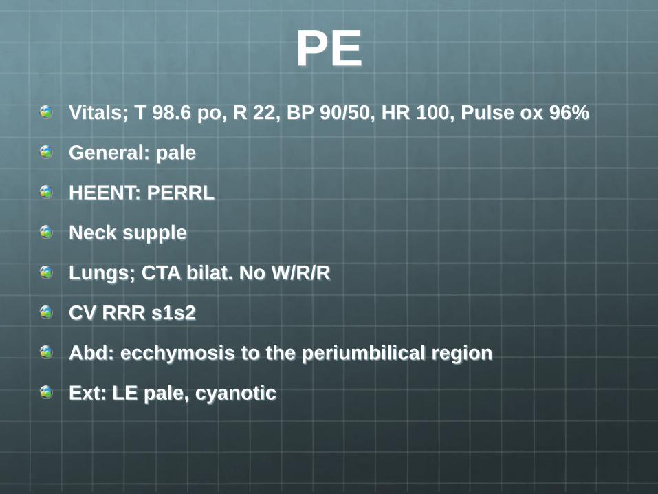
PE Vitals; T 98.6 po, R 22, BP 90/50, HR 100, Pulse ox 96%
General: pale
HEENT: PERRL
Neck supple
Lungs; CTA bilat. No W/R/R
CV RRR s1s2
Abd: ecchymosis to the periumbilical region
Ext: LE pale, cyanotic
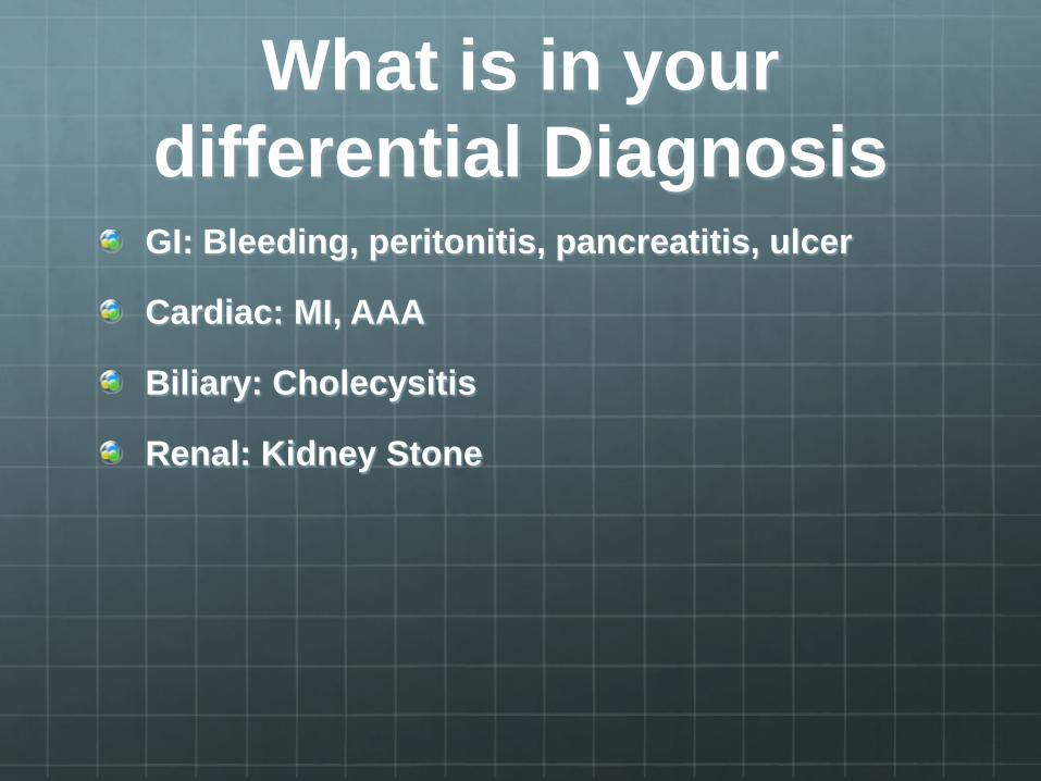
What is in your differential Diagnosis GI: Bleeding, peritonitis, pancreatitis, ulcer
Cardiac: MI, AAA
Biliary: Cholecysitis
Renal: Kidney Stone
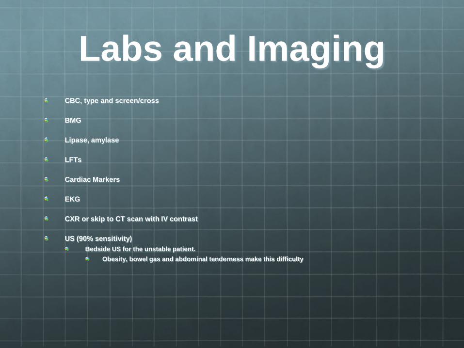
Labs and Imaging CBC, type and screen/cross
BMG
Lipase, amylase
LFTs
Cardiac Markers
EKG
CXR or skip to CT scan with IV contrast
US (90% sensitivity) Bedside US for the unstable patient.
Obesity, bowel gas and abdominal tenderness make this difficulty
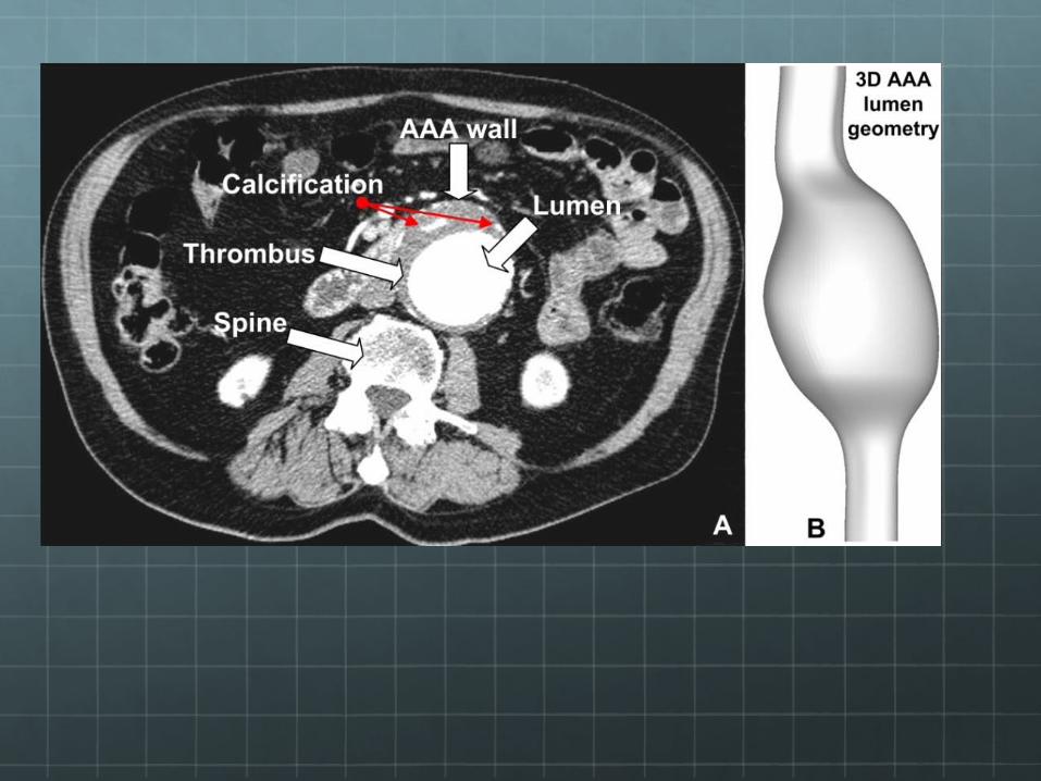
Pathophysiology AAA The Aortic wall is made of smooth muscle, elastin and collagen in concentric layers.
Mechanisms involved in the development of AAA
Proteolytic degradation of aortic wall connective tissue Inflammation and immune responses Biomechanical wall stress Molecular genetics
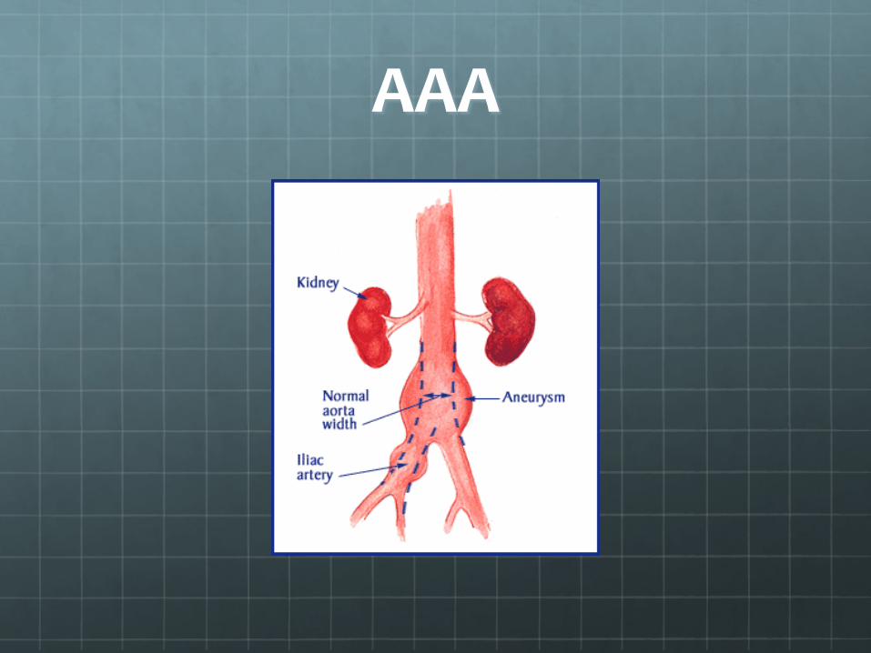
AAA
Pathophysiology AAA cont.
AAAs arise as a result of a failure of the major structural proteins (Elastin and Collagen)
Why this occurs is unknown GENETICS definitely predispose patients These proteins can gradually deteriorate with age. Certain conditions accelerate the process, including atherosclerosis, an excess of certain enzymes and in rare cases, infection.
Why does AAA rupture? Areas of high stress have been found in the AAAs (related to hemodynamics) and appear to correlate with the site of rupture
Aortic Abdominal Aneurysm
Defined as >3 cm
Repair if >5cm Concern for rupture
Aortoenteritc fistulas Consider in patients with unexplained UGI or LGI bleeding
Commonly involves the duodenum
Treatment O2, Cardiac monitoring and 2 lg bore IVs
All symptomatic aortic aneurysms require emergency surgical consult
Surgical treatment for ruptured aortic aneurysms ½ of pts with ruptured aneurysm who reach the operating room die.
Peri-operative B-blockers Reduces arrhthymias and myocardial ischemia, but not the rate of MI or mortality
Other Aneurysms Thoracic Aneurysm
Esophageal, tracheal, bronchial or neurological disorders
May compress or erode into surrounding structures
Refer and control BP
Extremity and visceral aneurysms
Popliteal, subclavian, femoral, iliac, renal, splenic, hepatic All require surgical reapir

Special Considerations
ER physicians in rural areas or small communities should not waste time with time-consuming diagnostics.
ALS protocol, fluids, blood, and blood products as needed with immediate transfer
Pearls 50% of ruptured AAA result in morbidity or mortality
Reduction depends on early detection
Many risk factors lead to AAA Most important are smoking, male gender, and family hx
Classic Triad of presentation Pain, pulsatile abdominal mass and shock
Usually the pain is stady and aggravating Lasts hours to days Unaffected by movement
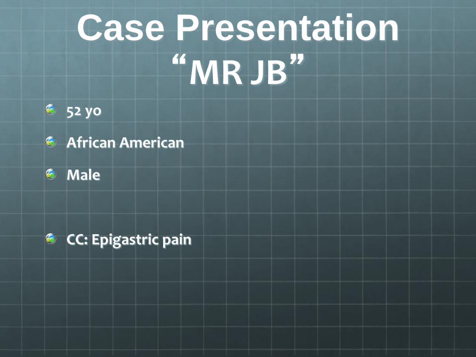
Case Presentation “MR JB”
52 yo
African American
Male
CC: Epigastric pain
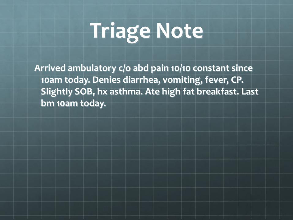
Triage Note Arrived ambulatory c/o abd pain 10/10 constant since
10am today. Denies diarrhea, vomiting, fever, CP. Slightly SOB, hx asthma. Ate high fat breakfast. Last bm 10am today.
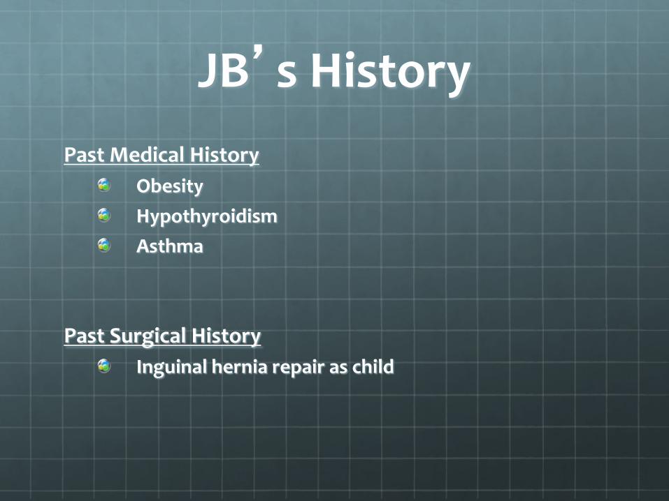
JB’s History Past Medical History
Obesity Hypothyroidism Asthma
Past Surgical History Inguinal hernia repair as child

JB’s History continued
Family history Mom: deceased MI. DM Dad: deceased…? Dehydration Sister: alive, unknown medical problems
Social History No tobacco No etoh No illicit drug use
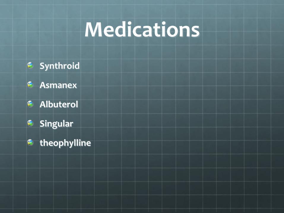
Medications Synthroid
Asmanex
Albuterol
Singular
theophylline
JB’s History expanded
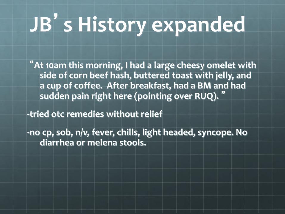
“At 10am this morning, I had a large cheesy omelet with side of corn beef hash, buttered toast with jelly, and a cup of coffee. After breakfast, had a BM and had sudden pain right here (pointing over RUQ). ”
-tried otc remedies without relief
-no cp, sob, n/v, fever, chills, light headed, syncope. No diarrhea or melena stools.

What else do you want to know?
Location: right upper quadrant/epigastric region
Severity: 10/10 with a smile on his face
Description: “can’t describe it, it’s just painful”
Timing: Onset: sudden over 2-3 minutes Duration: constant Frequency: never had pain like this before
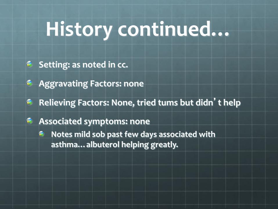
History continued… Setting: as noted in cc.
Aggravating Factors: none
Relieving Factors: None, tried tums but didn’t help
Associated symptoms: none Notes mild sob past few days associated with asthma…albuterol helping greatly.

Physical exam Neuro/HEENT: non focal
Cardiac: non focal
Pulmonary: no wheezing, slight decrease BS bibasilar
Abd: RUQ tenderness with guarding, no rebound tenderness. No CVA tenderness
MS/Derm: non focal
And… What are your Differentials
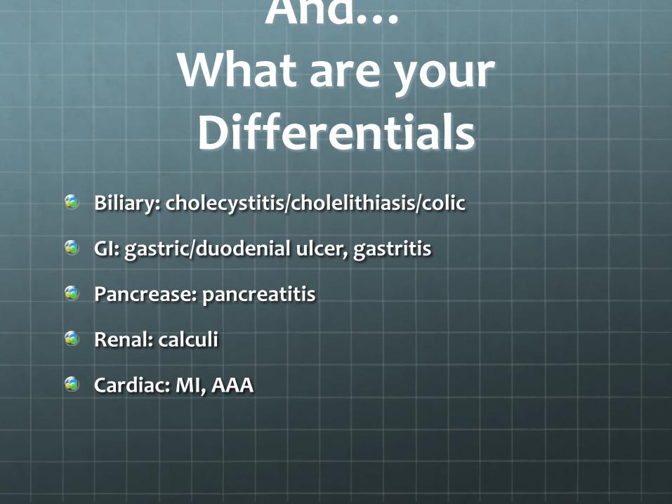
Biliary: cholecystitis/cholelithiasis/colic
GI: gastric/duodenial ulcer, gastritis
Pancrease: pancreatitis
Renal: calculi
Cardiac: MI, AAA
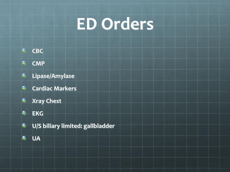
ED Orders CBC
CMP
Lipase/Amylase
Cardiac Markers
Xray Chest
EKG
U/S biliary limited: gallbladder
UA

And what did it all show Lab data non-contributory.
Chest xray negative
EKG: negative
US report “There is mild hydronephrosis of the right kidney” “There is a gallbladder polyp but there is no evidence of cholecystitis or cholelithiasis…” “The visualized portion of the abdominal aorta is at the upper limits of normal in diameter”

Now What…
? ? ?
CT ABD/Pelvis with Contrast…
Results “…are emphysematous changes in the lung bases and bil lower lung atelactasis/infiltrate… “spleen, pancreas, gallbladder, and adrenal glands are unremarkable…” “…there is an irregular linear low attenuation region visualized in the abdominal aorta…and could represent an intimal flap and is suspicious for aortic dissection…”
Hindsight CT Chest/abd: AAA Dissection Protocol
“Type B (Stanford classification) dissection of the thoracic aorta. There appears to be intramural hematoma at the level of the dissection…” “…begins in the distal aortic arch…the posterior component of the dissection in the proximal descending thoracic aorta represents the false lumen with the more ventral component representing the true lumen…” “…The dissection is not visualized slightly below the level of the renal arteries. There is no evidence of dissection extending into the iliac arteries…”
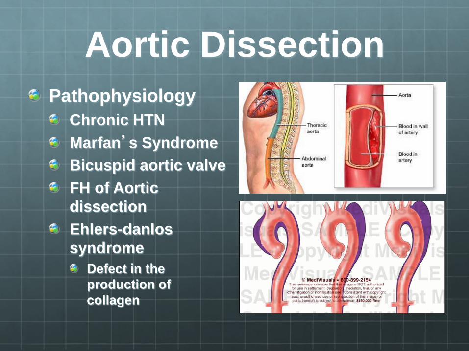
Pathophysiology of Aortic Dissection
The essential feature of aortic dissection is a tear in the intimal layer, followed by formation and propagation of a subintimal hematoma. This produces a false lumen which an reduce blood flow to the major arteries arising from the aorta.
Aortic Dissection Pathophysiology
Chronic HTN Marfan’s Syndrome Bicuspid aortic valve FH of Aortic dissection Ehlers-danlos syndrome
Defect in the production of collagen
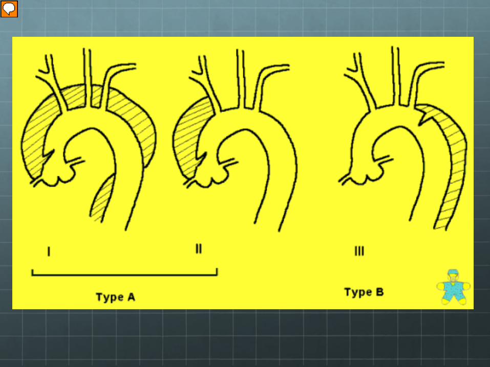
Type A or Type B Aortic Dissection
Type A: just distal to aortic valve
Type B: just distal to the left subclavian artery

Who gets AAA and what is the classical presentation?
Mostly men over 50 years of age
Asymptomatic A pulsating abd mass
MORE REVELANT TO THE ED: THE SYMPTOMATIC PT Chronic midabdominal or low back pain Described as ripping or tearing pain when they rupture
What is the classic presentation?
Severe, persistent chest pain of sudden onset, progressing to the abd and hips
+/- unequal pulses and BP

Case presentation 67 y/o female presents to the ER by ambulance with a cold, painful rt foot and ankle upon waking. Denies trauma. Denies previous episode. Unable to walk on rt leg. Denies sob, chest pain
Pt admits to recent bronchitis. Placed on antibiotics. She states her Coumadin level has been affected by it.
ROS: otherwise negative

History cont PMH/PSH: HTN, CABG, A fib, Hypercholesterolemia
Family history HTN: Mother and father DM Father Stroke father at 70 years of age
Social history No ETOH, Nonsmoker
Medications Asa, Coumadin, Zpak, Metoprolol, Zocor
Physical Exam PE: T 98.0, P 84, BP 140/90, R 16, Pulse ox: 100%
HEENT: PERRL Neck supple Lungs; CTA bilat. No W/R/R CV RRR Systolic murmur Abd: soft NT ND NABS Ext: rt foot cold compared to the left foot. No palpable pulse. Tenderness to touch dorsum of foot and ankle
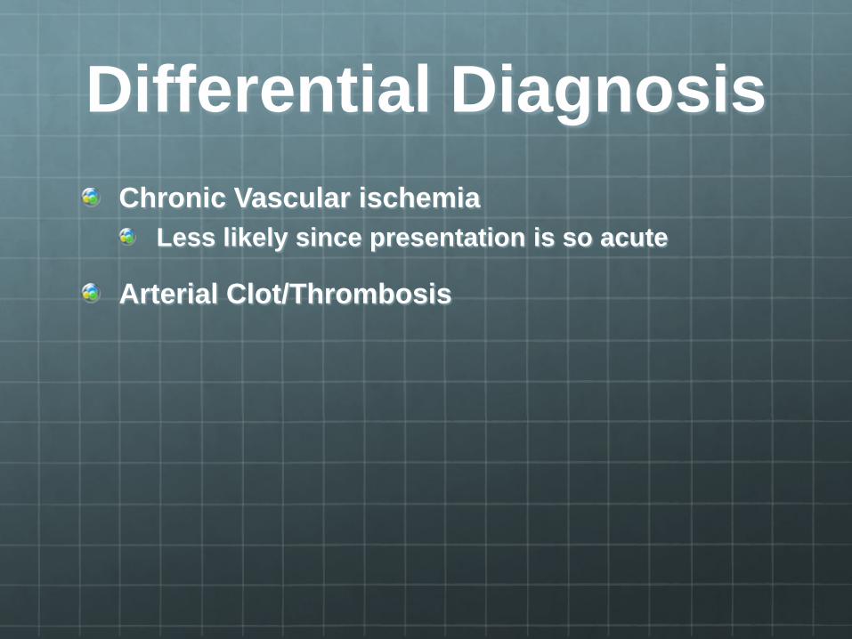
Differential Diagnosis Chronic Vascular ischemia
Less likely since presentation is so acute
Arterial Clot/Thrombosis

Labs and Imaging Bedside doppler to look for pulse
Arterial doppler
CBC, BMG, Cardiac Markers, EKG
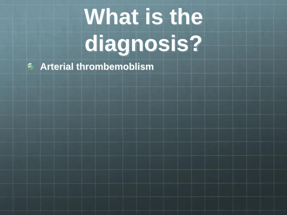
What is the diagnosis?
Arterial thrombemoblism
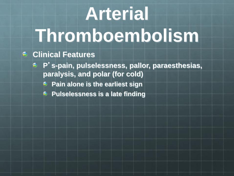
Arterial Thromboembolism
Clinical Features P’s-pain, pulselessness, pallor, paraesthesias, paralysis, and polar (for cold)
Pain alone is the earliest sign Pulselessness is a late finding
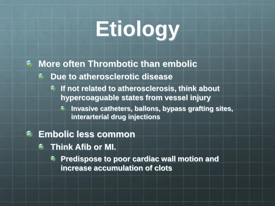
Etiology More often Thrombotic than embolic
Due to atherosclerotic disease If not related to atherosclerosis, think about hypercoaguable states from vessel injury
Invasive catheters, ballons, bypass grafting sites, interarterial drug injections
Embolic less common Think Afib or MI.
Predispose to poor cardiac wall motion and increase accumulation of clots

Management Emergent vascular consult- Admit
Check for Pulse with hand-held doppler
Ankle Brachial index if you detect a pulse with doppler in the affected limb
Arterial Doppler Despite the thinking that reperfusion should be established in 4-6 hours, tissue loss occurs in a significantly shorter amount of time.
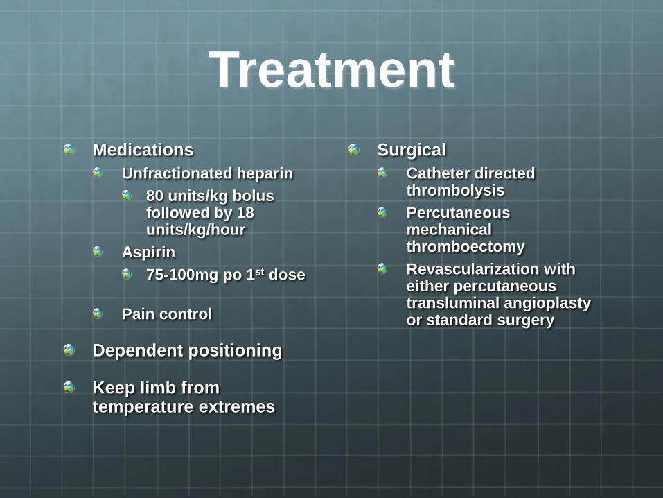
Treatment Medications
Unfractionated heparin 80 units/kg bolus followed by 18 units/kg/hour
Aspirin 75-100mg po 1st dose
Pain control
Dependent positioning
Keep limb from temperature extremes
Surgical Catheter directed thrombolysis Percutaneous mechanical thromboectomy Revascularization with either percutaneous transluminal angioplasty or standard surgery
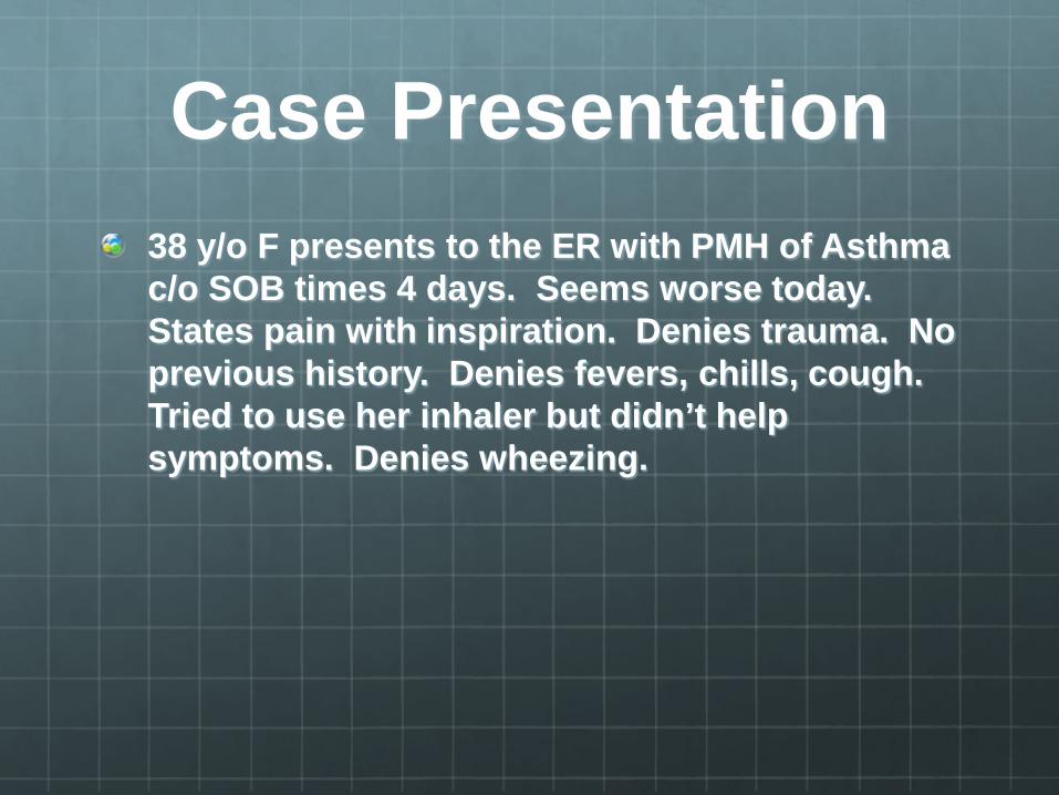
Case Presentation 38 y/o F presents to the ER with PMH of Asthma c/o SOB times 4 days. Seems worse today. States pain with inspiration. Denies trauma. No previous history. Denies fevers, chills, cough. Tried to use her inhaler but didn’t help symptoms. Denies wheezing.
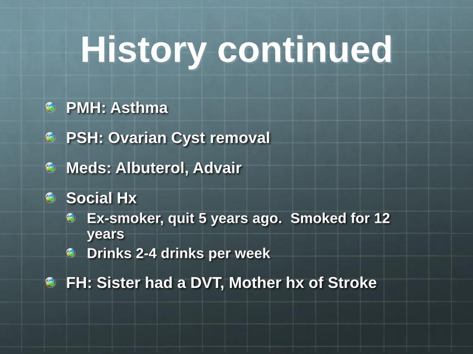
History continued PMH: Asthma
PSH: Ovarian Cyst removal
Meds: Albuterol, Advair
Social Hx Ex-smoker, quit 5 years ago. Smoked for 12 years Drinks 2-4 drinks per week
FH: Sister had a DVT, Mother hx of Stroke

What other questions would you like to ask
her? Recent travel
Recent surgery
FH of DVT or PE
Any leg pain
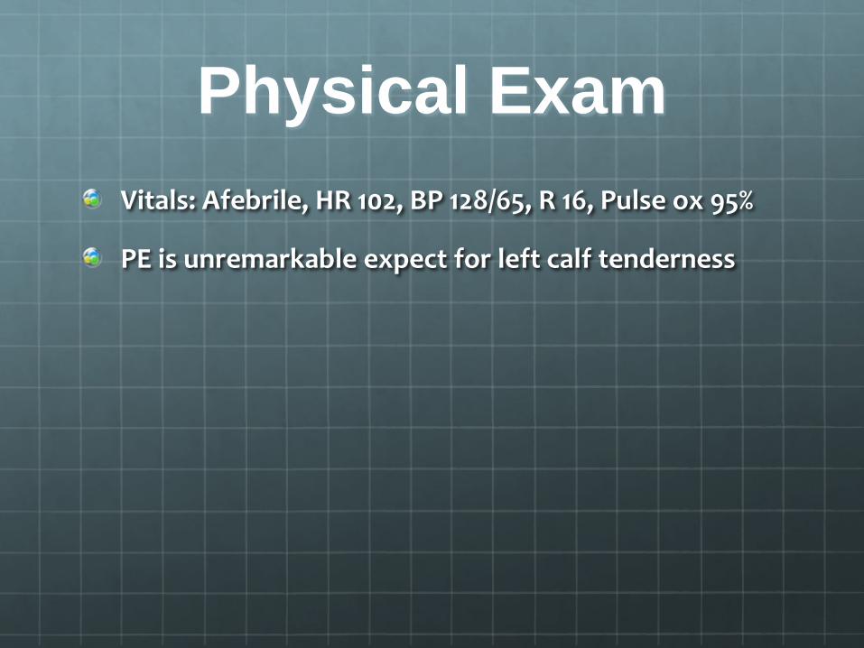
Physical Exam Vitals: Afebrile, HR 102, BP 128/65, R 16, Pulse ox 95%
PE is unremarkable expect for left calf tenderness
What is in your differential diagnosis?
Pulmonary: Asthma, Pneumothorax, Pneumonia, PE, Costochondritis
Cardiac: Carditis, less likely MI
GI: Reflux
Musculoskeletal: strain
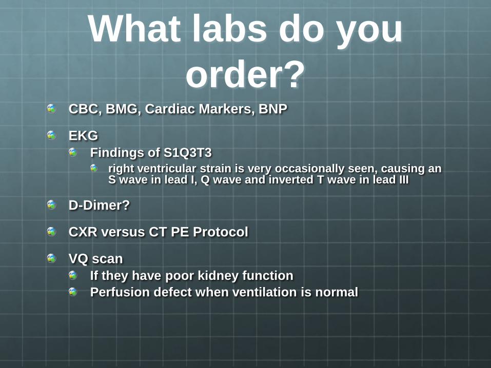
What labs do you order?
CBC, BMG, Cardiac Markers, BNP
EKG Findings of S1Q3T3
right ventricular strain is very occasionally seen, causing an S wave in lead I, Q wave and inverted T wave in lead III
D-Dimer?
CXR versus CT PE Protocol
VQ scan If they have poor kidney function Perfusion defect when ventilation is normal
CT PE Protocol- contrast enhanced pulmonary arteries
Positive for a filling defect consistent with a PE in rt middle lobar pulmonary artery
When to order a D-Dimer
D-Dimer is released when the fibrin in a clot is broken down by the body
Highly sensitive
Low specificity
In the low risk patient
Not in the high risk patient If they have enough risk factors, then go right to the CT scan
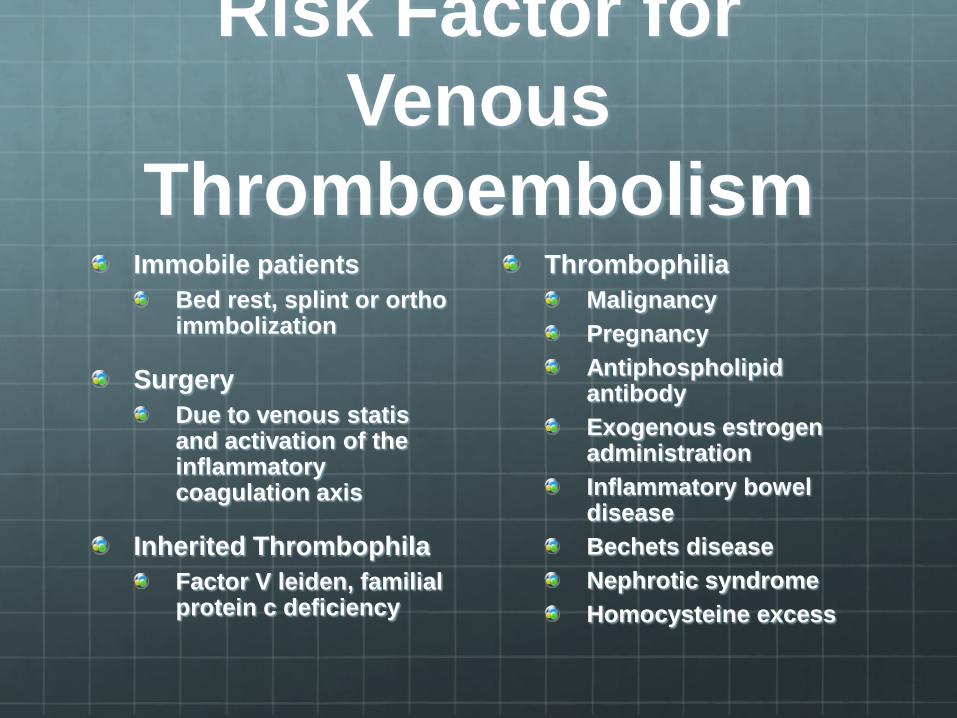
Risk Factor for Venous
Thromboembolism Immobile patients
Bed rest, splint or ortho immbolization
Surgery Due to venous statis and activation of the inflammatory coagulation axis
Inherited Thrombophila Factor V leiden, familial protein c deficiency
Thrombophilia Malignancy Pregnancy Antiphospholipid antibody Exogenous estrogen administration Inflammatory bowel disease Bechets disease Nephrotic syndrome Homocysteine excess
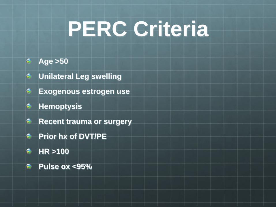
PERC Criteria Age >50
Unilateral Leg swelling
Exogenous estrogen use
Hemoptysis
Recent trauma or surgery
Prior hx of DVT/PE
HR >100
Pulse ox <95%
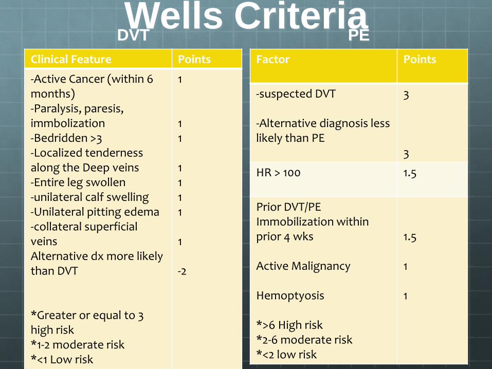
Wells Criteria DVT
Clinical Feature Points
-Active Cancer (within 6 months) -Paralysis, paresis, immbolization -Bedridden >3 -Localized tenderness along the Deep veins -Entire leg swollen -unilateral calf swelling -Unilateral pitting edema -collateral superficial veins Alternative dx more likely than DVT *Greater or equal to 3 high risk *1-2 moderate risk *<1 Low risk
1 1 1 1 1 1 1 1 -2
PE Factor Points
-suspected DVT -Alternative diagnosis less likely than PE
3 3
HR > 100 1.5
Prior DVT/PE Immobilization within prior 4 wks Active Malignancy Hemoptyosis *>6 High risk *2-6 moderate risk *<2 low risk
1.5 1 1
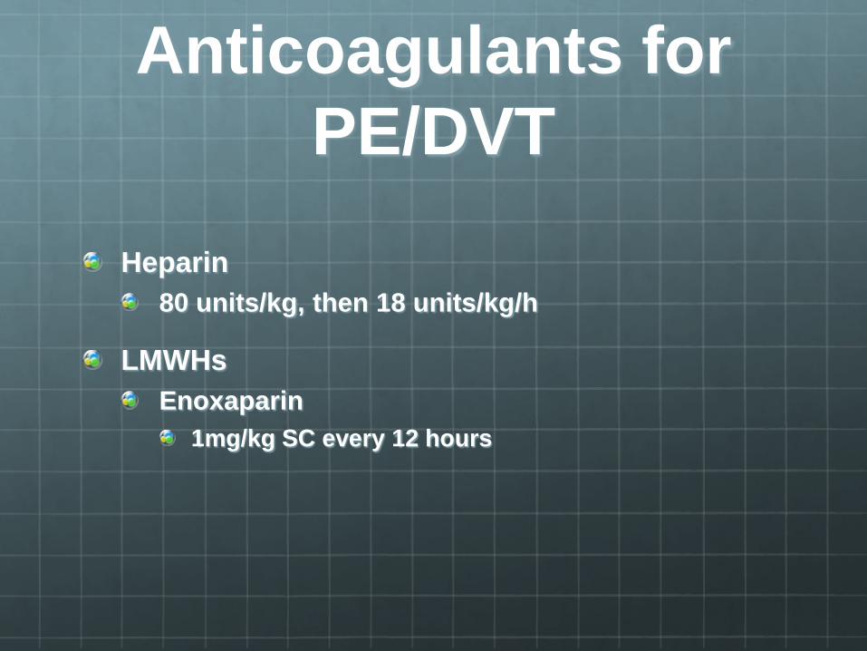
Anticoagulants for PE/DVT
Heparin 80 units/kg, then 18 units/kg/h
LMWHs Enoxaparin
1mg/kg SC every 12 hours

Hypertensive Urgency
Severe HTN without progressive target organ dysfunction
Usually stated as > or equal to 180/120
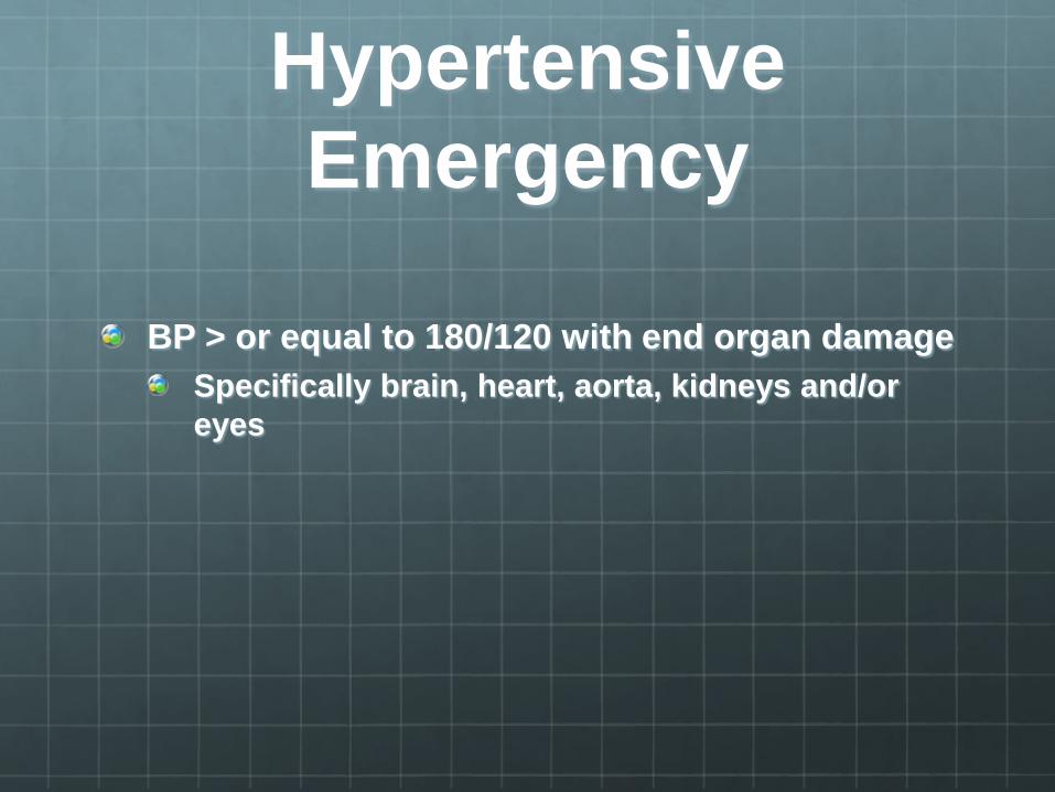
Hypertensive Emergency
BP > or equal to 180/120 with end organ damage Specifically brain, heart, aorta, kidneys and/or eyes
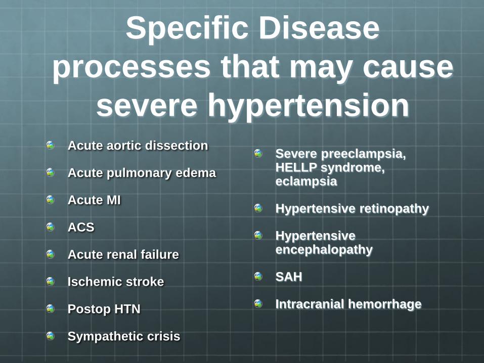
Specific Disease processes that may cause
severe hypertension Acute aortic dissection
Acute pulmonary edema
Acute MI
ACS
Acute renal failure
Ischemic stroke
Postop HTN
Sympathetic crisis
Severe preeclampsia, HELLP syndrome, eclampsia
Hypertensive retinopathy
Hypertensive encephalopathy
SAH
Intracranial hemorrhage

Treatment Common anti-hypertensives in ED
Labetalol Aortic dissection, Acute renal failure, SAH, intracranial hemorrhage, Ischemic stroke
Nitrogylercin AMI, hypertensive pulmonary edeam
Nicardipine Metoprolol
AMI
See table 61-4 for all treatment options Page 444 in tintinalli

Refernces Tintinalli pgs 430-463
Adams pgs 679-686


