Clot Retrieval in Lungs: Safer Treatment with Less Hospitalization Pulmonary embolism (PE) can quickly lead to death if the causative thrombus is not quickly resolved. And now, at the same time that COVID-19 has placed added attention on thrombosis treatment, specialty teams continue to put emphasis on newer and more expeditious ways to resolve blockage of pulmonary arteries. Direct withdrawal of the embolus via catheter is increasingly the solution. Last spring, Virtua became the first center in the region to make use of the FlowTriever device, which captures and withdraws a pulmonary embolus by suction and/or nitinol- mesh snare. This endovascular technique is for patients with submassive PE—or, in some cases, massive PE—if the patient is at high risk for other approaches. Patients with thrombi originating from DVT or post-surgery are typical candidates. “Percutaneous embolectomy allows us to remove large clots from large vessels, immediately restoring blood flow,” said Virtua interventional cardiologist Ibrahim Moussa, DO, FACC, FSCAI, RPVI. “We don’t have to risk use of thrombolytics and, since this is a minimally invasive, catheter-based procedure, we can better treat patients who are not candidates for open-chest surgery.” Suction, Mechanical Capture Withdraws Clot Patients with acute proximal PE have a main or lobar embolus. These patients with significant clot burden in central or segmental pulmonary arteries often have significant hypotensive and hemodynamic instability. They undergo CT with contrast and receive oxygen and anticoagulants. Patients in acute risk may be placed on ECMO. A decision for mechanical endovascular removal is based on size of the embolus and symptoms, including blood oxygen saturation, as well as right heart dysfunction. With access through the femoral vein, the team threads the FlowTriever catheter to the pulmonary arteries and the site of the blockage. Suction from the device pulls the clot out of the vessel. If the clot is adherent, the specialists can deploy a tool on the tip of the catheter that buries into the clot and expands with three mesh discs to drag the clot out. Patients have a shorter ICU and overall length of stay compared to those treated with thrombolytic drugs. Many require no ICU stay at all. Removal Prevents Later Complications The team may try to dislodge a fresh clot (e.g., one formed soon after surgery) with a slow thrombolytic drip and/or ultrasound. “But aged clots are hardened and difficult to protect patients from without removal,” said Dr. Moussa. A residual clot in the pulmonary arteries increases risk of heart failure, dyspnea, pulmonary hypertension, recurrent clotting, and death. The FlowTriever has a similar safety profile to embolism retrieval procedures in the heart, leg, or brain. “Surgical thrombectomy requires heart-lung bypass and carries high morbidity and mortality. And both systemic administration and direct infusion of thrombolytics at the site carry risk of major bleeding and intracranial hemorrhage,” says internist/pulmonologist, Emilio Mazza, MD, PhD, Chief of Critical Care Medicine at Virtua Memorial Hospital. “Being able to route these patients to Virtua Our Lady of Lourdes for FlowTriever is a great addition to our armamentarium for PE.” CARDIOVASCULAR MEDICINE TODAY APRIL 2021 Pulmonary blood flow before (left) and after (right) use of the first thrombectomy device purpose built and FDA indicated for mechanical removal of pulmonary emboli. Image courtesy of Inari Medical To contact the interventional cardiology service at Virtua, call the Virtua Transfer Center at 856-757-3284. Virtua stands ready to evaluate and treat your patients in locations that incorporate social distancing, safety, and cleaning protocols developed in the wake of COVID-19. To learn more about the safety precautions that are now a part Virtua’s standard procedures, visit: http://virtua.org/coronavirus Lung_Today_2020.indd 1 Lung_Today_2020.indd 1 4/5/2021 11:04:29 AM 4/5/2021 11:04:29 AM
Transcript
Clot Retrieval in Lungs: SaferTreatment with Less Hospitalization
Pulmonary embolism (PE) can quickly lead to death if the
causative thrombus is not quickly resolved. And now, at
the same time that COVID-19 has placed added attention
on thrombosis treatment, specialty teams continue to put
emphasis on newer and more expeditious ways to resolve
blockage of pulmonary arteries. Direct withdrawal of the
embolus via catheter is increasingly the solution.
Last spring, Virtua became the first center in the region to
make use of the FlowTriever device, which captures and
withdraws a pulmonary embolus by suction and/or nitinol-
mesh snare. This endovascular technique is for patients with
submassive PE—or, in some cases, massive PE—if the patient
is at high risk for other approaches. Patients with thrombi
originating from DVT or post-surgery are typical candidates.
“Percutaneous embolectomy allows us to remove large clots
from large vessels, immediately restoring blood flow,” said
Virtua interventional cardiologist Ibrahim Moussa, DO, FACC,
FSCAI, RPVI. “We don’t have to risk use of thrombolytics
and, since this is a minimally invasive, catheter-based
procedure, we can better treat patients who are not
candidates for open-chest surgery.”
Suction, Mechanical Capture Withdraws ClotPatients with acute proximal PE have a main or lobar
embolus. These patients with significant clot burden
in central or segmental pulmonary arteries often have
significant hypotensive and hemodynamic instability.
They undergo CT with contrast and receive oxygen and
anticoagulants. Patients in acute risk may be placed on
ECMO. A decision for mechanical endovascular removal
is based on size of the embolus and symptoms, including
blood oxygen saturation, as well as right heart dysfunction.
With access through the femoral vein, the team threads
the FlowTriever catheter to the pulmonary arteries and the
site of the blockage. Suction from the device pulls the clot
out of the vessel. If the clot is adherent, the specialists can
deploy a tool on the tip of the catheter that buries into the
clot and expands with three mesh discs to drag the clot
out. Patients have a shorter ICU and overall length of stay
compared to those treated with thrombolytic drugs. Many
require no ICU stay at all.
Removal Prevents Later ComplicationsThe team may try to dislodge a fresh clot (e.g., one formed
soon after surgery) with a slow thrombolytic drip and/or
ultrasound. “But aged clots are hardened and difficult to
protect patients from without removal,” said Dr. Moussa.
A residual clot in the pulmonary arteries increases risk of
MD, PhD, Chief of Critical Care Medicine at Virtua Memorial
Hospital. “Being able to route these patients to Virtua Our
Lady of Lourdes for FlowTriever is a great addition to our
armamentarium for PE.”
CARDIOVASCULAR MEDICINE TODAY
APRIL 2021
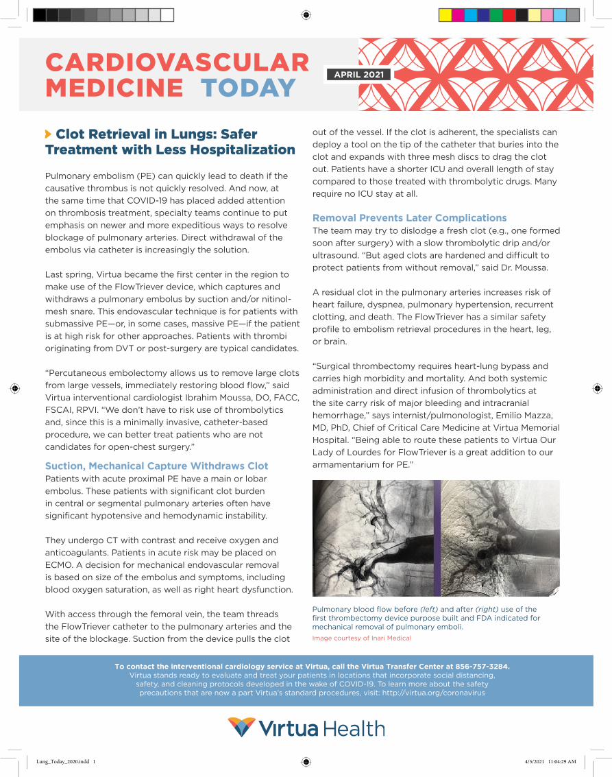
Pulmonary blood flow before (left) and after (right) use of the first thrombectomy device purpose built and FDA indicated for mechanical removal of pulmonary emboli.
Image courtesy of Inari Medical
To contact the interventional cardiology service at Virtua, call the Virtua Transfer Center at 856-757-3284. Virtua stands ready to evaluate and treat your patients in locations that incorporate social distancing,
safety, and cleaning protocols developed in the wake of COVID-19. To learn more about the safety precautions that are now a part Virtua’s standard procedures, visit: http://virtua.org/coronavirus
Lung_Today_2020.indd 1Lung_Today_2020.indd 1 4/5/2021 11:04:29 AM4/5/2021 11:04:29 AM
Even with three-dimensional echocardiography, the critical measure
of regional left ventricular (LV) function has never been easy to
assess with a high level of precision. But cardiac toxicity, whether
from chemo- or radiation therapies, or both, typically manifests
most profoundly in loss of LV ejection fraction (EF). A number of
tests can indicate LVEF decline, or the risk for it, but a newer
modality in echocardiography––strain echo––is proving most
revealing and informative in providing this information.
With the increased frame rates of current ultrasound equipment, the
strain test can characterize the elastic properties of the heart, using
myocardial deformation as a measure of strength of contraction.
During systolic function, twisting mechanics of the heart create
myocardial rotation. Thus, deformation of the ventricular wall takes
place in various dimensions, principal of which are longitudinal,
radial/circumferential and torsional. In this way, strain assesses
lengthening, shortening and thickening of the heart muscle. The
test can also quantify the velocity of deformation, or “strain rate.”
Progressive myocardial conditions first affect the subendocardial
fibers of the heart, those responsible for longitudinal motion.
Subepicardial fibers, responsible for more rotational dynamics,
temporarily compensate; but, as longitudinal and circumferential
functions both degrade, patients become more symptomatic.
Strain measures change in dimension normalized to an initial length.
Global longitudinal strain (GLS) turns out to be the best measure for
detecting subclinical LV dysfunction and can identify patients who
may be experiencing ventricular damage who do not have specific
electro-cardiographic changes or myocardial enzyme abnormalities.
Peak GLS in the range of -18 percent is normal for a healthy person,
and the lower the absolute value of strain below this number, the
more likely LVEF is abnormal or at risk. Those with a reduction in
1600 Haddon AvenueCamden, NJ 08103
Our Lady of LourdesMedical Center
NON-PROFIT ORGU.S. POSTAGE
PAIDPERMIT #36BELLMAWR
NJ 08031
Strain Detects Early Toxicity, Gives New Precision in Measuring Heart Injury
A publication of: Lourdes Health System1600 Haddon Avenue • Camden, NJ 08103 • www.lourdesnet.org1-888-LOURDES (1-888-568-7337)AVP, Communications: Carol Lynn Daly Publications Manager: Josh Bernstein • Writer: Russ Allen
absolute value of GLS to -16 percent or less are already
demonstrating abnormal myocardial mechanics suggestive of
damage. Strain is cost-efficient and is free of ionizing radiation.
“Strain is an especially important checkpoint for the patient at-risk
going into therapy or who is experiencing subclinical myocardial
damage during therapy.” said Lourdes cardiologist Geoffrey Zarrella,
DO, FACC. “Using this technology, we can identify individuals who
need pre-treatment with medications or other care adjustments to
prevent further heart damage.”
Change in global longitudinal strain (GLS) tracks with loss of EF for a
patient in this “bull’s eye” plot of strain values for each of the 17 myocardial
segments. The patient receiving cytotoxic chemotherapy had normalbaseline
strain and LVEF, but by 12 months met the criteria for cardiotoxicity.
*page 1 image: Endocardial longitudinal strain study of a patient with
coronary artery occlusion. Brown color indicates areas with impaired strain.
GLS was reduced in this patient to -15 percent.
Strain echocardiolographic image courtesy J. Am. Coll. Card., Volume 63, Issue 25 part A,July 2014, Thavendiranathan P., Poulin F., Lim K., et al.
Improved Methods for Closure of Refluxed Veins in Extremities
303 Lippincott DriveMarlton, NJ 08053
Vein issues, especially those that are cosmetic, have
a variety of new solutions. But common complaints
in the lower extremities are frequently related to or
accompanied by a more serious medical issue in,
or signaled by, vasculature of these limbs. Patients
frequently need the attention of specialists with
cardiology, phlebotomy, and vascular expertise.
Catheter Treatment Collapses Bulging VeinsSwelling in the legs is a typical presentation, but even
an ulcer may be inadequately attributed, for example, to
diabetes. Virtua’s vein team can use CT, MRI, ultrasound,
angiography, or venography to get to the cause, which
in the extremities is often manifested in the great
saphenous vein, where reflux can occur. In such cases,
the team emphasizes thermal ablation via laser or radio-
frequency catheter inserted through a small puncture.
Collapsing the vein in this way eliminates the backward
flow of blood from venous reflux. With such same-day
catheter-based interventions, patients may walk out
of the procedures and almost immediately return to
daily activities. Of course, symptoms may also come
from peripheral arterial disease. When medications are
inadequate for this condition, the vein team can perform
an angioplasty via the femoral artery, with placement of a
drug-coated stent. Virtua’s vein and vascular experts also
collaborate with wound services for nonhealing lesions.
Nonthermal Ablation Popular for Cosmetic ConcernsWhether their cosmetic vein concerns are connected
to more serious vascular or cardiac conditions or not,
patients are often willing to pursue solutions to the
appearance or discomfort issues that these problems
present. Varicose veins are a common complaint,
as are the related challenge of spider veins.
Ablation, through endovenous closure, to shut off and
shrink these veins is the preferred approach. For cosmetic
conditions, nonthermal ablation is particularly popular.
These are brief office procedures that spare patients
much postoperative pain and recovery. Among these
steps, updated methods of sclerotherapy serve to irritate
the lining of the errant vessel, causing it to swell and
stick together. Over several weeks or months, the vessel
turns into scar tissue that retracts and fades from view.
In addition, phlebectomy remains an option that has also
improved. In-office microphlebectomy is a minimally invasive
approach for residual veins that are too twisted or too close
to the surface for catheter treatment, or that are too large
to treat with sclerotherapy. The specialist removes the veins
percutaneously and closes the wound with surgical glue.
For vein and vascular consultation at Virtua, call 856-309-5869.
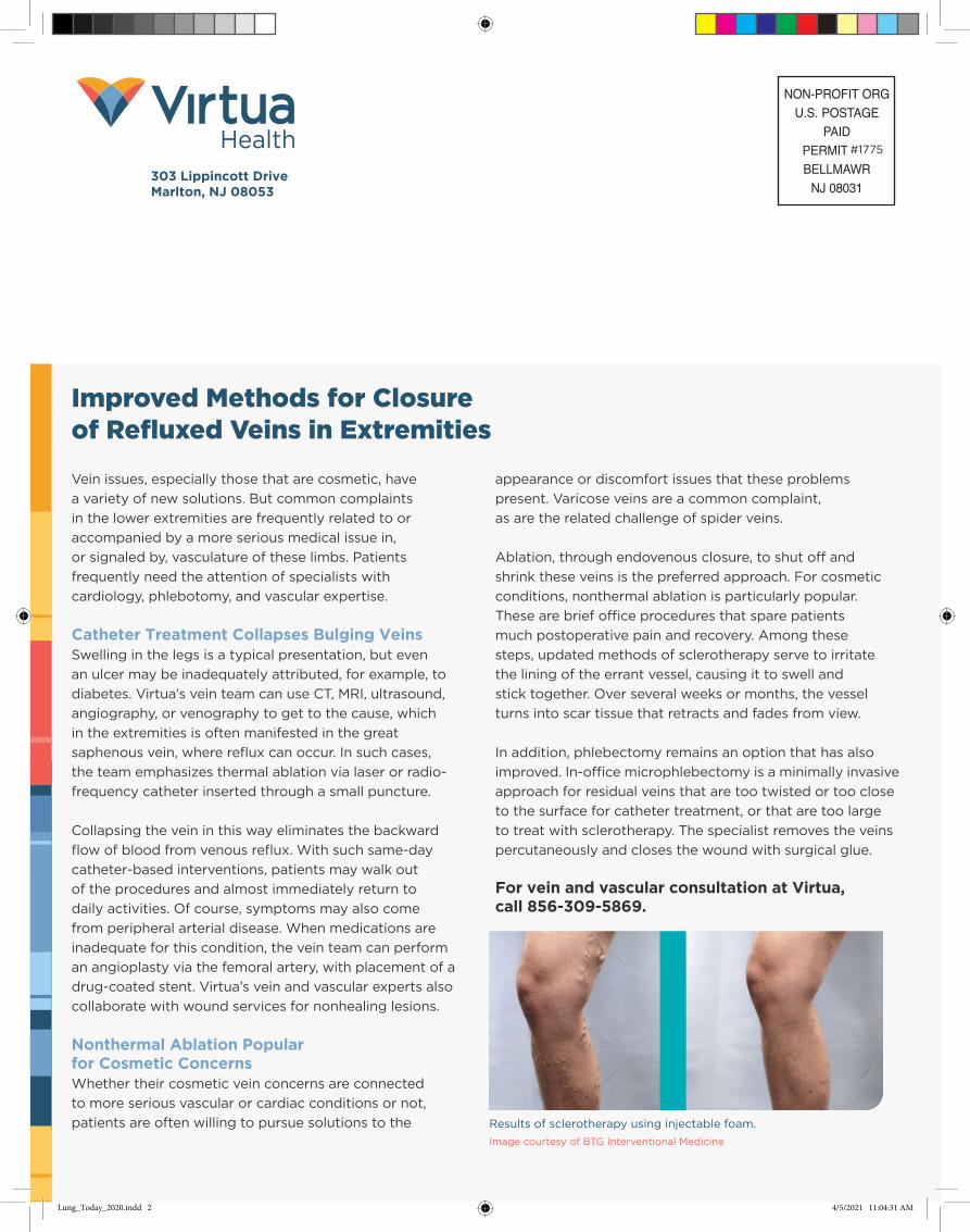
Results of sclerotherapy using injectable foam.
Image courtesy of BTG Interventional Medicine
#1775
Lung_Today_2020.indd 2Lung_Today_2020.indd 2 4/5/2021 11:04:31 AM4/5/2021 11:04:31 AM