Cardiovascular microRNA Webinar Cardiovascular microRNA Webinar Recent Work & Current Methods In Cardiovascular microRNA Research 2012‐12‐04 Christoph Eicken, PhD Head of Technical Services – Microarrays
Transcript
Cardiovascular microRNA WebinarCardiovascular microRNA WebinarRecent Work & Current Methods
In Cardiovascular microRNA Research
2012‐12‐04
Christoph Eicken, PhDHead of Technical Services – Microarrays
Agendag
Recent Studies
miRNA Intro
Current methods
Q & A
Case Studies
Q
microRNAs ‐What We Know
1. All miRNAs are small non‐coding RNAs, usually consisting of 20–22 nucleotides for animals and 20 24 nt for plantsnucleotides for animals and 20–24 nt for plants.
2. All miRNA precursors have a well‐predicted stem loop hairpin structure, and this fold‐back hairpin structure has a low free energy p gy
3. Many miRNAs are evolutionarily conserved, some from worm to human in animals, or from ferns to core eudicots or monocots in plants
4. Bind to complementary mRNA molecules and act as negative regulators of translation
5. High copy number
6. Expression is tissue (and developmental stage) specific
microRNAs ‐What We Know
1. Currently ‐ 25,141 mature miRNAs across 193 plant, animal, and y , p , ,
virus species (miRBase 19, Aug. 2012).
2. Mechanism is far reaching and complex – each miRNA may control many g p y ygenes and it is estimated that miRNAs regulate expression of up to 1/3 of all human genes.
3 O t b f t h th i d h i3. Operate by one of two hypothesized mechanisms:
– Complete pairing mRNA is degraded ‐ predominant in plantsg p p
– Imperfect pairing translation is repressed but mRNA remains intact ‐ predominant in animals
Milestones1993 – Lin‐4 was shown to encode two small RNA molecules (not protein)
• Small RNAs control developmental timing in C. elegans through negative regulation of lin‐14 gene. • Lee RC et al. 1993 The C. elegans heterochronic gene lin‐4 encodes small RNAs with antisense complementarity to lin‐14. Cell
75(5):843‐54. [article] 5(5) 8 3 5 [a t c e]
1998 – RNAi is observed for the first time – leads to Nobel Prize (2006)• Fire A, Mello CC et al. 1998 Potent and specific genetic interference by double‐stranded RNA in Caenorhabditis elegans. Nature
1998; 391:806‐811. [article]
Challenge to the Central Dogma of BiologyDNA > transcription > RNA > translation > protein
2000 L t 7 id tifi d i h d D hil ( h l l k l )2000 – Let‐7 was identified in humans and Drosophila. (Reinhart et al., Slack et al.)
2001 – Bartel, Tuschl, Ambros ‐ Discover large class of small regulatory RNAs, name them microRNA (miRNA)( )
• Lau NC et al. 2001 An abundant class of tiny RNAs with probable regulatory roles in Caenorhabditis elegans. Science 294(5543):858‐62. [abstract]
• Lagos‐Quintana M et al. 2001 Identification of novel genes coding for small expressed RNAs. Science 294(5543):853‐8. [abstract] • Lee RC et al. 2001 An extensive class of small RNAs in Caenorhabditis elegans. Science 294(5543):862‐4. [abstract]
2002 – Identification of Drosha reveals complete microRNA biogenesis pathway•pri‐miRNAs > Drosha > pre‐miRNAs (~70nt) > Dicer > mature miRNAs (~22nt) •Lee Y et al. (2002) The nuclear RNase III Drosha initiates microRNA processing. Nature 425(6956):415‐9. [abstract]
Milestones2004 – Microarrays are used to profile miRNA expression
• Several groups develop or modify existing gene expression microarrays for profiling miRNAs• Babak et al. 2004 [article], Barad et al. 2004 [article], Liu et al. 2004 [article], Nelson et al. [abstract], 2004, Thomson et al. 2004
[article][article].
2005 – Next‐Gen Sequencing is used for small RNA discovery and analysis• Green Lab ‐ uses Solexa (now Illumina) sequencing to identify novel small RNAs in Arabidopsis plants• Lu C et al. 2005 Elucidation of the small RNA component of the transcriptome. Science, 309: 1567–1569. [abstract]
2006 iRB li t S I tit t2006 – miRBase goes online at Sanger Institute• Depository for experimentally verified microRNAs: http://www.mirbase.org/• Griffiths‐Jones S et al. 2006 miRBase: microRNA sequences, targets and gene nomenclature. NAR 34(Database Issue):D140‐D144.
[article]
2008 – Circulating miRNAs detected in body fluids• miRNA signatures in serum and plasma provide cancer diagnosis / prognosis• Serum ‐ Chen X et al.(2008) Characterization of microRNAs in serum: A novel class of biomarkers for diagnosis of cancer and other ( ) g
diseases. Cell Res 18:997–1006. [article]• Plasma ‐Mitchell PS et al.(2008) Circulating microRNAs as stable blood‐based markers for cancer detection. PNAS USA 105:10513–
2005 ‐miRNA is essential for proper muscle development in Drosophila• miR 1 exerts post transcriptional control during myogenesis in Drosophila• miR‐1 exerts post‐transcriptional control during myogenesis in Drosophila • Sokol NS, Ambros V (2005) Mesodermally expressed Drosophila microRNA‐1 is regulated by Twist and is required in muscles during
larval growth. Genes Dev 19(19): 2343–2354.[article]• Kwon C, Han Z, Olson EN, Srivastava D. (2005) MicroRNA1 influences cardiac differentiation in Drosophila and regulates Notch
signaling. Proc Natl Acad Sci USA 102(52): 18986–18991. .[article]
2006 ‐Muscle microRNAs regulate myoblast proliferation and differentiation in skeletal muscle
• Chen JF et al. (2006) The role of microRNA‐1 and microRNA‐133 in skeletal muscle proliferation and differentiation. Nat Genet 38(2): 228–233. [article]
• KIM HK et al. (2006) Muscle‐specific microRNA miR‐206 promotes muscle differentiation. J Cell Biol 174(5): 677–687. [article]
2006 ‐ The expression of several miRNAs was identified as being regulated in response to cardiac muscle hypertrophy
• van Rooij E et al (2006) A signature pattern of stress responsive micro RNAs that can evoke cardiac hypertrophy and heart failure• van Rooij E et al. (2006). A signature pattern of stress‐responsive micro‐ RNAs that can evoke cardiac hypertrophy and heart failure. Proc Natl Acad Sci USA 103(48): 18255–18260. [article]
2008/2009 miRNAs are involved in Cardiomyocyte Apoptosis and2008/2009 ‐miRNAs are involved in Cardiomyocyte Apoptosis and Regeneration, Cardiac Conduction, Fibrosis
• Rane S – (2009) Circ Res 104: 879–886. [article], • Terentyev D (2009) Circ Res 104 514 521 [article]• Terentyev D – (2009) Circ Res 104: 514–521. [article], • van Rooij E – (2008) PNAS 105: 13027–13032. [article]
2010 ‐MicroRNAs regulate cardiac stem cell fateSl ij l (20 0) i d 99 l diff i i d lif i i h d i d di i• Sluijter JP et al. (2010) MicroRNA‐1 and ‐499 regulate differentiation and proliferation in human‐derived cardiomyocyte progenitor cells. Arterioscler Thromb Vasc Biol 30(4):859‐68 [article]
2010/2011 ‐ Circulating miRNAs may be novel biomarkers for coronary artery disease (CAD) and acute myocardial infarction (AMI)
• M. Hoekstra et al. (2010) The peripheral blood mononuclear cell microRNA signature of coronary artery disease. Biochem BiophysRes Commun, 394, 792–797. [article]
• V. Di Stefano et al. (2011) microRNAs as peripheral blood biomarkers of cardiovascular disease. Vascul Pharmacol, 55, 111–118. [article]
Why Study miRNAs in the di l ?Cardiovascular System?
1. Several miRNA genes are specifically expressed or highly enriched in skeletal and/or cardiac muscle, the so‐called muscle miRNAs.
2. miRNAs are essential for proper muscle development and exert post‐transcriptional control during myogenesistranscriptional control during myogenesis.
3. Muscle miRNAs regulate myoblast proliferation and differentiation in skeletal muscle.
4. Circulating miRNAs may be novel biomarkers for coronary artery disease (CAD) and acute myocardial infarction (AMI).
5. Dysregulation of miRNAs can contribute to cardiovascular related diseases and disorders: CAD, AMI, arrhythmia, hypertrophy and fibrosis.
6. miRNAs are likely associated with other muscle‐related diseases and are potential therapeutic targets.
miRNAs in the Cardiovascular System: Clinical PotentialM l l Di i / Bi kMolecular Diagnostics / Biomarkers – Identification of specific miRNAs or miRNA expression based signatures that can act as biomarkers for various diseases/pathologies. /p g
1. Biomarkers in body fluids: serum, plasma, exosomes, & HDL particles
2 M k t d d t il d li i l di i2. Make accurate and detailed clinical diagnosis
3. Potential to determine prognosis and predict treatment efficacy
Rosetta Genomics' miRview Lung Assay:Rosetta Genomics miRview Lung Assay:Assay Shown to Accurately Differentiate Between the Four Main Types of Lung Cancer
miRNAs in the Cardiovascular System: Clinical Potential
Drug Discovery / Therapeutics – Identification of miRNAs that play essential roles in disease to act as drugs or possible p y g ptherapeutic targets.
1. miRNAs as vascular regulating drugs
2. miRNAs as drug targets
3 St d f iRNA t d t d t i f ti t th ti li3. Study of miRNAs to understand response to infections, stress, other stimuli
2010 Review: MicroRNAs in Cardiovascular Disease and disordersCardiovascular Disease and disorders
Review ‐ Small EM, Frost RJ, Olson EN. (2010)MicroRNAs add a new dimension to cardiovascular disease. Circulation. 2010
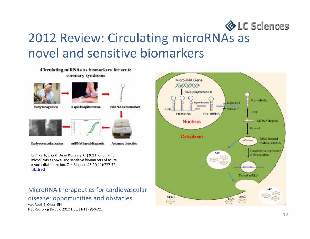
2012 Review: Extracellular/circulating gmicroRNAs and their potential role in cardiovascular disease
Cellular releases (A) and uptake (B) of miRNAs. (A) Extracellular miRNAs may be contained within vesicles, including microvesicles, exosomes, and apoptotic bodies, as well as within proteins such as
16
Zhu H, Fan GC. (2012) Extracellular/circulating microRNAs and their potential role in cardiovascular disease. Am J Cardiovasc Dis 1(2):138‐149. [article]
exosomes, and apoptotic bodies, as well as within proteins such as Ago2, HDL, and other RNA‐binding proteins. (B) Extracellular miRNAs may potentially interact with recipient cells via direct fusion, internalization, receptor‐mediated interactions and other possible mechanisms.
Colin C. Pritchard, Heather H. Cheng & Muneesh TewariColin C. Pritchard, Heather H. Cheng & Muneesh TewariNature Reviews Genetics 13, 358‐369 (May 2012) .[abstract]
• High through‐put method – 100s of samples analyzed per month
• Streamlined handling – can be easily sequence knowledge.
• Increased dynamic range and tunable sensitivity.
g yautomated
• Straightforward data analysis
Sh d i 5 d • Can detect structural variations such as gene fusions and alternative splicing events.
• Short turn‐around time – 5 days
• Lower cost
• A truly digital solution (absolute abundance vs relative abundance).
Small RNA Sequencing and Data AnalysisComprehensive RNA Sequencing ExperimentComprehensive RNA Sequencing Experiment
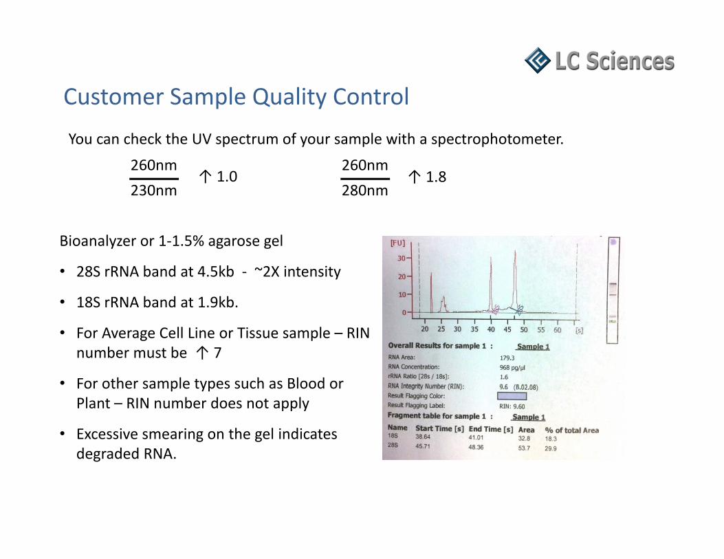
• Sample QC
• Sample preparationp p p
• Library preparation
• High‐throughput sequencing
• Advanced bioinformatics analysis
• High level customer support
Sequencing Run
Small RNA Sequencing and Data Analysisq g
Instrument: Illumina Genome Analyzer GAIIx
Length of Reads: 35 bases
Number of Reads: ~20‐30 Million
Data Output: ~0.7‐1.0 Gb
d ( d ) lBar‐coding (Indexing) Samples:
• We recommend 3 per lane, Max 4 per lane
• The total number of reads does not change with bar‐coding
• Sacrifice sequencing depth for lower cost
Total Reads / LaneNumber of
Samples / LaneReads/ Sample
Samples / Lane30 M 1 30 M30 M 2 15 M30 M 3 10 M30 M 4 7 5 M30 M 4 7.5 M30 M 5 6 M30 M 6 5 M
Reg. Experimental Design
Sample Replicates for Expression Studies
Biological Replicates – Still Very Important
Sample Replicates for Expression Studies
o og ca ep cates St e y po ta t
• For experiments performed with a small number of biological replicates, significant results may be due to biological diversity between individuals and may not be reproducible it is impossible to know whether expressionmay not be reproducible ‐ it is impossible to know whether expression patterns are specific to the individuals in the study or are a characteristic of the test condition.
f f ff• There is no statistical significance for a difference observed between 2 samples.
• There is no magic to RNA‐Seq. These ideas are widely accepted for DNA g q y pmicroarray experiments, where a large number of biological replicates are now required to justify scientific conclusions.
Hansen KD, Wu Z, Irizarry RA, Leek JT. 2011 Sequencing technology does not eliminate biological variability. Nat Biotechnol 29:572–573. [abstract]
Reads mapped to Reads un-mappedReads mapped to mammalian mirs in miRBase
Reads un-mapped to mammalian mirsin miRBaseACGT101‐miR
v3.5 Software
mirs mapped to species genome
mirs un-mapped to species genome
Reads un-mapped to mRNA, Rfam, and repbase
Reads mapped to mRNA, Rfam, and repbase
othersGroup 1 othersKnown species miRNAs
Reads mapped to species genome
Reads un-mapped to species genome
Reads mapped to species genome
Reads un-mapped to species genome
Group 4 no hitGroup 2 Group 3 G oup o tG oup G oup 3
Known miRNAscandidate species
miRNAs
Candidate species miRNAs genome
inconsistent with miRBase
Potentially novel miRNAs
IsomiRs
Small RNA Sequencing and Data Analysis
IsomiRs
Can explain discrepancies array data vs qRT‐PCR validation
Basic Research / Discovery
Characterization and discovery of novel miRNAs and moRNAs in JAK2V617FCharacterization and discovery of novel miRNAs and moRNAs in JAK2V617F mutated SET2 cells
To gain insights into a possible role of microRNAs in myeloproliferative neoplasms, they performedin myeloproliferative neoplasms, they performed short RNA massive sequencing and extensive bioinformatic analysis in the JAK2V617F‐mutated SET2 cell line.
O ll 652 k t iRNAOverall, 652 known mature miRNAs were detected, of which 21 were highly expressed, thus being responsible of most of miRNA‐mediated gene repression. microRNA putative targets were enriched in specific signaling g p g gpathways, providing information about cell activities under massive posttranscriptional regulation. The majority of miRNAs were mixtures of sequence variants, called isomiRs, mainly because of alternative noncanonicalmainly because of alternative, noncanonical processing of hairpin precursors.
Overall, this study shed light on the complexity of microRNA‐mediated gene regulation in SET2
Relative isomiR contribution to the total read count of 21 most expressed miRNAs in SET2 cells. The total read count belonging to sequences not passing the stringent threshold for being considered as isomiRs is indicated in the white rectangle.
Bortoluzzi S, Bisognin A, Biasolo M, Guglielmelli P, Biamonte F, Norfo R, Manfredini R, Vannucchi AM. (2012) Characterization and discovery of novel miRNAs and moRNAs in JAK2V617F mutated SET2 cells. Blood 119(13), e120‐e130.[article]
cells and represents the basis for future studies in JAK2V617F‐mutated cellular models.
Acute myocardial infarction (MI) due to coronary artery occlusion is accompanied by a pathological remodeling response that includes hypertrophic cardiac growth and fibrosis, which impair cardiac contractility.
Show by miRNA microarray that MI inShow by miRNA microarray that MI in mice and humans results in the dysregulation of specific miRNAs, which are similar to but distinct from those involved in hypertrophy and heart failure. A th MI l t d iRNAAmong the MI‐regulated miRNAs are members of the miR‐29 family, which are down‐regulated in the region of the heart adjacent to the infarct.
Down‐regulation of miR‐29 with anti‐miRs in vitro and in vivo induces the expression of collagens, whereas over‐expression of miR‐29 in fibroblasts reduces collagen expression Conclude that miR 29 acts as
MiRNA profiling in response to MI. (A) Masson Trichrome staining of mouse heart sections (B) Microarray analysis (C) Real‐time PCR analysis of specific miRNAs in response to MI in sham operated animals (D) Real‐time PCR analysis of miRNAs in human heart samples (E) Northern blot analysis
van Rooij E, Sutherland LB, Thatcher JE, DiMaio JM, Naseem RH, Marshall WS, Hill JA, Olson EN. (2008) Dysregulation of microRNAs after myocardial infarction reveals a role of miR‐29 in cardiac fibrosis. Proc Natl Acad Sci USA 105(35), 13027‐32.[article]
expression. Conclude that miR‐29 acts as a regulator of cardiac fibrosis and represents a potential therapeutic target for tissue fibrosis in general.
Identification of a miRNA signature of renal ischemia reperfusion injury
Renal ischemia reperfusion injury (IRI) is associated with significant morbidity and mortality
Identification of a miRNA signature of renal ischemia reperfusion injury
morbidity and mortality.
miRNA microarray expression profiling conducted on RNA prepared from IRI, sham, and naive kidneys of mice revealed nine miRNAs that are differentially expressed following IRI when compared with sham controls.controls.
The differentially expressed miRNAs could be placed into four groups based on their expression tt Th fi di d fi
G d i JG G X St h K J i h A T lli SG
patterns. These findings define a molecular fingerprint of renal injury.
Identification of a miRNA signature following renal IRI. (A) Heat map of miRNAs differentially expressed in the kidneys of mice after IRI or sham operation. (B) Plots of above microarray
Godwin JG, Ge X, Stephan K, Jurisch A, Tullius SG, Iacomini J. (2010) Identification of a microRNA signature of renal ischemia reperfusion injury. Proc Natl Acad Sci USA 107(32), 14339‐44. [abstract]
data expressed as the mean signal intensity ± SE.
The differentially expressed miRNAs could be placed into four groups based on their expression patterns following IRI: group 1 was rapidly up‐regulated, group 2 also up‐regulated, group 3 was rapidly down‐regulated, group 4 was also down‐regulated.
Molecular Diagnostics / Biomarkersin Body Fluids – BloodPeripheral blood microRNA signatures can be utilized to identify patients exhibiting atherosclerotic CAD
Used real‐time PCR‐based profiling toUsed real time PCR based profiling to determined the microRNA signature of peripheral blood mononuclear cells (PBMCs) from stable and unstable CAD patients and unaffected controls.
The presence of CAD in general coincided with a marked 5‐fold increase in the relative expression level of miR‐135a, and a 4‐fold decrease in the expression of miR‐147 in pPBMCs from CAD patients as compared to controls.
Unstable angina pectoris patients could be discriminated from stable patients based
(A) The relative expression level of several “housekeeping” microRNAs was determined in PBMCs from coronary artery disease patients (CAD; black bars) and unaffected controls (Control; white bars). Data are expressed as fold change compared to control and represent means + SEM of three (control) or six (CAD) different RNA pools derived from a respective total of 20 and 50
discriminated from stable patients based upon their relatively high expression level of a cluster of three microRNAs including miR‐134, miR‐198, and miR‐370, suggesting that the microRNA signatures can be used to identify
subjects. (B) The relative expression level of microRNAs that were differentially expressed between PBMCs from coronary artery disease patients (CAD; black bars) and unaffected controls (Control; white bars). P < 0.01, P < 0.001 (two‐way ANOVA) (C) The calculated ratio of
microRNA‐135a and microRNA‐147 expression levels in PBMCs from CAD patients (black bar) and controls (white bar) P < 0 01 (t test)
Hoekstra M et al. (2010) The peripheral blood mononuclear cell microRNA signature of coronary artery disease. BiochemBiophys Res Commun 394(3), 792‐97. [abstract]
patients at risk for acute coronary syndromes. patients (black bar) and controls (white bar). P < 0.01 (t‐test).
Drug Discovery / Therapeutics miRNAs as Novel Treatments
Mi RNA 210 i i i i hibi i d i di f i
Using microRNA microarrays, showed that microRNA‐210 was highly expressed in live mouse HL 1
MicroRNA‐210 can improve angiogenesis, inhibit apoptosis, and improve cardiac function in a murine model of myocardial infarction
210 was highly expressed in live mouse HL‐1 cardiomyocytes compared with apoptotic cells after 48 hours of hypoxia exposure. Gain‐of‐function and loss‐of‐function approaches were used to investigate microRNA‐210 therapeutic potential in vitro.
Adult FVB mice underwent intramyocardial injections with minicircle vector carrying microRNA‐210 precursor, minicircle carrying microRNA‐scramble, or sham surgery At 8 weeks echocardiography showedsham surgery. At 8 weeks, echocardiography showed a significant improvement of left ventricular fractional shortening in the minicircle vector carrying microRNA‐210 precursor group compared with the minicircle carrying microRNA‐scramble control.
MicroRNA‐210 represents a potential novel therapeutic approach for treatment of ischemic heart disease.
Evaluation of cardiac function after MI after miR‐210 treatment. A, Representative echocardiogram of mice with LAD ligation after injection of MC‐210, MC‐Scr, or sham group at Week 8. B, Quantitative analysis of left ventricular FS among the 3 groups. C, Representative Masson trichrome staining of explanted heart at Week 8 showed increased wall thickness for the MC‐210 group, confirming the positive functional imaging data seen in echocardiography D TUNEL staining of an explanted
Hu S, Huang M, Li Z, Jia F, Ghosh Z, Lijkwan MA, Fasanaro P, Sun N, Wang X, Martelli F, Robbins RC, Wu JC. (2010) MicroRNA‐210 as a novel therapy for treatment of ischemic heart disease. Circulation122(11 Suppl), S124‐31. [article]
imaging data seen in echocardiography. D, TUNEL staining of an explanted heart demonstrated significantly reduced apoptotic cells in MC‐210 group compared with the MC‐Scr control group. E, Immunofluorescence staining of CD31 endothelial marker (green) demonstrated increased neovascularization in the myocardium after MC‐210 delivery compared with the MC‐Scr control.
Drug Discovery / Therapeutics miRNAs as Drug Targets
l l f d l fMicroRNA‐24 regulates vascularity after myocardial infarction
Myocardial infarction leads to cardiac remodeling and development of heart failureremodeling and development of heart failure. Insufficient myocardial capillary density after myocardial infarction has been identified as a critical event in this process.
Showed that microRNA‐24 (miR‐24) is enriched in cardiac endothelial cells and considerably upregulated after cardiac ischemia. MiR‐24 induces endothelial cell apoptosis & abolishes endothelial capillary network formation
Low‐dose antagomir‐24 treatment preferably leads to miR‐24 silencing in endothelial cells in vitro and in vivo. A, Uptake of Cy3‐labeled antagomirs in human umbilical vein endothelial cells (10 μg/mL, 24 hours). B, Primary deposition of Cy3‐labeled t i (C 3 A t ) t l t l t d d th li l ll dh i l l 1
endothelial capillary network formation.
Blocking of endothelial miR‐24 limited myocardial infarct size of mice via prevention of endothelial apoptosis and enhancement of
l i hi h l d d di antagomirs (Cy3‐Ant.) to platelet and endothelial cell adhesion molecule‐1 (Pecam1)–positive endothelial capillaries after injection of a low dose (5 mg/kg) or homogenous cardiac uptake after treatment with 80 mg/kg (high dose). C, MiR‐24 expression in fractionated cardiomyocytes and cardiac endothelial cells 14 days after myocardial infarction (MI). Animals were treated at day 0 and day 2 with either 5 mg/kg scrambled antagomir (Scr) or antagomir against miR‐24 (Ant24). n=3 to 6 experiments/animals per group. Data are mean and SEM; ***P<0.005.
vascularity, which led to preserved cardiac function and survival.
The findings indicate that miR‐24 acts as a critical regulator of endothelial cell apoptosis
Fiedler J, Jazbutyte V, Kirchmaier BC, Gupta SK, Lorenzen J, Hartmann D et al. (2011) MicroRNA‐24 regulates vascularity after myocardial infarction. Circulation 124, 720–730. [article]
p / p g p ;g p pand angiogenesis and is suitable for therapeutic intervention in the setting of ischemic heart disease.
Drug Discovery / Therapeutics miRNAs as Drug Targets
l f b h bDownregulation of miRNA‐29 by antisense inhibitors protects against myocardial ischaemia‐reperfusion injury
Pioglitazone (PIO), a peroxisome proliferator‐ti t d t (PPAR) i t t tactivated receptor (PPAR)‐γ agonist, protects
against myocardial ischaemia–reperfusion (IR) injury.
Evaluated the expression changes of miRNAs in the rat heart after PIO administration using miRNA arrays and found that miR‐29a and c levels decreased remarkably after 7‐day treatment with PIO.
Antagomirs against miR‐29a or ‐29c significantly reduced myocardial infarct size and apoptosis in hearts subjected to IR injury.
These finding show that modulation of miRNAs can be achieved by pharmacological intervention and provide a rationale for the development of miRNA‐based strategies for the attenuation of IR injury. Fig 1 ‐ (A) Relative expression of miR‐29a 16 h after the third injection of
antagomir (anta‐29a) or missense antagomir (misanta‐29). (B) Relative expression of miR 29c 16 h after the third injection (C) Effect of anta 29aexpression of miR‐29c 16 h after the third injection. (C) Effect of anta‐29a, anta‐29c, and misanta‐29 on myocardial IS. (D) % of TUNEL‐positive‐stained cardiomyocytes in the ischaemic area of antagomir‐pre‐treated mice hearts subjected to IR. (E) Representative staining pattern. Arrows indicate TUNEL‐positive nuclei which are stained blue.
Ye Y, Hu Z, Lin Y, Zhang C, Perez‐Polo JR. (2010) Downregulation of microRNA‐29 by antisense inhibitors and a PPAR‐{gamma} agonist protects against myocardial ischaemia‐reperfusion injury. Cardiovasc Res 87(3), 535‐44.
CompanyOverview• Global ‐ Genomics & Proteomics Services Provider