21
Cardiovascular Cardiovascular physiology. physiology. Dr James Ker Dr James Ker
| Date post: | 17-Dec-2015 |
| Category: |
Documents |
| Upload: | franklin-tate |
| View: | 235 times |
| Download: | 0 times |
Cardiovascular physiology.Cardiovascular physiology.
Dr James KerDr James Ker
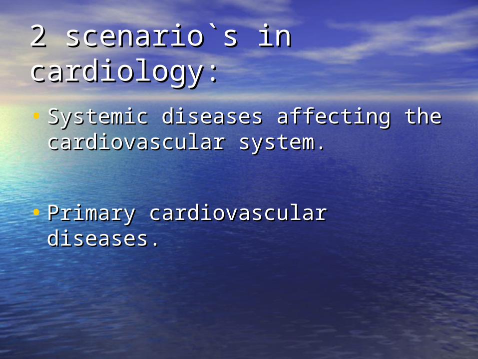
2 scenario`s in cardiology:2 scenario`s in cardiology:
• Systemic diseases affecting the Systemic diseases affecting the cardiovascular system.cardiovascular system.
• Primary cardiovascular diseases.Primary cardiovascular diseases.
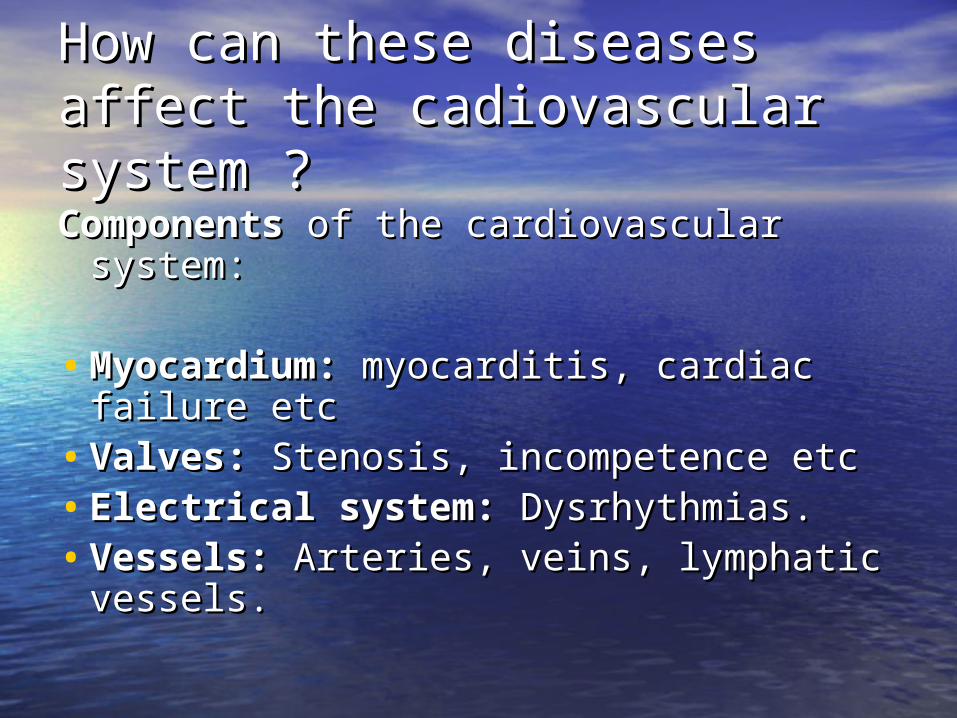
How can these diseases affect How can these diseases affect the cadiovascular system ?the cadiovascular system ?
Components Components of the cardiovascular of the cardiovascular system:system:
• Myocardium: Myocardium: myocarditis, cardiac myocarditis, cardiac failure etcfailure etc
• Valves: Valves: Stenosis, incompetence etcStenosis, incompetence etc• Electrical system: Electrical system: Dysrhythmias.Dysrhythmias.• Vessels: Vessels: Arteries, veins, lymphatic Arteries, veins, lymphatic
vessels.vessels.

Physiological disturbances:Physiological disturbances:
• Disturbances in the following may Disturbances in the following may occur:occur:
• Blood pressureBlood pressure
• Cardiac rhythmCardiac rhythm
• Valvular functionValvular function
• Cardiac systole and diastoleCardiac systole and diastole
• Blood flowBlood flow
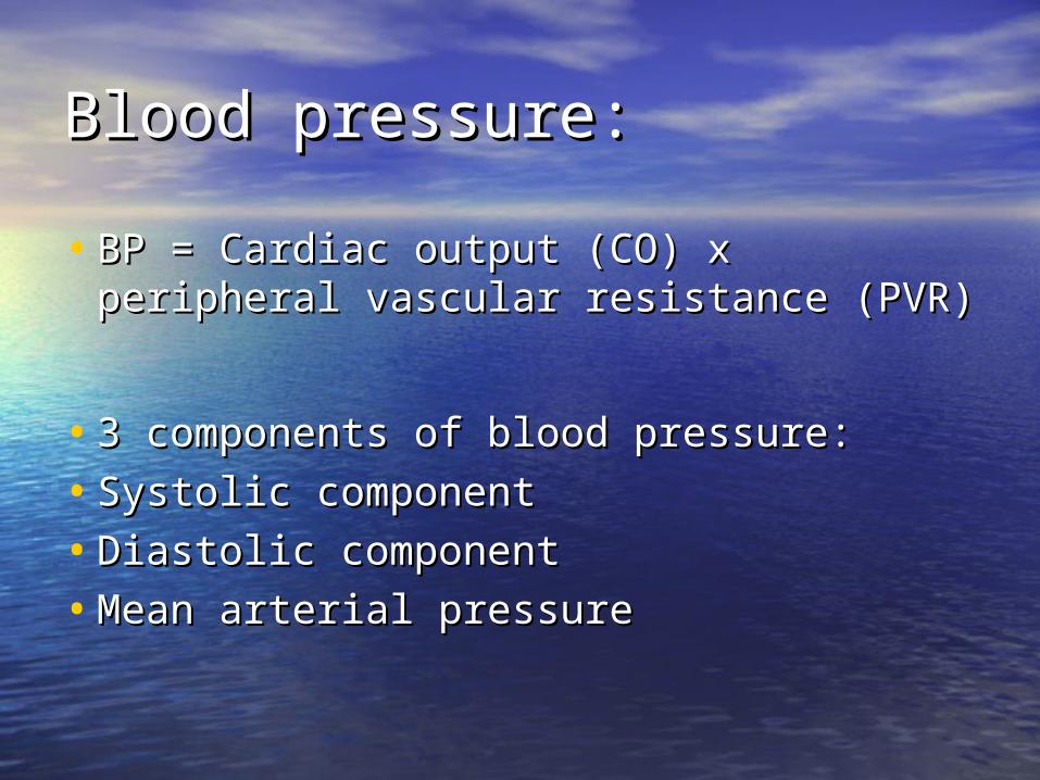
Blood pressure:Blood pressure:
• BP = Cardiac output (CO) x BP = Cardiac output (CO) x peripheral vascular resistance (PVR)peripheral vascular resistance (PVR)
• 3 components of blood pressure:3 components of blood pressure:
• Systolic componentSystolic component
• Diastolic componentDiastolic component
• Mean arterial pressureMean arterial pressure

• BP = 120/80 mmHgBP = 120/80 mmHg
• Mean arterial pressure = (S + 2D)/3 Mean arterial pressure = (S + 2D)/3 or or
• D + 1/3 PPD + 1/3 PP
• Pulse pressure=S – D (120-80=40 Pulse pressure=S – D (120-80=40 mmHg)mmHg)
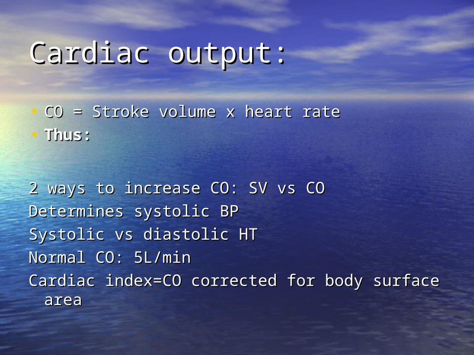
Cardiac output:Cardiac output:
• CO = Stroke volume x heart rateCO = Stroke volume x heart rate
• Thus:Thus:
2 ways to increase CO: SV vs CO2 ways to increase CO: SV vs CO
Determines systolic BPDetermines systolic BP
Systolic vs diastolic HTSystolic vs diastolic HT
Normal CO: 5L/minNormal CO: 5L/min
Cardiac index=CO corrected for body surface Cardiac index=CO corrected for body surface areaarea

Causes of increased CO:Causes of increased CO:
• FeverFever
• AnaemiaAnaemia
• HyperthyroidismHyperthyroidism
• PregnancyPregnancy
• ExerciseExercise
• Etc..Etc..
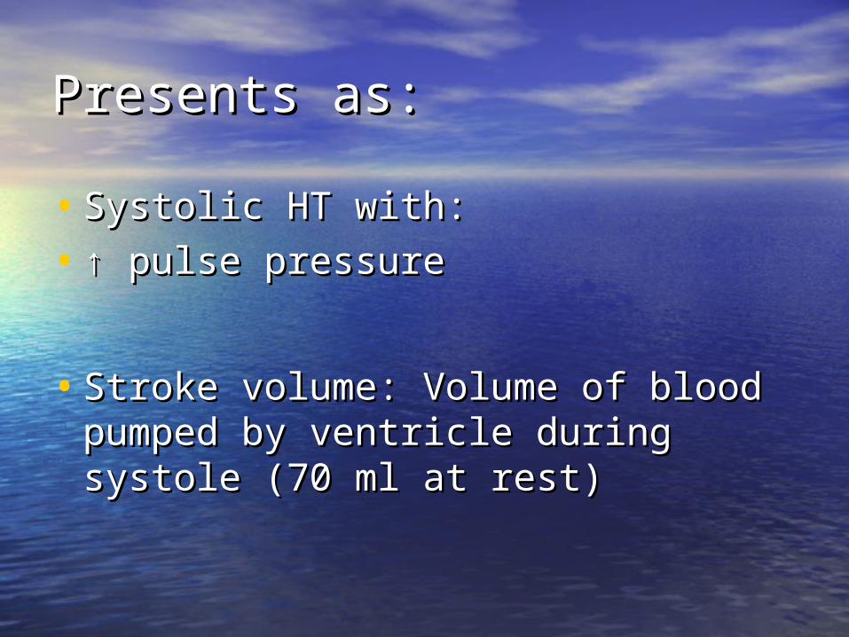
Presents as:Presents as:
• Systolic HT with:Systolic HT with:
• ↑ ↑ pulse pressurepulse pressure
• Stroke volume: Volume of blood Stroke volume: Volume of blood pumped by ventricle during systole pumped by ventricle during systole (70 ml at rest)(70 ml at rest)
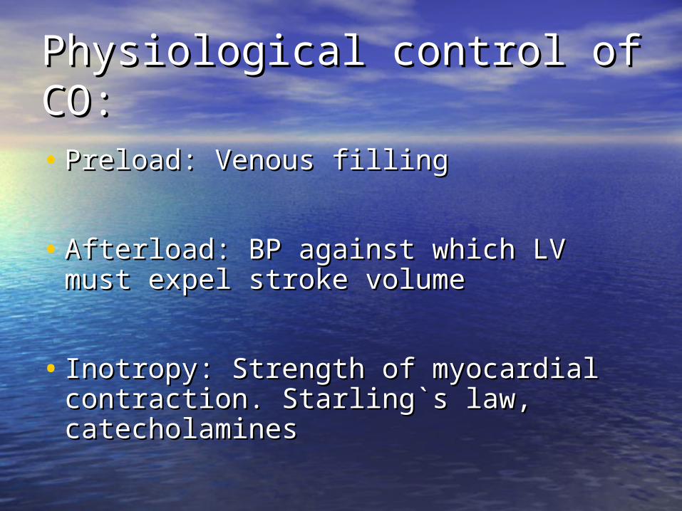
Physiological control of CO:Physiological control of CO:
• Preload: Venous fillingPreload: Venous filling
• Afterload: BP against which LV must Afterload: BP against which LV must expel stroke volumeexpel stroke volume
• Inotropy: Strength of myocardial Inotropy: Strength of myocardial contraction. Starling`s law, contraction. Starling`s law, catecholaminescatecholamines

Physiological control of heart Physiological control of heart rate:rate:
• Intrinsic: SA nodeIntrinsic: SA node
• Extrinsic:Extrinsic:
Hormonal: thyroid, catecholaminesHormonal: thyroid, catecholamines
Neurological: Autonomic nervous Neurological: Autonomic nervous systemsystem
Fever, electrolytesFever, electrolytes
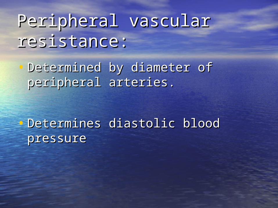
Peripheral vascular Peripheral vascular resistance:resistance:
• Determined by diameter of Determined by diameter of peripheral arteries.peripheral arteries.
• Determines diastolic blood pressureDetermines diastolic blood pressure

Control of PVR:Control of PVR:
• Hormonal: AT II, endothelins, NO, Hormonal: AT II, endothelins, NO, bradykinin, catecholamine etcbradykinin, catecholamine etc
• Neurological: Autonomic nervous Neurological: Autonomic nervous systemsystem
• Myogenic/Local.Myogenic/Local.
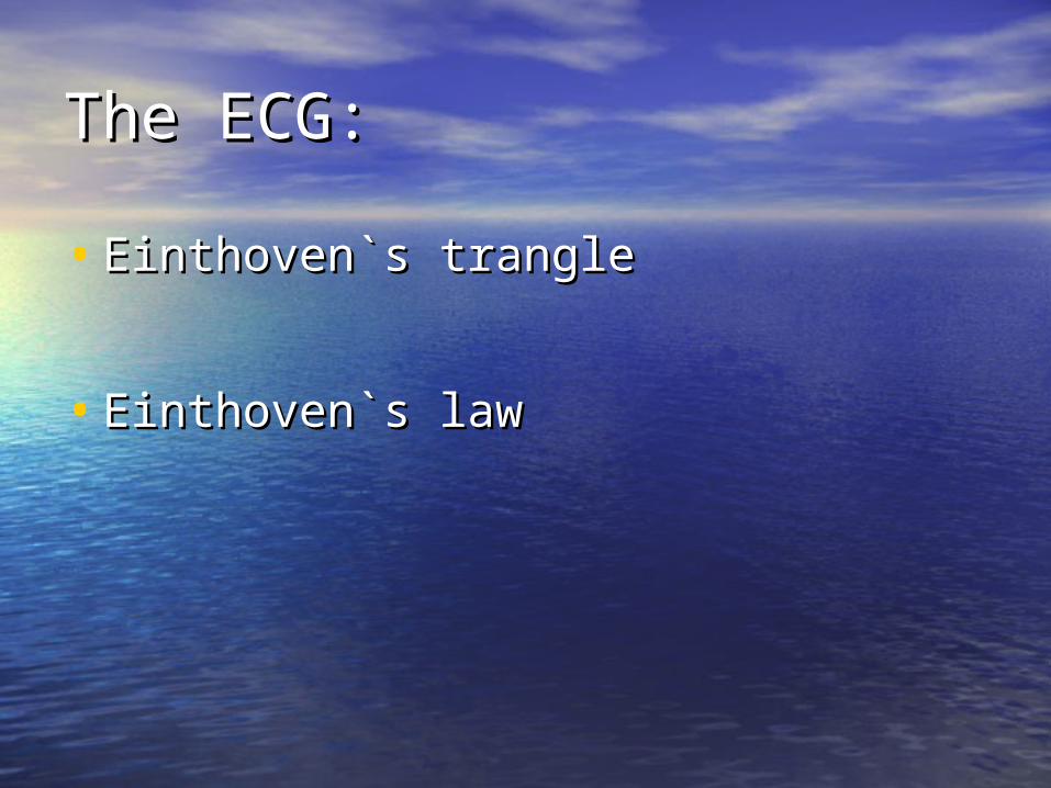
The ECG:The ECG:
• Einthoven`s trangleEinthoven`s trangle
• Einthoven`s lawEinthoven`s law
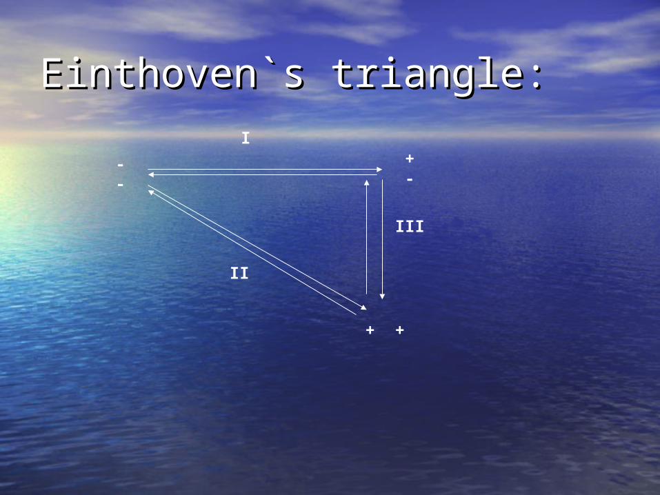
Einthoven`s triangle:Einthoven`s triangle:I
II
III
+-
--
+ +
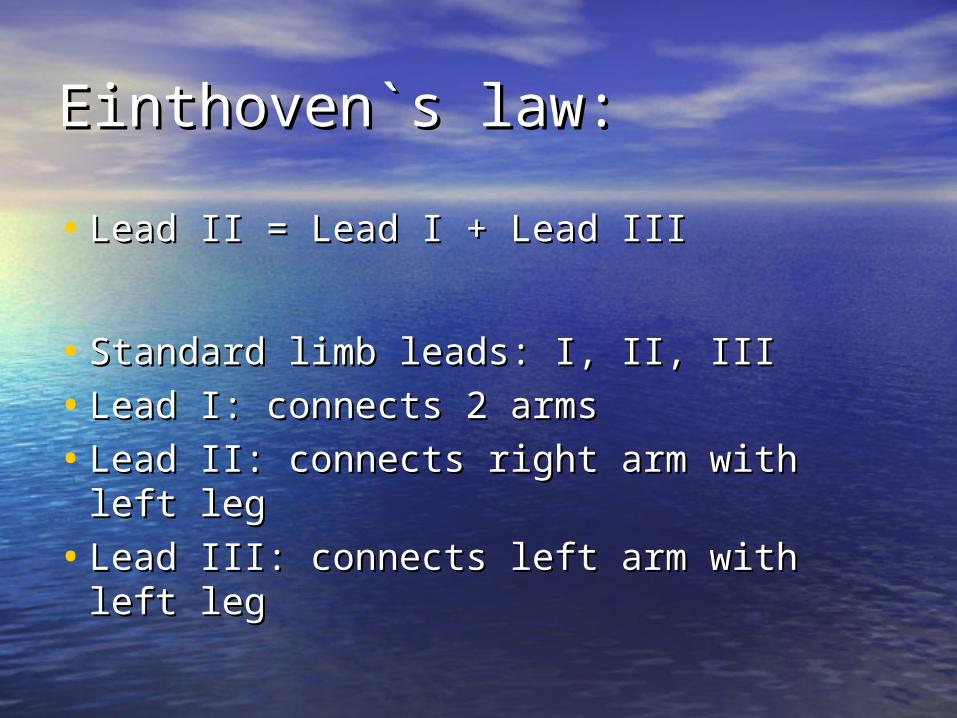
Einthoven`s law:Einthoven`s law:
• Lead II = Lead I + Lead IIILead II = Lead I + Lead III
• Standard limb leads: I, II, IIIStandard limb leads: I, II, III
• Lead I: connects 2 armsLead I: connects 2 arms
• Lead II: connects right arm with left legLead II: connects right arm with left leg
• Lead III: connects left arm with left legLead III: connects left arm with left leg

aV leads:aV leads:
• Limb leads are Limb leads are bipolarbipolar
• Unipolar Unipolar limb leads=aV leads: aVR, limb leads=aV leads: aVR, aVL and aVFaVL and aVF
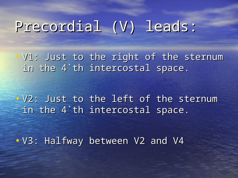
Precordial (V) leads:Precordial (V) leads:
• V1: Just to the right of the sternum in V1: Just to the right of the sternum in the 4`th intercostal space.the 4`th intercostal space.
• V2: Just to the left of the sternum in V2: Just to the left of the sternum in the 4`th intercostal space.the 4`th intercostal space.
• V3: Halfway between V2 and V4V3: Halfway between V2 and V4
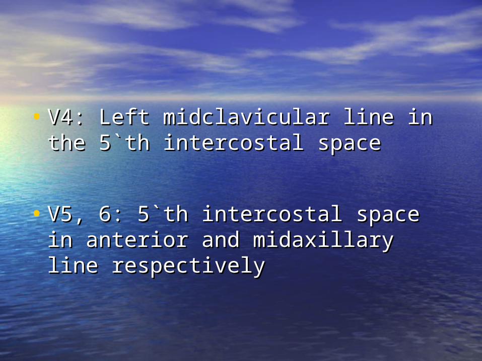
• V4: Left midclavicular line in the 5`th V4: Left midclavicular line in the 5`th intercostal spaceintercostal space
• V5, 6: 5`th intercostal space in V5, 6: 5`th intercostal space in anterior and midaxillary line anterior and midaxillary line respectivelyrespectively
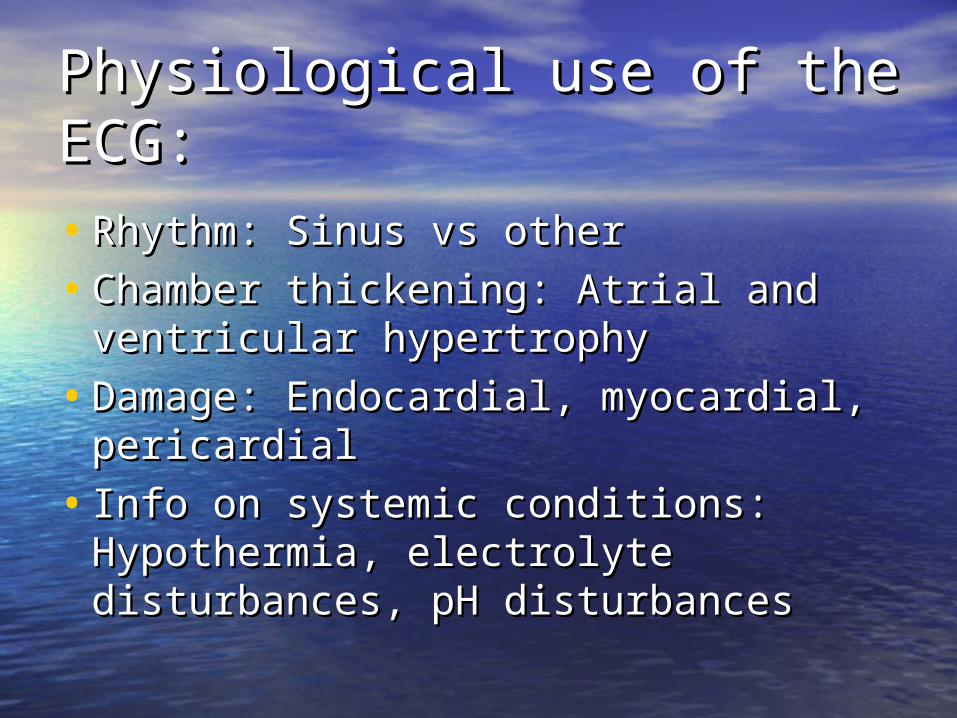
Physiological use of the Physiological use of the ECG:ECG:
• Rhythm: Sinus vs otherRhythm: Sinus vs other
• Chamber thickening: Atrial and Chamber thickening: Atrial and ventricular hypertrophyventricular hypertrophy
• Damage: Endocardial, myocardial, Damage: Endocardial, myocardial, pericardialpericardial
• Info on systemic conditions: Info on systemic conditions: Hypothermia, electrolyte Hypothermia, electrolyte disturbances, pH disturbancesdisturbances, pH disturbances
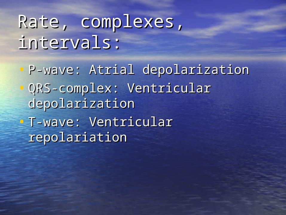
Rate, complexes, intervals:Rate, complexes, intervals:
• P-wave: Atrial depolarizationP-wave: Atrial depolarization
• QRS-complex: Ventricular QRS-complex: Ventricular depolarizationdepolarization
• T-wave: Ventricular repolariationT-wave: Ventricular repolariation


![A Review of the Effect of Diet on Cardiovascular Calcification...Cardiovascular (CV) calcification is a systemic disease [1] and is an independent predictor of CV events and all-cause](https://static.documents.pub/doc/80x56/605021b4da8d6a6688584923/a-review-of-the-effect-of-diet-on-cardiovascular-calcification-cardiovascular.jpg)


