14
Cardiovascular System Heart & Blood Vessels (bv) Transport O 2 , nutrients, hormones, cell wastes, etc…
| Date post: | 27-Dec-2015 |
| Category: |
Documents |
| Upload: | alexina-phillips |
| View: | 214 times |
| Download: | 0 times |

Cardiovascular System
Heart & Blood Vessels (bv) Transport O2, nutrients,
hormones, cell wastes, etc…
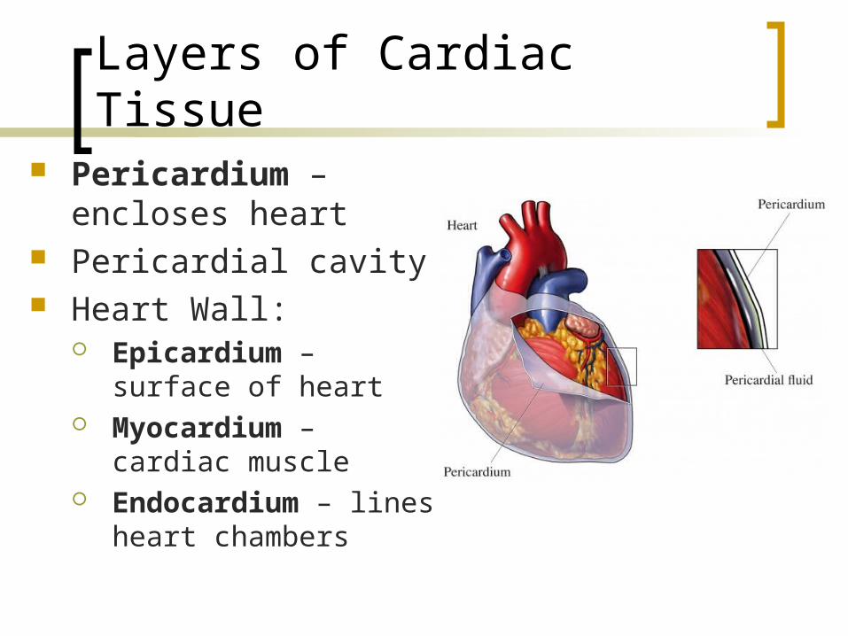
Layers of Cardiac Tissue Pericardium – encloses
heart Pericardial cavity Heart Wall:
Epicardium – surface of heart
Myocardium – cardiac muscle
Endocardium – lines heart chambers
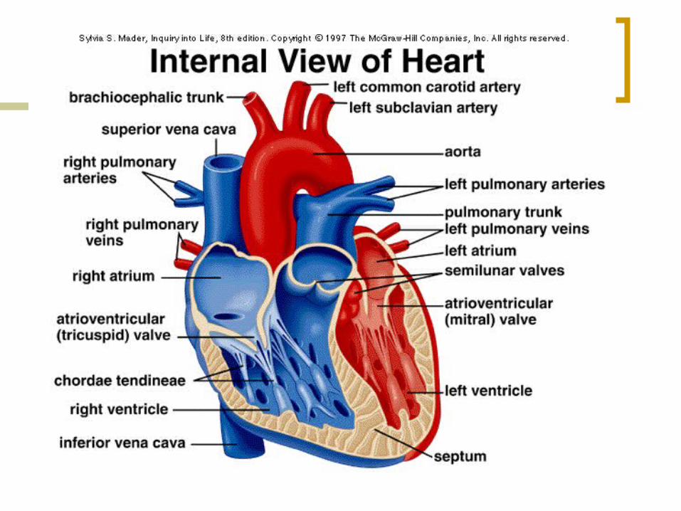

Valves
Atrioventricular (AV valves) – located between the atrial and ventricular chambers on each side Prevent backflow into the atria when
ventricles contract Bicuspid (Mitral) valve – left AV
valve Tricuspid valve – right AV valve
Semilunar valves – guard 2 large arteries leaving the ventricular chambers
Pulmonary Aortic
AV valves open during heart relaxation and closed during ventricle contraction
Semilunar valves closed during heart relaxation and open during ventricle contraction

Circulation
Systemic circulation – from left side of the heart through the body tissues and back to the right side of the heart
Pulmonary circulation – from right side of the heart to the lungs and back to the left side of the heart

Setting the Rhythm of Heart
Heart pumps about 6,000 quarts of blood per day! Setting the Rhythm Can contract spontaneously and
independently Atrial cells 60 times/min Ventricular cells 20-40 times/min ANS nerves can accelerate or decelerate Nodal system – special tissue
Depolarizes in atria-ventricle direction Causes heart to beat as coordinated
unit Sinoatrial (SA) Node – “pacemaker”
found in right atrium Impulse spreads from SA to
Atrioventricullar (AV) node and then the atria contract to Purkinje fibers causing ventricles to contract to eject blood from heart into arteries
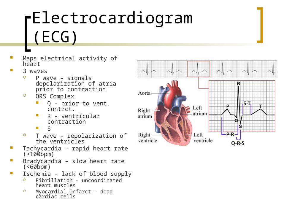
Electrocardiogram (ECG) Maps electrical activity of heart 3 waves
P wave – signals depolarization of atria prior to contraction
QRS Complex Q – prior to vent. contrct. R – ventricular contraction S
T wave – repolarization of the ventricles
Tachycardia – rapid heart rate (>100bpm)
Bradycardia – slow heart rate (<60bpm)
Ischemia – lack of blood supply Fibrillation – uncoordinated heart
muscles Myocardial Infarct – dead cardiac cells
Cardiac Cycle
Cardiac cycle – events of 1 heartbeat 0.8sec Atria contract simultaneously as they relax,
ventricles contract Systole – contraction of ventricles Diastole – relaxation of ventricles
Heart Sounds “lub-dup” pause “lub” – closing of AV valves (long & loud) “dup” – closing of semi-lunar valves (short & sharp) Murmurs – abnormal heart sounds
Vital Signs
Pulse Expansion & recoil of artery Normal 70-76 bpm @ rest
Blood Pressure Force blood exerts against inner walls of bv and keeps
blood flowing continuously 2 measurements (mmHg)
Systolic – pressure in arteries @ peak of ventricular contractions (110 – 140)
Diastolic – pressure when ventricles are relaxing (75 – 80)

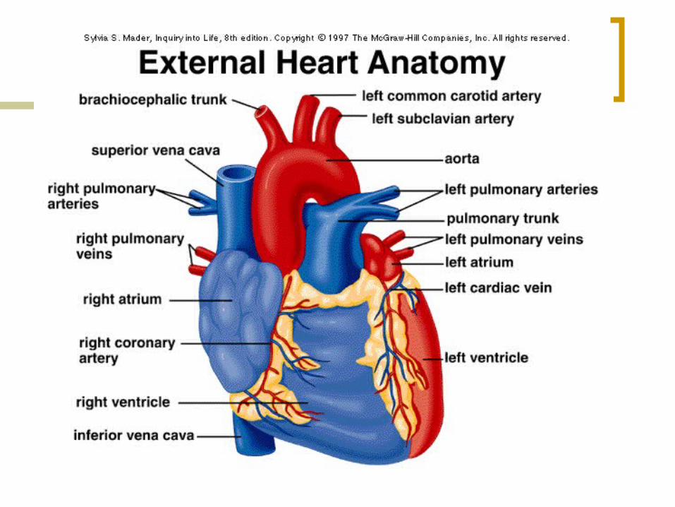
Blood Vessels Blood Vessels – closed
transport system Aorta – blood leaves heart Arteries – thick walls, red
O2 rich Arterioles – branch off of
arteries Capillary beds – found in
tissues, 1 cell layer for easy gas exchange
Venules – drain capillary beds
Veins – drain venules, low pressure, thin walls Have valves to prevent
backflow Vena cava – enters heart

Capillary Exchange
Substances move to/from body cells according to their conc. gradient
Pulmonary gas exchange (see pic)
Systemic gas exchange…
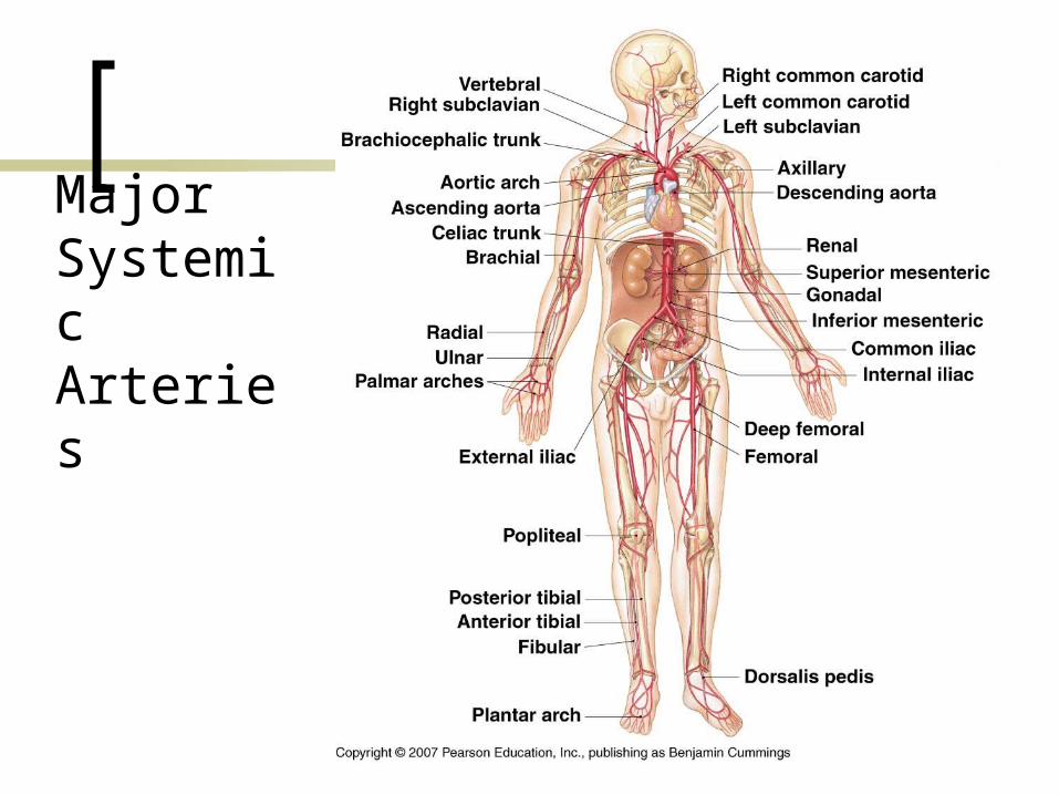
Major Systemic
Arteries

Major Systemic Veins


