48
Kathy Roche, Airway Clinical Nurse Specialist, TSCUH Dorothy Goslin, ENT/Airway CNM2 TSCUH Care of the child with a Tracheostomy

Kathy Roche, Airway Clinical Nurse Specialist, TSCUH
Dorothy Goslin, ENT/Airway CNM2 TSCUH
Care of the child with a Tracheostomy

• Extends from cricoid cartilage to carina
• Length: – 4cm in FT infant – 11-13cm in adult
• Diameter:
– 3.6mm in FT infant – 12-23mm in adult
• Composed of smooth muscle supported by 16-20 horse-shoe shaped cartilagenous rings
Anatomy & Physiology of Trachea:


• 1. Upper Airway Obstruction- causes
– Congenital:-
– Sub-glottic stenosis
– Bilateral Vocal cord paralysis
– Pierre Robin Sequence (Micrognathia/Macroglossia)
– Tracheomalacia/Bronchomalacia
Indications for Paediatric Tracheostomy

• Acquired :-
• Subglottic stenosis
• Trauma
• Inflammation( acute epiglottitis)
• Tumour
Upper Airway Obstruction

• 2. Ventilation insufficiency:-
• - To reduce dead space
• - Prolonged intubation
• - Pulmonary disease
• - Neurological disease
Indications cont’d
• 3. Protection of tracheobronchial tree:-
• -Aspiration risk
• - Neurological disease
• - Head + Neck surgery
• -Trauma
• - Coma (e.g. Post head injury)
Indication’s cont’d

• Neo or Ped
• Single or Double lumen
• Cuffed or Uncuffed
• Fenestrated or Unfenestrated
• PVC or Silicone
• Flextend or Customised tubes
Considerations when choosing a tube
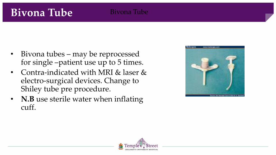
• Bivona tubes – may be reprocessed for single –patient use up to 5 times.
• Contra-indicated with MRI & laser & electro-surgical devices. Change to Shiley tube pre procedure.
• N.B use sterile water when inflating cuff.
Bivona Tube Bivona Tube

• Reduced dead space by approx 70%
• Easier breathing
• Loss of filtering warming & humidification of inspired air
• Reduction or loss of voice
Physiological changes post tracheostomy

• Immediate:-
• Haemorrhage or haematoma
• Anaesthetic problems
• Damage to local structures
- Pneumothorax
- Recurrent Laryngeal Nerve
- Oesophagus
Complications of Paediatric Tracheostomy

• Early:-
• - Tube dislodgement
• - False passage
• - Surgical Emphysema
• - Obstruction of tube or trachea
• - Infection
• - Haemorrhage
Complications cont’d

• Late:-
• -Granulations
• -Suprastomal Collapse
• -Chest Infection
• -Colonization with MRSA/Pseudomonas
• -Dysphagia/Speech delay
• -Laryngotracheal stenosis
Complications cont’d

Granulation tissue around stoma

• Safety- care with handling, not to dislodge tube
• Oxygen/Ambu bag/ MIE circuit, suction machine to accompany child
• Emergency case/bag with child
• Stay sutures labelled?
• Post op Chest X-Ray requested
Transfer from theatre to ICU/HDU

• Why was the tracheostomy performed?
• Is the child intubatable or has (s)he a patent upper airway?
• Is the tube patent?
• Is the tube secure?
Nursing Documentation
• Aims:-
• - Maintain Patent Airway
• - Facilitate removal of pulmonary secretions
• - Provide adequate humidification
• - Care of stoma
• - Early recognition of complications
Nursing Care
• Avoid Accidental Decannulation
• Avoid Tube Blockage
• N.B. Children with a tracheostomy require constant supervision at all times
Nursing Concerns

• Oxygen supply with connection tubing suitable for tracheostomy
• Ambu bag/ MIE circuit if required
• Suction machine with appropriate size suction catheters
• Emergency bag (refer to contents list)
• Emergency tracheostomy surgical pack at bedside (CUH) < 4weeks
• Humidification system-Fisher Pakel/HME
• Daily check list completed
Bedside Equipment
• How do you know that the tube is patent
Tube patency

• secretions visible at the trach port
• Noisy respirations
• Tachycardia/tachypnoea
• Restlessness in babies
• Older child will communicate
• Increased respiratory distress
• Colour changes
• Desaturation
Indications for suctioning

• Hand Washing/Gloves/Goggles
• Duration- 5 seconds per suction ( may suction 2-3 times using same catheter only if secretion loose and clear if secretions thick and coated replace suction catheter per suction)
• Depth – refer to tube measurement (outer diameter of the obturator from entry point to exit point of the tube)
• Pressure – no greater 60-80mmHg <4 weeks, 80-100mmHg 4 weeks up to 3 years,100-120mmHg
>3 years when suctioning.
• Size of suction catheter- double size of tube
Suctioning
• The nose & pharynx provides 75% of normal humidification, which a tracheostomy tube bypasses
• Therefore there is loss of warming, moistening and filtering
• This results in bronchonstriction reducing airflow, a dehydrated respiratory tract, impaired mucociliary function,sputum retention & atelectasis
Humidification
(Woodrow, 2002)

• Heat Moisture Exchange Filters (HME)
• Heated humidity ( all new trache’s )
• Nebulisation
• Hydration
How do we provide humidity?

• Trap warmth + moisture of expired air in cylinder coil.
• Inspired air is warmed + humidified through the coil.
• HME acts as macroscopic particle trap
• Change once wet
Heat + Moisture Exchanger
• Inspect stoma daily
• Vigilant cleaning with gauze and Nacl around stoma
• Wash the skin under the ties with warm soapy water, rinse and pat dry thoroughly
• Apply padding under the ties and skin protector if altered skin integrity evident.
• Trachi dress if required otherwise stoma is left exposed
• Observe for redness, swelling or discharge
• Avoid the use of powder and cotton wool
Stoma and Skin Care

• Common problems:
• Redness
• Dryness
• Excoriation
• Granulation tissue
• Fissures and skin breakdown
Stoma and Skin Care

Skin excoriation

3 Days later

Skin Inflammation


Stoma excoriation

• Marpac tapes
• Older child may consider Velcro tapes
• Tapes changed PRN, wet ties provide a medium for skin breakdown
• Rule of thumb! Tip of finger only should fit between neck + ties with head flexed forward
Securing Tracheostomy Tube

• Preparation
• Position
• Proficiency
• Demeanour/Reassurance
Routine Tracheostomy tube change

• Shiley tubes changed weekly, Bivona tubes changed monthly.
• Always a 2 person procedure except in emergency
• Remember to remove obturator once new tube inserted
Routine tracheostomy tube change

• Early detection is crucial
• Signs to observe for may include the following
failure to pass suction catheter
failure to ventilate
retractions
pallor/cyanosis
desaturation
respiratory arrest
Tube Blockage

• Shout for help
• Cut ties and remove blocked/dislodged tube
• Insert new tube as quickly as possible, and remember to remove introducer
• Assess for breathing
• If child not breathing follow BLS sequence
Management of Blocked Tube

• Early involvement of parents with tape changing, suctioning and tube changes
• Encourage parents to take child off ward once training + Basic Life Support completed
• Phased discharge is important where feasible.
• Rooming in with child prior to discharge (24hrs-48hrs) encourage parents to take child out for hours from the hospital
Parental Education

• The family must develop the skills needed to competently and independently provide tracheostomy care for their child.
• At ward level the staff nurse plays a huge role in encouraging the confidence and the skills in the parents to enable them to take their child home
Parental education cont’d

• Aim is to get child home as soon as possible
• Multidisciplinary approach
• Start discharge process early- identify link nurse/discharge coordinator
• Early involvement of community care team + local hospital
• Ensure family supports are in place prior to discharge
Discharge Planning

• Tracheostomy Info Booklet
• Parent Workbook
• Tracheostomy Awareness Group (TAG)
• Link with another family
• Internet/resources
• Family support is vital, Medical Social Worker, Speech + Language Therapist, Counselling services +/- Psychology + GP
Parental Support

• Regular Microlaryngobronchscopy (MLB) + re-evaluation in OPD as necessary.
• Decannulation- Two methods
• 1. Ward decannulation.
• 2. Surgical decannulation- requires
airway reconstruction .
Follow up + Management

• Decannulation=removal of a tracheostomy tube
• Surgeon will consider decannulation when there is
satisfactory pulmonary function
an adequate airway (at least 50% of
normal)
adequate vocal cord function
Decannulation

• A Microlaryngobronchoscopy (MLB) to ensure that there has been resolution of the original lesion and no evidence of complications i.e. granuloma’s/supra stomal collapse
• Child will be off ventilation a minimum of 2 weeks
Assessment

• Emotional time for child/parents devastating if it fails !
• Ward decannulation is always called a ‘trial’ because there is no guarantee it will be successful
• Consider psychology for older child
Preparation of child/family

• Day 1- Downsize and TOSCA
• Day 2-Tracheostomy tube is capped
• Day 3-Removal of Tracheostomy tube
Ward Decannulation

• Parents are re trained Basic Life Support
• Follow up in ENT Outpatients
• PHN contacted
• Parents hold on to supplies & equipment until review in OPD
• Return to school within 1 week if well
• Swimming once stoma site is sealed
• Reduction or discontinuation of homecare package one week post decanulation
Post Decannulation

?
Questions


