79
| Date post: | 26-Oct-2014 |
| Category: |
Documents |
| Upload: | krishna-sigdel |
| View: | 256 times |
| Download: | 5 times |


INTRODUCTION
Caries activity refers to the increment of active lesion(new and recurrent lesions) over a stated period of time.
Caries activity measures the speed of progression of a carious lesion.
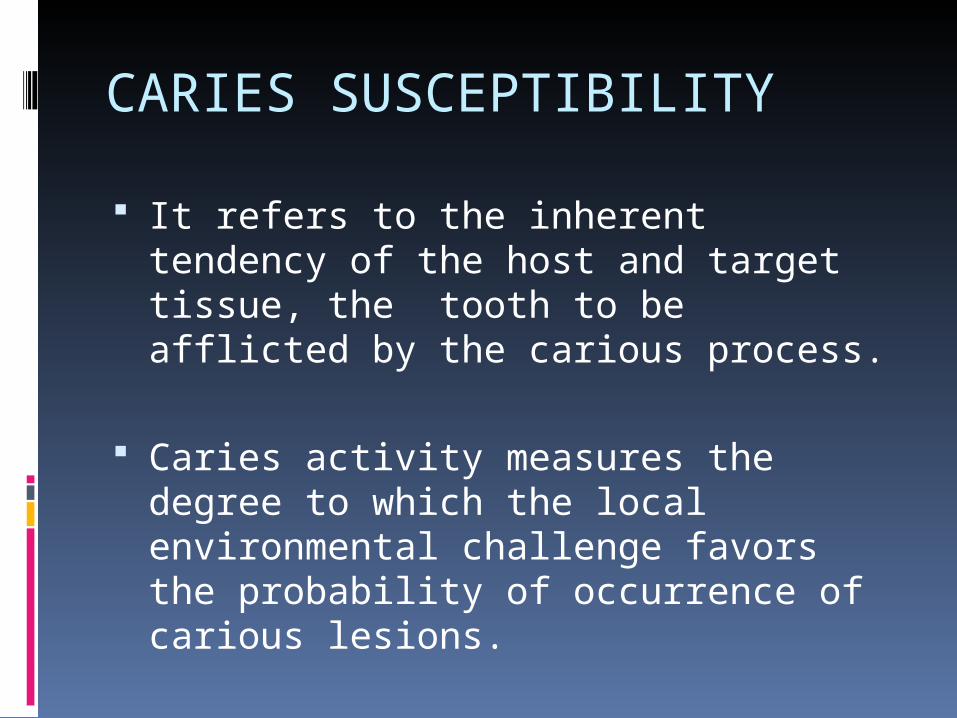
CARIES SUSCEPTIBILITY
It refers to the inherent tendency of the host and target tissue, the tooth to be afflicted by the carious process.
Caries activity measures the degree to which the local environmental challenge favors the probability of occurrence of carious lesions.

Uses of caries activity test
Identify the high-risks groups and individuals. Determine the need for personalized
preventive measures and motivate the individual.
Monitor the effectiveness of the oral health education programs.
Ensure a low level of caries activity before any restoration.
Aid in selection of patient for caries activity.

IDEAL REQUISITES
Should have sound theoretical basis.
Should have maximum correlation with clinical status.
Easy to perform.
Should be inexpensive and simple.

Should be adaptable for chair side.
Results should be accurate and reproducible.
Results should have good validity, reliability and feasibility.

TYPES OF CARIES ACTIVITY TEST MICROBIAL TEST FOR MUTANS
STREPTOCCOCI DETECTION.
MICROBIAL TEST FOR LACTOBACILLI DETECTION.
SNYDER TEST.
ALBAN TEST.
SALIVARY REDUCTASE TEST.

SWAB TEST
FOSDICK CALCIUM DISSOLUTION TEST.
SALIVARY BUFFER CAPACITY TEST.
DIP SLIDE METHOD.
ORA TEST.

MICROBIAL TEST FOR MUTANS STREPTOCCOCI
DETECTION Several methods are available to
measure the level of Mutans Streptococci in saliva, plaque etc.
Types of methods are as follows:1. Laboratory method.2. Chair-side method.3. Survey method.4. Selective method.5.Adherence method.

LABORATORY METHOD
Principle:
Measures the number of forming unit (CFU)/unit volume of
saliva by culturing the plaque samples from discrete
sites(occlusal fissures/proximal areas)for detecting and
quantitating S.Mutans colonies.
Incubation is done on Mitis Salivarius Agar(MSA) selective
streptococcal medium with addition of high concentration
of sucrose (20%) and 0.2 U bacitracin/ml (MSB) which
supresses the growth of non S.Mutans colonies.

LABORATORY METHOD
Saliva or plaque sample is collected.
It is then mixed with proper transport medium and the sample is sent to a microbiological laboratory.
Samples are directly placed the selective agar plates i.e(special petri dishes).
The agar plates are incubated at 37˚C for 48hrs in 95% at 5% CO2 gas mixture.

Mutans colonies on the plates are counted.
The results are expressed as the number of colony forming units per ml of saliva.
Most common type of selective agar plates used is mitis-salivarius-bacitracin agar.

RESULTS FOR LABORATORY METHODCaries Risk Or Activity 100,000 (105) S. mutans cells/ml of
saliva LOW. 100,000 to 500,000 (105 to 5X10⁵)
cells/ml MEDIUM. 500,000 (5X10⁵) or above cells/ml
HIGH

CHAIR-SIDE METHOD
Also known as “Strip mutans test” is based on the ability of streptococci to grow on hard surfaces.
use of the selective broth (High sucrose concentration in combination with bacitracin).
The Dentocult SM-strip mutans kit is used for estimation of mutans streptoccoci in saliva containing test strips, bacitracin discs, test tubes with broth, paraffin for chewing and a standard chart to evaluate the level of mutans after incubation.

The level of mutans streptococci is given as “Class” after comparision with chart.
Indicating low to high , equivalent to 106 mutans CFU per ml in saliva.
The mutans sreptococci colonies as small blue dots but the color can vary from dark blue to pale blue.

DENTOCULT STRIP MUTANS KIT

PROCEDURES
In case of dentocult SM Strip mutans test,bacitracin discs are added to the broth at least for 15 minutes before use.
Then, patient is asked to chew a piece of paraffin for 1 minute.
Plastic strip(test strip) is then taken in the mouth to become contaminated.

The strip is then withdrawn through closed lips, leaving a thin layer of saliva on the strip.
The strip then incubated in the selective broth.
After incubation for 48 hour at 35-37 degree centigrade, the strip with attached colonies is compared with the chart.

PROCEDURE

RESULTS FOR CHAIR SIDE METHOD
Strips for plaque(Square-tipped)
Test strips for stimulatedsaliva (round-tipped)
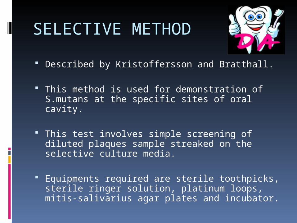
SELECTIVE METHOD
Described by Kristoffersson and Bratthall.
This method is used for demonstration of S.mutans at the specific sites of oral cavity.
This test involves simple screening of diluted plaques sample streaked on the selective culture media.
Equipments required are sterile toothpicks, sterile ringer solution, platinum loops, mitis-salivarius agar plates and incubator.
PROCEDURES
Plaques sample are collected from gingival 3rd of the tooth surface.
Toothpicks are inserted into the approximal spaces.
Then it is placed in Ringer’s solution. It is then shaken homogeneously. Contaminated slides are then pressed directly
against selective agar plates. Incubation is done at 37 degree centigrade for 72
hours. Then sites with or without mutans sreptococci can
be identified.

ADHERENCE METHOD
It categorizes salivary samples based on ability of S. mutans to adhere to glass surfaces when grown in sucrose containing broth.
Equipment required are tube to collect saliva, rack to hold the culture tubes, disposable pipettes, incubator and MSB(mitis- salivarius bacitracin ) broth.

PROCEDURES
Unstimulated saliva(0.1 ml) is inoculated in MSB broth.
Inoculated tubes are set at 60 degree angle and incubated aerobically at 37 degree centigrade for 24 hours.
After the growth has been observed, the supernatent medium is removed and the cells adhering to the glass surface are examined microscopically and scored.

RESULT OF ADHERENCE METHODVALUE INFERENCE
(-) No growth expressed
(+) A few deposits ranging from 1-10.
(++) Scattered deposits of smaller size.
(+++) Numerous minute deposits.

MICROBIAL TEST FOR LACTOBACILLI DETECTION
Introduced by Hadley in 1933.
-Introduced by Hadley in 1933.
Principle:Estimates the number of acidogenic and aciduric bacteria in patient’s saliva by counting the number of colonies appearing in tomato peptone agar plates(pH 5.0) after inoculation with sample of saliva.

PROCEDURES
Before breakfast the patient is asked to chew a paraffin and the saliva accumulated is collected in a sterile container and shaken for 2 mins to mix it.
saliva sample is diluted to 1:10 dilution by pipetting 1 ml of saliva into a 9 ml of sterile saline solution.
It is again diluted to 1:100.

0.4 ml of the diluted solution is spread on the agar plate and incubated at 37˚C for 3-4days.
Lactobacillus colonies are then counted using a colony counter or Quebec counter.
The number of lactobacilli/ml saliva is calculated by multiplying the number of colonies by the dilution factor.
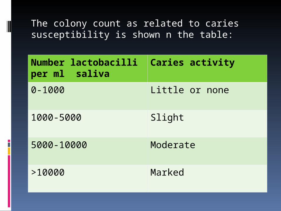
The colony count as related to caries susceptibility is shown n the table:
Number lactobacilli per ml saliva
Caries activity
0-1000 Little or none
1000-5000 Slight
5000-10000 Moderate
>10000 Marked
COLORIMETRIC SNYDER TEST
It measures the ability of salivary micro-organisms to form organic acid from carbohydrate medium.
Principle:
The Snyder test measures the rapidity of acid formation when a sample of stimulated saliva is inoculated into glucose agar adjusted to pH 4.7 to 5 with the help color indicator named bromocresol green.

EQUIPMENTS
Saliva collecting bottles Paraffin A tube of snyder glucose agar
containing bromocresol green Pipettes Incubator
PROCEDURE:
• Paraffin stimulated saliva is collected before breakfast.
• A tube of Snyder glucose agar is melted, cooled at 50˚C, shaken and 0.2 ml of saliva is pipetted in the tube.
• The agar is solidified and incubated at 37˚C.
• Amount of acid produced is detected by pH indicator and compared to an uninoculated control tube after 24, 48 & 72 hrs of incubation.

RESULT OF SNYDER TESTTime Time Time
24 hours 48 hours 72 hours
Color Yellow Yellow Yellow
Caries activity Marked activity
Definite activity
Limited activity
Color Green Green Green
Caries activity Continue test Continue test inactive

ALBAN TEST
Is a simplified version of the Snyder test.Main features:1. Softer medium are used that diffuse saliva
and acids without the necessity of melting the medium.
2. The patient simply has to spit directly into the tubes that contain the medium.

Materials required to prepare the Alban test medium:
1.Snyder agar2.A small scale to measure 60 grams3.A 2 litre Pyrex glass to melt the medium4.A funnel to dispense the medium into
test tubes5.Hundred 16mm test tubes with screw
caps.

PROCEDURE
• 60 gms of Snyder test agar is placed in 1 litre water and the suspension is boiled over a low flame.
• The melted agar is then distributed among the tubes. About 5ml/tube.
• These tubes are autoclave for 15 mins, cooled and stored in a refrigerator.

• 2 tubes of Alban medium are taken out and the patient is asked to expectorate saliva directly into the tubes.
• The tubes are labelled and incubated at 98.6˚F up to 4 days.
• The tubes are observed daily for the colour change from blue to green to definite yellow with decrease in pH.

RESULT OF ALBAN TEST
Color change Score
No color change
¾
Beginning of color change
+
One half color change
++
Three fourths color change
+++
Total color change to yellow
++++

SALIVARY REDUCTASE TEST
This test measures the activity of the reductase enzyme
present in the salivary bacteria.
Action:
This test measures the rate at which the indicator molecule Diazoresorcinol changes from blue to red to colourless on reduction by the mixed salivary flora.

PROCEDURE
The patient is asked to chew a paraffin and the saliva accumulated is collected directly into the collection tube(5ml).
Then sample collected is mixed with a fixed amount of diazoresorcinol.
Change in colour after 30 seconds and after 15 mins is taken as a measure of caries activity.

RESULT OF SALIVARY REDUCTASE TEST
COLOR TIME SCORE CARIES ACTIVITY
BLUE 15 minute 1 Non conductive
ORCHID 15 minute 2 Slightly conductive
RED 15 3 Moderately conductive
RED Immediately
4 Highly conductive
PINK OR WHITE
Immediately
5 Extremely conductive

SWAB TEST
• Grainger et al developed this test in 1965• Principle is similar to that of Snyder test.
Procedure:• Buccal surfaces of the teeth are swabbed with a
cotton applicator.
• The sample collected is incubated in the medium.
• The change in the pH after 48 hrs incubation period is read on a pH meter or using a color comparator.

RESULT OF SWAB TEST
pH Caries activity
4.1 Marked caries activity
4.2-4.4 Active
4.5-4.6 Slightly active
Over 4.6 Caries active

FOSDICK CALCIUM DISSOLUTION TESTPrinciple:
Measures the milligrams of powdered enamel dissolved in 4
hours by acid formed when the patient’s saliva is mixed with
glucose and powdered enamel.
Equipment :powdered human enamel, saliva collection bottles, sterile host tubes, test tube agitation equipment, and equipment for determining the calcium content of the saliva.

PROCEDURE• The patient is asked to chew paraffin wax and 25 ml
of the saliva is collected and part of it is analysed for calcium content.
• The remaining saliva is placed in an 8 inch sterile test tube with about 0.1 gm. of powdered human enamel.
• The tube is sealed and shaken for 4 hours at body temperature, after which it is again analysed for calcium content.
The use of paraffin to stimulate saliva increases the
concentration of the glucose by 5%. The amount of enamel
dissolution increases as the caries activity increases.

SALIVARY BUFFER CAPACITY TEST
Principle:
It measures the number of millimetres of acid required to lower the pH of saliva through an arbitrary pH interval, such as from pH 7.0 to
6.0 or the amount of acid or base necessary to bring colour indicators to their end point.

EQUIPMENT
1.pH meter 2.Titration equipment3.0.05 N lactic acid and 0.05 base 4.Paraffin5.Sterile glass jars containing oil.

LABORATORY METHOD
• 10 ml of stimulated saliva is collected under oil after 1 hour of eating.
• 5 ml is then measured in the beaker.
• pH of the saliva is adjusted to 7.0 by addition of lactic acid or base using a pH meter in room temperature.
• The level of lactic acid in graduated cylinder is re-recorded.
• pH of the sample is brought down to 6.0 by adding lactic acid.
• The amount (no of millimetres) of lactic acid needed to reduce the pH from 7 to 6 is the measure of buffer capacity.

CHAIRSIDE METHOD
This system differentiates between buffer capacities.
This was developed by Ericson and Bratthall.
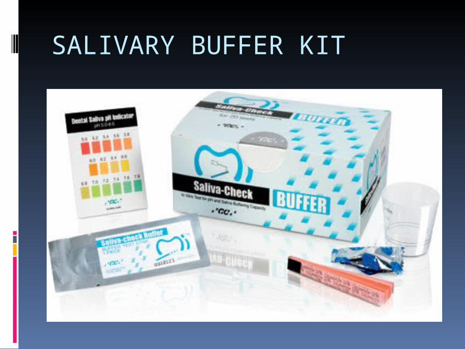
SALIVARY BUFFER KIT

PROCEDURES
A new and simplified method to estimate the salivary buffer capacity .
Dentobuff Strip, consists of a pH indicator paper that has been impregnated with acid.
A small volume of saliva is added to the strip and after 5 min the color of the strip is compared with a chart.
The colors have been chosen to indicate low, medium, or good buffer capacity.

RESULT OF BUFFER CAPACITY TESTBUFFER CAPACITY
FINAL pH VALUE
COLOR CHANGE
Low buffer capacity
4.0 or less Yellow color
Intermediate buffer capacity
4.5 to 5.5 Green color
Normal buffer capacity
6.0 or more Blue color

ORA TESTDeveloped by Rosenberg et al in 1989 for estimating oral microbial level.
Principle: Based on the oxygen depletion by
microorganisms in expectorated milk samples. In normal conditions the bacterial enzyme, aerobic dehydrogenase transfers electrons or protons to oxygen.
Once oxygen gets utilized by the aerobic organisms, methylene blue acts as an electron acceptor and gets reduced to leucomethylene blue. This reflects the metabolic activity of the aerobic organisms.

EQUIPMENT
1. Sterile beakers2. Sterilized milk3. Screw cap test tubes4. 0.1% aqueous solution of methylene
blue5. 10 ml disposable syringes6. Pipette7. Mirror8. Stop watch and test tube
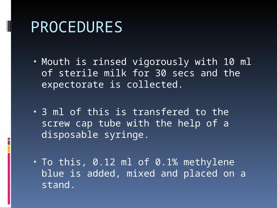
PROCEDURES
• Mouth is rinsed vigorously with 10 ml of sterile milk for 30 secs and the expectorate is collected.
• 3 ml of this is transfered to the screw cap tube with the help of a disposable syringe.
• To this, 0.12 ml of 0.1% methylene blue is added, mixed and placed on a stand.

• The tubes are observed every 10 mins for any color change at the bottom using a mirror.
• The time taken for the initiation of color change within 6 mm ring is recorded.
Higher the infection, lesser time taken for the color
change reflecting higher oral microbial levels.

DIP SLIDE METHOD FOR S.MUTANS COUNTDevised for the estimation of S. mutans levels in saliva.
Procedure:
• Saliva stimulated with paraffin is poured on a special plastic slide that is coated with MSA (Mitis salivarious agar) containing 20% sucrose.
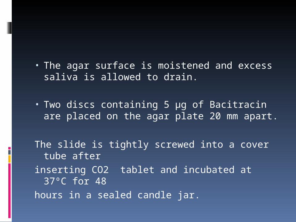
• The agar surface is moistened and excess saliva is allowed to drain.
• Two discs containing 5 µg of Bacitracin are placed on the agar plate 20 mm apart.
The slide is tightly screwed into a cover tube after
inserting CO2 tablet and incubated at 37ºC for 48
hours in a sealed candle jar.
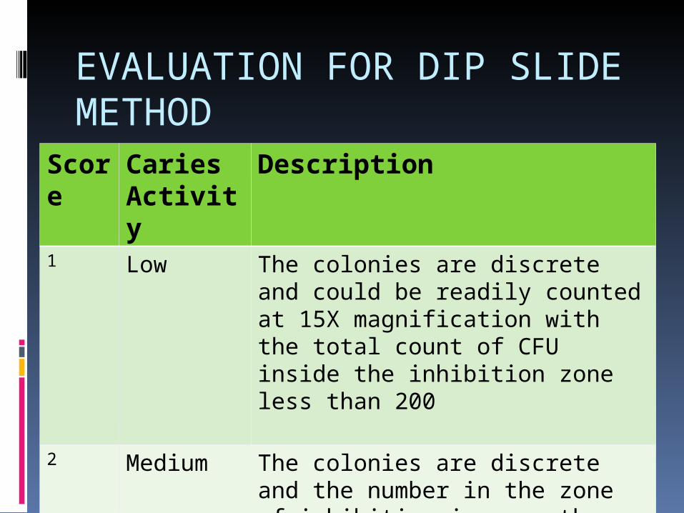
EVALUATION FOR DIP SLIDE METHOD
Score
Caries Activity
Description
1 Low The colonies are discrete and could be readily counted at 15X magnification with the total count of CFU inside the inhibition zone less than 200
2 Medium The colonies are discrete and the number in the zone of inhibition is more than 200 at 32X magnification

Score Caries Activity
Description
3 High The colonies are tiny and almost completely or totally cover the inhibition zone with the number of colonies uncountable even with 32X magnification.

CARIOGRAM It is a computer program showing a
graphical picture that illustrates a possible overall caries risk scenario.
It expresses to what extent different etiological factors of caries affect caries risk.
The cariogram identifies the caries risk factors for the individual and provides examples of preventive and treatment strategies to the clinician.

HOW IS A CARIOGRAM CREATED? The patient is examined and the data is
collected for some factors of direct relevance for caries including bacteria,diet and susceptibilty related factors.
The factors/variables are given score according to a predetermined scale and entered in a computer program.
The program presents a pie diagram which represents a following sectors.

1. The dark blue sector (DIET) is based on a combination of diet contents and diet frequency.
2. The red sector (BACTERIA) is based on the amount of plaque and mutans streptoccoci.

3. The light blue sector (SUSCEPTIBILITY) is based on a combination of fluoride program, saliva secretion and salivary buffer capacity.
4. The yellow sector (CIRCUMSTANCES) is based on a combination of caries experience and related diseases.

5. The green sector shows an estimation of (CHANCE OF AVOIDING CARIES). When the chance of avoiding caries is high, the caries risk is small and vice-versa.

CARIOGRAM

CONCLUSION
Till date no single caries activity test is sufficiently accurate or reliable to serve as a predictor of potential caries activity for the individual patient.
Since, caries activity tests measure a single parameter such as acid production, colony counts etc, the best predictors of caries activity would therefore result from the combined use of several selected tests.

REFERENCE
Nikhil Marwah 2nd edition. ‘Textbook Of Pediatric Dentistry’
Shova Tandon 2nd edition.’Textbook Of Pedodontics’
Soben Peter 4th edition. ‘ Essentials Of Preventive And Community Dentistry’
Internet sources

WHAT ARE THE USES OF CARIES ACTIVITY TEST?

Identify the high-risks groups and individuals.
Determine the need for personalized preventive measures and motivate the individual.
Monitor the effectiveness of the oral health education programs.
Ensure a low level of caries activity before any restoration.
Aid in selection of patient for caries activity.

WHAT IS A CARIOGRAM?

It is a computer program showing a graphical picture that illustrates a possible overall caries risk scenario.

IDENTIFY THE PICTURE.

WHAT IS THE MOST COMMAN TYPE OF AGAR USED FOR DETECTION OF MUTANS STREPTOCOCCI?

mitis-salivarius-bacitracin agar(MSB)


