Date post: | 14-May-2018 |
Category: |
Documents |
Upload: | vuongtuyen |
View: | 215 times |
Download: | 1 times |
0
Ph.D. Thesis
CAROTENOIDS ASSIST IN ASSEMBLY AND FUNCTIONS OF
PHOTOSYNTHETIC COMPLEXES IN CYANOBACTERIA
Özge Sözer
Supervisors: Dr. Zoltán Gombos & Dr. Mihály Kis
University of Szeged
Biological Research Centre
Hungarian Academy of Sciences Szeged
Szeged
2011
1
TABLE OF CONTENTS
0.0 PROLOG…………………………………………………………………………………..5
1.0 INTRODUCTION ............................................................................................................... 6
1.1 Photosynthesis.................................................................................................................. 6
1.1.1 Light Harvesting Antenna System ............................................................................. 7
1.1.2 Major photosynthetic complexes in thylakoid membrane ......................................... 8
1.1.2.1 Photosystem II ........................................................................................................ 8
1.1.2.2 Cytochrome b6f ..................................................................................................... 11
1.1.2.3 Photosystem I ....................................................................................................... 11
1.2 Carotenoids .................................................................................................................... 13
1.2.1 Chemical structure and some physiological properties of carotenoids……………13
1.2.2 Functional roles of carotenoids in photosynthesis ................................................... 15
1.2.2.1 Light harvesting .................................................................................................... 15
1.2.2.2 Photoprotection ..................................................................................................... 16
1.2.2.3 Membrane structure organization ......................................................................... 17
1.3 Model Organism: Cyanobacterium Synechocystis sp. PCC 6803 ................................ 18
1.4 Biosynthetic pathway of carotenoids in Synechocystis .................................................. 18
1.5 Localization of carotenoids in photosynthetic complexes ............................................. 21
1.5.1 Localization of β-carotene molecules in photosystem II ......................................... 21
1.5.2 Localization of a β-carotene molecule in cytochrome b6f ....................................... 23
1.5.3 Localization of β-carotene molecules in photosystem I .......................................... 23
1.6 Phosphatidylglycerol in photosynthesis ......................................................................... 24
1.7 Earlier studies on carotenid mutants in cyanobacteria ................................................... 25
2.0 AIMS.................................................................................................................................. 28
3.0 MATERIALS AND METHODS ....................................................................................... 29
3.1 Construction of Synechocystis ΔcrtH/B mutant ............................................................. 29
3.2 Organisms and growth conditions ................................................................................. 29
3.3 Chlorophyll content analysis.......................................................................................... 30
3.4 Carotenoid content analysis ........................................................................................... 30
3.5 Absorption spectroscopy ................................................................................................ 31
3.6 Low-temperature (77 K) fluorescence spectroscopy ..................................................... 31
3.7 RNA isolation and Northern blot analysis ..................................................................... 31
3.8 Radioactive labeling of the cells .................................................................................... 31
3.9 Thylakoid membrane isolation ...................................................................................... 32
3.10 Protein analyses ........................................................................................................... 32
3.11 Oxygen-evolving activity measurements ..................................................................... 33
2
3.12 Chl a fluorescence efficiency (Fv/Fm) and the maximum changes in P700 signal (Pm-
Po), and oxidation-reduction measurements of P700 kinetics ............................................. 34
4.0 RESULTS .......................................................................................................................... 35
4.1 Generation of carotenoid-less ΔcrtH/B strain in Synechocystis..................................... 35
4.2 Physiological consequences of the carotenoid deficiency ............................................. 36
4.2.1 Growth rate and light sensitivity ............................................................................. 36
4.3 Structural consequences of carotenoid deficiency ......................................................... 40
4.3.1 Level of the large protein subunits of photosynthetic complexes ........................... 40
4.3.2 Assembly of the photosynthetic complexes and their large protein subunits .......... 41
4.3.3 Stabilization of the small protein subunits binding to the large protein subunits in
photosystem II .................................................................................................................. 43
4.3.4 Assembly of the functioning photosystem II core complex .................................... 45
4.3.5 Level of genes encoding PSII large protein subunits .............................................. 47
4.4 Functional consequences of carotenoid deficiency........................................................ 48
4.4.1 Oxygen-evolving activity of the cells ...................................................................... 48
4.4.2 Maximal photosystem II efficiency in dark adapted state (Fv/Fm) ......................... 48
4.4.3 Maximal redox changes in P700 signal in dark adapted state (Pm-Po) .................. 49
4.4.4 Oxidation-reduction kinetics of P700 ...................................................................... 51
4.5 Phosphatidylglycerol (PG) depletion induces an increase in myxoxanthophyll and
echinenone biosynthetic activities in Synechocystis PCC 6803 cells .................................. 52
5.0 DISCUSSION .................................................................................................................... 57
5.1 Structural and functional consequences of the carotenoid deficiency in the
Synechocystis ....................................................................................................................... 57
5.2 Phosphatidylglycerol (PG) depletion induces an increase in myxoxanthophyll and
echinenone biosynthetic activities in Synechocystis PCC 6803 cells .................................. 65
6.0 CONCLUSIONS................................................................................................................ 68
AKNOWLEDGEMENTS........................................................................................................ 69
REFERENCES ........................................................................................................................ 70
ÖSSZEFOGLALÓ...................................................................................................................80
SUMMARY………………………………………………………………………………….85
PUBLICATIONS…………………………………………………………………………….90
3
ABREVIATIONS
Absmax Absorbance maximum
Chl Chlorophyll
CP43 and CP47 Photosystem II antenna chlorophyll-protein subunits
Cyt b6f Cytochrome b6f complex
D1 and D2 Photosystem II core complex protein subunits
DCMU 3-(3,4-dichlorophenyl)-1,1-dimethylurea
Fo Fluorescence yield of open PSII RCs in dark-adapted state
Fm Maximum fluorescence yield of closed PSII RCs in dark-
adapted state
Fv Maximum fluorescence change in dark-adapted state (Fm-Fo)
iD1 Intermediate form of D1 protein subunit after removal of part
of the C-terminal extension
HPLC High performance liquid chromatography
LAHG Light-activated heterotrophic growth
NPQ Non-photochemical quenching
OD Optical density
OEC Oxygen-evolving complex
PBS Phycobilisome
pD1 Precursor form of D1 protein subunit with complete C-terminal
extension
PG Phosphatidylglycerol
pgsA The mutant deficient in phosphatidylglycerol (PG) synthesis
Pm Maximum P700 signal in dark-adapted state
Po Minimum P700 signal in dark-adapted state
PsaA and PsaB Photosystem I core complex protein subunits
P680 Primary electron donor of PSII reaction center
P700 Primary electron donor of PSI reaction center
RC47 Intermediate PSII complex lacking inner antennae
CP43 protein subunit
RCC(1) and RCC(2) Monomeric and dimeric PSII complexes, respectively
PSI(1) and PSI(3) Monomeric and trimeric PSI complexes, respectively
4
RC Reaction center
RCa Intermediate PSII complex containing D1 (or iD1), D2 and
Cyt b-559 but lacking CP47 and CP43
ROS Reactive oxygen species
2D-BN/SDS-PAGE Two-dimensional blue native/sodium dodecyl sulfate
polyacrylamide gel electrophoresis
ΔcrtH The mutant deficient in cis- to trans- carotene isomerase
encoded by crtH gene in Synechocystis
ΔcrtH/B The mutant deficient in the phytoene synthase encoded by crtB
gene
5
0.0 PROLOG
Cyanobacteria are a fascinating and versatile group of bacteria of immense biological
importance. In earth history cyanobacteria as nitrogen and carbon-fixing organisms have a
special position and they play a significant role in Earth’s nitrogen and carbon cycles.
Cyanobacteria are able to perform nitrogen and carbon fixation, which are often competing
metabolic processes in the same cell. Moreover, their ancestors are the first oxygen-evolving
organisms, which were responsible for the oxygen in the atmosphere due to their
photosynthetic activity, which was invented 2.8 billion years ago. Cyanobacteria even
nowadays contribute up to 30% of the yearly oxygen production on Earth.
Carotenoids are ubiquitous compounds playing a role in a range of special processes.
Thus, they are regarded as essential compounds for life mainly due to their various roles in
photosynthesis. During the last couple of year interest in these pigments get into the focus of
plant biology. Recently, various genes involved in carotenoid biosynthesis of several
bacterial organisms have been indentified, which offers new opportunities for the genetic
engineering of carotenoid synthesis in vivo.
Complete genome sequences and the transformability of several cyanobacterial strains
allow us to generate various mutants and study the importance of carotenoids in
photosynthetic processes. We aimed to generate a carotenoid deficient mutant in order to
understand the structural and functional roles of carotenoids in photosynthesis.
6
1.0 INTRODUCTION
1.1 Photosynthesis
Photosynthesis is a process by which the energy of sunlight is converted into chemical
energy in higher plants, algae and cyanobacteria. Photosystem II (PSII), photosystem I (PSI),
cytochrome b6f, (Cyt b6f), and ATP-synthase are the four major protein complexes embedded
in the thylakoid membranes. Both PSI and PSII reaction centers (RCs), which carry out light-
driven electron transfer, are surrounded by light-harvesting antenna systems, which consist of
protein-pigment complexes. Both photosystems are coupled functionally by a plastoquinone
pool, the Cyt b6f complex, and plastocyanin or cytochrome c6 (Fig. 1).
Figure 1: Major photosynthetic complexes of thylakoid membrane of cyanobacteria
Major complexes (Photosystem II, cytochrome b6f, photosystem I and ATPsynthase) and
light driven electron transfer chain across the membrane. The figure was adapted from Dr.
Donald A. Bryant (1994) and modified by Dr. László Kovács.
7
1.1.1 Light Harvesting Antenna System
Photosynthetic organisms have additional peripheral light harvesting antenna
complexes containing different pigments, which are specifically associated with proteins in a
unique structure of photosynthetic apparatus in order to overcome the light limited conditions
by extending the spectral range of absorbance. The large protein antenna complexes of higher
plants contain chlorophyll a (Chl a), chlorophyll b and xanthophyll pigments. Some marine
organisms, such as diatoms and brown algae utilize xanthophylls, Chl a, and chlorophyll c
within a single light harvesting antenna complex. Red algae and cyanobacteria have
phycobilisomes. Although the various peripheral light-harvesting antennas differ significantly
among the photosynthetic organisms, their common functions are to absorb light energy and
to control dissipation of excess excitation energy under light stress, and to regulate energy
flow between the two photosystems.
In cyanobacteria, there are specific light harvesting antenna complexes,
phycobilisomes (PBSs), which consist of phycobiliproteins (Fig. 1). In the cyanobacterium
Synechocystis sp. PCC 6803 (hereafter Synechocystis) there are two blue-pigmented
phycobiliproteins: phycocyanin (Absmax 620 nm) and allophycocyanin (Absmax 650 nm).
Some cyanobacteria also have phycoerythrin, a red-pigmented phycobiliprotein (Absmax 560
nm). The PBSs contain core and rod substructures. Phycoerythrin and phycocyanin form the
rods, which efficiently absorb and transfer excitation energy to the PBS core, which consist
predominantly of allophycocyanin (Liu et al. 2005). The PBS core also contains terminal
emitters (Absmax 683 nm), which tunnel light energy from the PBSs to the RCs. The exact
interaction between the PBS and the photosystems is still under debate. It is known that a
PBS can also be decoupled from the Photosystems, leading to long-lived excitation
(Bittersmann and Vermaas 1991). PBS excitation energy distribution between the two
photosystems is regulated, in order to optimize photosynthetic performance (Mullineaux
2008). PBSs are also subject to carotene-triggered dissipation of absorbed energy as heat.
The phenomenon is known as non-photochemical quenching (NPQ) in cyanobacteria
(Rakhimberdieva et al. 2010)
8
1.1.2 Major photosynthetic complexes in thylakoid membrane
1.1.2.1 Photosystem II
The PSII complex has been conserved among cyanobacteria, algae and higher plants.
The X-ray crystallographic structure of the monomeric PSII complex from
Thermosynechococcus elongatus (T. elongatus) consists of 17 intrinsic and 3 extrinsic protein
subunits, 35 Chl a, 11 β-carotene, 2 haem, and one non-haem iron molecules, two calcium
ions, at least 25 integral lipids, 1-2 chloride ion(s), 3 plastoquinones and CaMn4 cluster
(Ferreira et al. 2004, Loll et al. 2005, Guskov et al. 2009, Guskov et al. 2010).
The membrane intrinsic part of monomeric PSII consists of the core complex protein
subunits D1 and D2, each containing 5 transmembrane α-helices which bind Chl, pheophytin
and plastoquinone co-factors (Rappaport and Diner 2008). In addition, there are intrinsic
antenna protein subunits CP47 and CP43, each containing 6 transmembrane α-helices, which
bind 26 Chl a and 9 β-carotene molecules (Loll et al. 2005). These four large protein subunits
are surrounded by 13 low-mass small protein subunits on the periphery of the PSII.
Cytochrome b-559 (Cyt b-559) consists of two transmembrane α-helices; PsbE and PsbF
represents two of those small protein subunits. Cyt b-559 is able to accumulate in the
membrane in the absence of D1 and D2 (Komenda et al. 2004). It seems to act as a nucleation
factor to initiate PSII assembly: the formation of the first Cyt b-559-D2 subcomplex (Fig. 2)
(Komenda et al. 2004, Komenda et al. 2008). After the integration of D1 and PsbI,
(Dobakova et al. 2007), an intermediate PSII complex, RCa is formed (Fig. 2) (Komenda et
al. 2008). In the absence of CP47, CP43 is unable to attach stably to the subcomplex
(Komenda et al. 2008), although in the absence of CP43, CP47 is able to attach to the
subcomplex and forms the next intermediate PSII complex, RC47 (Fig 2) (Komenda et al.
2004). RC47 is unable to oxidize water but is still able to drive oxidation of redox-active
tyrosine, TyrZ, in D1. The subsequent attachment of CP43 allows the formation of
monomeric PSII complex (RCC(1)) (Fig. 2), which is the starting point for the light-driven
assembly of the CaMn4 cluster together with the attachment of the extrinsic protein subunits
PsbO, PsbV and PsbU. They are located at the lumenal side of the core complex assembly
and contribute to the stability of the CaMn4 cluster (Fig. 2) (Roose et al. 2007, Enami et al.
2008). Finally two monomeric PSII complexes form a dimeric PSII complex (RCC(2)) (Fig.
2). Both monomeric and dimeric PSII complexes can be formed in whole cells and isolated
9
thylakoid membranes of Synechocystis and T. elongatus with dimeric form being the more
active form (Nowaczyk et al. 2006).
Figure 2: Assembly of PSII complex in Synechocystis.
The small protein subunits PsbE, PsbF, PsbH, PsbI and PsbK and the extrinsic protein
subunits PsbO, PsbU and PsbV are designated by the upper case letter. Cytochrome b-559
(cyt b-559) is composed of a heterodimer of PsbE and PsbF. Types of PSII complexes: RCa,
intermediate PSII complex containing either mature D1, intermediate D1 (iD1), or precursor
D1 (pD1) but lacking CP43 and CP47; RC47, intermediate PSII complex lacking CP43;
RCC(1), monomeric PSII complex; RCC(2), dimeric PSII complex. For clarity, assembly of
the cofactors and many of the small protein subunits is not included. The figure was adapted
from Nixon et al. (2010).
During biogenesis of PSII, D1, which is synthesized in most organisms as a precursor
protein, pD1, with a C-terminal extension (16 amino acid extension), must be cleaved to
allow the assembly of a functional CaMn4 cluster. During C-terminal extension processing,
the intermediate of D1, designated iD1 can be found (8 amino acid extension), (Komenda et
al. 2007). The iD1 is found mainly in PSII complexes in vivo (Komenda et al. 2004). C-
terminal extension is not required for assembly of the oxygen-evolving complex (OEC) of
PSII (Satoh and Yamamoto 2007). However, mutants either lacking the extension or
containing modified C-terminal extension are in general much more susceptible to
photoinhibition (Ivleva et al. 2000, Kuvikova et al. 2005).
10
Figure 3: Electron transport chain in PSII RC
Major participants: P680, (dark green); pheophytin, (light green); QA, QB, and QC (grey);
CaMn4 cluster (yellow) and tyrosine Z (red). The figure was created by the software
Discovery Studio Visualizer (DSV) (http://accelrys.com/products/discovery-
studio/visualization) and the PDB file 3BZ1 (Guskov et al. 2009).
Light energy captured by the light harvesting antenna complexes is transferred to PSII
RC. In the RC, light-induced charge separation takes place leading to the oxidation of P680
(P680+), which is a heterodimeric Chl a pair. The electron released from excited P680
(P680*) travels along the redox-active cofactors of the electron transfer chain. Pheophytin is
the first electron acceptor. Then two plastoquinone molecules QA and QB receive electrons
from pheophytin. The tightly bound QA acts as electron transmitter and the mobile QB is the
substrate of the quinone reductase part of PSII complex (Fig. 3). An additional plastoquinone
molecule (QC) with unresolved function has been located next to the QB binding site. After
two charge separation events, the doubly reduced QB (QB2-
) binds two protons from the
stromal side of the membrane yielding a fully reduced plastohydroquinone, PQH2.
P680+ has an exceptionally strong oxidizing power and is able to abstract electrons
via redox active tyrosine Z from the OEC, a heteronuclear CaMn4 cluster located at the
lumenal side of PSII (Fig. 3). After the accumulation of four positive charges, water is
oxidized to molecular oxygen. The oxidation of two water molecules in PSII results in the
11
release of four protons in the lumen. This contributes to the formation of the electrochemical
gradient that drives ATP synthesis.
1.1.2.2 Cytochrome b6f
The Cyt b6f complex, which is another large membrane-bound protein complex, lies
between PSII and PSI complexes in the thylakoid membrane. In general the native dimeric
form consist of eight protein subunits and several cofactors legated to the proteins (Kurisu et
al. 2003). Four of these are considered the large protein subunits and include Cyt f, the Rieske
iron-sulfur protein, Cyt b6, and subunit IV.
Electrons are transferred through mobile plastoquinone (PQH2) molecules from PSII
to the Cyt b6f complex. Cyt b6f donates the electrons to one of the lumenal electron carrier
proteins, plastocyanin, (or in some cases to Cyt c6), which carries the electron, to PSI.
Further, the reoxidation of two PQH2 by Cyt b6f also results in the release of an additional
four protons in the lumen, thereby contributing to the formation of the electrochemical
gradient that drives ATP synthesis.
1.1.2.3 Photosystem I
X-ray crystal structure of the monomeric PSI complex from T. elongatus exhibits 12
protein subunits which are bound to 127 cofactors performing light capturing and electron
transfer (Jordan et al. 2001, Fromme et al. 2001, Grotjohann and Fromme 2005, El-
Mohsnawy et al. 2010). These cofactors include 96 Chl a and 22 carotenoids, two
phylloquinones, three iron-sulfur cluster and four lipid molecules. The central part of the core
complex is formed by the heterodimer of the two large protein subunits PsaA and PsaB,
comprising 22 transmembrane helices, located in the center of the monomeric PSI complex.
They coordinate most of the Chl a, carotenoids and the redox cofactors of electron transport
(Amunts and Nelson 2009). PSI exists in vivo in monomeric and trimeric forms. The
trimeric PSI is the most abundant in the thylakoid membrane of T. elongatus and the
formation of trimeric form is essential for the growth of the cells at low light intensity
(Grotjohann and Fromme 2005). Trimeric PSI form also plays an important role in the
assembly and structural integrity of PSI (Grotjohann and Fromme 2005). Interaction of
antenna Chl a of the monomeric forms within the trimeric form leads to a larger amount of
long-wavelength Chl a, resulting in a higher photochemical activity of the trimeric form
under red or far-red illumination. The dynamic equilibrium between monomeric and trimeric
12
forms in the thylakoid membrane may indicate a transient monomeric population in the
course of biogenesis and could also be the basis for short-term adaptation of the cell to
changing environmental conditions (El-Mohsnawy et al. 2010).
PSI catalyzes the light driven electron transfer from plastocyanin, which is located on
the lumenal side of the thylakoid membrane, to ferredoxin, which is located on the stromal
side of the membrane. In some cyanobacteria and green algae, Cyt c6 can replace
plastocyanin as an electron donor to PSI. Excitation energy captured by antenna pigments is
funneled to P700, a heterodimer of Chl a pair in PSI RC where the charge separation takes
place. The electron is transferred from P700 stepwise to A and A0 (Chl a molecules), A1 (a
phylloquinone molecule) and from there subsequently to the iron-sulfur clusters, FX, FA and
FB. The latter mediates electron transfer to ferredoxin, which act as soluble electron carrier.
Ferredoxin transfers the electron to the ferredoxin NADP+-reductase enzyme (FNR), which
then finally reduces NADP+ to NADPH (Fig. 4). To complete the cycle, P700
+ is re-reduced.
Figure 4: Electron transport chain in PSI RC.
Major participants: P700, grey; A and A0, red and green, respectively; A1, pale green; FX, FA
and FB (magenta). The figure was created by the software DSV and the PDB file 1JB0
(Jordan et al. 2001).
13
1.2 Carotenoids
Carotenoids represent the most fascinating, abundant and widely distributed classes of
natural pigments. From photosynthetic organisms to anoxygenic bacteria through
cyanobacteria, algae and higher plants (Frank and Cogdell 1996), as well as numerous non-
photosynthetic bacteria and fungi (Goodwin 1980) produce carotenoid pigments. Until now,
more than 700 naturally-occurring carotenoid pigments have been identified (Britton et al.
2004).
1.2.1 Chemical structure and some physiological properties of carotenoids
Their structures are consistent with the assumption that they are produced by joining
together isoprene units, usually in a "head to tail" fashion. Isoprene is the common name for
2-methyl-1,3-butadiene. Most naturally occurring carotenoid pigments are tetraterpens (eight
isoprene units) (Fig. 5). A very important property of carotenoids, both from spectroscopic
and structural points of view, is the presence of double bonds in conjugated system which
forms the chromophore responsible for the molecule color. A conjugated double bond system
of a polyene longer than nine is responsible for the pigment properties of carotenoids (Fig. 5)
(Gruszecki and Strzalka 2005). The conjugated double bond system constitutes a rigid, rod-
like skeleton of carotenoid molecules. This feature seems to play a key role in the
stabilization function of carotenoids, both with respect to lipid membranes and proteins.
14
Figure 5: Chemical formulas of serial carotenoid pigments
All-trans- β-carotene, zeaxanthin, lutein, violaxanthin and 9-cis- violaxanthin
Carotenoids are classified in two subgroups, carotenes and xanthophylls. Carotenes are pure
hydrocarbons with cyclic ionone rings at one or at two sides. β-carotene which is responsible
for the orange color in plants and animals is the most known carotene (Fig. 5). In several
cases, the cyclic ionone rings at one or at both sides are complemented with oxygen
functional groups such as hydroxy, keto or epoxy (Fig. 5). These oxygenated carotene
derivatives are called xanthophylls. Since carotenes are hydrocarbons without any polar
group, they are fat-soluble and extremely hydrophobic, non-polar molecules. In contrast to
xanthophylls, which contain polar groups, carotenes are hardly soluble in water. Thus,
carotenes are expected to be restricted to hydrophobic region in the cell, such as the inner
core of membranes, except in association with a protein, which allows them to access the
aqueous phase. Obviously, polar functional groups alter the polarity of carotenoids and affect
their interactions with other molecules (Britton 1995a).
In general, carotenoids may exist as different stereoscopic isomers. Each double bond
can exist in trans- or cis- configuration (Goodwin 1980). In nature carotenoids exist primarily
in the thermodynamically more stable all-trans- configuration rather than in cis-
configuration. It has been well-documented that polar carotenoids, in the all-trans-
configuration, are linear, rigid molecules and they are effective modulators of the fluidity of
15
natural and model lipid membranes (Gruszecki and Strzalka 2005). The cis-isomers,
however, are not simple linear molecules. Their overall shape differs substantially from that
of the all-trans form, so their ability to fit into subcellular structures is greatly altered. The
tendency of cis-isomers to crystallize or aggregate is usually much less; therefore cis-isomers
may be more readily solubilized, absorbed, and transported than their all-trans- counterparts
(Widomska et al. 2009). Minor cis-isomers can be regarded as products of chemical, thermal
and photo isomerization (Molnar and Szabolcs 1993, Masamoto et al. 2004).
1.2.2 Functional roles of carotenoids in photosynthesis
1.2.2.1 Light harvesting
In photosynthesis, carotenoids act as accessory antenna pigments by absorbing light
energy in the blue-green region of the solar spectrum (approximately between 400-550 nm).
Absorption of UV radiation by a carotenoid molecule excites it from a vibrational level in the
electronic ground state (S0) to vibrational levels in the excited singlet state (1Car*) (all
electrons in the molecule are spin-paired), (1). Classically, carotenoid photophysics has been
interpreted in terms of two low-lying excited singlet states, called S2 and S1 (Frank and
Cogdell 1996). Due to the selection rules, the one photon allowed transition from the ground
state goes to S2. Then S2 internally is converted to S1 by dissipating the excess energy as heat
(Kandori et al. 1994). Then the decay from S1 to S0 occurs. Carotenoids can donate energy
from their both lowest singlet excited states S1 and S2 to the neighboring Chl and excite it to
the level of the singlet excited state (1Chl*), the so called singlet-singlet energy transfer (2).
The overall efficiency of this energy transfer varies from organism to organism. Singlet-
singlet energy transfer which depends on the antenna complex involved, is either from S2 to
the Chl Qx singlet excited state or from S1 to the Chl Qy singlet excited state (Ritz et al.
2000); in some complexes, both pathways are active (Macpherson et al. 2001).
Carotenoids orient nearby Chl in precise configurations that are designed for optimal
singlet energy capture and efficient migration of excitation energy with minimal energy
losses (van Amerongen and van Grondelle 2001). The Chl subsequently transfers the
excitation energy to the RC where it is used to induce a charge separation (3).
The above mechanisms involved in light-harvesting are briefly shown as follows:
16
Carotenoid+hv→1Carotenoid* (1)
Singlet–singlet energy transfer:
1Carotenoid*→
1Chl* (2)
Then,
1Chl* →
1P680* → P680
+/Pheo
- (3)
1.2.2.2 Photoprotection
Under excess light conditions carotenoids quench singlet excited state Chl (Horton et
al. 1996). The effect of excess light on a photosynthetic organism can be extremely harmful.
In fact, the absorption of excess light required for maximum CO2 fixation will lead to the
formation of a rather long lifetime triplet excited state Chl (3Chl*) (one set of electron spins is
unpaired) (1) which can react with molecular oxygen and can produce excited singlet oxygen
(1O2*) (5), a very reactive oxygen species (ROS) formed mainly in PSII RC (Knox and
Dodge 1985). The photooxidative damage may result in oxidation of lipids (Havaux et al.
1996), proteins and pigments (Formaggio et al. 2001), which can lead to photoinhibition of
the photosynthetic machinery and to photobleaching. At this point, carotenoids can act in two
ways: either by quenching the triplet excited state of Chl (2) (Schodel et al. 1999) or by
directly scavenging the singlet excited state oxygen, which can induce interaction of triplet
excited state of Chl with molecular oxygen (3), (Farmilo and Wilkinson 1973). This capacity
relies on the photoprotection property of the carotenoid triplet state (3Carotenoid*), whose
energy is sufficiently low to effectively quench triplet state Chl and singlet state oxygen
(Polivka and Sundstrom 2004).
In PSII RC, β-carotenes are not in close contact with P680, hence they cannot quench
triplet excited state of P680 (3P680*) by triplet-triplet transfer instead scavenge the singlet
excited state oxygen, which is formed by the interaction of the triplet excited state of P680
with molecular oxygen. In both cases triplet excited carotenoids are formed and then relax to
their ground state by heat dissipation (4). For an efficient triplet-triplet transfer between Chl
17
and carotenoid both molecules must be positioned in close contact with each other (Cogdell
and Frank 1987), which is maintained by the proteins, via coordinating pigments.
The above-mentioned mechanisms involved in carotenoid protection are briefly
summarized as follows:
Chl+hv→1Chl*→
3Chl* (1)
Triplet-triplet energy transfer:
3Chl*+Carotenoid→Chl+
3Carotenoid* (2)
Or,
1O2*+Carotenoid→
3Carotenoid* (3)
Then,
3Carotenoid*→ Carotenoid + heat → photoprotection (4)
Otherwise,
3Chl* + O2 →
1O2* + Chl photoinhibition (5)
1.2.2.3 Membrane structure organization
Several recent studies indicate that protection of biomembranes against oxidative
damage can be also rendered via modification of the physical properties of the lipid phase of
the membranes. The hydrophobic core of biomembranes composed of polyunsaturated fatty
acids is a potential target of ROS which may directly lead to membrane degradation. The
protection against ROS is one of the main biological functions of carotenoids. A direct effect
of carotenoid pigments particularly on structural and dynamic properties of lipid membrane is
a significant decrease in the lipid-membrane susceptibility to oxidative degradation (Cogdell
and Frank 1987, Gruszecki and Strzalka 2005). The effect of carotenoids on the modulation
of physical properties of lipid membranes is the result of the carotenoid-lipid interactions
therefore highly depends on the physiological properties of the lipids composing the
membrane and embedded carotenoids. The presence of hydroxyl groups which are anchored
at the opposite sides of lipid bilayers in the carotenoid molecule seems to significantly affect
18
the membrane properties by modulating membrane fluidity and forming penetration barrier of
small molecules, including oxygen (Berglund et al. 1999).
1.3 Model Organism: Cyanobacterium Synechocystis sp. PCC 6803
Cyanobacteria are remarkably resistant organisms that inhabited almost all
environments where light is available, ranging from freshwater, marine and terrestrial
environments to extreme environments such as hot springs, deserts and Antarctica.
They are regarded as the origin of plant chloroplasts originated by endosymbiosis due
to their ability to perform the oxygen-evolving photosynthesis, a characteristic of
photosynthetic eukaryotes. Cyanobacteria were formerly named as blue-green algae based on
the provisions of the Botanical Code, but they are in fact prokaryotes, lacking internal
organelles, and having cell wall (Rippka et al. 1979).
Cyanobacteria are amenable to genetic manipulation. Some strains are naturally
transformable. The transformability of cyanobacteria is determinant in the development of the
molecular biology of these organisms. Genomes of the most popular cyanobacteria have been
analyzed extensively and complete genome sequencing data for various strains are now
available. Their unique photosynthetic abilities combined with their usefulness as
experimental model organisms have made cyanobacteria popular photosynthetic subjects for
genetic and physiological studies.
Synechocystis is one of the most highly studied cyanobacteria which can grow both in
photoautotrophic and heterotrophic conditions in the absence of light. Complete genomic
sequences have been obtained for the Synechocystis by Dr. Satoshi Tabata and coworkers
(Nakamura et al. 1996). It is transformable at high efficiency and integrates DNA by
homologous double recombination. The photosynthetic apparatus is very similar to that found
in higher plants.
1.4 Biosynthetic pathway of carotenoids in Synechocystis
Farnesyl pyrophosphate (C15) (FPP) is synthesized from three isopentenyl
pyrophosphates (IPPs) by IPP isomerase. Then one IPP (C5-isoprenoid units) is added to
farnesyl pyrophosphate by geranylgeranyl pyrophosphate synthase (CrtE) to yield
geranylgeranyl pyrophosphate (C20) (GGPP). During a condensation of the two C20
compounds, the first carotene of 15-cis-phytoene (C40) is formed by phytoene synthase
(CrtB). Geranylgeranyl pyrophosphate synthase (CrtE) and phytoene synthase (CrtB)
19
(Martinez-Ferez et al. 1994) are the first enzymes of carotenoid biosynthesis in
Synechocystis (Fig. 6). The subsequent conversion of phytoene to β-carotene requires
desaturases and cyclases steps. Phytoene desaturase (CrtP), (Martinez-Ferez and Vioque
1992) converts 15-cis-phytoene to cis- -carotenes via 15-cis-phytofluene, -carotene
desaturase (CrtQ), (Bautista et al. 2005a) converts all-trans- -carotenes to cis-lycopene via
cis-neurosporene) (Fig. 6). Cis-to-trans carotene isomerase (CrtH) or light (Masamoto et al.
2001) converts cis-carotenes to all-trans-lycopene (Fig. 6).
All trans-lycopenes are cyclized to β-carotene via γ-carotene. There are two possible
branching points in the synthesis of β-carotene and its derivatives, and myxoxanthophyll.
One is at lycopene, where two lycopene cyclases may be present: lycopene β-cyclase, which
produces β-carotene, and lycopene monocyclase, which produces only γ-carotene for
myxoxanthophyll. The other possible branching point is at γ-carotene: lycopene cyclase
produces β-carotene via γ-carotene, which is also used for myxoxanthophyll synthesis. In any
case, the branching points are still unknown (Takaichi and Mochimaru 2007). β-carotene is
further modified by the enzymes such as β-carotene ketolase (CrtO) (Fernandez Gonzalez et
al. 1997) and β-carotene hydroxylase (CrtR) (Lagarde and Vermaas 1999). CrtO catalyzes β-
carotene to echinenone (Fernandez Gonzalez et al. 1997). CrtR catalyzes β-carotene to
zeaxanthin via β-cryptoxanthin, and echinenone to 3-hydroxyechinenone and deoxymyxol 2-
dimethyl-fucoside to myxoxanthophyll (Masamoto et al. 1998, Lagarde and Vermaas 1999,
Takaichi et al. 2001).
20
Figure 6: The pathway of carotenoid biosynthesis in Synechocystis General carotenoid biosynthesis pathway together with accumulated end-products: β-
carotene, zeaxanthin, echinenone, 3’hydoxyechonenone and myxoxanthophyll. Blue line
indicates the inactivation of crtB gene. On the right side: A putative carotenogenesis pathway
from 15-cis-phytoene to all-trans-lycopene. Red line indicates the inactivation of crtH gene.
21
1.5 Localization of carotenoids in photosynthetic complexes
1.5.1 Localization of β-carotene molecules in photosystem II
Twelve carotenoids were found in monomeric PSII complex of T. elongates by X-
ray crystallographic analysis (Guskov et al. 2009) All carotenoid molecules were identified as
β-carotene in all-trans-configuration (Loll et al. 2005, Guskov et al. 2009). Recently, the
absorption spectroscopy of redissolved crystals confirmed 11 β-carotene molecules (Guskov
et al. 2010), the missing one β-carotene in the structure of the monomeric PSII is located
actually at the monomer-monomer interface in dimeric PSII and therefore may detach due to
its exposed localization in the monomer (Guskov et al. 2010). The best characterized β-
carotene molecule is in the monomeric PSII which is close D2 and Cyt b-559 (Guskov et al.
2010). The almost identical structure of monomeric and dimeric PSII is also reflected by the
location of the tetrapyrrole cofactors and the non-heme iron. In agreement with this finding,
the presence of 11 β-carotene at positions expected from dimeric PSII could also be
confirmed in monomeric form (Guskov et al. 2010). As five of all β-carotene molecules in
the dimeric PSII are located at the monomer-monomer interface it is remarkable that only one
of these five molecules is missing in the monomeric form (Fig. 7). This β-carotene is found in
a bridging position across the monomer-monomer interface (Fig. 7), (Guskov et al. 2010).
Figure 7: Stromal side view of β-carotene molecule localization on the monomer-
monomer interface
Red frame emphasizes 5 β-carotene molecules (orange) located on the monomer-monomer
interface. The figure was created by the software DSV and the PDB files 3BZ1 and 3BZ2
(Guskov et al. 2009)
22
Only two β-carotenes are structurally related to the D1 and D2 protein subunits, five
are associated with CP47 and four with CP43 (Fig. 8). Most of the β-carotene molecules
associated with CP47 are clustered at the monomer-monomer interface between CP47 of one
monomer and D1 and PsbT of the other monomer. A number of β-carotene molecules at the
outside of the dimeric PSII core complex is nestled between transmembrane helices of the
large and small membrane intrinsic protein subunits, so that PsbF, PsbH, PsbJ, PsbK and
PsbZ are apparently involved in carotenoid binding (Fig. 8) (Muh et al. 2008).
Three Chl a in CP47 and two Chl a in CP43 are in close contact with carotenoids.
Thus, an efficient quenching of excited triplet states become possible (Muh et al. 2008). Five
carotenoid molecules are in close contact with each other and are possibly coupled for fast
electron and/or exciton transfer (Guskov 2009).
Figure 8: Stromal side view of β-carotene and membrane-intrinsic protein subunits
localization in the monomeric PSII complex.
Orange, β-carotene; red, D1; navy, D2; light green CP47; dark green, CP43; purple, PsbH;
light blue, PsbF; magenta, PsbE, orange, PsbT; dark red, PsbJ; black, PsbK; dark blue, PsbZ;
violet, PsbI; grey, PsbM. The figure was created by the software DSV and the PDB file 3BZ1
(Guskov et al. 2009).
23
1.5.2 Localization of a β-carotene molecule in cytochrome b6f
The crystallographic structure of the Cyt b6f complex from the filamentous
cyanobacterium Mastigocladus laminosus, Nostoc sp. PCC 7120 exhibited stoichiometrically
bound Chl a and β-carotene at a ratio of approximately one molecule of each per Cyt f
(Baniulis et al. 2009). A 9-cis-β-carotene molecule which is in agreement with Raman spectra
results is inserted near the center of the transmembrane region between the PetL and PetM
helices, in contact with the PetB, PetE, and PetG helices (Kurisu et al. 2003). It is oriented
obliquely to the membrane plane and Chl a. It is too far from Chl a for effective quenching of
the excited triplet state Chl a (Cramer et al. 2006).
1.5.3 Localization of β-carotene molecules in photosystem I
Twenty two carotenoids have been identified in the structure of monomeric PSI from
T. elongatus (Jordan et al. 2001, Fromme et al. 2001, Grotjohann and Fromme 2005) (Fig. 9).
They were modelled as β-carotene (Jordan et al. 2001) which has been previously shown as
the dominant carotenoid in PSI (Palsson et al. 1998). Seventeen carotenoid molecules showed
the all-trans configuration, 2 are 9-cis, and 3 contain one of 9, 9’-cis 9, 13’-cis and 13-cis
bonds. The ‘fit-into-space’ consideration would be the easiest explanation for the
incorporation of the cis-carotenoids. Other options may include the possibilities that there
may be a different efficiency for quenching for the cis- and trans-carotenoids, due to the
higher energy level for the cis- compared to the trans- carotenoids (Grotjohann and Fromme
2005).
According to crystallographic structure of T. elongatus (Jordan et al. 2001,
Grotjohann and Fromme 2005), six carotenoid clusters are present in PSI. Four of six clusters
are associated with PsaA and PsaB, and contain three, three, two and two carotenoids,
respectively. Cluster five contains six carotenoid molecules and interacts with PsaA, PsaB,
PsaF and PsaJ and is located peripherally, on the outside of the complex. Cluster six contains
also six carotenoids and is near a trimeric axis and participates in hydrophobic contacts with
PsaA, PsaB, PsaI, PsaL and PsaM.
24
Figure 9: Lumenal side view of the β-carotene molecules localization in the monomeric
PSI complex
Orange, β-carotene; red, PsaA; blue, PsaB; light blue, PsaL; magenta, PsaI, yellow, PsaM and
dark green, chlorophyll a pigments. The figure was created by the software DSV and the
PDB file 1JB0 (Jordan et al. 2001).
The majority of the carotenoids are deeply inserted into the hydrophobic region of
membrane, and only a few of the head groups located closer to the stromal or luminal side. In
PSI, all carotenoids are in direct vicinity to Chl a head groups and they are in van der Waals
contacts with them. More than 60 of the 96 Chl a are in close contact with carotenoids,
thereby facilitating efficient triplet quenching and energy transfer (Fromme et al. 2001).
1.6 Phosphatidylglycerol in photosynthesis
. Lipid-protein and lipid-pigment interactions play an important role in the regulation
of pigment-protein complexes embedded in thylakoid membrane (Gruszecki and Strzalka
2005). Thylakoid membrane of cyanobacteria possesses a characteristic membrane lipid
composition that consists of glycolipids and phospholipid. In cyanobacteria and in the
thylakoid membrane of higher plants, phosphatidylglycerol (PG) is the only phospholipid.
25
The recent X-ray crystallographic structure of T. elongatus showed that there are PG
molecules in PSII complex localized between CP43 and D1 (Guskov et al. 2009) as well as in
PSI (Jordan et al. 2001).
1.7 Earlier studies on carotenoid mutants in cyanobacteria
The major carotenoids in Synechocystis are β-carotene; its hydroxyl derivative,
zeaxanthin; its keto derivative, echinenone and 3’hydroxyechinenone and the carotenoid
glycoside, myxoxanthophyll (Britton et al. 2004, Takaichi and Mochimaru 2007) (Fig. 6). In
the ΔcrtO mutant, absence of echinenone affected photosynthetic electron transport to only a
small extent (Fernandez Gonzalez et al. 1997). In contrast, ΔcrtR lacking both zeaxanthin
synthesis and myxoxanthophyll accumulation resulted in strong photoinhibition of overall
photosynthetic electron transport as well as the PSII activity under high light treatment
(Schäfer et al. 2005). However, ΔcrtR showed that these two missing carotenoids are not
required for the assembly of RCs (Steiger et al. 1999). In the double mutant ΔcrtRO (Schäfer
et al. 2005) the effect was additive. The oxygen-evolving activity and maximum quantum
yield of PSII dramatically dropped under high-light treatment. Moreover, the remaining β-
carotene and modified myxoxanthophyll are not able to protect the photosynthetic machinery.
ΔcrtH, cis- to trans-isomerase mutant (Masamoto et al. 2001), under light-activated
heterotrophic growth (LAHG) conditions (described in materials and methods), produced
primarily cis-lycopenes and small amounts of all-trans carotenes, which are intermediate
carotenes for all-trans lycopene synthesis, but no xanthophylls. Under light conditions,
however, this mutant produced all carotenoids which are typical for wild-type strain
(Masamoto et al. 2001). ΔcrtH can accumulate β-carotene under light conditions. However,
the mutant cells grown under LAHG condition did not contain detectable levels of either β-
carotene or D1 protein subunit and there were no oxygen-evolving activity of PSII observed.
β-carotene and D1 protein subunit appeared and a high level of PSII activity was detected
after the cells were transferred to continuous light condition. The PSI activity of ΔcrtH cells
were almost the same as those of WT cells, both before and after transfer to continuous light.
These results suggest that β-carotene is required for the assembly of PSII but not for that of
PSI (Masamoto et al. 2004). Moreover, genetic manipulation of the carotenoid synthesis
suggested that PSII assembly requires the presence of a carotenoid with at least one β-
ionylidene ring, which may play a structural role in early stages of assembly (Bautista et al.
2005b).
26
Cyanobacteria are well protected against stress-mediated photooxidation. They
possess a well-balanced antioxidative system to cope with photoinhibition. Due to their
antioxidative properties, carotenoids are essential for the protection of the photosynthetic
apparatus against different stress conditions. The substitutions of the β-carotene molecule
determine antioxidative properties (Britton 1995a). In general, hydroxy carotenoids
efficiently scavenge peroxy radicals (Woodall et al. 1997b) whereas keto derivatives are
more efficient in quenching of reactive singlet oxygen (Woodall et al. 1997a) and possess
efficient stability against peroxy radicals and photooxidation (Steiger et al. 1999). In
Synechocystis, genetically modified cells containing over-expressed zeaxanthin were more
efficient in photoprotection than the WT (Schäfer et al. 2005). Similar results obtained on
Synechococcus PCC 7942 also support the efficient antioxidant activity of zeaxanthin, since
UV-B stress increased the level of zeaxanthin (Gotz et al. 1999). Under high-light treatment
of T. elongatus, the level of zeaxanthin was the highest and level of myxol glycoside greatly
increased while there were only minor alterations in the level of β-carotene (Kilian et al.
2007). Investigations on the photoprotective function of myxoxanthophyll in the
cyanobacterium Plectonema boryanum cells exposed to UV radiation revealed a reduced
efficiency of energy transfer to PSII RCs and an elevated level of myxoxanthophyll (Ivanov
et al. 2000). Another stress factor that can generate elevated levels of carotenoids is low
temperature stress. Low temperature stress increased the myxoxanthophyll content of
Cylindrospermopsis raciborskii cells (Várkonyi et al. 2002). Carotenoids are known to affect
the structure of photosynthetic membranes (Gruszecki and Strzalka 2005). Stress-induced
carotenoid accumulation was observed in both thylakoid and cytoplasmic membranes
(Masamoto and Furukawa 1997, Masamoto et al. 1999).
Other photoprotective mechanism involves down-regulation of photosynthesis by
increasing thermal dissipation of the energy absorbed by the PSII antenna. While this process
has been well studied in plants, the equivalent process in cyanobacteria was only recently
discovered (Kerfeld 2004, Wilson et al. 2006). The orange carotenoid protein (OCP) binding
3’hydroxyechinenone as well as echinenone (Punginelli et al. 2009) is the key inducer of this
photoprotective mechanism. Light causes structural changes within both carotenoids and the
proteins, leading to the conversion of an orange inactive form into a red active form. The
activated red form induces an increase of energy dissipation leading to a decrease in the
fluorescence of the PBSs, the cyanobacterial antenna, and thus of the energy arriving to the
RCs. The OCP, which senses light and triggers photoprotection, is a unique example of a
27
photoactive protein containing a carotenoid as the photoresponsive chromophore in
cyanobacteria (Wilson et al. 2010).
Studies on Synechocystis cells demonstrated that PG plays an important role in
oxygen evolution (Hagio et al. 2000), in the formation of dimeric PSII (Sakurai et al. 2003),
and in the formation of trimeric PSI (Domonkos et al. 2004). PG depletion resulted in a
decrease of the Chl content and a suppression of electron transport from QA to QB, which may
lead to over-reduction of QA and to the generation of QA2-
(Gombos et al. 2002, Laczkó-
Dobos et al. 2008) indicating the general slowdown of the photosynthetic processes. PG plays
an essential role against low temperature stress (Murata et al. 1982, Murata 1983, Moon et al.
1995) and high light stress (Vass et al. 1992).
28
2.0 AIMS
Changes in global environment might lead to stress conditions in photosynthetic
organisms and limit the efficiency of photosynthesis. Understanding photosynthesis and its
protective mechanisms might lead to the development of plant, algae and cyanobacteria with
improved growth characteristics even under stress conditions. Cyanobacteria, our model
organisms, are regarded as an origin of the plant chloroplast; therefore they are excellent to
study higher plant photosynthesis. Carotenoids are very important protective agents in
photosynthesis due to their anti-oxidative properties against photooxidative damage. Their
structural and functional features in protection mechanisms are not completely elucidated yet.
Complete genome sequences and the transformability of several cyanobacterial strains allow
us to generate various mutants and study the importance of carotenoids in photosynthetic
processes. Therefore, the aims of my thesis are:
I. To generate the first viable oxygenic photosynthetic prokaryotic in the
cyanobacterium Synechocystis sp. PCC 6803, which is completely deficient in
carotenoid synthesis in order to study, for the first time, in vivo:
i. The structural consequences of carotenoid deficiency on the assembly of the
photosynthetic complexes
ii. The functional consequences of carotenoid deficiency on the activity of the
photosynthetic reaction centers
II. Studying how carotenoids respond to stress conditions other that light stress; to this
end PG depletion was induced in the Synechocystis sp. PCC 6803 pgsA strain.
29
3.0 MATERIALS AND METHODS
3.1 Construction of Synechocystis ΔcrtH/B mutant
In order to generate the carotenoid deficient mutant, partial carotenoid deficient ΔcrtH
cells were used as a host strain due to their adaptation to partial carotenoid deficiency and
LAHG conditions (check in 3.2) that could increase the probability of transformation.
Restriction enzymes BglII and NotI were used to cut the cosmid clone cs0798
(http://genome.kazusa.or.jp/cyanobase/Synechocystis/map/Chr/orf16) provided by S. Tabata.
A 7.7 kb BglII fragment containing the phytoene synthase encoding crtB gene of
Synechocystis was cloned into the BamHI site of pMPMA2 (Mayer 1995). A part of the crtB
gene coding region was removed by ApaI-HindIII digestion and replaced with an omega
cassette. Transformants were selected under LAHG conditions on BG11 agar plates
supplemented with glucose and increasing amount of spectinomycin by several restreaking of
single colonies. Complete segregation of ΔcrtH/B cells was confirmed by PCR using the
primers crtBup (5’-CGGTGCCCAACTTTTACCTA-3’), and crtBdown (5’-
TCACCTAAGGGGAAACATCG-3’).
3.2 Organisms, growth conditions and light treatment
The mutant strains used in this study were derived from the glucose-tolerant strain of
Synechocystis ΔcrtH, and ΔcrtH/B strains were grown at 30 oC in BG11 medium (Allen
1968) supplemented with 5 mM HEPES buffer (pH 7.5) and 10 mM glucose under light-
activated heterotrophic growth (LAHG) conditions (Anderson and McIntosh 1991). In
LAHG, the cells grown in the dark were supplied by white fluorescent lights for 10 min once
a day at an intensity of 15 μmol photons m–2
s–1
. For low-light treatment the cells were treated
at 35 µmol m-2
s-1
(50 µmol photons m-2
s-1
only for protein analysis) and for high-light
treatment the cells were treated at 500 µmol photons m-2
s-1
. Forty µg ml-1
kanamycin and 40
µg ml-1
spectinomycin, and 20 µg ml-1
kanamycin were added to the medium of ΔcrtH/B and
ΔcrtH cells, respectively. Cultures were aerated on a gyratory shaker at 100 rpm.
Synechocystis pgsA mutant cells were grown photoautotrophically in BG11 medium
(Allen 1968) supplemented with 5 mM HEPES-NaOH (pH 7.5), 20 μg ml-1
kanamycin and
20 μM dioleoyl-PG (18:1/18:1 PG) (P-9664, Sigma, St Louis, MO, USA) at 30°C under
continuous illumination at a light intensity of 30 μmol photons m-2
s-1
. PG depletion was
30
achieved by washing the cells twice with PG-free medium and keeping them afterwards in
PG-free medium. Cultures were aerated on a gyratory shaker at 100 rpm.
3.3 Chlorophyll content analysis
Chlorophyll content analysis for HPLC measurements – Chl concentration was
measured by absorbance at 665 nm, using a 90% methanol extract (Lichtenthaler 1987).
Chlorophyll content analysis for protein measurements – Sedimented cells or
membranes were extracted with 100% methanol and the Chl content of the extract was
calculated from the absorbances at 666 nm and 720 nm (Welburn and Lichtenthaler 1984).
3.4 Carotenoid content analysis
Synechocystis cells and subcellular fractions isolated from them were extracted with
acetone:methanol (7:2 v/v) and centrifuged for 3 min at 4°C in a Sigma K-18 centrifuge
(20,000×g). The supernatant fluid was evaporated under nitrogen gas and the extracted
pigments were dissolved in HPLC-grade ethanol.
Pigments were separated on a Prostar HPLC system (Varian, Miami, FL, USA),
equipped with a photodiode array spectrophotometric detector Tidas I (World Precision
Instruments, Sarasota, FL, USA) and a Nucleosil 100 C18 reversed phase column, 5 μm
particle size (Technokroma, Barcelona, Spain), using the solvent system described by
Lagarde and coworkers (Lagarde et al. 2000). Samples (100 μl) were filtered through a
stainless steel filter (φ = 0.22 μm) and loaded on the column equilibrated with Solvent A
(acetonitril:water:triethylamine, 9:1:0.01 by vol.). The column was eluted with Solvent B
(ethyl acetate 100%), by a three-step gradient (0-40% B for 10 min, 40-60% B for 10 min,
60-100% B for 3 min) followed by an isocratic hold (2 min) at 100% B. During separation, a
constant flow rate of 1.5 ml min-1
was ensured. The absorption spectra of the eluates (380–
800 nm) were recorded every 0.2 s.
Carotenoid derivatives were identified on the basis of both their absorption spectra
and their retention times. The relative content of each pigment was estimated by a
comparison of peak areas on chromatograms recorded at 440 nm. The concentrations of
carotenoid species and chlorophyll were calculated from Beer–Lambert's law using their
specific extinction coefficients at 440 nm (Mantoura and Llewellyn 1983) and at 665 nm
(Lichtenthaler 1987) (Only the echinenone was calculated at 460 nm (Britton 1995b)). Values
are means ± SD from at least three individual experiments.
31
3.5 Absorption spectroscopy
Absorption spectra of cell suspensions were recorded with a UV-3000 (Shimadzu,
Japan) spectrophotometer. The cell densities were adjusted to an identical optical density at
730 nm. The absorption spectra of cell suspensions were scanned in the visible region from
400 nm to 800 nm. All these absorption spectra were taken at room temperature and were not
corrected for spectral sensitivity.
3.6 Low-temperature (77 K) fluorescence spectroscopy
Low temperature steady-state fluorescence emission spectra (600 to 800 nm) were
recorded at 77 K using an Aminco Bowman Series 2 luminescence spectrometer (Spectronic
Unicam, USA). The cells were excited at 435 nm. Spectra were corrected for the sensitivity
of the photomultiplier and normalized to the maximum of PSI emission around 725 nm.
3.7 RNA isolation and Northern blot analysis
Total RNA from Synechocystis WT and ΔcrtH/B cells grown under LAHG conditions
was isolated using the hot phenol method (Mohamed and Jansson 1989), with the following
modification: the first phenol extraction was done in a boiling water bath for 3 min. Northern
blot analysis was performed as described by Kis and coworkers (Kis et al. 1998). DNA
probes were generated by PCR using Synechocystis genomic DNA and gene-specific primers
as shown: psbA gene; F:5'-GACATCGACGGTATCCGTGAG-3,
R:5'ACAGCAGGAGCGGTCAAAG-3'. psbDII gene; F:5'-
TGTCCTCGACGATTGGCTAAAG-3', R:5'-AAACCGACGATACCCACAGAAC-3'.
psbDIC gene; F:5'-CTTGGTGGTCGGGAAATG-3', R:5'-GTGAAGGCTTGGGATTGG-3'.
psbB gene; F:5' TGCCCACATCGTTCTATC-3', R: 5'-TGCGGAATACACCATCAG-3'. All
membranes were probed also with the rnpB as loading and transfer control.
3.8 Radioactive labeling of the cells
For radioactive labeling, cells containing 75 μg of Chl were resuspended in 250 μl of
BG11 in a microcentrifuge tube, shaken at 50 µmol photons m-2
s-1
for 15 min and then a
mixture of [35
S]Met and [35
S]Cys (Trans-label, MP Biochemicals, Irvine, USA) was added
(final specific activity 400 µCi ml-1
). The suspension was exposed to 50 µmol photons m-2
s-1
white light for 15 min (pulse), then chloramphenicol (1 mg ml-1
final concentration) and a
32
mixture of cold Met and Cys (5 mM final concentration) were added and incubation
continued for an additional 15 min (pulse-chase). Afterwards the cells were frozen in liquid
nitrogen and used for isolation of thylakoid membranes.
3.9 Thylakoid membrane isolation
Isolation of thylakoid membranes of ΔcrtH/B cells -Thylakoid membranes were
prepared by breaking the cells using glass beads according to Komenda et al. (2005) with the
following modifications: the cells were washed, broken and resuspended in 25 mM
MES/NaOH, pH 6.5, containing 10 mM CaCl2, 10 mM MgCl2 and 25% glycerol. Glass beads
were subsequently removed by filtering and thylakoid membranes were obtained by
differential centrifugation
Isolation of thylakoid and cytoplasmic membranes of pgsA cells- Thylakoid and
cytoplasmic membranes were isolated (Murata and Omata 1988), with some modifications.
The 0.2% lysozyme-treated (37°C, 2 h) and pelleted cells were disrupted with 0.1 mm glass
beads in a Bead Beater homogenizer (Biospec Products, Bartlesville, OK, USA) in the
presence of 1 mM phenylmethylsulfonyl fluoride (PMSF) protease inhibitor. The disrupted
cells were treated with 0.1% DNase for 15 min, and the unbroken cells were removed by
centrifugation (10 min, 7,000×g). The membrane vesiculi were ultracentrifuged in a
discontinuous sucrose density gradient (130,000×g, 16 h, 4°C). After flotation centrifugation,
the cytoplasmic membranes formed a yellow band in the 30% sucrose layer, and the
thylakoid membranes formed a green band at the interface between the 39 and 50% sucrose
layers.
Cytosolic fractions were isolated from PG-supplemented and PG-depleted cells that
were suspended in a buffer containing 50 mM MES (pH 6.5), 2 mM ε-amino-caproic acid, 5
mM EDTA, 1 mM PMSF and 1 mM benzamidine. Cells were disrupted with 0.1 mm glass
beads in a Bead Beater homogenizer. Intact cells, cell walls and all membranes were removed
by ultracentrifugation (45 min, 145,000× g, 4°C). Pigments were extracted from lyophilized
supernatants.
3.10 Protein analyses
2D-BN/SDS-PAGE - For the analysis of protein complexes, the isolated membranes
were solubilized with dodecyl-β-D-maltoside (DM/Chl = 40:1 w/w) and analyzed by blue-
native electrophoresis at 4 ºC in a 5 to 14% polyacrylamide gel according to Schägger and
33
coworkers (Schagger et al. 1994). Samples with 6 µg Chl content were loaded onto the gel.
The protein composition of the complexes was assessed by a 2nd
electrophoresis in a
denaturing 12 to 20% linear gradient polyacrylamide gel containing 7 M urea (Komenda et
al. 2002). The lanes from the native gel were excised along their entire length, incubated for
30 min in 25 mM Tris/HCl, pH 7.5 containing 1% SDS (w/v) and placed on top of the
denaturing gel. Proteins separated in the gel were stained by Coomassie Blue.
Western blot analysis of PSI and PSII large protein subunits – Samples containing 1 µg
Chl were loaded onto the denaturating gel (described above) and the separated proteins were
transferred onto a PVDF membrane. Membranes were incubated with specific primary
antibodies and then with a secondary antibody-horseradish peroxidase conjugate (Sigma, St.
Louis, USA). The primary antibodies used in this study were raised in rabbits against: (i)
residues 58 to 86 of the spinach D1 polypeptide, (ii) the last 12 residues of the D2
polypeptide from Synechocystis, (iii) residues 380 to 394 of barley CP47; (iv) the whole
isolated CP43 from Synechocystis; and (v) the last 14 residues of the PsbI protein from
Synechocystis.
For autoradiography, the gel or the membrane with labeled proteins was visualized on
X-ray films exposed at room temperature for 2 to 3 days or on Phosphorimager plates (GE
Healthcarotenoide, Vienna, Austria) overnight. Quantitation of bands was done using
ImageQuant 5.2 software (GE Healthcarotenoide, Vienna, Austria).
Western blot analysis of Cyt b6f large protein subunits– SDS-PAGE was performed
according to the standard procedure (Schagger and von Jagow 1987), using 12% gels. In each
lane a sample containing 3 µg Chl was loaded. Proteins separated by SDS-PAGE were
transferred to nitrocellulose membranes (Protran BA 85; Schleicher &
Schuell, Keene, NH)
according to Towbin et al. (Towbin et al. 1979). Blots were probed with rabbit polyclonal
antibodies raised against Cyt b6f protein subunits of Synechocystis (anti-Cyt b6 and anti-Cyt
f). Blots were developed by using goat anti-rabbit secondary antibodies
conjugated with
alkaline phosphatase according to the standard nitroblue tetrazolium/5-bromo-4-chloro-3-
indolyl phosphate staining protocol (Ausubel et al. 1995).
3.11 Oxygen-evolving activity measurements
Oxygen-evolving activity in whole cells was measured with a Clark-type oxygen
electrode (Hansatech Instruments, Kings Lynn, U.K.) as described by Gombos and co-
workers (Gombos et al. 2002). PSII oxygen-evolving activity was measured from H2O to
34
parabenzoquinone (artificial acceptor), at a concentration of 500 μM. The cells were washed
with BG11 medium and re-suspended in fresh BG11 medium for the measurement of oxygen
evolution. An incandescent lamp equipped with a red optical filter was the light source. This
arrangement was used for all the oxygen evolution measurements at a saturating light
intensity of 500 μmol photons m-2
s-1
. The Chl concentration of the cells was adjusted to 5 μg
ml-1
.
3.12 Chlorophyll a fluorescence measurements (Fv/Fm) and the changes in P700 signal
(Pm-Po), and oxidation-reduction measurements of P700 kinetics
Redox changes of P700 and Chl a fluorescence were measured by a Dual-PAM-100
Measuring System (Heinz Walz GmbH, Germany) equipped with DUAL-E Measuring Head
(difference of intensities of 830 nm and 875 nm) with P700 Near Infra Red Emitter (720 nm)
and DUAL-DR (red) Measuring Head (620 nm). The same amount of sample (equivalent to
20 μg Chl concentration) was filtered onto a Whatman GF/C glass-fiber disc. Three
independent repetitions were made for each type of measurement.
Maximum PSII yield, (Fv/Fm) was determined after 20 min dark-adaptation to allow
relaxation of the photosynthetic electron transport and determination of the fluorescence yield
of open PSII RCs (Fo). Maximal fluorescence yield of close PSII RCs in dark adaptate state
(Fm) is detected during a 2000 µmol photons m-2
s-1
saturation pulse. Using these parameters,
the following ratios were calculated. Fv (Fm-Fo), variable fluorescence, indicates the
maximum fluorescence change in dark-adapted cells. Fv/Fm=(Fm-Fo)/Fm is a sensitive
indicator of maximum photosynthetic efficiency of PSII in the dark-adapted state.
Maximum changes of P700 signals (Pm-Po) were determined after 20 min dark
adaptation that allows reduction of P700 (P0). Then, P700 was oxidized by far red light pre-
illumination for 10 seconds and then maximum signal level was induced by 20000 µmol
photons m-2
s-1
actinic red light in the presence of far red light. Maximum changes of P700
signal levels between P700 fully reduced (P0) and P700 fully oxidized (Pm) were recorded on
a millisecond time scale.
Oxidation-Reduction Kinetics of P700 was determined by 53 µmol photons m-2
s-1
continuous actinic red light illumination after 20 min dark adaptation at room temperature.
P700 absorbance changes were recorded on a millisecond time scale. Linear electron
transport was inhibited by the addition of 100 μM 3-(3,4-dichlorophenyl)-1,1-dimethylurea
(DCMU).
35
4.0 RESULTS
4.1 Generation of carotenoid-less ΔcrtH/B strain in Synechocystis
For the generation of the carotenoid deficient mutant, partial carotenoid deficient
ΔcrtH cells were used as a host strain. ΔcrtH cells grown in dark contain only cis-lycopenes
and a small amount of all-trans-carotenes. On the other hand, in light conditions the crtH
gene, encoding cis to trans carotene isomerase, can be activated by photoisomerization and
ΔcrtH cells contain all carotenoids typical for WT (Masamoto et al. 2001). Their adaptation
to partial carotenoid deficiency and dark-grown conditions could increase the probability of
successful survival of the carotenoid-less mutant. Phytoene synthase encoded by the gene
crtB catalyses the first synthetic reaction of carotenoid biosynthesis. It produces 15-cis-
phytoene. The fully segregated, carotenoid-less mutant cells lacking the crtB gene were
obtained by transformation of the ΔcrtH mutant (Fig 6).
Figure 10: Genetic inactivation of crtB (A) Physical map of the Synechocystis genome fragment containing crtB. In the inactivated
strain the omega cassette replaces a 140-bp-long ApaI-HindIII fragment. Black arrows
indicate position of PCR primers used for checking the complete segregation. (B) PCR
analysis of WT and ΔcrtH/B transformants. Sizes of the fragments containing WT and mutant
allele are indicated on the right and size marker (M) on the left.
36
Transformation was performed by insertion of an omega cassette that provides
spectinomycin resistance (Fig. 10A) as a selection marker. Complete segregation of the
ΔcrtH/B mutant (Fig. 10B) could be carried out only under light-activated heterotrophic
growth (LAHG) conditions. In LAHG conditions cells are grown in the presence of 5-10 mM
glucose in the dark with brief, 5-10 min daily illumination (Anderson and McIntosh 1991).
To prove the absence of carotenoids in the mutant, elution profiles of the extracted
pigments of WT and ΔcrtH/B cells grown under LAHG conditions were compared by high-
performance liquid chromatography (HPLC) (Fig. 11). The peaks corresponding to
carotenoid and Chl a were identified according to both their absorption spectra and retention
times (Fig. 11A). WT cells contained myxoxanthophyll, zeaxanthin, echinenone, β-carotene
and Chl a, while in cells of ΔcrtH/B only Chl a was found, neither carotenoids nor any of
their intermediates were detectable (Fig. 11B). The same results were obtained with cells
illuminated for 24 hours (data not shown).
Figure 11: Pigment analysis by of WT (A) and ΔcrtH/B (B) cells by high-performance
liquid chromatography
The cells were grown under LAHG conditions. The HPLC chromatograms were recorded at
440 nm. Carotenoid derivatives were identified on the basis of both their absorption spectra
and their retention times. Abbreviations: β-Car, β-carotene; Myx, myxoxanthophyll; Zea,
zeaxanthin; Ech, echinenone; Chl, chlorophyll and Chl iso, chlorophyll isomers.
4.2 Physiological consequences of the carotenoid deficiency
4.2.1 Growth rate and light sensitivity
The growth rate of ΔcrtH and ΔcrtH/B cells was similar to that of WT cells under
LAHG conditions. In 3 days the cell density of all strains gradually increased from 0.2 to 0.8-
37
0.9 (optical density recorded at 730 nm, OD730) under LAHG conditions (Fig. 12). Following
3 days of culturing the cells were exposed to a light intensity of 35 µmol photons m-2
s-1
. WT
and ΔcrtH cells adapted to light and kept growing. Under LAHG and photomixotrophic
growth conditions ΔcrtH and WT cells gave similar growth profiles indicating that disruption
of the crtH gene had no serious effect on growth (Masamoto et al. 2001). In contrast, the
ΔcrtH/B cells stopped growing and the cells bleached and died.
Figure 12: Growth curve of Synechocystis WT, ΔcrtH and ΔcrtH/B cells WT (squares), ΔcrtH (triangles) and ΔcrtH/B (circles) cells grown at 30 °C under LAHG
conditions for 3 days and then illuminated at 35 µmol photons m-2
s-1
. Data derived from a
representative experiment are shown.
WT and ΔcrtH cells have typical green coloration both under LAHG and light
conditions (Fig. 13A, 13B). However, ΔcrtH/B cells grown under LAHG conditions had
bluish color, indicating the presence of phycobiliproteins and suppression of Chls (Fig. 13A).
Following a short-term light treatment at 35 µmol photons m-2
s-1
the ΔcrtH/B cells were
photo-bleached (Fig. 13B). These results suggested that the carotenoid-less mutant is
extremely light sensitive.
38
Figure 13: Effect of light on the pigmentation of WT, ΔcrtH and ΔcrtH/B cells.
(A): Synechocystis WT, ΔcrtH and ΔcrtH/B cells grown at 30 ºC at the end of 3rd
day under
LAHG conditions. (B): Synechocystis WT, ΔcrtH and ΔcrtH/B cells after a 48 hour light
treatment at 35 µmol photons m-2
s-1
.
4.2.2 Spectroscopic properties
Absorption spectra of Synechocystis WT cells taken at room temperature showed four
distinct regions. The absorption range between 435 and 450 nm corresponds to Sorret region
of Chl a. The absorption ranges of 400 to 550 nm, 550 to 650 nm and 650 to 700 nm belong
to visible regions of carotenoids, phycobiliproteins and Chl a, respectively (Fig. 14). In the
absorption spectra of the ΔcrtH/B cells there was a sharp decrease of absorption in the 450 to
550 nm region corresponding to carotenoids, confirming the complete absence of carotenoids
in the cells (Fig. 14, red arrow). Furthermore, ΔcrtH/B cells exhibited a decrease in
absorption with a shoulder at around 435 and 680 nm corresponding to Chl a (Fig. 14, navy-
blue arrows). One-hour high light treatment at 500 µmol photons m-2
s-1
had no effect on the
Chl a absorption profile of the WT cells (Fig. 14A dashed line). However, Chl a content of
ΔcrtH/B cells significantly decreased after one hour high light treatment (Fig. 14B solid line
at around 680 nm) compared to that of the cells grown under LAHG conditions (Fig. 14B
dashed line at around 680).
39
Figure 14: Absorption spectra of WT and ΔcrtH/B cells
(A) WT and (B) ΔcrtH/B cells grown under LAHG conditions (LAHG, solid line) and after
one hour illumination at 500 µmol photons m-2
s-1
white light (1hHL, dashed line). The
spectra were taken at an identical absorbance of 750 nm. Blue and red arrows indicate the
changes in absorption bands of Chl a and carotenoid, respectively.
Low temperature Chl fluorescence emission spectra of WT and crtH/B cells (OD730
~ 0.8) were recorded. Chl a was excited at 435 nm. The emission spectrum of WT cells
showed a minor band at around 685 nm that might correspond to PSII and a major band at
725 nm, which corresponds to PSI-associated Chl a (Fig. 15A). The fluorescence spectrum of
crtH/B cells was distinctly different from that of WT cells. It showed three bands at 650, at
approximately 685 and at 723 nm (Fig. 15A). The latter is the highest peak corresponding to
PSI-associated Chl a and it is shifted by 2-3 nm to the blue region comparing with that of
WT, most probably as a consequence of missing carotenoids. The peak at 650 nm is emitted
by phycocyanin (MacColl and Guard-Friar 1987) and the peak at around 685 nm is emitted
either by PSII-associated Chl a or by APC-B and APC-LCM (Rakhimberdieva et al. 2010),
terminal emitters of PBSs. These two increased fluorescence emission peaks suggested that
the linear electron transfer between PBS and PSI could not function efficiently. During
thylakoid isolation process, PBSs are easily detached from the membrane. To distinguish
between PBS-specific and Chl-specific fluorescence properties, emission spectra of isolated
and well washed thylakoid membranes from WT and ΔcrtH/B cells were measured (Fig.
15B). Majority of the large fluorescence emission peaks at 650 and at around 685 nm was
suppressed in the spectra of the thylakoid membranes of ΔcrtH/B cells, indicating that these
40
bands were related to PBS terminal emitters emitting at 683 nm at 77 K (Rakhimberdieva et
al. 2010).
Figure 15: Low-temperature (77 K) fluorescence emission spectra of WT and ΔcrtH/B
cells
(A) Whole cells and isolated thylakoid membranes (B) of WT (solid lines) and crtH/B
(dashed lines) cells cultured under LAHG conditions. The excitation wavelength was 430 nm.
The spectra were recorded at 77 K, corrected for the sensitivity of the photomultiplier and
normalized to long wavelength maxima.
4.3 Structural consequences of carotenoid deficiency
4.3.1 Level of the large protein subunits of photosynthetic complexes
The accumulation of PSII as well as PSI and Cyt b6f protein in carotenoid deficient
ΔcrtH/B cells grown in LAHG conditions were investigated by semi-quantitative western blot
analysis (Fig.16).The level of PSI antenna protein subunits, PsaA and PsaB and Cyt b6f
protein subunits, Cyt b6 and Cyt f of the ΔcrtH/B cells were similar to that of WT (Fig. 16).
On the contrary, the levels of PSII core complex protein subunits D1 and D2, in ΔcrtH/B
were much lower in comparison with those in the WT. Antenna protein subunit CP47 was
hardly detectable and CP43 was completely absent (Fig. 16). The effect of low light on the
accumulation of PSII, Cyt b6f and PSI protein subunits was also investigated in WT and
ΔcrtH/B cells. After an exposure to 50 µmol photons m2 s
-1 for an hour, a further decrease in
the amount of PSII protein subunits, D1, D2 and CP47 was detected while the level of PSI
and Cyt b6f protein subunits did not change significantly in the carotenoid deficient mutant
(Fig. 16) (Lupinkova and Komenda 2004).
41
Figure 16: Western blot analysis of large protein subunits of photosynthetic complexes
in WT and ΔcrtH/B cells
Cells were grown under LAHG conditions followed by 1 hour illumination at a light intensity
of 50 μmol photons m-2
s-1
. One μg of Chl per lane was loaded onto the gel. Blots were
incubated with specific antibodies and protein subunits of PSII and PSI were detected by
peroxidase-conjugated secondary antibodies while Cyt b6f protein subunits by alkaline
phosphatase-conjugated secondary antibodies. Stained bands of ATP synthase protein
subunits are shown to document equal protein loading.
4.3.2 Assembly of the photosynthetic complexes and their large protein subunits
Accumulation of PSII subunit proteins were severely affected by the absence of
carotenoids. Whereas, on the basis of the reason, which might be attributed either to the lack
of the synthesis of protein subunits or to the lack of assembly..These effects were
characterized in crtH/B cells by 2D gel electrophoresis. The first dimension was a blue
native PAGE and denaturing PAGE was used as the second dimension (2D BN/SDS-PAGE).
Protein levels were studied by autoradiography and immunoblotting. These techniques have
recently been used for the characterization of the protein content of various Synechocystis
mutants (Dobakova et al. 2007, Sobotka et al. 2008, Laczkó-Dobos et al. 2008). The protein
complexes detected in WT cells grown under LAHG conditions were similar to those present
42
in WT cells cultivated under photoheterotrophic growth conditions (Dobakova et al. 2007).
Most of the PSI complexes existed in a trimeric form in WT cells and contribution of
monomeric form did not exceed 30%. In contrast to the WT cells, the monomeric PSI form
was predominant in cells of ΔcrtH/B and it had a faster mobility (Komenda et al. 2004).
About 90% of PSII protein subunits of WT cells accumulated in monomeric and dimeric
forms and only a small amount of these protein subunits, less than 10%, was present in RC47,
intermediate PSII complex lacking CP43 (Komenda et al. 2004). In the carotenoid-less
ΔcrtH/B cells both monomeric and dimeric PSII complexes were absent and only a negligible
amount of RC47 was detected by Coomassie staining. Cyt b6f complexes were found
exclusively in the form of monomeric complexes in WT. The level of the monomeric form of
the Cyt b6f complex in the ΔcrtH/B was similar to that in the WT (Fig. 17, blue-native PAGE
and Coomassie staining).
The labeling patterns of WT and ΔcrtH/B large protein subunits were shown on the
autoradiogram of 2D gel electrophoresis (Fig. 17). The D1 was the most intensively labeled
protein subunit in both strains. In WT cells, the D1 bands were present in both monomeric
and dimeric PSII complexes and a small amount in RC47 while the majority of the D1 bands
accumulated in mainly RC47 in the ΔcrtH/B, and also in monomeric PSII complex and in
RCa (Fig 17., 3 arrows). The RCa is an intermediate PSII complex lacking both inner antenna
CP47 and CP43. It contains processed D1 as well as iD1, an incompletely processed
precursor of D1 (Komenda et al. 2004). In contrast to typical labeling patterns of WT protein
subunits, the majority of the ΔcrtH/B protein subunits were detected on the unassembled part
(U.P), (Fig. 17). In addition, the overall labeling of the ΔcrtH/B proteins was 5-10 times less
intensive than that of the WT proteins, Thus, in order to see the protein labeling of ΔcrtH/B,
the gel was exposed for five times longer period than that of WT.
43
Figure 17: 2D-BN/SDS-PAGE of WT and ΔcrtH/B cells Cells were grown under LAHG conditions and radioactively labeled with [
35S] Met and [
35S]
Cys. The gels were stained by Coomassie Blue and exposed to Phosphorimager plates.
Designation of complexes: PSI(3) and PSI(1), trimeric and monomeric PSI complexes,
respectively; and RCC(2) and RCC(1), dimeric and monomeric PSII complexes, respectively.
RC47 is an intermediate PSII complex lacking CP43, RCa is an intermediate PSII complex
lacking both CP43 and CP47. U.P. indicates unassembled part. Boxes define positions of the
large protein subunits of the monomeric Cyt b6f complex. Arrow 1, 2 and 3 designates D1
protein in RCC(1), RC47 and RCa complexes, respectively in the ΔcrtH/B. Each sample
contained 6 μg of Chl.
4.3.3 Stabilization of the small protein subunits binding to the large protein subunits in
photosystem II
There are 13 low-mass small protein subunits with less than 10 kDa in each monomer
of PSII complex from T. elongatus (Guskov et al. 2009). It has been shown that small protein
subunits can bind to the large protein subunits and many of them have been shown to have
44
stabilization function in the PSII assembly (Promnares et al. 2006, Komenda et al. 2007,
Dobakova et al. 2007, Muh et al. 2008). Analysis of the PSII complex demonstrated that all
large protein subunits may exist in two forms with slower and faster electrophoretic
mobilities (Komenda et al. 2004). The faster bands contain only the large protein subunits
while the slower bands usually contain both large protein subunits together with binding
small protein subunits. For instance, the slowly migrating native pD1 contains not only pD1
but also PsbI small protein subunit (Dobakova et al. 2007), or the slower migrating CP47
band contains not only CP47 but also PsbH small protein subunit, as well as Hlips or Scps
(Promnares et al. 2006). Hlips (High Light Induced Proteins) or Scps (Small Cap-like
Proteins), are single helix proteins with sequence similar to regions of the plant Chl a/b
binding proteins (CAB family proteins), and they are mostly induced under stress conditions
such as high irradiance or low temperature (Funk and Vermaas 1999, He et al. 2001).
On the autoradiogram of 2D gel electrophoresis of ΔcrtH/B, small amount of
unassembled CP43, CP47, D2 and pD1, the D1 precursor, were detected (Fig. 17-
autoradiogram U.P. region and shown more detailed in Fig.19). To evaluate a possible effect
of carotenoid deficiency on the binding of small protein subunits to large protein subunits in
PSII, we compared the electrophoretic mobilities of unassembled forms of large protein
subunits such as CP47, CP43 and D2 in the blue-native gel of the ΔcrtH/B with using two
control strains; ΔYCF48 and ΔPsbK. The ΔYCF48, which is an early PSII assembly mutant,
accumulates large amounts of unassembled CP47, CP43 and D2 due to the low availability of
D1 caused by the absence of the assembly factor YCF48 (Komenda et al. 2008). In the blue-
native gel the majority of these unassembled large protein subunits migrated as the slower
band (Fig. 18, designated without inverted comma - u.CP47, u.CP43 and u.D2). This
indicated the presence of small subunit proteins bound to large unassembled large protein
subunits in ΔYCF48. On the other hand, the unassembled band of CP43 migrated as the
faster band in the blue-native gel of ΔPsbK strain (Ikeuchi et al. 1991) (Fig. 18, designated
with inverted comma, u.CP43’) indicating that the slower band of unassembled CP43 of
ΔYCF48 contained at least PsbK small protein subunit (Fig.18). Interestingly, the faster band
of unassembled CP43 also prevailed in ΔcrtH/B (Fig. 18), as well as that of the majority D2
and CP47 (Fig. 18, designated u.D2’ and u.CP47’, respectively). These indicated the absence
of small protein subunits bound to large unassembled protein subunits in PSII complex of
ΔcrtH/B in the absence of carotenoids.
45
Figure 18: Autoradiogram of pulse-labeled membrane proteins of ΔYCF48, ΔPsbK and
ΔcrtH/B mutants separated by 2D-BN/SDS-PAGE Six μg of Chl were loaded onto each gel. After separation the gels were stained, dried and
exposed on PhosphorImager plates. Designation of the PSII complexes is the same as in Fig.
17. Moreover, pD1 indicates unprocessed D1 precursor and iD1 indicates incompletely
processed D1 intermediate. The apostrophe designates unassembled proteins with unusually
fast mobility in the BN gel most probably lacking bound small subunits.
4.3.4 Assembly of the functional PSII core complex
Taking into account the effect of carotenoid depletion on the migration of particular
antenna protein subunits CP43 and CP47, we were interested in whether the absence of
carotenoids also affected the assembly of PSII core complex protein subunits D1 and D2.
Therefore, we chased the labeled proteins in the presence of chloramphenicol, a protein
synthesis inhibitor, and followed their incorporation into complexes by 2D analysis (Fig. 19).
Most of the unassembled D2 and pD1 (Fig. 17-pulse) disappeared with a concomitant
increase in the labeling of D2 and D1 in RC47. This showed that they were efficiently
inserted into the intermediate PSII complexes (Fig. 19-pulse-chase). This reflected partly the
transformation of RCa into RC47, and partly the maturation of iD1 into D1, in the remaining
46
RCa. The amount of weakly labeled protein subunits in monomeric PSII form also
significantly decreased during the chase experiment. However, we could not detect any
labeled protein subunits in the region of putative dimeric PSII form. This indicated that
monomeric form was unstable and was rapidly converted into RC47, which was the main
accumulated intermediate PSII complex as identified by Coomassie staining (Fig. 17) and
immunoblotting (Fig. 19). Recently it was suggested that PsbI, a small protein subunit of
PSII, stabilizes binding of CP43 into the PSII complex (Dobakova et al. 2007). According to
X-ray crystallographic measurements, PsbI small protein subunit is bound to D1 in the
vicinity of a β-carotene molecule (Loll et al. 2005b, Guskov et al. 2009). Immunodetection
using specific antibodies confirmed the presence of PsbI in RC47 (Fig. 19-blot). Since, no
increase in the level of unassembled CP43 was observed during the chase experiment we
believe that the absence of β-carotene was the reason why CP43 is quickly detached from
monomeric PSII complex and probably degraded. In ΔcrtH/B, RC47 contained another small
subunit protein PsbH (Fig. 19-blot), which stabilizes CP47 and facilitates its binding to the
D1-D2 heterodimer (Komenda et al. 2002, Komenda et al. 2005).
47
Figure 19: Autoradiogram and Western blot analysis of protein subunits of ΔcrtH/B
cells separated by 2D-BN/SDS-PAGE Cells of the ΔcrtH/B grown under LAHG conditions were labeled with [
35S] Met and [
35S]
Cys (pulse experiment), then cold Met and cold Cys together with chloramphenicol were
added and incubation continued (pulse-chase experiment) as described in Materials and
Methods. Blot only for the pulse experiment is shown as it was identical to that of the pulse-
chase experiment. Designation of the PSII complexes is the same as in Fig. 17. The
apostrophe designates unassembled proteins with unusually fast mobility in the BN gel. Each
loaded sample contained 3 μg of Chl.
4.3.5 Level of genes encoding PSII large protein subunits
Carotenoid deficiency seriously affected the accumulation of the major PSII proteins.
However, it was not clear whether transcriptional levels of genes encoding these proteins
were affected. To this end Northern blot analysis of WT and ΔcrtH/B cells grown under
LAHG conditions was used. Steady-state levels of psbA, psbB, psbDII and psbDIC gene
transcripts encoding D1, CP47, D2 and D2 together with CP43 protein, respectively, were
48
detected by gene specific probes. This analysis indicated that the transcription efficiencies of
these genes were similar in WT and the ΔcrtH/B cells (Fig. 20).
Figure 20: Northern hybridization analyses of genes encoding large protein subunits of
PSII
psbA, psbDII, psbDIC and psbB genes in WT and ΔcrtH/B grown under LAHG conditions.
Ten μg of total RNA was loaded per lane. All membranes were probed also with the rnpB
(RNase P RNA gene) as a loading and transfer control.
4.4 Functional consequences of carotenoid deficiency
4.4.1 Oxygen-evolving activity of the cells
Oxygen-evolving activity from H2O to 1,4-parabenzoquinone, an artificial electron
acceptor of PSII, was 220 µmol O2 mg chl-1
h-1
in WT cells grown under LAHG conditions.
When the cells were transferred to the light at 35 µmol photons m-2
s-1
light intensity, the activity
increased from 220 to 260 and to 480 µmol O2 mg chl-1
h-1
in 3 and 48 hour, respectively.
crtH cells showed no photosynthetic oxygen-evolving activity from H2O to 1,4-
parabenzoquinone, however they had active PSI related oxygen uptake (Masamoto et al.
2004) under LAHG conditions. After 48 hours of illumination, the oxygen evolving activity
of crtH cells detected at 390 µmol O2 mg chl-1
h-1
which was similar to that of the WT cells
indicating recovery of crtH cells upon illumination. In contrast, crtH/B cells showed no
PSII activity measured from H2O to CO2 and from H2O to 1,4-parabenzoquinone either under
LAHG conditions or after 3 or 48 hour of illumination.
4.4.2 Maximum photosystem II efficiency in dark-adapted state (Fv/Fm)
Measurements of Chl a fluorescence on Synechocystis WT, ΔcrtH and ΔcrtH/B cells
grown under LAHG conditions were performed following illumination at a light intensity of
35 µmol photons m-2
s-1
for 0, 2, 10, 24 and 48 hours. Cell cultures were dark adapted for 20
min before each measurement to allow the determination of fluorescence intensity when all
PSII RCs are open (Fo), i.e. they are capable of transferring electrons from the water
49
oxidizing complex to the QA electron acceptor before a saturation pulse applied to determine
the maximum fluorescence intensity of closed PSII RCs (Fm). Based on these two
parameters, the Fv/Fm parameter was calculated that is a sensitive indicator of photosynthetic
efficiency in dark-adapted cells. WT, ΔcrtH and ΔcrtH/B cells grown under LAHG
conditions showed no variable fluorescence (Fv) (Fig. 21A). PSII-recovery in both WT and
ΔcrtH cells started within 2 hours of light treatment. The initial rapid increase in Fv/Fm
values of WT and ΔcrtH accounted within 24 hours which was nearly 70 % of those in light
grown forms. Fv/Fm values in WT and ΔcrtH cells grown in the light (at 35 µmol photons m-
2 s
-1 light intensity) were about 0.35 and 0.3, respectively (Fig. 21A, ∞). In Synechocystis
Fv/Fm values are rather low (~0.3–0.4) (Ruengjitchatchawalya et al. 2005, Wilson et al.
2006) compared to those of higher plants. In contrast to the WT and ΔcrtH cells, in ΔcrtH/B
cells no PSII activity was induced by illumination (Fig 21A).
4.4.3 The maximum redox changes in P700 signals in dark-adapted state (Pm-Po)
Measurement of the redox changes in P700 were performed in Synechocystis WT,
ΔcrtH and ΔcrtH/B cells grown under LAHG conditions after light treatment for 0, 2, 10, 24
and 48 hours at 35 µmol photons m-2
s-1
. Cells were dark-adapted for 20 min before each
measurement for maintaining the P700 in the reduced form (all RCs are open) to determine
the minimum P700 signal level (Po). The changes in the level of active PSI centers were
determined by applying a far-red pre-illumination for 10 seconds and the maximum P700
signal level (Pm) was induced by applying saturating light. The measurements demonstrated
the presence of active P700 centers in carotenoid-less ΔcrtH/B under LAHG conditions.
Exposure of cells for 24 hours to light significantly decreased PSI activity and almost no
signal was detected after 48 hours of illumination (Fig. 21B). In Figure 21, it was
demonstrated that in ΔcrtH/B cells there was neither PSII activity (Fig. 21A) nor PSI activity
at the end of the light treatment (Fig 21B, 48 hours). Nevertheless, PSII-less mutant (psbDI,
psbDII and psbC genes encoding D2 and CP43 proteins, respectively were blocked) having
carotenoid but lacking PSII activity showed a constant PSI activity and typical green color
even when the cells were grown in the light (Figure 22). These results indicate that
carotenoid deficiency was the reason for the photoinhibition of PSI in ΔcrtH/B.
50
Figure 21: Photosynthetic activity of WT, ΔcrtH and ΔcrtH/B cells
(A) Maximum PSII efficiency (Fv/Fm) and (B) Maximum changes in P700 signals (Pm-Po),
were determined for WT, ΔcrtH and ΔcrtH/B under LAHG conditions after exposure to 35
µmol photons m-2
s-1
light for 0, 2, 10 24 and 48 hours for both measurements. The data from
three independent measurements were averaged. Blue; WT, red; ΔcrtH, dark green; ΔcrtH/B,
0; cells grown under LAHG conditions; ∞; cells grown under continuous light.
Figure 22: (A) The maximum changes in P700 signals for ΔcrtH/B and PSII-less
mutants. (B) The effect of light on pigmentation of ΔcrtH/B and PSII-less mutants
ΔcrtH/B cells grown under LAHG conditions were collected after 48 hours at 35 µmol
photons m-2
s-1
light treatment and PSII-less cells grown in continuous light of 35 µmol
photons m-2
s-1
. Delete the rest of the sentence. The data from three independent
measurements were averaged.
51
4.4.4 Oxidation-reduction kinetics of P700
Oxidation-reduction of P700 displays complex kinetics before it reaches the final
steady-state level, as observed in several species of algae (Maxwell and Biggins 1977) and
cyanobacteria (Ruengjitchatchawalya et al. 2005, Sas et al. 2006). In light grown WT cells,
the main features of this oxidation kinetics can be described by a rapid initial decrease in the
absorption (a) due to a fast photooxidation of P700 (b), followed by an almost complete,
transient re-reduction (c), and then a second, slow oxidation to the final steady-state level (d).
The transient reduction is caused by the electron flow from the reduced intersystem electron
transport components, such as the plastoquinone (PQ) pool, the Cyt b6f complex, the
plastocyanin, and the Cyt c553. The slow oxidation of P700 reflects the depletion of this
electron pool as a consequence of the rate-limiting step of PQH2 oxidation at the Cyt b6f
complex. The dark reduction of P700+ (e) by intersystem electron transfer components after
illumination follows first-order kinetics, in accordance with a single rate-limiting step
between the two photosystems (Hartwig 2001). Figure 23A shows the P700 kinetics of WT
cells in the absence and presence of 100 μM DCMU. DCMU blocks QA to QB electron
transfer and inhibits the transient reduction of P700 by preventing linear electron flow
towards PSI..
The oxidation-reduction kinetics of P700 was measured for dark-adapted WT, ΔcrtH
and ΔcrtH/B cells grown under LAHG conditions and following 0, 2, 10, 24 and 48 hours of
light treatment at 35 µmol photons m-2
s-1
. The cells were dark-adapted for 20 min and then
measured. In WT, ΔcrtH and ΔcrtH/B cells grown under LAHG conditions the extent of
absorbance changes (Fig 23B, black lines) were similar to that of WT cells grown in light in
the presence of DCMU (Fig 23A) indicating that there is no transient reduction of P700 by
electrons originating from PSII in any of the strains under LAHG conditions. The oxidation
kinetics of P700 in the dark-adapted ΔcrtH is similar to that of the WT. In both strains,
transient reduction can be observed after 2 h of illumination. The transient reduction of P700
in ΔcrtH compared to that in WT was slightly delayed and its initial rate was slightly lower.
Its final steady-state level, though, was almost the same after 48 h illumination. The oxidation
of P700 in the ΔcrtH/B cells follows remarkably different kinetics. A transient reduction
pattern was not observed in ΔcrtH/B cells after illumination. These data strongly suggest that,
in contrast to PSII-recovery in WT and ΔcrtH cells exposed to light, in ΔcrtH/B the rate-
limiting step was found at the donor side of P700 indicating absence of functional PSII (Fig.
23B).
52
Figure 23: Oxidation-reduction kinetics of P700
(A) Typical oxidation-reduction kinetics in WT and WT+100μM DCMU. a, b, c, d and e
indicate the consecutive steps of oxidation-reduction kinetics. (B) Transient reduction
kinetics of WT, ΔcrtH and ΔcrtH/B cells. The cells grown under LAHG conditions and after
exposed to 50 µmol photons m-2
s-1
light for 2, 10 24 and 48 hours for both measurements.
The data from 3 independent measurements were averaged.
53
4.5 Phosphatidylglycerol (PG) depletion induces an increase in myxoxanthophyll and
echinenone biosynthetic activities in Synechocystis PCC 6803 cells
In order to study the role of carotenoids in protection against other physiological
stress we used a phosphatidylglycerol (PG)-deficient Synechocystis pgsA mutant (Hagio et al.
2000). We measured the changes in carotenoid content and composition during longer term
PG depletion (Laczko-Dobos et al. 2008, Domonkos et al. 2009, Bogos et al. 2010). In order
to analyze and localize the carotenoid accumulation in the PG-deficient mutant we isolated
thylakoid and cytoplasmic membrane fractions either from PG-supplemented or from PG-
depleted cells. The separation of cytoplasmic and thylakoid membrane layers by
ultracentrifugation on a discontinuous sucrose density gradient revealed that the PG-depleted
cells (–PG14day and –PG21day) contained higher amount of carotenoids than the PG-
supplemented (+PG) cells, especially in the upper part of the gradient which contained the
water-soluble non-membrane fraction of the cells (Fig. 24).
Figure 24: Accumulation of carotenoids induced by PG depletion
Isolation of thylakoid and cytoplasmic membranes from Synechocystis pgsA cells by sucrose
gradient centrifugation. The loaded samples were normalized on the wet weight of the
pelleted cells. +PG, PG-supplemented cells; -PG14d and –PG21d, PG-depleted cells on 14th
and 21st days of growth, respectively; CM, cytoplasmic membrane; TM, thylakoid
membrane; NMf, non-membrane fraction.
54
Figure 25: HPLC analysis of the pigment composition of Synechocystis pgsA cells
(A) 3D image of the chromatogram, (B) chromatogram recorded at 440 nm, (C)
chromatogram recorded at 460 nm. myx, myxoxanthophyll; zea, zeaxanthin; ech, echinenone;
βcar, β-carotene; Chl, chlorophyll.
55
Figure 26: Changes in carotenoid content upon PG depletion
Amounts of carotenoid species relative to the Chl content (A) of whole cells and (B) of
thylakoid membranes in PG-supplemented and PG-depleted pgsA cells. +PG, cells grown in
the presence of PG; –PG, cells grown in the absence of PG for 21 days ; β-car, β-carotene;
zea, zeaxanthin; ech, echinenone; myx, myxoxanthophyll; other car, unidentified carotene
derivatives. Error bars represent ±SD (n = 3).
We analyzed the carotenoid species extracted from pgsA cells and from isolated
membrane fractions by HPLC (Fig 25). In pgsA cells, myxoxanthophyll, zeaxanthin,
echinenone and β-carotene were identified on the basis of their absorption spectra and their
retention times (Fig 25). Since the difference in retention times between Chl a and
echinenone was too small, the echinenone content was calculated from the absorbance at the
maximum (460 nm) of its spectrum. The total amount of carotenoid species was calculated
from the HPLC chromatogram on Chl basis in PG-supplemented and PG-depleted pgsA cells
as well as in thylakoid membranes (Fig. 26). The amounts of β-carotene and zeaxanthin
decreased in the PG-depleted samples compared to those of the PG-supplemented samples.
The amount of myxoxanthophyll increased approximately 3-fold in all PG-depleted samples,
and the amount of echinenone was twice as high in the PG-depleted thylakoid membranes
56
and to a lesser extent in whole cells than in PG-supplemented cells. Relative amount of the
individual carotenoid species was also estimated from their peak areas from the HPLC
chromatograms. The proportion of myxoxanthophyll and echinenone in the carotenoids of
PG-depleted whole cells, isolated membranes and cytosol proved to be higher than in the
corresponding PG-supplemented controls (data not shown).
57
5.0 DISCUSSION
5.1 Structural and functional consequences of the carotenoid deficiency studied by the
Synechocystis ΔcrtH/B cells
The structural and functional consequences of the complete lack of carotenoids were
studied in a carotenoid-less mutant of the oxygenic photosynthetic organism, Synechocystis.
The mutant was generated in ΔcrtH mutant cells by inactivation of the crtB gene encoding
phytoene synthase, which is responsible for the first committed reaction toward carotenoid
biosynthesis (Fig. 27).
Figure 27: Inactivation of the crtB gene, encoding phytoene synthase, in ΔcrtH mutant
in the carotenoid biosynthetic pathway in Synechocystis
ΔcrtH was used as a host strain for its better transformability compared to WT.
Under photoautotrophic growth conditions carotenoid composition and growth rate of the
ΔcrtH mutant cells were very similar to those of the WT (Breitenbach et al. 1998, Masamoto
et al. 2001, Masamoto et al. 2004). Therefore, ΔcrtH was more suitable substitute than WT in
construction of the carotenoid-less mutant ΔcrtH/B. Under LAHG conditions complete
segregation of the ΔcrtH/B mutant occurred and HPLC analysis demonstrated that the mutant
cells contained no carotenoid derivatives (Fig. 11). The bluish color of the mutant cells
indicated that phycobiliprotein synthesis and accumulation was not significantly suppressed,
58
in contrast to chlorophyll synthesis and accumulation (Fig. 12). When the cells were exposed
to light, the ΔcrtH/B cells gradually bleached and died (Fig. 12). This light susceptibility
suggested that carotenoids are essential for the cells in the photoprotection mechanism.
As it was mentioned above, pigment availability is a major factor in the formation and
function of photosynthetic complexes. In the monomeric PSII complex there are 12
carotenoid pigments identified in T. elongatus as β-carotene in all-trans configuration
(Guskov 2009). Previously it has been suggested that β-carotene is vital for synthesis of D1
protein in the green alga, Chlamydomonas reinhardtii (Trebst and Depka 1997) as well as in
the cyanobacterium, Synechocystis (Masamoto et al. 2004). Moreover, genetic manipulation
of the carotenoid synthesis suggested that PSII assembly requires the presence of a carotenoid
with at least one β-ionylidene ring, which may play a structural role in early stages of
assembly (Bautista et al. 2005b). On the other hand, it is interesting to note that carotenoid-
less mutants of purple bacteria with type-2 RCs have normal photosynthesis. These RCs
assemble and function properly, although their stability decrease compared to those of the
WT (Ouchane et al. 1997). Therefore, in these phototrophic bacteria carotenoids are not
essential for structure and function of type-2 RCs..Since photosynthetic complexes are
evolutionarily well conserved among cyanobacteria and higher plants, our results demonstrate
the general importance of carotenoids in the assembly of photosynthetic complexes and
photoprotection of photosynthetic machinery against changing environmental conditions.
In the carotenoid-less ΔcrtH/B mutant, amount of the large protein subunits of PSII
was strongly suppressed as detected by western blotting (Fig. 16). D1 and D2 core complex
protein subunits were present at lower level than those of the WT under LAHG conditions.
After the cells were exposed to light, even lower level of D1 and D2 protein subunits was
detected indicating photooxidation of those proteins in the absence of carotenoids. CP47 and
especially CP43 Chl-binding antenna protein subunits were most affected. This is in
agreement with the latest X-ray crystallographic model of PSII structure (Guskov et al. 2009,
Guskov et al. 2010) which shows that most of the β-carotene molecules are located in the
vicinity of the transmembrane α-helices of CP47 and CP43.
For ΔcrtH/B, 2D gel analyses of protein subunits in combination with their
radioactive labeling provided clues for the explanation of why no active PSII complexes were
assembled. The results show that dimeric PSII complex is not formed and monomeric PSII
complex could only be detected by radioactive labeling (Fig. 17, 19). Its amount significantly
decreased after chase experiment together with chloramphenicol treatment, which is a protein
synthesis inhibitor, suggesting that monomeric PSII complex could actually be formed,
59
however, it was very unstable (Fig 19, chase). RC47 appeared as the most dominant complex,
which is an intermediate of PSII complex lacking CP43 that is easily detectable by 2D gel
electrophoresis (Fig 17). The increase in the level of RC47 after chase experiments can be a
response to the decrease in the level of monomeric PSII form by detachment of CP43.
Concomitantly an increase in the level of RCa was also observed, which is also intermediate
PSII lacking both CP47 and CP43. It is known that PsbI and PsbH small protein subunits
stabilize CP43 and CP47 in PSII complex, respectively (Komenda et al. 2002, Komenda et al.
2005, Dobakova et al. 2007). They were detected in RC47 by immunoblotting following 2D
gel separation (Fig. 19). These findings confirmed that not the absence of these two small
protein subunits but the absence of β-carotene resulted in instability of CP47 and especially
CP43 in the PSII complex of crtH/B mutant. These results suggest the importance of
carotenoids for PSII biogenesis. However, presence of RC47 and RCa in carotenoid-less cells
indicated that at least PSII core complex consisting of D1-D2 heterodimer can be formed and
the 2 β-carotenes (Guskov et al. 2009) in the vicinity of the D1-D2 heterodimer are not
primarily responsible for the PSII core complex assembly (Fig. 28).
Figure 28: Two β-carotene molecules located in the PSII core complex. Two β-carotene molecules named as carD1 and car D2 according to Guskov et al., 2009. The
figure was created with the software DSV and by the PDB file 3BZ1 (Guskov et al. 2009).
D1 protein was the most intensively labeled protein in RC47, as well as in monomeric
PSII complex and RCa. An intense radioactive labeling of pulse-chased D1 protein pointed to
a rapid functional D1 turnover in the crtH/B and it was in line with the selective
60
replacement of the D1 protein during PSII repair. However, other newly synthesized PSII
proteins such as D2, CP43 and CP47 were detected mainly in the unassembled proteins
(U.P.) region and could only transiently be detected by the autoradiogram.
Recent X-ray structural models of cyanobacterial PSII (Ferreira et al. 2004, Loll et al.
2005, Guskov et al. 2009) have demonstrated the presence of β-carotenes at the interface
between initial transmembrane helixes of PSII large protein subunits and small protein
subunits outside the heterodimer D1-D2. This applies to D2-Cyt b559 pair and to CP47-
PsbM/PsbT pair and especially to CP43-PsbK/PsbZ pair. It has been shown previously that
there are two parallel bands of unassembled PSII large protein subunits on the BN PAGE
which are slower and faster electrophoretic mobility protein bands (Dobakova et al. 2007,
Komenda et al. 2008). The mobility differences between the bands rise from the
absence/presence of PSII small protein subunits.. Additional PSII small protein subunits seem
to stabilize the PSII large protein subunits in the complex (Komenda et al. 2005, Dobakova et
al. 2007, Komenda et al. 2008). In ΔcrtH/B, bands were found in faster electrophoretic
mobility of radioactively labeled unassembled CP47, CP43 and D2 large protein subunits.
These findings indicate that carotenoid deficiency negatively affected the stability of binding
small protein subunits to large protein subunits in PSII (Fig. 18). Data from Northern blot
analyses indicated that the transcription of genes encoding PSII large protein subunits is not
affected by elimination of carotenoids under LAHG conditions (Fig. 20).
The elimination of carotenoids suppressed the synthesis of PSII proteins in LAHG
conditions and the synthesis of the overall membrane proteins during light treatment. This
pointed to a general detrimental effect of the absence of carotenoids on the protein subunits.
The regulation mechanism behind this suppression has not been elucidated yet. However, it
has been shown that oxidative stress related to the action of ROS inhibits the elongation step
of D1 translation via oxidation of the elongation factor G (Kojima et al. 2007). We assume
that there may be a similar reason for inhibited translation in the ΔcrtH/B mutant, since lack
of carotenoids severely decreased the ability of cells to scavenge ROS and to prevent the
inactivation of translation factors.
Cyt b6f complexes possess specific carotenoid-binding sites, suggesting an important
structural role for these pigment molecules (Wenk et al. 2005). Despite this expectation,
ΔcrtH/B cells showed a growth rate similar to that of the WT under LAHG conditions and 2D
protein analysis revealed similar accumulation of monomeric Cyt b6f complex in both WT
and ΔcrtH/B cells (Fig. 17). This finding indicates that carotenoids are not essential for the
assembly of functional Cyt b6f complexes.
61
PSI complex can be assembled in the absence of carotenoids.. Interestingly, PSI in the
ΔcrtH/B was mostly present in the monomeric form while the prevailing form in WT cells is
the trimeric form. Blue native gel experiments revealed that the trimer/monomer ratio of PSI
drastically decreased in the mutant. On the other hand, we cannot exclude that trimeric form
exist in the mutant cells and they are destabilized and decomposed during gel analysis. We
suggest that carotenoids have an important stabilization function in PSI trimerization domain.
This is in accordance with the fact that trimerization domain is rich in β-carotene (Fig. 29)
(Grotjohann and Fromme 2005).
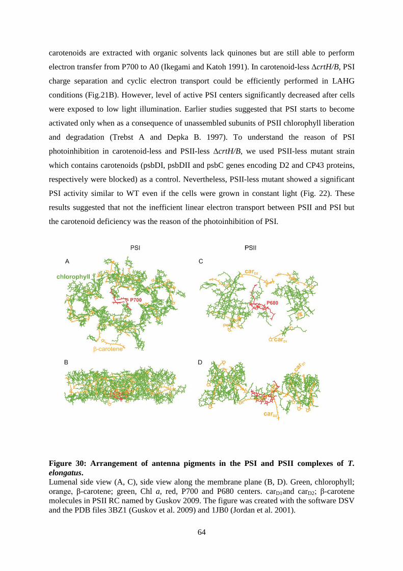
The striking difference between the antenna system in PSI and PSII is the lack of the
central antenna domain in PSII. Whereas the PSI central antenna domain contains 96 Chl a
and 22 carotenoid molecules carrying the electron transport chain in PSI RC. In contrast, the
PSII RC contains only the D1-D2 heterodimer, 2 β-carotene and 6 Chl a molecules (Fig. 28
and Fig. 30). The absence of the central antenna domain in PSII may be the response to its
ability for water splitting as an unlimited electron source, which leads to the process of D1
turnover. This process may have hindered the fusion of the RC domain and the antenna
domain in PSII,(Fig. 30). Definitely, among photosynthetic membrane complexes in ΔcrtH/B
mutant, PSII was the most severely affected complex by the absence of carotenoids. Indeed,
PSII-dependent oxygen-evolving activity of carotenoid-less crtH/B cells measured by using
an artificial electron acceptor 1,4-parabenzoquinone was undetectable indicating the absence
of functional PSII complexes. Additionally, the absence of variable Chl a fluorescence and
the absence of transient reduction in P700 kinetics confirmed that in ΔcrtH/B cells, no active
PSII centers are present (Fig 21A, 23).
62
Figure 29: Luminal side view of trimeric PSI complex (A) Trimerization domain, rich in β-carotene, shown in black circle and (B) in magnified
view. Yellow, carotenoid, green, chlorophyll, light blue, PsaL; violet, PsaI and yellow PsaM.
The figure was created with the software DSV and the PDB file 1JB0 (Jordan et al. 2001).
63
PSII is known to experience radiation damages even at very low illumination
intensities. The main ROS produced in the PSII RC is the very reactive singlet oxygen. This
is due to the generation of singlet oxygen during charge recombination between P680+/Pheo
-
and to the fact that carotenoid molecules in PSII RC are located too far from P680 to be able
to efficiently quench the triplet state Chl (Telfer 2002) (Fig. 28). On the other hand, if the
carotenoids were closer, they would probably be immediately oxidized by P680+ because of
its strong oxidizing potential (Telfer 2002). For this reason PSII RC is poorly protected from
the action of singlet oxygen and an efficient mechanism of PSII repair has evolved in order to
maintain the photochemical capacity. When light is in excess, the D1 protein is rapidly
degraded and resynthesized to reestablish a fully active PSII RC (Baena-Gonzalez and Aro
2002). A similar mechanism is not known for PSI. After radiation damages its recovery
requires several days (Kudoh and Sonoike 2002). Furthermore, the repair from
photoinhibition does not require the turnover of a single polypeptide, as in the case of D1, but
involves degradation and resynthesis of the whole PSI complex. Although the mechanisms
and localization are different, the D1 protein might also be considered as a safety fuse: in
fact, once D1 is degraded, not only charge separation but also singlet oxygen production are
stopped thus protecting the remaining PSII core complex from oxidation damage and
photobleaching (Bergantino et al. 2003). Thus, it appears that in both complexes some
sacrificial proteins exist functioning like a safety fuse for the rest of the structure. In the case
of PSI large antenna Chl-protein domain while in the case of PSII is the D1 protein of the RC
to play this role. It is worth mentioning that P700+ is not such a strong oxidizer as P680
+, and
no massive charge recombination has been reported in PSI. This is probably one major reason
explaining why different photoprotection strategies evolved in PSI and PSII. It is interesting
to point out that, despite the differences, in both cases the antenna systems are fundamental in
photoprotection. Even under high light conditions, large antenna system works very
efficiently as the excited triplet state Chl a cannot be detected in intact PSI (Grotjohann and
Fromme 2005). In addition, PSI photoprotection is mainly mediated by ROS scavenging
enzymes (e.g., superoxide dismutase and ascorbate peroxidase) instead of carotenoid
scavenging. These enzymes efficiently detoxify reactive species such as hydrogen peroxide
(H2O2), superoxide (O2-), hydroxyl radicals (OH) produced at the reducing side of PSI (Asada
1999). Therefore, PSI is generally believed to be less sensitive to light stress and its
carotenoid related photoprotection mechanisms were less investigated. Norflurazon (inhibitor
of phytoene synthase)-treated Scenedesmus obliquus cells lacking carotenoids still contain
some PSI and charge separation can be detected (Romer et al. 1995) . Samples in which all
64
carotenoids are extracted with organic solvents lack quinones but are still able to perform
electron transfer from P700 to A0 (Ikegami and Katoh 1991). In carotenoid-less ΔcrtH/B, PSI
charge separation and cyclic electron transport could be efficiently performed in LAHG
conditions (Fig.21B). However, level of active PSI centers significantly decreased after cells
were exposed to low light illumination. Earlier studies suggested that PSI starts to become
activated only when as a consequence of unassembled subunits of PSII chlorophyll liberation
and degradation (Trebst A and Depka B. 1997). To understand the reason of PSI
photoinhibition in carotenoid-less and PSII-less ΔcrtH/B, we used PSII-less mutant strain
which contains carotenoids (psbDI, psbDII and psbC genes encoding D2 and CP43 proteins,
respectively were blocked) as a control. Nevertheless, PSII-less mutant showed a significant
PSI activity similar to WT even if the cells were grown in constant light (Fig. 22). These
results suggested that not the inefficient linear electron transport between PSII and PSI but
the carotenoid deficiency was the reason of the photoinhibition of PSI.
Figure 30: Arrangement of antenna pigments in the PSI and PSII complexes of T.
elongatus. Lumenal side view (A, C), side view along the membrane plane (B, D). Green, chlorophyll;
orange, β-carotene; green, Chl a, red, P700 and P680 centers. carD1and carD2; β-carotene
molecules in PSII RC named by Guskov 2009. The figure was created with the software DSV
and the PDB files 3BZ1 (Guskov et al. 2009) and 1JB0 (Jordan et al. 2001).
65
5.2 Phosphatidylglycerol (PG) depletion induces an increase in myxoxanthophyll and
echinenone biosynthetic activities in Synechocystis PCC 6803 cells
Investigation of pigment-lipid interaction in a membrane environment is important to
understand the primary mechanism of antioxidant activity and bioavailability of carotenoids.
Our study has demonstrated the importance of pigment-lipid interactions to drive efficient
photosynthesis. Phosphatidylglycerol (PG) is the only phospholipid found in photosynthetic
reaction centers. PG depletion resulted in a remarkable suppression of photosynthetic
oxygen-evolving activity and impaired PSII activity by probably generation of ROS as shown
earlier for pgsA (Hagio et al. 2000) and PALcdsA (Laczkó-Dobos et al. 2008) mutants. Here
we studied how carotenoids respond to stress conditions induced by PG depletion in the PG-
deficient mutant of Synechocystis, pgsA.
In Synechocystis, carotenoids are synthesized via a branched pathway initiating from
common precursors (Fig 6). Lycopene is a central molecule that is present in the synthetic
pathway of all carotenoids and their intermediates. Our results suggest PG depletion
significantly increased the synthesis of two major carotenoids, myxoxanthophyll and
echinenone and on the other hand, the suppression of β-carotene and zeaxanthin synthesis
was observed. This differential effect of PG starvation could be explained by differences
between the metabolic pathways leading to the biosynthesis of the different carotenoids
(Lagarde et al. 2000). Synthesis towards myxoxanthophyll and a group of β-carotenes,
echinenone and zeaxanthin, is branched at γ-carotene (Fig. 31). The second branching point is
at β-carotene, where the synthesis of echinenone and that of zeaxanthin diverge (Fig 30). Our
results suggest that these steps are regulated differently. The increase in the relative content
of echinenone can be explained by its higher stability compared with those of other
carotenoid species in Synechocystis (Steiger et al. 1999), whereas its absolute increase in the
cytoplasmic membrane can be due to enhanced biosynthesis. However, the higher amount of
myxoxanthophyll, a less stable carotenoid species comparing to zeaxanthin and echinenone,
seems to indicate enhanced biosynthesis of this compound.
66
Figure 31: Flow sheet of carotenoid biosynthesis in Synechocystis.
Arrow 1 indicates non-regulated or down-regulated catalytic processes, and arrows 2, 3
indicate up-regulated catalytic processes in carotenoid biosynthesis.
The two up-regulated carotenes, myxoxanthophyll and echinenone, are special
protective agents. In Synechocystis, myxoxanthophyll, which was preferentially synthesized,
under high light stress provided the strongest protection and echinenone was the most stable
carotenoid under photo-oxidative conditions (Steiger et al. 1999). Investigations on the
photoprotective function of myxoxanthophyll in cyanobacterium Plectonema boryanum cells
under UV radiation stress revealed a reduced efficiency of energy transfer to PSII RCs and an
elevated level of myxoxanthophyll (Ivanov et al. 2000). Low temperature stress increased the
myxoxanthophyll content of Cylindrospermopsis raciborskii cells (Várkonyi et al. 2002).
Myxoxanthophyll is also required for the formation of normal cell wall structure and it
stabilizes thylakoid membranes (Mohamed et al. 2005). Induction of myxoxanthophyll
biosynthesis by high light exposure in Synechocystis has been demonstrated by Mohamed and
Vermaas (2004). However, very little is known about the biological function of both
myxoxanthophyll and echinenone.
Increased myxoxanthophyll and echinenone content of the PG-depleted cells was
detected in the membranes; nevertheless, a significant amount of carotenoids was also found
in the cytosol. The accumulation of carotenoids was demonstrated in the upper non-
membrane layer of the sucrose gradient (Fig. 24). Accordingly, we found increased
67
myxoxanthophyll and echinenone contents in the cytosol of PG-depleted cells. Since
carotenoids are not water-soluble molecules, they should be bound to proteins in the cytosol.
In cyanobacteria, orange carotenoid-binding proteins were found to maintain the carotenoids
in the aqueous phase (Kerfeld 2004). Carotenoids occur in all parts of the cyanobacterial cell.
Earlier studies suggested that carotenoid-binding proteins were present in cytoplasmic
membranes (Bullerjahn and Sherman 1986). Carotenoids were also localized to outer
membranes (Jurgens and Weckesser 1985). The carotenoids in the outer membrane were
found to be oriented (Jurgens and Mantele 1991).
The increased amount of myxoxanthophyll pointed to its protection role against
slowdown of photosynthetic processes generated by PG depletion. High amounts of
echinenone and myxoxanthophyll, whose synthesis were up-regulated by PG depletion (Fig.
31, arrow 2 and 3), seem to protect the structure and function of the photosynthetic apparatus
of cyanobacterial cells. On the other hand, the synthesis of β-carotene and zeaxanthin was
non-regulated or down-regulated in the PG-depleted cells (Fig. 31, arrow 1). Synthesis
towards myxoxanthophyll and a group of β-carotene, echinenone and zeaxanthin, is branched
at γ-carotene. The second branching point is at β-carotene, where the synthesis of echinenone
and that of zeaxanthin diverge. These findings suggested that these steps are regulated
differently by PG-depleted stress conditions.
68
6.0 CONCLUSIONS
I. We have successfully generated a viable carotenoid-less mutant in an oxygenic
prokaryotic species, Synechocystis. ΔcrtH/B cells were extremely light sensitive and could
grow only under LAHG conditions, demonstrating the protective role of carotenoids against
light stress.
Carotenoid-less mutant of Synechocystis, ΔcrtH/B, allowed us study the structural and
functional consequences of the complete carotenoid deficiency on photosynthesis here for the
first time, in vivo.
i. The structural consequences of the carotenoid deficiency on the assembly of
photosynthetic complexes were studied in ΔcrtH/B:
Dimeric and monomeric PSII complex could not be formed. Instead,
intermediate PSII complexes lacking CP43 and lacking CP43/CP47 were
found.
D1 was the most intensively labeled protein in ΔcrtH/B indicating D1 turnover
was active.
Small protein subunits binding to the unassembled PSII large protein subunits
were not stable without carotenoids.
PSI and Cyt b6f complexes could be assembled, however trimeric/monomeric
form of PSI ratio significantly decreased.
ii. The functional consequences of the carotenoid deficiency on the activity of
photosynthetic reaction centers.were studied in ΔcrtH/B cells:
There were no active PSII centers.
PSI centers were active. however they could not maintain their activity in the
absence of carotenoids even under low light.
II. Phosphatidylglycerol (PG) depletion leads to a significant increase in the level of both
echinenone and myxoxanthophyll suggesting their protection functions under stress
conditions. Here we show that the increase of carotenoid content is a response to different
stress conditions, not only to high light stress, however, the level of specific carotenoids
induced by stress conditions against oxidative damage regulated differently.
69
ACKNOWLEDGEMENT
I am thankful to my supervisors, Dr. Zoltán Gombos & Dr. Mihály Kis, whose
encouragement, guidance and support from the initial to the final level enabled me to develop
an understanding of the subject.
I warmly thank to members of the Lipid Function and Structure Group, BRC, for the
valuable support and friendly environment during my PhD studies.
During this work I have collaborated with Dr. Josef Komenda, Dr. László Kovács and
Dr. Przemyslaw Malec those I have great regard for.
My sincere thanks are to Dr. Tibor Janda and Dr. Szilvia Tóth for their detailed
review, constructive criticism and excellent advice.
I owe my loving thanks to my husband E. Serkan Sozer. Without his encouragement
and understanding it would not have been easy for me to finish this work.
Özge Sözer
70
REFERENCES
Allen, M.M. (1968) Simple conditions for growth of unicellular blue-green algae on plates. J.
Phycol. 4: 1-4.
Amunts, A. and Nelson, N. (2009) Plant photosystem I design in the light of evolution.
Structure 17: 637-650.
Anderson, S.L. and McIntosh, L. (1991) Light-activated heterotrophic growth of the
cyanobacterium Synechocystis sp. strain PCC 6803: a blue-light-requiring process. J.
Bacteriol. 173: 2761-2767.
Asada, K. (1999) The water-water cycle in chloroplasts: Scavenging of active oxygens and
dissipation of excess photons. Annual Review of Plant Physiology and Plant Molecular
Biology 50: 601-639.
Ausubel, F., Brent, R., Kingston, R.E., Moore, D.D., Seidman, J.G., Smith, J.A. (1995)
Short protocols in molecular biology. -John Wiley, New York.
Baena-Gonzalez, E. and Aro, E.M. (2002) Biogenesis, assembly and turnover of
photosystem II units. Philosophical Transactions of the Royal Society of London Series B-
Biological Sciences 357: 1451-1459.
Baniulis, D., Yamashita, E., Whitelegge, J.P., Zatsman, A.I., Hendrich, M.P., Hasan,
S.S. (2009) Structure-function, stability, and chemical modification of the cyanobacterial
cytochrome b(6)f complex from Nostoc sp PCC 7120. J. Biol. Chem. 284: 9861-9869.
Bautista, J.A., Rappaport, F., Guergova-Kuras, M., Cohen, R.O., Golbeck, J.H., Wang,
J.Y. et al. (2005a) Biochemical and biophysical characterization of photosystem I from
phytoene desaturase and carotene desaturase deletion mutants of Synechocystis sp. PCC
6803. J. Biol. Chem. 280: 20030-20041.
Bautista, J.A., Tracewell, C.A., Schlodder, E., Cunningham, F.X., Brudvig, G.W., and
Diner, B.A. (2005b) Construction and characterization of genetically modified Synechocystis
sp. PCC 6803 Photosystem II core complexes containing carotenoids with shorter -
conjugation than -carotene. J. Biol. Chem. 280: 38839-38850.
Bergantino, E., Segalla, A., Brunetta, A., Teardo, E., Rigoni, F., Giacometti, G.M. et al.
(2003) Light- and pH-dependent structural changes in the PsbS subunit of photosystem II.
Proc. Natl. Acad. Sci. U. S. A. 100: 15265-15270.
Berglund, A.H., Nilsson, R., and Liljenberg, C. (1999) Permeability of large unilamellar
digalactosyldiacylglycerol vesicles for protons and glucose - influence of -tocopherol, -
carotene, zeaxanthin and cholesterol. Plant Physiology and Biochemistry 37: 179-186.
Bittersmann, E. and Vermaas, W. (1991) Fluorescence lifetime studies of cyanobacterial
photosystem II mutants. Biochim. Biophys. Acta. 1098: 105-116.
Bogos, B., Ughy, B., Domonkos, I., Laczko-Dobos, H., Komenda, J., Abasova, L. et al.
(2010) Phosphatidylglycerol depletion affects photosystem II activity in Synechococcus sp
PCC 7942 cells. Photosynth. Res. 103: 19-30.
71
Breitenbach, J., Fernandez-Gonzalez, B., Vioque, A., and Sandmann, G. (1998) A
higher-plant type carotene desaturase in the cyanobacterium Synechocystis PCC6803.
Plant Mol. Biol. 36: 725-732.
Britton G, Liaaen-Jensen S and Pfander H. (2004) Carotenoids Handbook. Birkhauser
Verlag, Basel-Boston-Berlin
Britton, G. (1995a) Structure and properties of carotenoids in relation to function. FASEB J.
9: 1551-1558.
Britton, G. (1995b) UV/visible spectroscopy. In Carotenoids: spectroscopy. Edited by
Britton, G., Liaaen-Jensen, S., and Pfander, H. pp. 13-63. Birkhauser Verlag, Basel.
Bryant D.A..(1994) The Molecular Biology of Cyanobacteria. Kluwer Academic Publishers,
Dordrecht, The Netherlands
Bullerjahn, G.S. and Sherman, L.A. (1986) Identification of a carotenoid-binding protein
in the cytoplasmic membrane from the heterotrophic cyanobacterium Synechocystis sp strain
PCC6714. J. Bacteriol. 167: 396-399.
Cogdell, R.J. and Frank, H.A. (1987) How carotenoids function in photosynthetic bacteria.
Biochim. Biophys. Acta 895: 63-79.
Cramer, W.A., Zhang, H.M., Yan, J.S., Kurisu, G., and Smith, J.L. (2006)
Transmembrane traffic in the cytochrome b(6)f complex. Annual Review of Biochemistry 75:
769-790.
Dobakova, M., Tichy, M., and Komenda, J. (2007) Role of the PsbI protein in photosystem
II assembly and repair in the cyanobacterium Synechocystis sp. PCC 6803. Plant Physiol.
145: 1681-1691.
Domonkos, I., Malec, P., Laczko-Dobos, H., Sozer, O., Klodawska, K., Wada, H. et al.
(2009) Phosphatidylglycerol depletion induces an increase in myxoxanthophyll biosynthetic
activity in Synechocystis PCC6803 cells. Plant Cell Physiol. 50: 374-382.
Domonkos, I., Malec, P., Sallai, A., Kovacs, L., Itoh, K., Shen, G.Z. et al. (2004)
Phosphatidylglycerol is essential for oligomerization of photosystem I reaction center. Plant
Physiol. 134: 1471-1478.
El-Mohsnawy, E., Kopczak, M.J., Schlodder, E., Nowaczyk, M., Meyer, H.E.,
Warscheid, B. et al. (2010) Structure and function of intact photosystem I monomers from
the cyanobacterium Thermosynechococcus elongatus. Biochemistry 49: 4740-4751.
Enami, I., Okumura, A., Nagao, R., Suzuki, T., Iwai, M., and Shen, J.R. (2008)
Structures and functions of the extrinsic proteins of photosystem II from different species.
Photosynth. Res. 98: 349-363.
Farmilo, A. and Wilkinson, F. (1973) On the mechanism of quenching of singlet oxygen in
solution. Photochem. Photobiol. 18: 447-450.
72
Fernandez Gonzalez, B., Sandmann, G., and Vioque, A. (1997) A new type of
asymmetrically acting carotene ketolase is required for the synthesis of echinenone in the
cyanobacterium Synechocystis sp. PCC 6803. J. Biol. Chem. 272: 9728-9733.
Ferreira, K.N., Iverson, T.M., Maghlaoui, K., Barber, J., and Iwata, S. (2004)
Architecture of the photosynthetic oxygen-evolving center. Science 303: 1831-1838.
Formaggio, E., Cinque, G., and Bassi, R. (2001) Functional architecture of the major light-
harvesting complex from higher plants. J Mol Biol 314: 1157-1166.
Frank, H.A. and Cogdell, R.J. (1996) Carotenoids in photosynthesis. Photochem.
Photobiol. 63: 257-264.
Fromme, P., Jordan, P., and Krauss, N. (2001) Structure of photosystem I. Biochim.
Biophys. Acta. 1507: 5-31.
Funk, C. and Vermaas, W. (1999) A cyanobacterial gene family coding for single-helix
proteins resembling part of the light-harvesting proteins from higher plants. Biochemistry 38:
9397-9404.
Gombos, Z., Varkonyi, Z., Hagio, M., Iwaki, M., Kovacs, L., Masamoto, K. et al. (2002)
Phosphatidylglycerol requirement for the function of electron acceptor plastoquinone Q(B) in
the photosystem II reaction center. Biochemistry 41: 3796-3802.
Goodwin, T.W. (1980) Carotenoids in green and purple photosynthetic bacteria. Journal of
Scientific & Industrial Research 39: 682-688.
Gotz, T., Windhovel, U., Boger, P., and Sandmann, G. (1999) Protection of photosynthesis
against ultraviolet-B radiation by carotenoids in transformants of the cyanobacterium
Synechococcus PCC7942. Plant Physiol 120: 599-604.
Grotjohann, I. and Fromme, P. (2005) Structure of cyanobacterial Photosystem I.
Photosynth. Res. 85: 51-72.
Gruszecki, W.I. and Strzalka, K. (2005) Carotenoids as modulators of lipid membrane
physical properties. Biochim Biophys Acta 1740: 108-115.
Guskov, A., Gabdulkhakov, A., Broser, M., Glockner, C., Hellmich, J., Kern, J. et al.
(2010) Recent progress in the crystallographic studies of photosystem II. Chemphyschem 11:
1160-1171.
Guskov, A., Kern, J., Gabdulkhakov, A., Broser, M., Zouni, A., and Saenger, W. (2009)
Cyanobacterial photosystem II at 2.9-Å resolution and the role of quinones, lipids, channels
and chloride. Nat. Struct. Mol. Biol. 16: 334-342.
Hagio, M., Gombos, Z., Varkonyi, Z., Masamoto, K., Sato, N., Tsuzuki, M. et al. (2000)
Direct evidence for requirement of phosphatidylglycerol in photosystem II of photosynthesis.
Plant Physiol. 124: 795-804.
Hartwig, A. (2001) Zinc finger proteins as potential targets for toxic metal ions: differential
effects on structure and function. Antioxid. Redox. Signal. 3: 625-634.
73
Havaux, M., Tardy, F., Ravenel, J., Chanu, D., and Parot, P. (1996) Thylakoid membrane
stability to heat stress studied by flash spectroscopic measurements of the electrochromic
shift in intact potato leaves: influence of the xanthophyll content. Plant, Cell Environ. 19:
1359-1368.
He, Q., Dolganov, N., Bjorkman, O., and Grossman, A.R. (2001) The high light-inducible
polypeptides in Synechocystis PCC6803. Expression and function in high light. J. Biol.
Chem. 276: 306-314.
Horton, P., Ruban, A.V., and Walters, R.G. (1996) Regulation of light harvesting in green
plants. Annu. Rev. Plant Physiol Plant Mol. Biol. 47: 655-684.
Ikegami, I. and Katoh, S. (1991) Antenna chlorophyll-a has a structural role in stabilizing
the functional conformation of P-700 Chlorophyll-Protein Complexes. Biochim. Biophys.
Acta. 1059: 275-280.
Ikeuchi, M., Eggers, B., Shen, G.Z., Webber, A., Yu, J.J., Hirano, A. et al. (1991)
Cloning of the psbK gene from Synechocystis sp. PCC 6803 and characterization of
photosystem II in mutants lacking PSII-K. J. Biol. Chem. 266: 11111-11115.
Ivanov, A.G., Miskiewicz, E., Clarke, A.K., Greenberg, B.M., and Huner, N.P. (2000)
Protection of photosystem II against UV-A and UV-B radiation in the cyanobacterium
Plectonema boryanum: the role of growth temperature and growth irradiance. Photochem
Photobiol 72: 772-779.
Ivleva, N.B., Shestakov, S.V., and Pakrasi, H.B. (2000) The carboxyl-terminal extension of
the precursor D1 protein of photosystem II is required for optimal photosynthetic
performance of the cyanobacterium Synechocystis sp PCC 6803. Plant Physiol. 124: 1403-
1411.
Jordan, P., Fromme, P., Witt, H.T., Klukas, O., Saenger, W., and Krauss, N. (2001)
Three-dimensional structure of cyanobacterial photosystem I at 2.5 Å resolution. Nature 411:
909-917.
Jurgens, U.J. and Mantele, W. (1991) Orientation of carotenoids in the outer-membrane of
Synechocystis PCC 6714 (Cyanobacteria). Biochim. Biophys. Acta. 1067: 208-212.
Jurgens, U.J. and Weckesser, J. (1985) Carotenoid-containing outer-membrane of
Synechocystis sp strain PCC6714. J. Bacteriol. 164: 384-389.
Kandori, H., Sasabe, H., and Mimuro, M. (1994) Direct determination of a lifetime of the
S2 state of -carotene by femtosecond time-resolved fluorescence spectroscopy. Journal of
the American Chemical Society 116: 2671-2672.
Kerfeld, C.A. (2004) Structure and function of the water-soluble carotenoid-binding proteins
of cyanobacteria. Photosynth. Res. 81: 215-225.
Kilian, O., Steunou, A.S., Fazeli, F., Bailey, S., Bhaya, D., and Grossman, A.R. (2007)
Responses of a thermophilic Synechococcus isolate from the microbial mat of octopus spring
to light. Appl. Environ. Microbiol. 73: 4268-4278.
74
Kis, M., Zsiros, O., Farkas, T., Wada, H., Nagy, F., and Gombos, Z. (1998) Light-induced
expression of fatty acid desaturase genes. Proc. Natl. Acad. Sci. U. S. A 95: 4209-4214.
Knox, J.P. and Dodge, A.D. (1985) Singlet oxygen and plants. Phytochemistry 24: 889-896.
Kojima, K., Oshita, M., Nanjo, Y., Kasai, K., Tozawa, Y., Hayashi, H. et al. (2007)
Oxidation of elongation factor G inhibits the synthesis of the D1 protein of photosystem II.
Mol. Microbiol. 65: 936-947.
Komenda, J., Lupinkova, L., and Kopecky, J. (2002) Absence of the psbH gene product
destabilizes photosystem II complex and bicarbonate binding on its acceptor side in
Synechocystis PCC 6803. Eur. J. Biochem. 269: 610-619.
Komenda, J., Reisinger, V., Muller, B.C., Dobakova, M., Granvogl, B., and Eichacker,
L.A. (2004) Accumulation of the D2 protein is a key regulatory step for assembly of the
photosystem II reaction center complex in Synechocystis PCC 6803. J. Biol. Chem. 279:
48620-48629.
Komenda, J., Tichy, M., and Eichacker, L.A. (2005) The PsbH protein is associated with
the inner antenna CP47 and facilitates D1 processing and incorporation into PSII in the
cyanobacterium Synechocystis PCC 6803. Plant Cell Physiol. 46: 1477-1483.
Komenda, J., Kuvikova, S., Granvogl, B., Eichacker, L.A., Diner, B.A., and Nixon, P.J.
(2007) Cleavage after residue Ala352 in the C-terminal extension is an early step in the
maturation of the D1 subunit of photosystem II in Synechocystis PCC 6803. Biochimica et
Biophysica Acta-Bioenergetics 1767: 829-837.
Komenda, J., Nickelsen, J., Tichy, M., Prasil, O., Eichacker, L.A., and Nixon, P.J. (2008)
The cyanobacterial homologue of HCF136/YCF48 is a component of an early photosystem II
assembly complex and is important for both the efficient assembly and repair of photosystem
II in Synechocystis sp. PCC 6803. J. Biol. Chem. 283: 22390-22399.
Kudoh, H. and Sonoike, K. (2002) Irreversible damage to photosystem I by chilling in the
light: cause of the degradation of chlorophyll after returning to normal growth temperature.
Planta 215: 541-548.
Kurisu, G., Zhang, H., Smith, J.L., and Cramer, W.A. (2003) Structure of the cytochrome
b6f complex of oxygenic photosynthesis: tuning the cavity. Science 302: 1009-1014.
Kuvikova, S., Tichy, M., and Komenda, J. (2005) A role of the C-terminal extension of the
photosystem II D1 protein in sensitivity of the cyanobacterium Synechocystis PCC 6803 to
photoinhibition. Photochem. Photobiol. Sci. 4: 1044-1048.
Laczkó-Dobos, H., Ughy, B., Tóth, S.Z., Komenda, J., Zsiros, O., Domonkos, I. (2008)
Role of phosphatidylglycerol in the function and assembly of Photosystem II reaction center,
studied in a cdsA-inactivated PAL mutant strain of Synechocystis sp. PCC6803 that lacks
phycobilisomes. Biochim. Biophys. Acta 1777: 1184-1194.
Lagarde, D. and Vermaas, W. (1999) The zeaxanthin biosynthesis enzyme -carotene
hydroxylase is involved in myxoxanthophyll synthesis in Synechocystis sp PCC 6803. FEBS
Lett. 454: 247-251.
75
Lagarde, D., Beuf, L., and Vermaas, M. (2000) Increased production of zeaxanthin and
other pigments by application of genetic engineering techniques to Synechocystis sp strain
PCC 6803. Appl. Environ. Microbiol. 66: 64-72.
Lichtenthaler, H.K. (1987) Chlorophylls and carotenoids: pigments of photosynthetic
biomembranes. Methods Enzymol. 148: 350-382.
Liu, L.N., Chen, X.L., Zhang, Y.Z., and Zhou, B.C. (2005) Characterization, structure and
function of linker polypeptides in phycobilisomes of cyanobacteria and red algae: An
overview. Biochimica et Biophysica Acta-Bioenergetics 1708: 133-142.
Loll, B., Kern, J., Saenger, W., Zouni, A., and Biesiadka, J. (2005) Towards complete
cofactor arrangement in the 3.0 A resolution structure of photosystem II. Nature 438: 1040-
1044.
Lupinkova, L. and Komenda, J. (2004) Oxidative modifications of the Photosystem II D1
protein by reactive oxygen species: From isolated protein to cyanobacterial cells. Photochem.
Photobiol. 79: 152-162.
MacColl, R. and Guard-Friar, D. (1987) Phycobiliproteins. CRC Press, Boca Raton, FL
Macpherson, A.N., Arellano, J.B., Fraser, N.J., Cogdell, R.J., and Gillbro, T. (2001)
Efficient energy transfer from the carotenoid S-2 state in a photosynthetic light-harvesting
complex. Biophysical Journal 80: 923-930.
Mantoura, R.F.C. and Llewellyn, C.A. (1983) The rapid-determination of algal chlorophyll
and carotenoid pigments and their breakdown products in natural waters by reverse-phase
High-Performance Liquid Chromatography. Anal. Chim. Acta 151: 297-314.
Martinez-Ferez, I., FernandezGonzalez, B., Sandmann, G., and Vioque, A. (1994)
Cloning and expression in Escherichia coli of the gene coding for phytoene synthase from the
cyanobacterium Synechocystis sp. PCC6803. Biochim. Biophys. Acta. 1218: 145-152.
Martinez-Ferez, I.M. and Vioque, A. (1992) Nucleotide sequence of the phytoene
desaturase gene from Synechocystis sp. PCC 6803 and characterization of a new mutation
which confers resistance to the herbicide norflurazon. Plant Mol. Biol. 18: 981-983.
Masamoto, K. and Furukawa, K. (1997) Accumulation of zeaxanthin in cells of the
cyanobacterium, Synechococcus sp. strain PCC 7942 grown under high irradiance. J. Plant
Physiol. 151: 257-261.
Masamoto, K., Misawa, N., Kaneko, T., Kikuno, R., and Toh, H. (1998) -carotene
hydroxylase gene from the cyanobacterium Synechocystis sp. PCC6803. Plant Cell Physiol.
39: 560-564.
Masamoto, K., Zsiros, O., and Gombos, Z. (1999) Accumulation of zeaxanthin in
cytoplasmic membranes of the cyanobacterium Synechococcus sp. strain PCC7942 grown
under high light condition. J. Plant Physiol. 155: 136-138.
Masamoto, K., Wada, H., Kaneko, T., and Takaichi, S. (2001) Identification of a gene
required for cis-to-trans carotene isomerization in carotenogenesis of the cyanobacterium
Synechocystis sp. PCC 6803. Plant Cell Physiol. 42: 1398-1402.
76
Masamoto, K., Hisatomi, S., Sakurai, I., Gombos, Z., and Wada, H. (2004) Requirement
of carotene isomerization for the assembly of photosystem II in Synechocystis sp. PCC 6803.
Plant Cell Physiol. 45: 1325-1329.
Maxwell, P.C. and Biggins, J. (1977) Kinetic behavior of P-700 during induction of
photosynthesis in algae. Biochim. Biophys. Acta. 459: 442-450.
Mayer, M.P. (1995) A new set of useful cloning and expression vectors derived from
pBlueScript. Gene 163: 41-46.
Mohamed, A. and Jansson, C. (1989) Influence of light on accumulation of photosynthesis-
specific transcripts in the cyanobacterium Synechocystis 6803. Plant Mol. Biol. 13: 693-700.
Mohamed, H.E. and Vermaas, W. (2004) Slr1293 in Synechocystis sp strain PCC 6803 is
the C-3 ',4 ' desaturase (CrtD) involved in myxoxanthophyll biosynthesis. J. Bacteriol. 186:
5621-5628.
Mohamed, H.E., van de Meene, A.M.L., Roberson, R.W., and Vermaas, W.F.J. (2005)
Myxoxanthophyll is required for normal cell wall structure and thylakoid organization in the
cyanobacterium, Synechocystis sp strain PCC 6803. J. Bacteriol. 187: 6883-6892.
Molnar, P. and Szabolcs, J. (1993) (Z/E)-Photoisomerization of C40-Carotenoids by Iodine.
Journal of the Chemical Society-Perkin Transactions 2261-266.
Moon, B.Y., Higashi, S.I., Gombos, Z., and Murata, N. (1995) Unsaturation of the
membrane lipids of chloroplasts stabilizes the photosynthetic machinery against low-
temperature photoinhibition in transgenic tobacco plants. Proc. Natl. Acad. Sci. U. S. A. 92:
6219-6223.
Muh, F., Renger, T., and Zouni, A. (2008) Crystal structure of cyanobacterial photosystem
II at 3.0 Å resolution: A closer look at the antenna system and the small membrane-intrinsic
subunits. Plant Physiology and Biochemistry 46: 238-264.
Mullineaux, C.W. (2008) Phycobilisome-reaction centre interaction in cyanobacteria.
Photosynth. Res. 95: 175-182.
Murata, N., Sato, N., Takahashi, N., and Hamazaki, Y. (1982) Compositions and
positional distributions of fatty acids in phospholipids from leaves of chilling-sensitive and
chilling-resistant plants. Plant Cell Physiol. 23: 1071-1079.
Murata, N. (1983) Molecular species composition of phosphatidylglycerols from chilling-
sensitive and chilling-resistant plants. Plant Cell Physiol. 24: 81-86.
Murata, N. and Omata, T. (1988) Isolation of cyanobacterial plasma membranes. Methods
Enzymol. 167: 245-251.
Nakamura, Y., Miyajima, N., Hirosawa, M., Kaneko, T., Tabata, S. (1996) Cyanobase:
The Genome Database for Synechocystis sp. strain PCC 6803 in Proc. Seventh Workshop on
Genome Informatics; 214-215.
77
Nixon, P.J., Michoux, F., Yu, J.F., Boehm, M., and Komenda, J. (2010) Recent advances
in understanding the assembly and repair of photosystem II. Annals of Botany 106: 1-16.
Nowaczyk, M.M., Hebeler, R., Schlodder, E., Meyer, H.E., Warscheid, B., and Rogner,
M. (2006) Psb27, a cyanobacterial lipoprotein, is involved in the repair cycle of photosystem
II. Plant Cell 18: 3121-3131.
Ouchane, S., Picaud, M., Vernotte, C., and Astier, C. (1997) Photooxidative stress
stimulates illegitimate recombination and mutability in carotenoid-less mutants of Rubrivivax
gelatinosus. EMBO J. 16: 4777-4787.
Palsson, L.O., Flemming, C., Gobets, B., van Grondelle, R., Dekker, J.P., and
Schlodder, E. (1998) Energy transfer and charge separation in photosystem I: P700 oxidation
upon selective excitation of the long-wavelength antenna chlorophylls of Synechococcus
elongatus. Biophysical Journal 74: 2611-2622.
Polivka, T. and Sundstrom, V. (2004) Ultrafast dynamics of carotenoid excited states -
from solution to natural and artificial systems. Chemical Reviews 104: 2021-2071.
Promnares, K., Komenda, J., Bumba, L., Nebesarova, J., Vacha, F., and Tichy, M.
(2006) Cyanobacterial small chlorophyll-binding protein ScpD (HliB) is located on the
periphery of photosystem II in the vicinity of PsbH and CP47 subunits. J. Biol. Chem. 281:
32705-32713.
Punginelli, C., Wilson, A., Routaboul, J.M., and Kirilovsky, D. (2009) Influence of
zeaxanthin and echinenone binding on the activity of the orange carotenoid protein. Biochim
Biophys Acta 1787: 280-288.
Rakhimberdieva, M.G., Elanskaya, I.V., Vermaas, W.F.J., and Karapetyan, N.V. (2010)
Carotenoid-triggered energy dissipation in phycobilisomes of Synechocystis sp PCC 6803
diverts excitation away from reaction centers of both photosystems. Biochimica et Biophysica
Acta-Bioenergetics 1797: 241-249.
Rappaport, F. and Diner, B.A. (2008) Primary photochemistry and energetics leading to the
oxidation of the (Mn)4Ca cluster and to the evolution of molecular oxygen in Photosystem II.
Coordination Chemistry Reviews 252: 259-272.
Rippka, R., Deruelles, J., Waterbury, J.B., Herdman, M., and Stanier, R.Y. (1979)
Generic assignments, strain histories and properties of pure cultures of cyanobacteria. J. Gen.
Microbiol. 111: 1-61.
Ritz, T., Damjanovic, A., Schulten, K., Zhang, J.P., and Koyama, Y. (2000) Efficient
light harvesting through carotenoids. Photosynth. Res. 66: 125-144.
Romer, S., Senger, H., and Bishop, N.I. (1995) Characterization of the carotenoidless strain
of Scenedesmus obliquus, mutant C-6E, a living photosystem I model. Bot. Acta 108: 80-86.
Roose, J.L., Kashino, Y., and Pakrasi, H.B. (2007) The PsbQ protein defines
cyanobacterial Photosystem II complexes with highest activity and stability. Proc. Natl.
Acad. Sci. U. S. A. 104: 2548-2553.
78
Ruengjitchatchawalya, M., Kovacs, L., Mapaisansup, T., Sallai, A., Gombos, Z.,
Ponglikitmongkol, M. et al. (2005) Higher plant-like fluorescence induction and
thermoluminescence characteristics in cyanobacterium, Spirulina mutant defective in PQH(2)
oxidation by cyt b(6)/f complex. J. Plant Physiol. 162: 1123-1132.
Sakurai, I., Hagio, M., Gombos, Z., Tyystjarvi, T., Paakkarinen, V., Aro, E.M. et al.
(2003) Requirement of phosphatidylglycerol for maintenance of photosynthetic machinery.
Plant Physiol. 133: 1376-1384.
Sas, K.N., Kovacs, L., Zsiros, O., Gombos, Z., Garab, G., Hemmingsen, L. et al. (2006)
Fast cadmium inhibition of photosynthesis in cyanobacteria in vivo and in vitro studies using
perturbed angular correlation of gamma-rays. Journal of Biological Inorganic Chemistry 11:
725-734.
Satoh, K. and Yamamoto, Y. (2007) The carboxyl-terminal processing of precursor D1
protein of the photosystem II reaction center. Photosynth. Res. 94: 203-215.
Schäfer, L., Vioque, A., and Sandmann, G. (2005) Functional in situ evaluation of
photosynthesis-protecting carotenoids in mutants of the cyanobacterium Synechocystis
PCC6803. J. Photochem. Photobiol. B-Biol. 78: 195-201.
Schagger, H. and von Jagow, G. (1987) Tricine-sodium dodecyl sulfate-polyacrylamide gel
electrophoresis for the separation of proteins in the range from 1 to 100 kDa. Anal. Biochem.
166: 368-379.
Schagger, H., Cramer, W.A., and von Jagow, G. (1994) Analysis of molecular masses and
oligomeric states of protein complexes by blue native electrophoresis and isolation of
membrane-protein complexes by 2-dimensional native electrophoresis. Anal. Biochem. 217:
220-230.
Schodel, R., Irrgang, K.D., Voigt, J., and Renger, G. (1999) Quenching of chlorophyll
fluorescence by triplets in solubilized light-harvesting complex II (LHCII). Biophysical
Journal 76: 2238-2248.
Sobotka, R., Duerhring, U., Komenda, J., Peter, E., Gardian, Z., Tichy, M. (2008)
Importance of the cyanobacterial GUN4 protein for chlorophyll metabolism and assembly of
photosynthetic complexes. J. Biol. Chem. 283: 25794-25802.
Steiger, S., Schafer, L., and Sandmann, G. (1999) High-light-dependent upregulation of
carotenoids and their antioxidative properties in the cyanobacterium Synechocystis PCC
6803. J. Photochem. Photobiol. B-Biol. 52: 14-18.
Takaichi, S., Maoka, T., and Masamoto, K. (2001) Myxoxanthophyll in Synechocystis sp.
PCC 6803 is myxol 2'-dimethyl-fucoside, (3R,2'S)-myxol 2'-(2,4-di-O-methyl-alpha-L-
fucoside), not rhamnoside. Plant Cell Physiol 42: 756-762.
Takaichi, S. and Mochimaru, M. (2007) Carotenoids and carotenogenesis in cyanobacteria:
unique ketocarotenoids and carotenoid glycosides. Cell Mol Life Sci 64: 2607-2619.
Telfer, A. (2002) What is -carotene doing in the photosystem II reaction centre? Philos
Trans R Soc Lond B Biol Sci 357: 1431-1439.
79
Towbin, H., Staehelin, T., and Gordon, J. (1979) Electrophoretic transfer of proteins from
polyacrylamide gels to nitrocellulose sheets: procedure and some applications. Proc. Natl.
Acad. Sci. U. S. A 76: 4350-4354.
Trebst, A. and Depka, B. (1997) Role of carotene in the rapid turnover and assembly of
photosystem II in Chlamydomonas reinhardtii. FEBS Lett 400: 359-362.
Van Amerongen, H. and Van Grondelle, R. (2001) Understanding the energy transfer
function of LHCII, the major light-harvesting complex of green plants. Journal of Physical
Chemistry B 105: 604-617.
Varkonyi, Z., Masamoto, K., Debreczeny, M., Zsiros, O., Ughy, B., Gombos, Z. et al.
(2002) Low-temperature-induced accumulation of xanthophylls and its structural
consequences in the photosynthetic membranes of the cyanobacterium Cylindrospermopsis
raciborskii: An FTIR spectroscopic study. Proc. Natl. Acad. Sci. U. S. A. 99: 2410-2415.
Vass, I., Styring, S., Hundal, T., Koivuniemi, A., Aro, E.M., and Andersson, B. (1992)
Reversible and irreversible intermediates during photoinhibition of photosystem 2. Stable
reduced Qa species promote chlorophyll triplet formation. Proc. Natl. Acad. Sci. U. S. A. 89:
1408-1412.
Welburn, A.R. and Lichtenthaler, H. (1984) Formula and program to determine total
carotenoids and Chl a and b of leaf extracts in different solvents. In Advances in
Photosynthesis Research. Edited by Sybesma, C. pp. 9-12. Martinus Nijhoff/Dr. W. Junk
Publishers, The Hague.
Wenk, S.O., Schneider, D., Boronowsky, U., Jager, C., Klughammer, C., de Weerd, F.L.
et al. (2005) Functional implications of pigments bound to a cyanobacterial cytochrome
b(6)f complex. FEBS J. 272: 582-592.
Widomska, J., Kostecka-Gugala, A., Latowski, D., Gruszecki, W.I., and Strzalka, K.
(2009) Calorimetric studies of the effect of cis-carotenoids on the thermotropic phase
behavior of phosphatidylcholine bilayers. Biophysical Chemistry 140: 108-114.
Wilson, A., Ajlani, G., Verbavatz, J.M., Vass, I., Kerfeld, C.A., and Kirilovsky, D.
(2006) A soluble carotenoid protein involved in phycobilisome-related energy dissipation in
cyanobacteria. Plant Cell 18: 992-1007.
Wilson, A., Kinney, J.N., Zwart, P.H., Punginelli, C., D'Haene, S., Perreau, F. (2010)
Structural determinants underlying photoprotection in the photoactive Orange Carotenoid
Protein of cyanobacteria. J. Biol. Chem. 285: 18364-18375.
Woodall, A.A., Britton, G., and Jackson, M.J. (1997a) Carotenoids and protection of
phospholipids in solution or in liposomes against oxidation by peroxyl radicals: relationship
between carotenoid structure and protective ability. Biochim Biophys Acta 1336: 575-586.
Woodall, A.A., Lee, S.W., Weesie, R.J., Jackson, M.J., and Britton, G. (1997b) Oxidation
of carotenoids by free radicals: relationship between structure and reactivity. Biochim
Biophys Acta 1336: 33-42.
80
ÖSSZEFOGLALÓ
Karotinoidok részvétele a cianobaktériumok fotoszintetikus komplexeinek
összeszerelődésében és működésében
A fotoszintézis alapvető biofizikai és biokémiai folyamat, amelynek során a napfény
energiája az élethez nélkülözhetetlen kémiai energiává alakul át. A cianobaktériumok a
legősibb ismert oxigéntermelő, fotoszintetizáló, prokarióta szervezetek, melyek az
endoszimbiózis elmélete szerint a növényi kloroplasztisz ősének tekinthetők. A
cianobaktériumok még napjainkban is Földünk éves oxigéntermelésének közel 30%-át
biztosítják.
A cianobaktériumok igen ellenálló, a környezeti változásokhoz jól alkalmazkodó
szervezetek, melyek hatékonyan védekeznek a reaktív oxigéngyökök (ROS) fotoszintetikus
aktivitást csökkentő, fotoinhibíciót eredményező fotooxidatív károsító hatásai ellen is. A
karotinoidok működése nélkülözhetetlen a védekezéshez. A fénykárosodástól azáltal védik a
sejtet, hogy képesek kioltani a gerjesztett szinglet és triplet állapotú klorofillt, valamint
befogni a szinglet oxigént, amely rendkívül káros hatású ROS (Knox és Dodge 1985).
Nélkülözhetetlen védőszerepük mellett a karotinoidok járulékos fénybegyűjtő pigmentként is
működnek (Frank és Cogdell 1996) és részt vesznek a membránok felépítésében is (Cogdell
és Frank1987, Gruszecki és Strzalka 2005).
A crtB gén által kódolt fitoén-szintáz enzim katalizálja a karotinoidok
bioszintézisének első lépését, a fitoén kialakítását. A fitoénből további enzimek
közreműködésével különböző karotin- és xantofillmolekulák képződnek.
Az egyik legintenzívebben tanulmányozott cianobaktérium, a Synechocystis sp. PCC
6803 jellemző karotinoidjai a következők: β-karotin, hidroxiszármazéka a zeaxantin,
ketoszármazékai az echinenon és a 3’-hidroxiechinenon, valamint a mixoxantofill, amely egy
karotinoid glikozid (Britton és mtsai 2004, Takaichi és Mochimaru 2007). Az újabb
röntgenkrisztallográfiás analízisek feltárták, hogy a cianobakteriális fotoszintetikus
komplexekben a β-karotin az egyetlen karotinoidfajta, de az összes többi karotinoid is
megtalálható a cianobaktériumok tilakoidmembránjában, a citoplazmamembránban és a
citoszolban. Különböző stresszkörülmények hatására mennyiségük megnő a sejtben. A β-
karotin-molekulán levő szubsztituensek kémiai természete meghatározza a molekula
antioxidatív tulajdonságát, szerepét a fénykárosodás elleni védelemben (Britton 1995a). A
hidroxi-karotinoidok általában jók a peroxidgyökök inaktiválásában (Woodall és mtsai
81
1997b), míg a ketoszármazékok hatékonyabbak a reaktív szinglet oxigén kioltásában
(Woodall és mtsai 1997a), és legstabilabbnak bizonyultak a peroxidgyökök és a fotooxidáció
károsításával szemben (Steiger és mtsa 1999).
Röntgenkrisztallográfiás analízissel (Guskov és mtsai 2009) 12 karotinoidot
azonosítottak Thermosynechococcus elongatus PSII komplexében monomerenként. A PSII a
fotooxidatív stresszel szemben a leginkább érzékeny fotoszintetikus komplex. Korábbi
tanulmányok arra engednek következtetni, hogy a β-karotin elengedhetetlenül szükséges a D1
fehérje felhalmozódásához Chlamydomonas reinhardtii zöld algában (Trebst és Depka 1997)
és Synechocystis PCC 6803 cianobaktériumban is (Masamoto és mtsai 2004). Tehát a
karotinoidok jelenléte fontos a működőképes PSII felépüléséhez.
A T. elongatus PSI komplexek szerkezetében 22 karotinoidmolekulát azonosítottak
monomerenként (Jordan és mtsai 2001, Fromme és mtsai 2001, Grotjohann és Fromme
2005). A PSI általában kevésbé érzékeny a fénystresszre, és a karotinoidok részvétele a
fénykárosodás elleni védelemben a PSI esetén kevéssé ismert. A fitoén-szintáz gátló
norflurazonnal történt kezelés hatására karotinoidmentessé vált Chlamydomonas reinhardtii
sejtek még tartalmaztak PSI komplexeket, melyekben töltésszeparációt is megfigyeltek
(Romer és mtsai 1995). A tilakoidmembránokból szerves oldószerrel kivonták a
karotinoidokat és a kinonokat, ezután is még megtörtént az elektronszállítás a P700 és az A0
között (Ikegami és Katoh 1991).
A fotoszintézis folyamatai a PSI- és PSII-reakciócentrumokban játszódnak le, melyek
a tilakoidmembránba ágyazott pigment-fehérje komplexek. A lipid-fehérje és lipid-pigment
kölcsönhatások fontos szerepet játszanak a fotoszintézis szabályozásában. A
cianobaktériumok tilakoidmembránja jellegzetes lipidösszetételű, mely glikolipideket és egy
foszfolipidet tartalmaz. A cianobaktériumokban és a magasabbrendű növények
tilakoidmembránjában egyetlen foszfolipid fordul elő, a foszfatidil-glicerin (PG). A T.
elongatus fotoszintetikus komplexeinek röntgenkrisztallográfiás szerkezetvizsgálata PG-
molekulákat mutatott ki a PSII komplex CP43 és D1 fehérjéi között (Guskov és mtsai 2009),
valamint a PSI szerkezetében is (Jordan és mtsai 2001). A PG eltávolítása a sejtekből a
klorofilltartalom csökkenését és a fotoszintetikus folyamatok lassulását okozta. A QA és QB
közti elektrontranszport gátlása volt megfigyelhető, ami a QA túlredukálódásához és QA-2
képződéséhez vezet (Gombos és mtsai 2002, Laczko-Dobos és mtsai 2008). A PG
esszenciális az alacsony hőmérsékleti stressz (Murata és mtsai 1982, Murata 1983, Moon és
mtsai 1995) és a fénystressz elleni védekezésben (Vass és mtsai 1992).
82
A globális környezet változása egyre gyakrabban idéz elő olyan
stresszkörülményeket, melyek a fotoszintetizáló szervezetekre hatva csökkentik a
fotoszintézis hatékonyságát. A stresszkörülmények között a fotoszintézisben működő
védekező mechanimusok megismerésének igénye a különböző körülmények között eltérő
fejlődési sajátosságokkal rendelkező növények, algák és cianobaktériumok kutatásához
vezetett. A cianobaktériumok a növényi kloroplasztisz ősének tekinthetők, így kiváló
modellszervezetek a magasabbrendű növények fotoszintézisének tanulmányozásához. Az
antioxidáns tulajdonságokkal bíró karotinoidok fontos szerepet játszanak a
stresszkörülmények elleni harcban, de a védekezésben elengedhetetlen szerkezeti és
funkcionális sajátosságaik csak részben felderítettek. Ezért munkám során a következő
célokat tűztem ki:
I. Felderíteni, hogy in vivo, a fotoszintetikus apparátusban a karotinoidok hiánya
milyen (i) szerkezeti változásokat okoz, és ez milyen (ii) funkcionális következményekkel
jár. Ennek érdekében az első olyan oxigéntermelő, fotoszintetikus prokarióta (Synechocystis
sp. PCC 6803 cianobaktérium) mutánst létrehozni, melyben a karotinoidok szintézise teljes
mértékben gátolt.
II. Ezenkívül a karotinoidok védő szerepét tanulmányozni olyan stresszkörülmények
között, melyet a PG hiánya váltott ki a Synechocystis sp. PCC 6803 pgsA mutánsban.
A fitoén-szintáz enzimet kódoló crtB gént inaktiváltuk a részlegesen
karotinoidhiányos ΔcrtH mutánsban. Így létrejött egy teljes mértékben karotinoidmentes
kettős mutáns, a ΔcrtH/B, amely csak sötétben, glükóztartalmú tápoldatban volt fenntartható.
HPLC analízissel igazoltuk, hogy a ΔcrtH/B sejtek semmilyen karotinoidszármazékot nem
tartalmaznak. A ΔcrtH/B sejtkultúra kékes színe arra utalt, hogy a sejtekben a fikobilinek a
domináns pigmentek. A karotinoidok hiánya nem érintette szignifikánsan a fikobiliproteinek
szintézisét és felhalmozódását, a klorofill szintézisét és akkumulációját viszont gátolta.
Fénykezelés hatására a mutáns sejtek fokozatosan kifehéredtek, elvesztették
pigmentációjukat és elpusztultak, igazolva, hogy a karotinoidok nélkülözhetetlen elemei a
fotoprotekciónak.
Western blot analízissel kimutattuk, hogy a karotinoidmentes ΔcrtH/B mutánsban a
PSII nagy fehérjealegységeinek szintje igen alacsony, míg a PSI és a citokróm-b6f fehérjéinek
mennyisége nem csökkent. LAHG körülmények között a D1 és D2 fehérjealegységek szintje
sokkal alacsonyabb volt, mint a vad típusban. Miután a sejteket megvilágítottuk, a D1 és D2
fehérjék mennyisége tovább csökkent, jelezve a karotinoidok hiányában bekövetkező
fotooxidációt. A CDP47 és különösen a CP43 klorofillkötő antennaalegységek szintje volt a
83
leginkább érintett. Ez a megfigyelés összhangban van a PSII legutóbbi
röntgenkrisztallográfiás szerkezeti modelljével, mely szerint a β-karotin molekulák többsége
a CP47 és CP43 alegységek transzmembrán α-hélixeinek közelében helyezkedik el. A
ΔcrtH/B sejtek fehérje alegységeinek 2D-gélanalízise radioaktív „pulse-chase” jelöléssel
kombinálva megadta a kulcsot annak magyarázatához, hogy miért nem épülnek fel aktív PSII
komplexek a mutánsban. Az eredmények megmutatták, hogy PSII-dimerek nem képesek
kialakulni β-karotin hiányában, sőt még a nagyon instabil PSII-monomerek is csak radioaktív
jelöléssel voltak detektálhatóak. A CP47 és különösen a CP43 alegységek nem kötődtek
stabilan a PSII komplexben, ezért csak egy intermedier PSII komplex, az RC47 (melyből a
CP43 hiányzik) volt képes felhalmozódni gélelektroforézissel detektálható mennyiségben. A
ΔcrtH/B sejtek tilakoidjában a D1 fehérje volt a radioaktívan leginkább jelölődött fehérje,
ami jelzi, hogy a D1 fehérje „steady state turnovere” karotinoidok hiányában is fennmaradt.
A radioaktívan jelölődött D1 fehérje leginkább az RC47 komplexben volt detektálható,
valamint a monomer PSII-ben és az RCa-ban, amely egy olyan intermedier PSII komplex,
ahonnan a CP43 és a CP47 is hiányzik. Miután a „chase” kísérletet kloramfenikolkezeléssel,
azaz a fehérjeszintézis gátlásával kombináltuk, a PSII-monomerek eltűntek a gélből, ami a
komplex instabilitására és fényérzékenységére utal. Sőt β-karotin hiányában azoknak a kis
fehérje alegységeknek a kapcsolódása sem stabil, amelyek a nagy PSII-alegységekhez
kötődnek a komplexek összeszerelődése során. A PSII komplexszel ellentétben, a PSI és a
citokróm-b6f komplexek felépülnek, bár a trimer/monomer PSI aránya szignifikánsan
csökkent karotinoidok hiányában.
Kétségtelenül a PSII volt a karotinoidok hiányában leginkább érintett fotoszintetikus
membránkomplex a ΔcrtH/B mutánsban. A karotinoidhiányos mutáns sejtek valóban nem
tartalmaztak működőképes PSII komplexeket, amit a PSII-függő oxigénfejlesztő aktivitás
mérésével igazoltunk. Továbbá a PSII klorofill-a fluoreszcencia mérései és a P700 tranziens
redukciókinetikájának mérései megerősítették, hogy a ΔcrtH/B sejtekben nem alakultak ki
aktív PSII-reakciócentrumok. Ezzel szemben a PSI-hez köthető töltésszeparáció és ciklikus
elektrontranszport működik a LAHG-körülmények között nevelt ΔcrtH/B sejtekben is.
Azonban, miután a sejteket megvilágítottuk, az aktív PSI-reakciócentrumok mennyisége
szignifikánsan csökkent. Azért, hogy megértsük a PSI fotoinhibíciójának okát a
karotinoidmentes és aktív PSII-mentes ΔcrtH/B sejtekben, méréseket végeztünk egy másik
PSII-hiányos mutáns törzsben, melyben a D2 és a CP43 alegységek szintézise gátolt. A
karotinoidokat is tartalmazó PSII-hiányos mutáns konstans PSI-aktivitást mutatott, még akkor
is, ha a sejteket folyamatos fényen neveltük. Az eredmények arra utalnak, hogy nem a
84
kevésbé hatékony lineáris elektrontranszport, hanem a karotinoidok hiánya vezetett a PSI
fotoinhibíciójához.
A Synechocystis sp. PCC 6803 pgsA sejtekben a foszfatidil-glicerin kiürítése
csökkentette a PSII aktivitását, valószínűleg a ROS fokozott termelődése következtében.
Ezért megmértük a sejtek karotinoidtartalmának és -összetételének változását a PG
kiürítésének ideje alatt. A karotinoidok analízise és a felhalmozódás helyének meghatározása
céljából tilakoid- és citoplazmamembrán-frakciókat izoláltunk mind a PG-vel kiegészített
tápoldatban nevelt, mind a PG-hiányos sejtekből. A citoplazma- és tilakoidmembránokat
lépcsős cukorgradiensen ultracentrifugálással elválasztva szemmel látható volt, hogy a PG-
hiányos sejtek több karotinoidot tartalmaztak, mint a PG-vel kiegészített tápoldatban nevelt
sejtek, különösen a gradiens felső rétegében, ahol a sejtek vízoldékony, membránt nem
tartalmazó frakciója gyűlt össze. HPLC-vel határoztuk meg a pgsA sejtekből és az izolált
membránfrakciókból extrahált karotinoidokat. Abszorpciós spektrumuk és retenciós idejük
alapján mixoxantofil, zeaxantin, echinenon és β-karotin karotinoidokat azonosítottunk. A PG-
vel kiegészített tápoldatban nevelt mintákhoz képest a PG-hiányos mintákban csökkent a β-
karotin és a zeaxantin mennyisége. A mixoxantofil mennyisége kb. háromszorosára nőtt a
PG-hiányos mintákban, míg az echinenon mennyisége kétszer nagyobb volt a PG-hiányos
tilakoidmembránban és kisebb mértékben nőtt az egész sejtben, mint a PG-vel kiegészített
mintákban. Az egyes karotinoidfajták arányát is kiszámítottuk a HPLC-kromatogramokból a
csúcsok területe alapján. A mixoxantofil és echinenon karotinoidok aránya magasabb volt a
PG-hiányos sejtekben, izolált membránokban és a citoszolban is mint a PG-vel kiegészített
mintákban. Ezen eredmények alapján arra következtethetünk, hogy a PG-kiürülés
következtében fellépő fotooxidatív károsodás egyes specifikus karotinoidok szintjének
növekedését indukálta. Ezt a specifikus növekedést valószínűleg a karotinoidok
bioszintézisének a stressz hatására megváltozott regulációja okozta.
Vizsgálataink alapján levonhatjuk azt a következtetést, hogy a karotinoidok
nélkülözhetetlen résztvevői a működőképes fotoszintetikus komplexek felépülésének,
különös tekintettel a PSII-re. Karotinoidok hiányában a ΔcrtH/B sejtek rendkívül
fényérzékenyek, még alacsony intenzitású fénykezelés is erős fotoinhibíciót okoz. Másrészt
kimutattuk, hogy a karotinoidok védő szerepe nem csak fénystressz esetén, de másfajta
stresszkörülmények között is nélkülözhetetlen. A PG-kiürülés okozta fotooxidatív károsodás
specifikus karotinoidok mennyiségének a megnövekedését eredményezte nemcsak a
fotoszintetikus membránokban, de a citoplazmamembránban és a citoszolban is.
85
SUMMARY
Photosynthesis is a vital process by which the energy of sunlight is converted into
chemical energy needed for life. Cyanobacteria are the oldest known oxygenic photosynthetic
prokaryotic organisms regarded as ancestors of plant chloroplast according to the
endosymbiosis theory. Cyanobacteria contribute up to 30% of the yearly oxygen production
on Earth.
Cyanobacteria are remarkably resistant organisms to changes in environmental
conditions and very well protected against photooxidative damage generated by reactive
oxygen species (ROS) whose attacks might result in the decline of the photosynthetic activity
(photoinhibition). Carotenoids are indispensible components in the protection. Their
photoprotective role is related to their ability to quench excited singlet-state and triplet-state
chlorophyll, and scavenge the singlet-state oxygen, that are highly destructive ROS (Knox
and Dodge 1985). Besides this very essential protective role, carotenoids act as accessory
light harvesting pigments (Frank and Cogdell 1996) and regulate the membrane organization
(Cogdell and Frank 1987, Gruszecki and Strzalka 2005).
Phytoene synthase encoded by the crtB gene, which is the first committed step in
carotenoid biosynthesis pathway, produces phytoene which is converted to various carotenes
and xanthophylls. In Synechocystis sp. PCC 6803, one of the most studied cyanobacteria, the
major carotenoids are as follows; β-carotene; its hydroxyl derivative, zeaxanthin; its keto
derivative, echinenone and 3’hydroxyechinenone and the carotenoid glycoside,
myxoxanthophyll (Britton et al. 2004, Takaichi and Mochimaru 2007). Recent X-ray
structural analysis revealed that β-carotene is the only carotenoid identified in the
photosynthetic complexes of cyanobacteria. Besides β-carotene, all other carotenoids are
found in the thylakoid membrane, cytoplasmic membrane and cytosol in cyanobacteria. The
increase in their level is detected under various stress conditions. The substitutions of the β-
carotene molecule determine the antioxidative properties against photooxidative stress
(Britton 1995a). In general, hydroxy carotenoids are good in inactivating peroxy radicals
(Woodall et al. 1997b) whereas keto derivatives are more efficient in quenching of reactive
singlet oxygen (Woodall et al. 1997a) and possess the best stability against peroxy radicals
and photooxidation (Steiger et al. 1999).
Twelve carotenoids were found in monomeric PSII complex of Thermosynechococcus
elongatus (T. elongatus) by X-ray crystallography (Guskov et al. 2009). PSII is believed to be
86
the most sensitive photosynthetic complex to photooxidative stress which is likely due to the
absence of central antenna domain in PSII. Earlier studies suggested that β-carotene is vital
for accumulation of D1 in the green alga Chlamydomonas reinhardtii (Trebst and Depka
1997) as well as in Synechocystis PCC 6803 (Masamoto et al. 2004). Therefore, carotenoid
availability seems to be a major factor in the assembly of functional PSII.
Twenty two carotenoids have been identified in the structure of monomeric PSI from
T. elongatus (Jordan et al. 2001, Fromme et al. 2001, Grotjohann and Fromme 2005). PSI is
generally less sensitive to light and its carotenoid-triggered photoprotection is less known.
Norflurazon (inhibitor of phytoene synthase)-treated Scenedesmus obliquus cells lacking
carotenoids still contain some PSI and charge separation can be detected (Romer et al. 1995).
Samples in which all carotenoids are extracted with organic solvents lack the quinones but
are still able to perform electron transfer from P700 to A0 (Ikegami and Katoh 1991).
Photosynthetic processes occur in PSI and PSII RC composed of pigment-protein
complexes embedded in the thylakoid membrane. Lipid-protein and lipid-pigment
interactions play an important role in the regulation of photosynthesis. Thylakoid membranes
of cyanobacteria possess a characteristic membrane lipid composition that consists of
glycolipids and phospholipids. In cyanobacteria and in the thylakoid membrane of higher
plants, phosphatidylglycerol (PG) is the only phospholipid. The recent X-ray crystallographic
structure of T. elongatus showed that there are PG molecules in the PSII complex localized
between CP43 and D1 (Guskov et al. 2009) as well as in PSI (Jordan et al. 2001). PG
depletion results in a decrease of the Chl content of cells and in a general slowdown of
photosynthetic processes. A suppression of electron transport from QA to QB was detected,
which may lead to over-reduction of QA and to the generation of QA2-
(Gombos et al. 2002,
Laczkó-Dobos et al. 2008). PG plays an essential against low-temperature stress (Murata et
al. 1982, Murata 1983, Moon et al. 1995) and high light stress (Vass et al. 1992).
Changes in global environment might lead to stress conditions in photosynthetic
organisms and limit the efficiency of photosynthesis. Understanding photosynthesis and its
protective mechanisms might lead to the development of plant, algae and cyanobacteria with
improved growth characteristics even under stress conditions. Cyanobacteria, our model
organisms, are regarded as an origin of the plant chloroplast; therefore they are excellent to
study higher plant photosynthesis. Carotenoids are very important protective agents in
photosynthesis due to their anti-oxidative properties against photooxidative damage. Their
structural and functional features in protection mechanisms are not completely elucidated yet.
Complete genome sequences and the transformability of several cyanobacterial strains allow
87
us to generate various mutants and study the importance of carotenoids in photosynthetic
processes. Therefore, the aims of my thesis are:
I. Generation of a viable oxygenic photosynthetic prokaryotic mutant in
cyanobacterium Synechocystis that is completely deficient in carotenoid synthesis. The aim of
creating this mutant is to study (i) the structural and (ii) the functional consequences of
carotenoid deficiency on photosynthesis for the first time, in vivo.
II. Studying how carotenoids respond to stress conditions other than light stress; to this
end PG depletion was induced in the Synechocystis sp. PCC 6803 pgsA strain.
The crtB gene encoding phytoene synthase was inactivated in the partially
carotenoid-less mutant ΔcrtH. Thus, a carotenoid-less double mutant, ΔcrtH/B, was
produced. ΔcrtH/B cells are extremely light sensitive and only grown in light activated
heterotrophic growth LAHG conditions (in the presence of 5-10mM glucose and in the dark
with an exposure of 5 µmol photons m-2
s-1
white light for 10 min/day). HPLC analysis
demonstrated that the ΔcrtH/B cells contained no carotenoid derivatives. The bluish color of
the ΔcrtH/B cells indicated that phycobiliproteins were the most dominant pigments and their
synthesis and accumulation was not significantly suppressed. On the other hand, chlorophyll
synthesis and accumulation was suppressed by carotenoid deficiency. The mutant cells
gradually bleached and died in the light indicating that carotenoids are indispensible
components in photoprotection mechanism against light stress.
In the carotenoid-less ΔcrtH/B mutant levels of the large protein subunits of PSII
were strongly suppressed, however, not those of PSI and Cyt b6f as detected by western
blotting. D1 and D2 core-complex protein subunits were present at lower level than those of
the WT under LAHG conditions. After the cells were exposed to light, even lower level of
D1 and D2 protein subunits could be detected indicating photooxidation of those proteins in
the absence of carotenoids. The level of CP47 and especially CP43 Chl-binding antenna
protein subunits were the most affected. This is in agreement with the latest X-ray
crystallographic model of PSII structure, which shows that most of the β-carotene molecules
are located in the vicinity of the transmembrane α-helices of CP47 and CP43. 2D gel analyses
of protein subunits of ΔcrtH/B in combination with their radioactive pulse-chase labeling
provided clues how to explain why no active PSII complexes were assembled. The results
show that, dimeric PSII complex could not be formed in the absence of β-carotene, even
though very unstable monomeric PSII complex could be detected only by radioactive
labeling. CP47 and especially CP43 protein subunits were not stable in the PSII complex.
Therefore, only the intermediate PSII complex, RC47 lacking CP43 was able to accumulate
88
to the level detectable by gel electrophoresis. The D1 protein subunit was the most labeled
protein in crtH/B indicating that the steady state D1 turnover was functioning in the
carotenoid deficient mutant. D1 labeling could be detected mostly in RC47 as well as in
monomeric PSII complex and RCa, intermediate PSII complex lacking both CP43 and CP47
protein subunits. After chase experiment together with chloramphenicol treatment which is a
protein synthesis inhibitor, monomeric PSII complex disappeared indicating instability and
light susceptibility of the complex. Moreover, small protein subunits binding to the
unassembled PSII large protein subunits were not stable without β-carotene. In contrast to
PSII, PSI and Cyt b6f could be assembled, however the ratio of trimeric/monomeric PSI
complexes was significantly decreased in the carotenoid deficiency.
Definitely, among photosynthetic membrane complexes in the ΔcrtH/B mutant, PSII
was the most severely affected complex by the absence of carotenoids. Indeed, PSII-
dependent oxygen-evolving activity was not detected in the carotenoid-less crtH/B cells
under all conditions demonstrating the absence of functional PSII complexes. Moreover,
measurements of PSII Chl a fluorescence and transient reduction kinetics of P700 confirmed
that in ΔcrtH/B cells no active PSII centers are formed. In contrast, PSI charge separation and
cyclic electron transport could be efficiently performed in ΔcrtH/B cells grown under LAHG
conditions. However, changes in the level of active PSI centers significantly decreased after
cells were exposed to low light illumination. To understand the reason of PSI photoinhibition
in carotenoid-less and active PSII-less ΔcrtH/B, we made use of PSII-less mutant strain
(psbDI, psbDII and psbC genes encoding D2 and CP43 proteins, respectively were blocked).
Nevertheless, PSII-less mutants, containing carotenoids showed a constant PSI activity even
if the cells were grown in the constant light. These results suggested that not the inefficient
linear electron transport between PSII and PSI but the carotenoid deficiency was the reason
for the photoinhibition of PSI.
Phosphatidylglycerol (PG) depletion suppressed the PSII activity in Synechocystis
PCC6803 pgsA mutant cells most likely caused by elevated ROS production. Therefore, the
changes in carotenoid content and composition during longer-term PG depletion were
measured. In order to analyze and localize the carotenoid accumulation in PG-deficient
mutant we isolated thylakoid and cytoplasmic membrane fractions either from PG-
supplemented or from PG-depleted cells. The separation of cytoplasmic and thylakoid
membrane layers by ultracentrifugation on a stepwise sucrose density gradient revealed that
the PG-depleted cells (–PG14day and –PG21day) contained higher amount of carotenoids
89
than the PG-supplemented (+PG) cells, especially in the upper part of the gradient, which
contained a water-soluble non-membrane fraction of the cells. We analyzed the carotenoid
species extracted from pgsA cells and from isolated membrane fractions by HPLC. In pgsA
cells, myxoxanthophyll, zeaxanthin, echinenone and β-carotene were identified on the basis
of their absorption spectra and their retention times. The amounts of β-carotene and
zeaxanthin decreased in the PG-depleted samples compared to those of the PG-supplemented
samples. The amount of myxoxanthophyll increased approximately 3-fold in all PG-depleted
samples, and the amount of echinenone was twice as high in the PG-depleted thylakoid
membranes and to a lesser extent in whole cells than in PG-supplemented cells. Relative
amount of the individual carotenoid species was also estimated from their peak areas from the
HPLC chromatograms. The proportion of myxoxanthophyll and echinenone in the
carotenoids of PG-depleted whole cells, isolated membranes and cytosol proved to be higher
than in the corresponding PG-supplemented controls. These results suggested that
photooxidative damage mediated by PG depletion induced the level of specific carotenoids.
This is most likely the result of differently regulated biosynthetic steps of carotenoids under
stress conditions.
We can conclude that carotenoids are indispensable components for the assembly and
the function of photosynthetic complexes especially that of PSII shown for the first time, in
vivo. In the absence of carotenoids cells were extremely light sensitive and exposure to the
low light resulted in strong photoinhibition. On the other hand we demonstrate that
carotenoid accumulation is a response to different stress conditions other than high light
stress as well, however, the increase in the level of specific carotenoids induced by stress
conditons are regulated differently.
90
PUBLICATIONS
Source of the publication of the thesis
Domonkos I, Malec P, Laczkó-Dobos H, Sozer O, Klodawska K, Wada H, Strzalka K,
Gombos Z. Phosphatidylglycerol depletion induces an increase in myxoxanthophyll
biosynthetic activity in Synechocystis PCC6803 cells Plant Cell Physiol. 2009; 50(2):374-82
Sozer O, Komenda J, Ughy B, Domonkos I, Laczkó-Dobos H, Malec P, Gombos Z, Kis M.
Involvement of carotenoids in the synthesis and assembly of protein subunits of
photosynthetic reaction centers of Synechocystis sp. PCC 6803 Plant Cell Physiol. 2010;
51(5):823-35
Sozer O, Kovács L, Kis M, Gombos Z
Involvement of carotenoids in the activity of photosynthetic reaction centers of Synechocystis
sp. PCC 6803 (In preparation)
Other publications
Sozer O, Kis M, Gombos Z, Ughy B
Proteins, glycerolipids and carotenoids in the functional photosystem II architecture
Frontiers in Bioscience 2011; 16: 619-643
Laczkó-Dobos H, Todinova S.J, Sozer O, Komenda J, Kis M, Sallai A, Dobrikova A.G,
Ughy B, Debreczeny M, Gombos Z, Apostolova E.L, Domonkos I. Identification of thylakoid
membrane thermal transitions in Synechocystis sp. PCC6803 photosynthetic mutants
(accepted)