21
| Date post: | 16-Dec-2015 |
| Category: |
Documents |
| Upload: | gwendoline-george |
| View: | 214 times |
| Download: | 0 times |
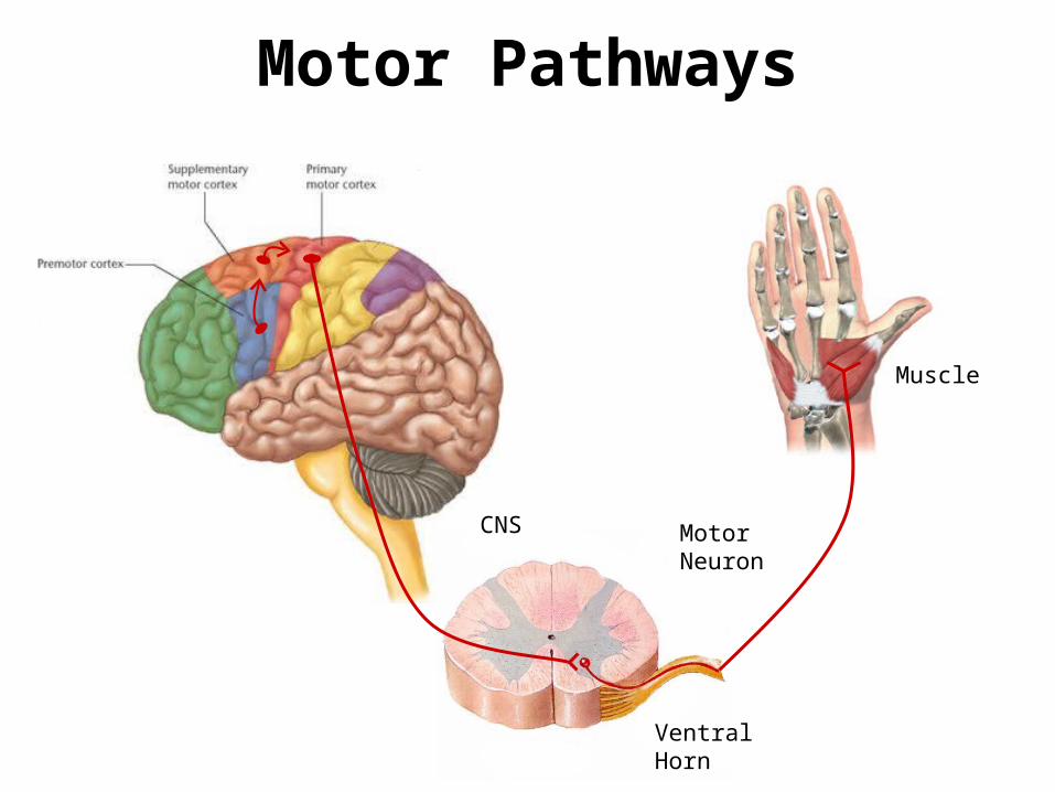
Motor Pathways
Ventral Horn
CNS Motor Neuron
Muscle
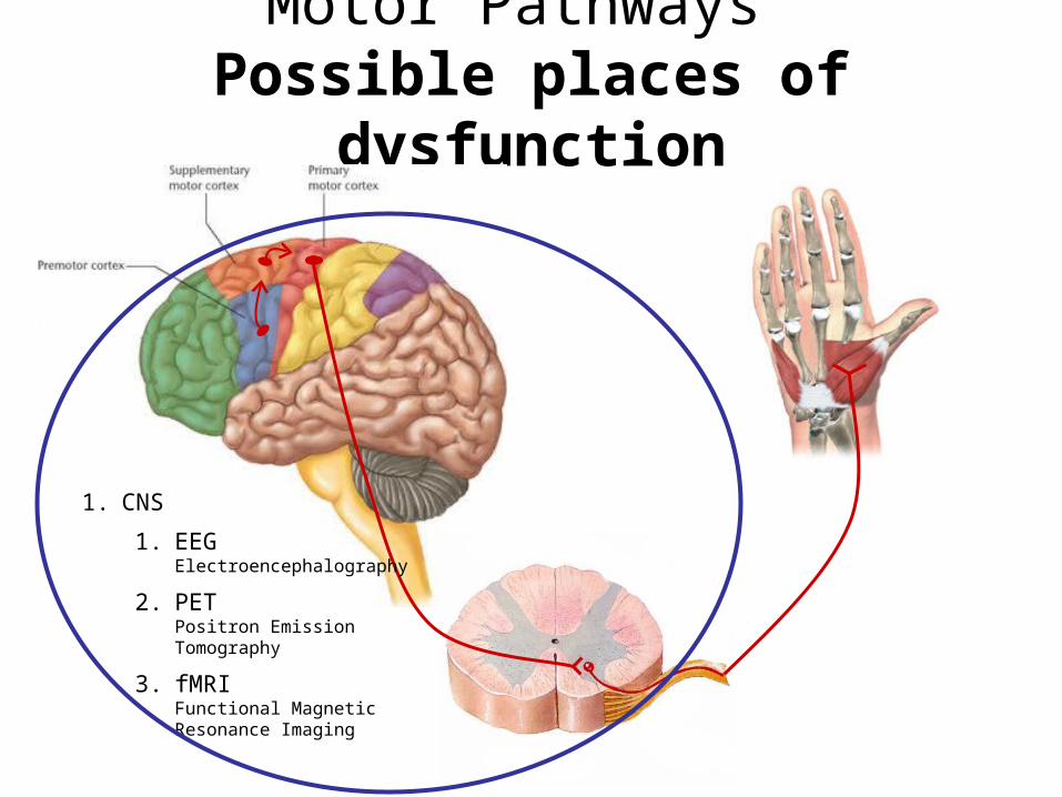
Motor Pathways Possible places of dysfunction
1. CNS
1. EEGElectroencephalography
2. PETPositron Emission Tomography
3. fMRIFunctional Magnetic Resonance Imaging
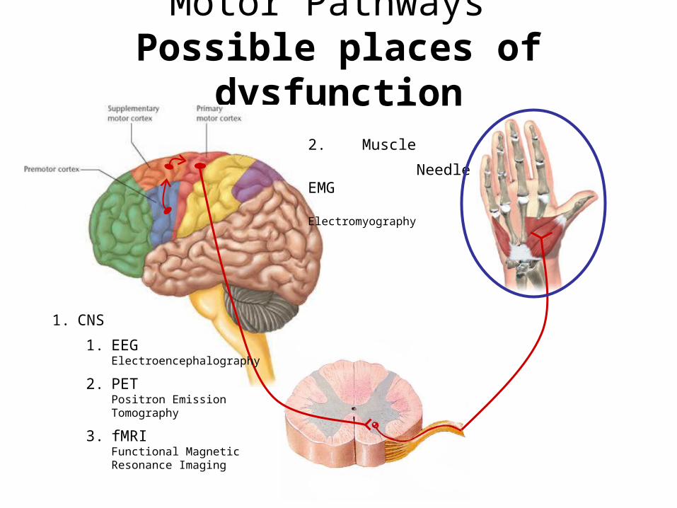
Motor Pathways Possible places of dysfunction
2. Muscle
Needle EMG Electromyography
1. CNS
1. EEGElectroencephalography
2. PETPositron Emission Tomography
3. fMRIFunctional Magnetic Resonance Imaging
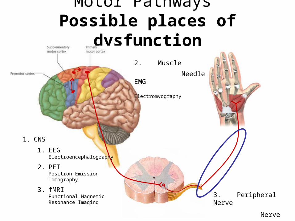
Motor Pathways Possible places of dysfunction
2. Muscle
Needle EMG Electromyography
1. CNS
1. EEGElectroencephalography
2. PETPositron Emission Tomography
3. fMRIFunctional Magnetic Resonance Imaging 3. Peripheral Nerve
Nerve conduction study
Peripheral Nerve
• Made up of:- Sensory nerves
- Motor nerves
- Myelin
- Glial cells
MEDIAN ULNAR
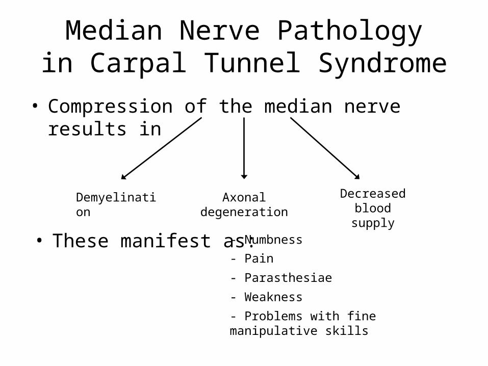
Median Nerve Pathologyin Carpal Tunnel Syndrome
• Compression of the median nerve results in
Demyelination Axonal degeneration
Decreased blood supply
• These manifest as: - Numbness
- Pain
- Parasthesiae
- Weakness
- Problems with fine manipulative skills
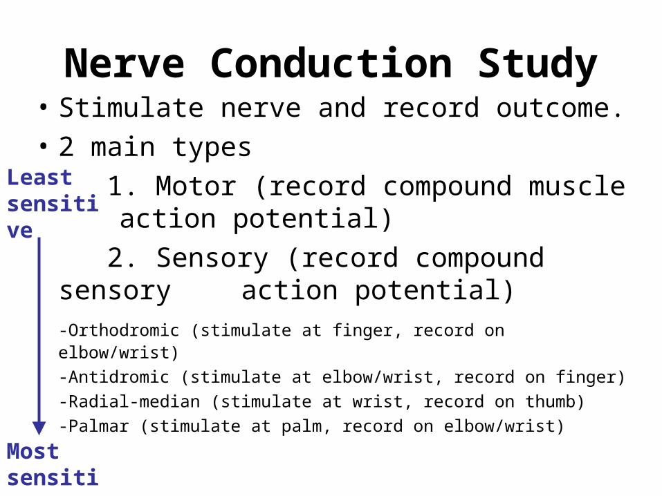
Nerve Conduction Study• Stimulate nerve and record outcome.
• 2 main types
1. Motor (record compound muscle action potential)
2. Sensory (record compound sensory action potential)
-Orthodromic (stimulate at finger, record on elbow/wrist)
-Antidromic (stimulate at elbow/wrist, record on finger)
-Radial-median (stimulate at wrist, record on thumb)
-Palmar (stimulate at palm, record on elbow/wrist)Most sensitive
Least sensitive
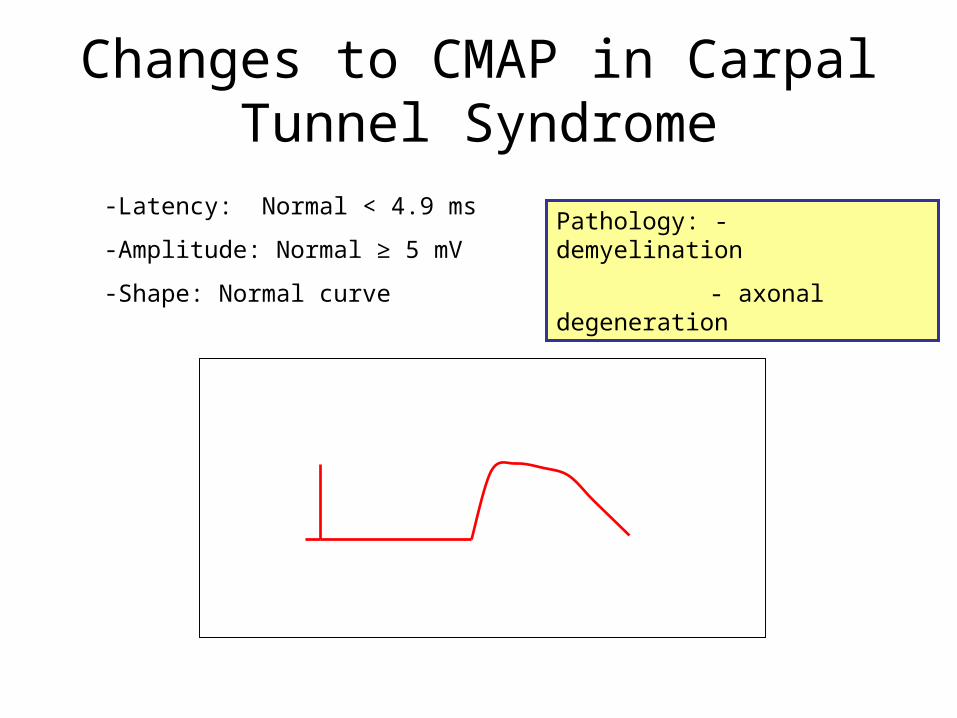
Changes to CMAP in Carpal Tunnel Syndrome
-Latency: Normal < 4.9 ms
-Amplitude: Normal ≥ 5 mV
-Shape: Normal curve
Time
Response amplitude
Pathology: - demyelination
- axonal degeneration
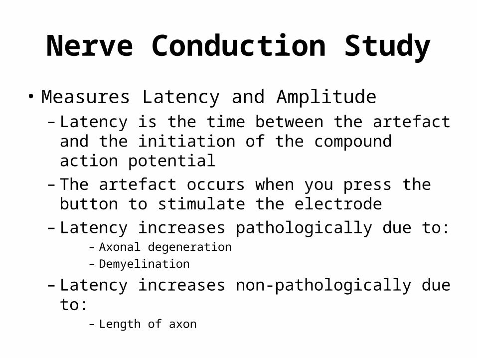
Nerve Conduction Study
• Measures Latency and Amplitude– Latency is the time between the artefact and
the initiation of the compound action potential– The artefact occurs when you press the button
to stimulate the electrode– Latency increases pathologically due to:
– Axonal degeneration– Demyelination
– Latency increases non-pathologically due to:– Length of axon
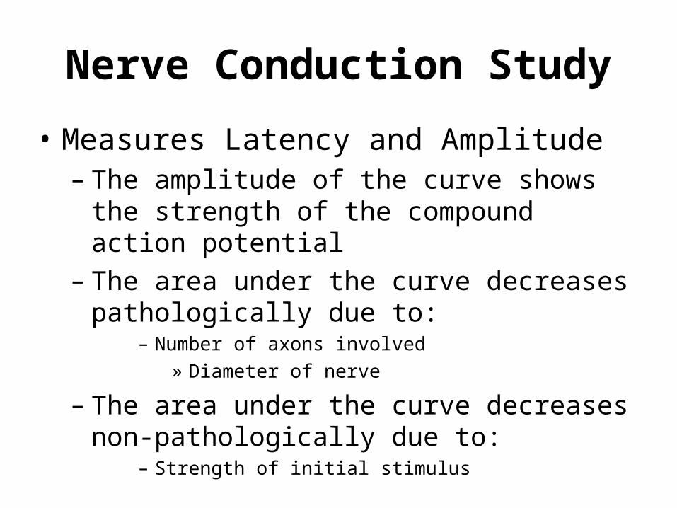
Nerve Conduction Study
• Measures Latency and Amplitude– The amplitude of the curve shows the strength
of the compound action potential– The area under the curve decreases
pathologically due to:– Number of axons involved
» Diameter of nerve
– The area under the curve decreases non-pathologically due to:
– Strength of initial stimulus
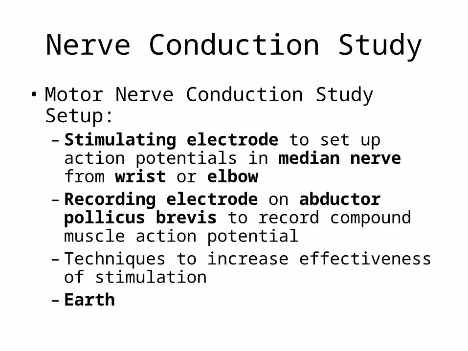
Nerve Conduction Study
• Motor Nerve Conduction Study Setup:– Stimulating electrode to set up action
potentials in median nerve from wrist or elbow
– Recording electrode on abductor pollicus brevis to record compound muscle action potential
– Techniques to increase effectiveness of stimulation
– Earth
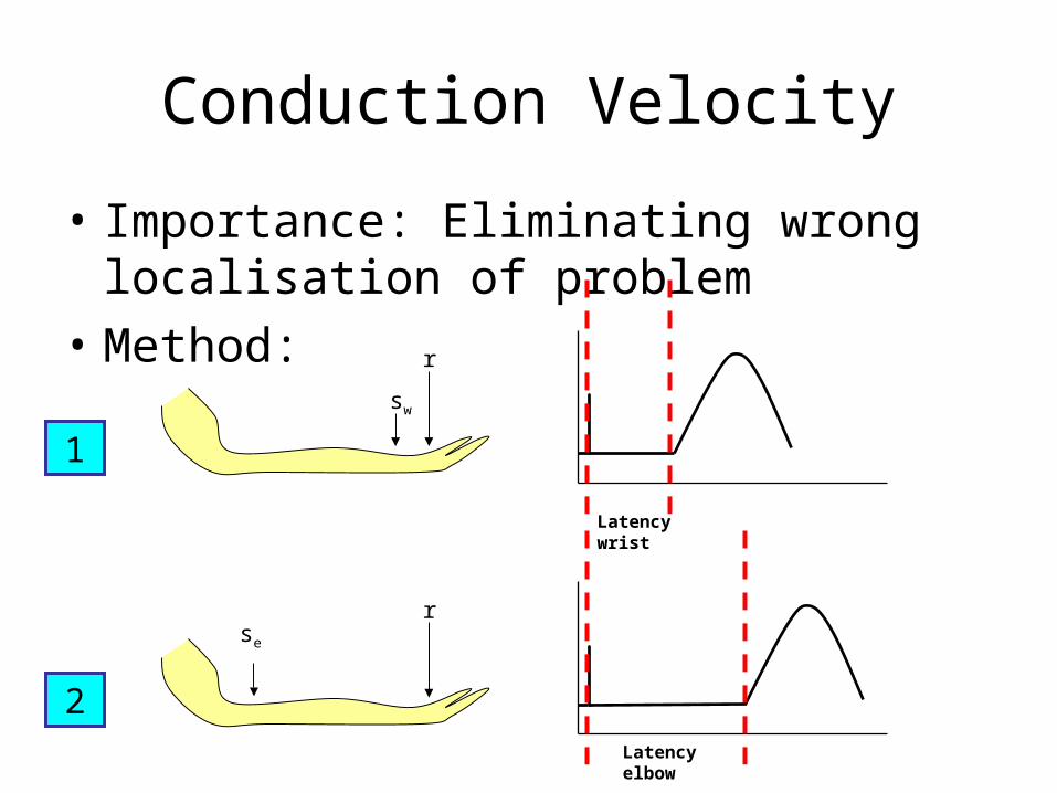
Conduction Velocity
• Importance: Eliminating wrong localisation of problem
• Method:
1
sw
r
Latency wrist
2
se
r
Latency elbow
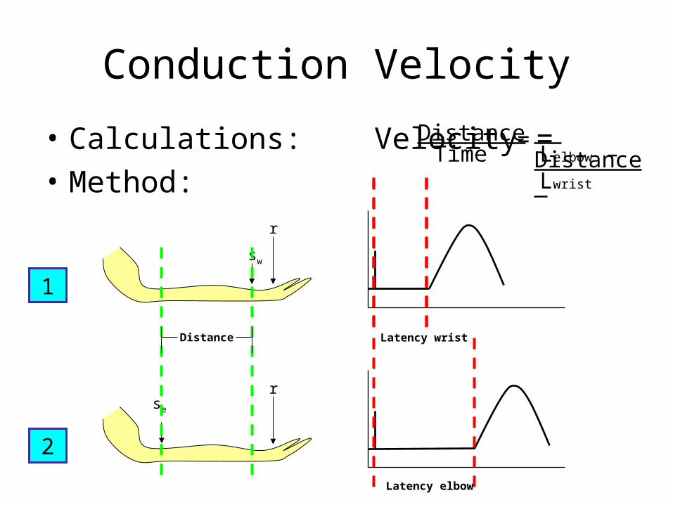
Conduction Velocity
• Calculations: Velocity =
• Method:
1
sw
r
Latency wrist
2
se
r
Latency elbow
Distance
DistanceTime
Distance Lelbow – Lwrist
=
Conduction Velocity
• Normal value > 50 m/s
• Other sources of error
– stimulus position
- estimation of nerve course
- latency measurement
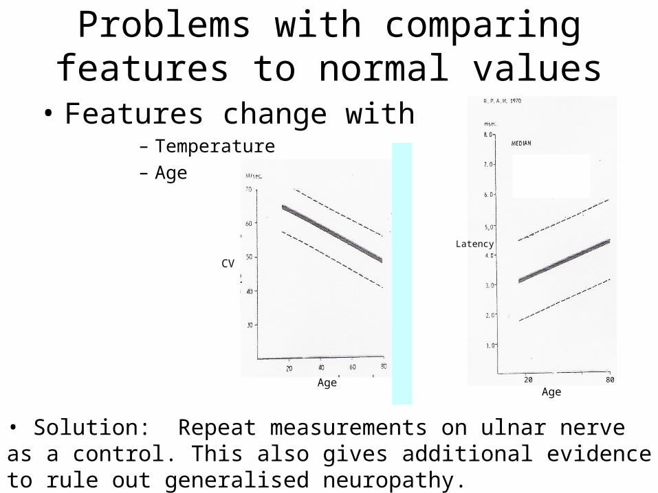
Problems with comparing features to normal values
• Features change with– Temperature – Age
Age20 80
Latency
Age
CV
• Solution: Repeat measurements on ulnar nerve as a control. This also gives additional evidence to rule out generalised neuropathy.
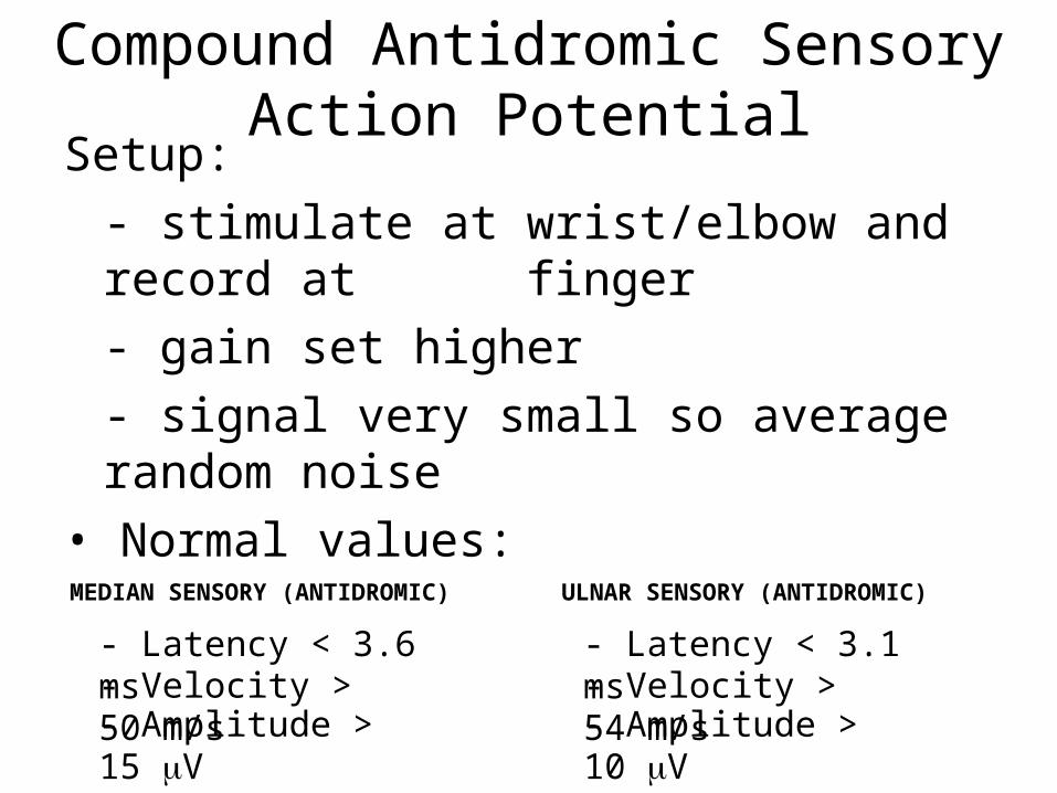
Compound Antidromic Sensory Action Potential
Setup:
- stimulate at wrist/elbow and record at finger
- gain set higher
- signal very small so average random noise
• Normal values:
- Latency < 3.6 ms- Velocity > 50 m/s- Amplitude > 15 V
- Latency < 3.1 ms- Velocity > 54 m/s- Amplitude > 10 V
MEDIAN SENSORY (ANTIDROMIC) ULNAR SENSORY (ANTIDROMIC)
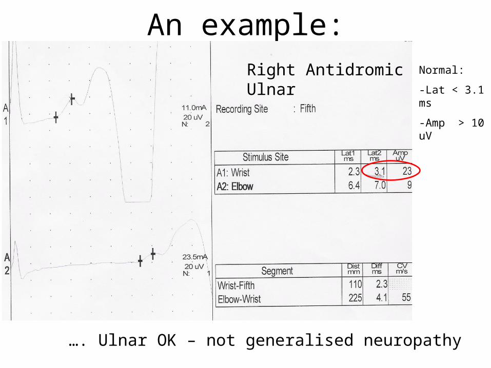
An example:Right Antidromic Ulnar Normal:
-Lat < 3.1 ms
-Amp > 10 uV
…. Ulnar OK – not generalised neuropathy

An example:Right Antidromic Median Normal:
-Lat <3.6 ms
-Amp >15 uV
-CV >50 m/s
…. In median nerve, evidence of demyelination and axonal degeneration between wrist and finger, but not between wrist and elbow. Suggestive of carpal tunnel syndrome.
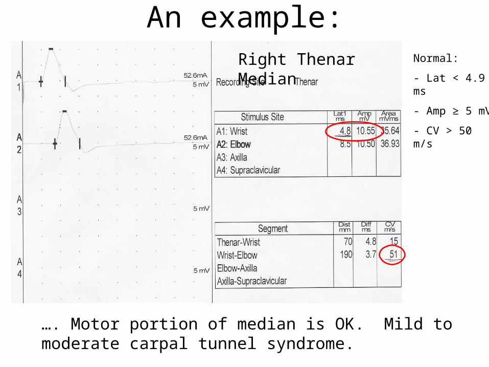
An example:Right Thenar Median Normal:
- Lat < 4.9 ms
- Amp ≥ 5 mV
- CV > 50 m/s
…. Motor portion of median is OK. Mild to moderate carpal tunnel syndrome.
Take home message!
• In carpal tunnel syndrome there will be a longer latency between the wrist and the finger/thenar eminence but NOT between the elbow and the wrist
• The ulnar nerve will not be effected in carpal tunnel syndrome because it does not pass through the carpal tunnel