9

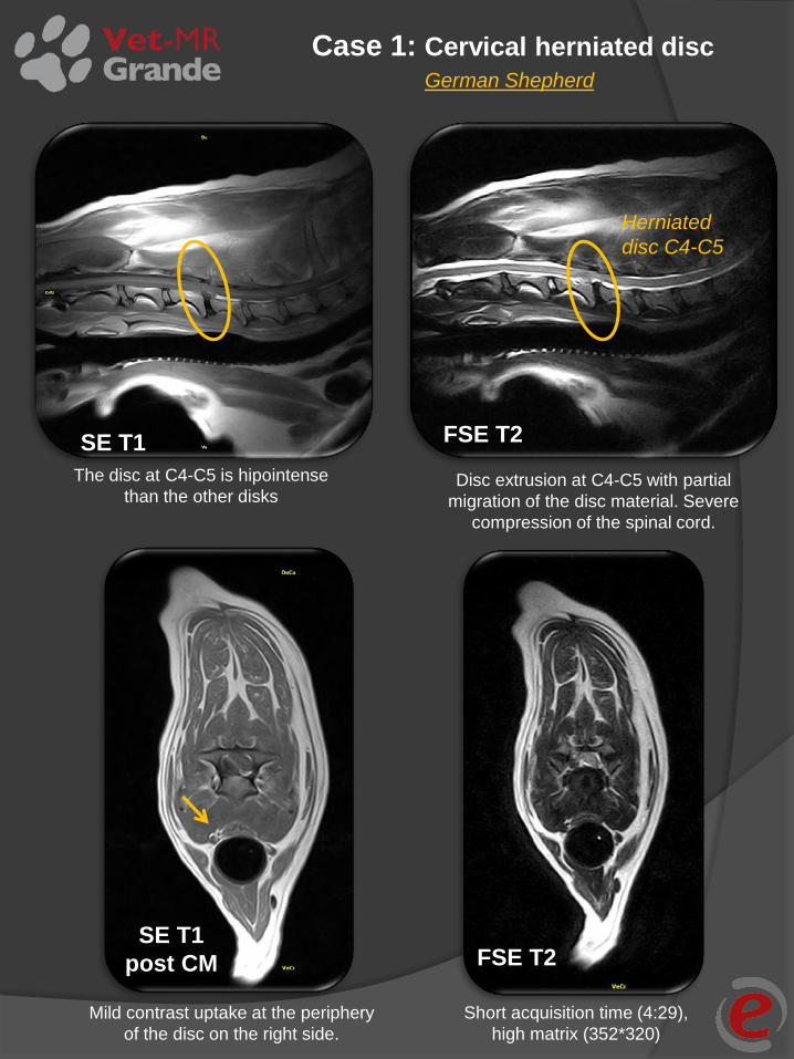
Case 1: Cervical herniated disc
Herniated
disc C4-C5
SE T1
Disc extrusion at C4-C5 with partial
migration of the disc material. Severe
compression of the spinal cord.
SE T1
post CM
FSE T2
The disc at C4-C5 is hipointense
than the other disks
FSE T2
Mild contrast uptake at the periphery
of the disc on the right side.
Short acquisition time (4:29),
high matrix (352*320)
German Shepherd

SE T1 FSE T2
FSTIR
The fat signal is completely suppressed,
note the ramification of the vessels in the
paravertebral soft tissue.
Irregular ventral profile of the L7-S1
intervertebral disc.
Sagittal plane, cranial is on the left.
Case 2: Lumbar Spine Italian Hound

FSE T2
SE T1
SE T1 FSE T2
Disc extrusion at T11-T12 left side. Severe compression of the spinal cord.
T11-T12 and T12-T13 calcificated discs. T11-T12 disc extrusion.
Case 3: Thoracic herniated disc Dachshund

GE STIR SE T1
post CM
FSE T2 SE T1
post CM
High contrast uptake of the left
side soft tissue of the neck.
Hyperintense signal of the left side
soft tissue of the neck from C5 to T2.
Diffuse signal hyperintesity around
the left spinal nerve of C6-C7.
High contrast uptake.
Suspect involvement of the
brachial plexus left side.
Case 4: Contrast uptake (neck) English Setter

FLAIR
SE T1
FSE T2
Severe hydrocepahlus of the
lateral ventriculs. The CSF signal
is completely suppressed.
Crowding of the cerebellum at the
foramen magnum.
Case 5: Hydrocephalus of the lateral ventricules
Mongrel

FLAIR
FSE T2
FLAIR FSE T2
Hyperintense signal at the brain
stem, no mass effect is detected.
Hyperintense signal at the brain stem, right
side.
The signal of the lateral ventriculi is
completely depressed, note the high
hyperintense signal at the brain stem,
right side.
Case 6: Brain stem hyperintensity Jack Russel

SE T1
SE T1
post CM FSE T2
SE T1 post CM FSE T2
Left side deviation of the
cerebrum falx.
High hyperintense signal of
the mass at the frontal lobe,
right side.
Intracranial space occupying mass at the
frontal lobe.
Omogeneous contrast uptake of
the mass, peripheral uptake of
the meningis of the right
temporal lobe.
Case 7: Intracranial mass at the frontal lobe
Schnauzer


