Correction of a Class III malocclusion withover 20 mm of space to close in the maxillaby using miniscrews for extra anchorageK. Hero BreuningTiel, the Netherlands
Unilateral closure of maxillary extraction spaces in patients with Class III malocclusion can be challenging.This case report describes the closure of first premolar and first molar extraction spaces in a patient with aClass III dental relationship. Two miniscrews were used for intraoral skeletal anchorage. The Class III dentalrelationship was corrected; a positive vertical overbite was achieved; occlusion of the canines, premolars,and molars was improved; and the extraction spaces were closed. (Am J Orthod Dentofacial Orthop 2008;
133:459-69)
Unilateral closure of a large maxillary extractionspace in a patient with a Class III dentalrelationship can be a serious orthodontic chal-
lenge. The use of miniscrews for skeletal anchorageduring orthodontic treatment, originally introduced byCreekmore and Eklund,1 allows large extraction spacesto be closed during treatment, without detriment to theposition of the anterior teeth.
DIAGNOSIS AND ETIOLOGY
The patient, a boy aged 13 years 6 months, wasreferred by his dentist for correction of his malocclu-sion (Figs 1-4). Clinical evaluation indicated a skeletalClass I pattern and a midline shift of the mandible by 3mm to the right. Overbite and overjet were approxi-mately –1 mm, and the right canine was out ofocclusion; the occlusion of the right canines and thefirst molar was a Class III relationship. There was nocrowding in either dental arch.
The facial photographs show a mild Class III facialappearance, a chin deviation of 3 mm to the right, andincompetent lips at rest. The patient reported no historyof mandibular prominence in the family. Intraoralphotographs and dental casts showed the extent of the2 extraction spaces in the maxillary arch. The pan-oramic radiograph showed all teeth present, but therewas extensive caries in the maxillary left first premolarand first molar. The patient’s general practitioner
thought that both teeth were beyond repair and shouldbe extracted. The resulting large extraction spaceswould need to be restored with implants or bridges, orclosed orthodontically.
The cephalometric tracing and analysis (Fig 4,Table I) showed an SNA angle of 77.6°, indicating aretrognathic maxillary complex. The ANB angle, how-ever, was normal at 3°. The maxillary incisors wereretroclined, and the mandibular incisors were slightlyproclined. The Wits appraisal indicated a normal ClassI relationship.
TREATMENT OBJECTIVES
The treatment objectives for this patient were tocorrect the Class III incisor relationship; achieve apositive vertical overbite; improve the occlusion of thecanines, premolars, and molars; close the extractionspaces; finish; and retain.
TREATMENT ALTERNATIVES
Correction of the malocclusion would involve tra-ditional orthodontic treatment with fixed appliances inboth arches. A combined orthodontic/orthognathic ap-proach after growth was initially considered. The pa-tient had a mild skeletal Class III relationship, butcephalometric measurements were within the normalrange, and no prominent jaws were reported in thefamily. Development of a more severe Class III skeletalrelationship was thought unlikely; therefore, delayingthe start of treatment to observe the need for surgicalcorrection of the skeletal relation was not indicated.
Closure of extraction spaces closure was the mostchallenging aspect, particularly because of the Class IIIincisor relationship. Correction of the occlusion withoutclosing the spaces was, of course, an option, but this
treatment plan would require prosthetic restoration of the
American Journal of Orthodontics and Dentofacial OrthopedicsMarch 2008
460 Breuning
dentition after orthodontic treatment. Because of thepatient’s age, alveolar growth after orthodontic treatmentwould be expected; temporary prosthetic devices wouldbe needed to fill the spaces in the maxillary arch untildental implants could be used for a definitive solution.
If feasible, closure of 1 extraction space or bothwould be the treatment of choice.
TREATMENT PLAN
Orthodontic mechanics with significantly reducedfriction in the molar and premolar regions were desir-able because less friction would help reduce the forceneeded to close the extraction spaces. Molar andpremolar tubes were used (Fig 5).
Two miniscrews (1 between the maxillary leftlateral incisor and the canine; the second just distal tothe canine) were placed to significantly enhance ante-rior anchorage during protraction of the maxillary leftsecond premolar and the second molar. Interarch elastictraction would be used to help with correction ofoverjet, overbite, and occlusion.
TREATMENT PROGRESS
The maxillary teeth were bonded with a .022-inpreadjusted self-ligating bracket system. In the maxil-
Fig 1. Pretreatment extra
lary incisors and canines, interactive self-ligating
brackets were used. In the maxillary premolars andmolars, bonded tubes were placed. Tip and torquevalues used in this patient are listed in Table II.
A continuous .014-in nickel-titanium-seleniumwire, with a dimple between the maxillary centralincisors to prevent sliding, was used during the firstphase of treatment.
After leveling, which took only 8 weeks, a .016 �.025-in nickel-titanium-selenium wire was used forfurther correction of rotations, tip, and torque of themaxillary teeth. One month later, a panoramic radio-graph was taken to help determine the position of theminiscrews (Fig 6).
Miniscrews (diameter, 1.6 mm; length, 10 mm)were considered ideal for this patient, and he wasreferred to the maxillofacial surgeon for placement of 2miniscrews under local anesthesia. One was placedbetween the maxillary left lateral incisor and thecanine, and the second just distal to the canine.
Immediately after surgery, a ligature was used toconnect the miniscrew to the maxillary left canine toincrease the anchorage value of this tooth (Fig 7). Anickel-titanium spring was then used to provide gentleforces to protract the second premolar and the secondmolar simultaneously. Those teeth had been previously
nd intraoral photographs.
linked together with a steel ligature. At the next visit, even
American Journal of Orthodontics and Dentofacial OrthopedicsVolume 133, Number 3
Breuning 461
with the miniscrew to stabilize the position of the canine,some distalization was observed (Fig 8). Because of thisloss of anchorage, the mechanical system was changed.Direct loading of the mesial miniscrew with the samenickel-titanium spring might have helped to protract thesecond premolar and the second molar, but without the
Fig 2. Pretrea
Fig 3. Pretreatment panoramic radiograph.
tment dental casts.
reciprocal distalizing force on the canine.
Fig 4. Pretreatment cephalometric tracing.
American Journal of Orthodontics and Dentofacial OrthopedicsMarch 2008
462 Breuning
As a result of the direction of the applied traction,an open bite started to develop (Fig 9). It was decidedat this stage to use a sliding hook with a long armplaced distal to the second premolar to provide theforce to protract it. For protraction of the second molar,a nickel-titanium spring connected to the distal minis-crew and the second molar was used.
For evaluation purposes, a panoramic radiograph wastaken (Fig 10). Controlled protraction of the secondpremolar and the second molar (without tipping of theroots) was confirmed. Extraoral photographs were used toevaluate facial changes (Fig 11). Fixed appliances wereplaced in the mandible to correct the sagittal and verticalrelationships between the arches. Protraction of the secondpremolar and the second molar was finally hindered by theposition of the miniscrews, and they were removed,without anesthetic, during a regular appliance check visit.No pain or discomfort was reported, either during or afterremoval. Once the miniscrews had been removed, finalcorrection of the malocclusion occlusion was achievedwith the help of interarch elastics.
At the end of treatment, the maxillary dental mid-
Fig 5. Brackets and tubes (Hero system) used in thispatient.
Table I. Pretreatment and posttreatment cephalometricvalues
Pretreatment Posttreatment
SNA angle 77.6° 78.3°SNB angle 74.5° 74.9°ANB angle 3.0° 3.4°Y-axis 70.2° 72.2°U1 to ANS-PNS 98.6° 100.3°SOB 0.3 mm 3.0 mmVOB �1.0 mm 1.6 mmU1 to FOP �0.4 mm �1.1 mmL1 to FOP �0.6 mm 0.5 mmLI to MP 97.3° 99.5°SN to MP 35.6° 35.2°Wits appraisal 0.1 mm 0.7 mm
line was positioned in line with the facial midline, but
the mandibular dental midline was not coordinated withthe maxillary midline, because of the underlying man-dibular asymmetry.
The panoramic radiograph (Fig 12) shows goodposition of the roots with no root resorption evident.The maxillary left third molar should erupt in occlusionwith the mandibular second molar. The second man-dibular molar overerupted slightly during the first fewweeks after treatment; the overeruption was identifiedat the first posttreatment check visit, and a fixed retainerwas used to prevent further eruption (Fig 13).
The posttreatment cephalometric tracing and ceph-alometric values are shown in Figure 14 and Table I.Superimposed tracings (Fig 15) show the differences inthe position of the maxillary left second molar beforetreatment. The maxillary superimposition shows theposition of the left second molar and the most promi-nent maxillary incisor, and the mandibular superimpo-
Fig 6. Panoramic radiograph during treatment.
Table II. Torque, angulation, and tip values
Torque Angulation Rotation
MaxillaCentral incisors �12° �5°Lateral incisors �8° �9°Canine with hooks �2° �9°First and second premolar
tubes (� hooks)�7° �0°
First and second molar tubes(� hooks)
�10° �0° 10°
MandibleAnterior teeth �1° 0°Canines with hooks �2° �7°First premolar �11° 0°Second premolar tubes with
hook�17° 0°
First molar tubes with hooks �20° 0°Second molar tubes with hooks �20° 0°
sition shows the position of the first molar and most
media
owing
raph
American Journal of Orthodontics and Dentofacial OrthopedicsVolume 133, Number 3
Breuning 463
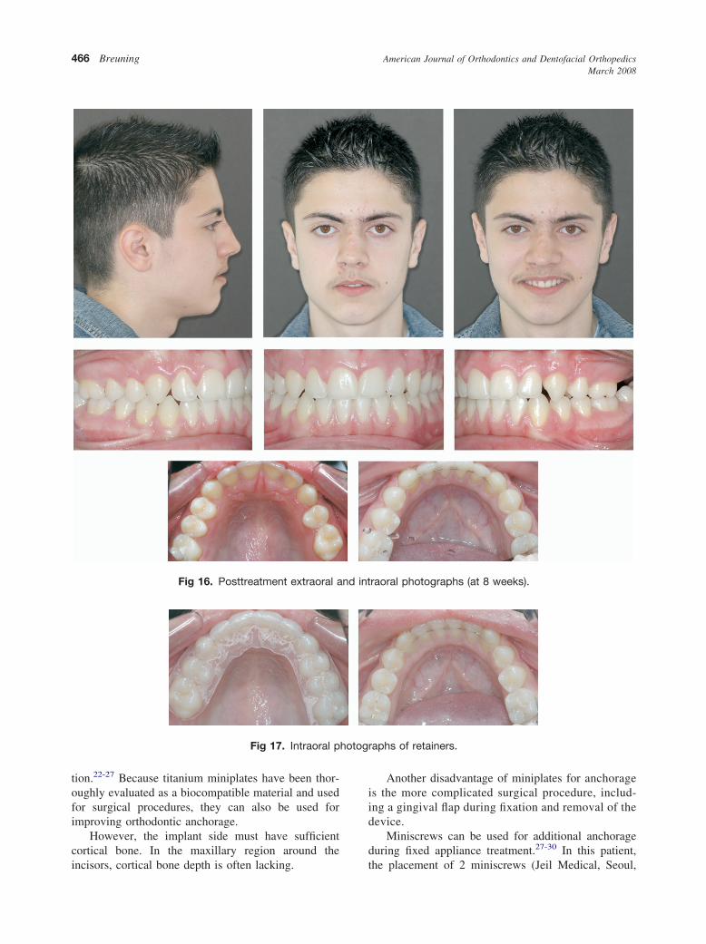
prominent incisor. Posttreatment extraoral and intraoralphotos were taken during the first retention visit, 8weeks after removal of the appliance (Fig 16). Reten-tion consisted of a fixed retainer in the mandibular archand an Essix (Raintree, Seoul, Korea) retainer in the
Fig 7. Intraoral photographs im
Fig 8. Intraoral photograph sh
Fig 9. Intraoral photog
Fig 10. Panoramic radiograph showing miniscrews androots during protraction.
maxillary arch (Fig 17). During settling, the occlusion
changed. Plaster study models taken 6 months post-treatment (Fig 18) show coordinated midlines.
tely after miniscrew placement.
loss of anchorage of canine.
showing bite opening.
Fig 11. Frontal extraoral photograph during treatment.
The total active treatment time was 1 year 6 months.
American Journal of Orthodontics and Dentofacial OrthopedicsMarch 2008
464 Breuning
DISCUSSION
The main problem in treating this patient was theexcessive extraction spaces in 1 side of the maxillaryarch with no crowding and no overjet. Closing thisextraction space, without a detrimental effect on themaxillary arch form, and correcting the interarch rela-tionships were extremely tall orders.
The etiology of this malocclusion was mainlygenetic, with a small retropositioned maxilla. Treat-ment was complicated by the extreme dental decay.
The patient needed medication early in his life forsevere asthma; this might have contributed to thedecay, but the medical history was not clear. He wasnot a regular dental patient. Before any orthodontictreatment was attempted, dietary habits and oral hy-giene were discussed with him and his parents. Duringactive treatment, the patient was asked to use a fluoriderinse daily to prevent further tooth decay.
Anchorage
At least a quarter of the extraction space is routinelyoccupied by the anchor teeth during orthodontic treat-ment unless measures are taken to supplement thisanchorage.2 Absolute anchorage was needed to prevent
Fig 12. Posttreatment panoramic radiograph.
Fig 13. Closeup of retention of the mandibular leftsecond molar to prevent eruption.
retraction of the maxillary incisors and worsening of
the Class III incisor relationship. Ideally, the maxillaryincisors would be moved forward during orthodontictreatment to correct overjet and overbite.
Anchorage loss is related to the forces needed toprotract the premolar and the second molar. If a bracketsystem with lower friction is used, less force willtheoretically be needed to protract the teeth, and there-fore less anchorage loss can be expected.3
Self-ligating brackets were introduced in the mid-1930s in the form of the Russell attachment (United StatesPatent 6302688). Two types of self-ligating brackets arepopular: those with a self-ligating clip that does not pressagainst the wire (passive), such as the Damon and SmartClip brackets, and those with a clip that presses against thewire if a larger wire is used (interactive), such as Speed,System R, and Time brackets.
In this patient, a combination of passive and inter-active brackets was used. A combination of rectangularwire and interactive brackets (Time; American Orth-odontics, Sheboygan, Wis) was used to correct thetorque of the incisors, overjet, and overbite. Passivebrackets (Nr 098-1510C, Nr 098-1511C; Hero tubes)were used to protract the premolar and the molar in themaxillary arch. Elastic forces were also used to coun-
Fig 14. Cephalometric tracing 6 months after treatment.
teract tongue and lip pressure and close the bite.
American Journal of Orthodontics and Dentofacial OrthopedicsVolume 133, Number 3
Breuning 465
Several studies demonstrated significant decreasesin friction for self-ligating brackets, compared withconventional bracket designs, by using elastics or steelligatures.4-7
The use of premolar and molar tubes in this patientcontributed to a mechanical system that required lowforces to move the teeth. When temporary anchoragedevices are used, a low force system is recommended.8,9
It has been suggested that reduced friction can helpto shorten treatment time, especially in extractionpatients in whom tooth translation is achieved bysliding mechanics.
Anchorage alternatives
Extraction of the right first premolars in the maxillaand the mandible was also considered, but, because ofthe patient’s Class III profile, cephalometric values,dental relationships, and the absence of crowding in thearches, this treatment alternative was rejected. Closingof all spaces in the left side of the maxilla duringcorrection of the occlusion, without compensating ex-tractions, required additional anchorage. A facemask toprovide this extra anchorage was considered, but,because of the patient’s age, sufficient compliance inusing it during treatment could not be guaranteed, andhe was not keen about this approach. Furthermore,anchorage was needed only on the left side of themaxilla, so it was a unilateral problem.
The use of a standard dental implant in the firstpremolar region for temporary anchorage during orth-odontic tooth movement to protract the maxillary leftsecond molar, and then as a permanent abutment for toothreplacement, was reported and was considered.10-15
The patient’s age would have necessitated addi-tional time for osseointegration, and this, in addition tothe costs of the implant and the crown, meant that thisapproach was rejected.
Several temporary anchorage devices for orthodonticanchorage have been introduced.16-18 In this patient,several temporary anchorage devices could have beenused. A palatal implant was described for absolute anchor-age during orthodontic treatment and can be used forvarious orthodontic procedures such as en-masse retrac-tion of the 6 anterior teeth or canine distalization withoutanchorage loss.19-21 The position of a palatal implant isnot the most suitable place for skeletal anchorage toprotract a second premolar and molar, and this treatmentoption was rejected in this patient. There were otherdisadvantages to a palatal implant for this patient, such asthe extra time needed for its integration, the costs of theimplant, and the discomfort of surgical placement andremoval.
Fig 15. Superimposed tracing before and after treat-ment. Maxillary superimposition shows the position ofthe maxillary left second molar and the mandibularincisors, before and after treatment.
Miniplates can be used to help protract the denti-
nd int
hotog
American Journal of Orthodontics and Dentofacial OrthopedicsMarch 2008
466 Breuning
tion.22-27 Because titanium miniplates have been thor-oughly evaluated as a biocompatible material and usedfor surgical procedures, they can also be used forimproving orthodontic anchorage.
However, the implant side must have sufficientcortical bone. In the maxillary region around the
Fig 16. Posttreatment extraoral a
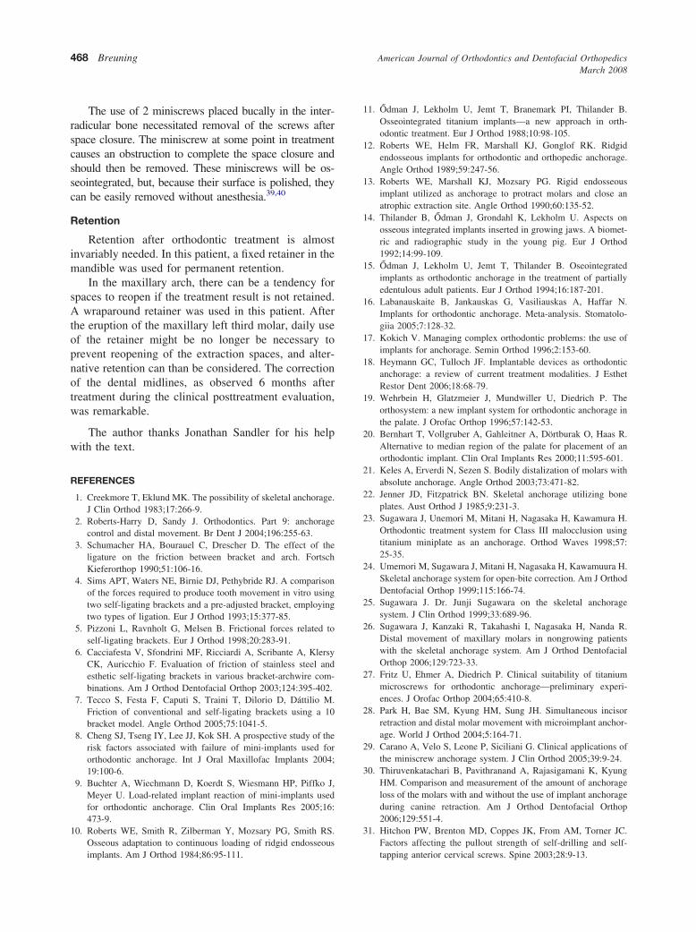
Fig 17. Intraoral p
incisors, cortical bone depth is often lacking.
Another disadvantage of miniplates for anchorageis the more complicated surgical procedure, includ-ing a gingival flap during fixation and removal of thedevice.
Miniscrews can be used for additional anchorageduring fixed appliance treatment.27-30 In this patient,
raoral photographs (at 8 weeks).
raphs of retainers.
the placement of 2 miniscrews (Jeil Medical, Seoul,
6 mo
American Journal of Orthodontics and Dentofacial OrthopedicsVolume 133, Number 3
Breuning 467
Korea) was considered the most suitable treatmentoption. The surgical procedure required only localanesthetic, no gingival flap was required, miniscrewsare relatively inexpensive, and miniscrews can beremoved in the orthodontic office without anesthesia.
Surgical procedure
When orthodontic treatment including miniscrewsis planned, a protocol for their use should be followed,but there is no widely accepted protocol yet.31-38
Our team’s procedure for placing miniscrews usu-ally uses these guidelines. The patient rinses with achlorhexide solution for at least 1 minute before sur-gery. Local anesthetics are administered, and self-tapping screws are used without predrilling, unless thecortical bone is excessively thick. In that case, predrill-ing of the cortical bone only is sufficient. Manualscrewdrivers are always used, and the junction of theattached gingiva and the reflected mucosa is consideredto be the ideal site. The miniscrew is placed at an angleof up to 45° to the cortical plate. Primary stability,indicated by the screw’s resistance to lateral movement,is desirable. If primary stability is not achieved, a largerscrew, a different location, or an additional screw
Fig 18. Dental casts,
should be considered. After surgery, the patient is
instructed on the maintenance of the miniscrews andreferred to the orthodontist for the application oforthodontic force. Immediate loading with a gentleforce from a nickel-titanium spring (within severalhours of surgery) is recommended.
In our opinion, the orthodontist is responsible forpostoperative instruction on screw cleaning and whatthe patient should do in case of screw failure.
Relatively low forces should be used, and thereforesliding mechanics and bracket systems with low fric-tion are recommended. Intermittent forces should cer-tainly be avoided. Continuous forces (nickel-titaniumsprings, nickel-titanium wires, power chain, E-links)can all be used effectively.
Treatment after the surgical procedure
A nickel-titanium coil spring (with a constant forceof 100 g) was used to protract the second premolar andthe second molar. In this patient, the initial treatmentmechanics and the strengthening of the anchorage ofthe maxillary left canine were not outstandingly suc-cessful. Changing the force mechanism to direct load-ing of the miniscrew was successful. Changing theforce mechanism again was also needed to correct the
nths after treatment.
open bite caused by the vertical component of traction.
American Journal of Orthodontics and Dentofacial OrthopedicsMarch 2008
468 Breuning
The use of 2 miniscrews placed bucally in the inter-radicular bone necessitated removal of the screws afterspace closure. The miniscrew at some point in treatmentcauses an obstruction to complete the space closure andshould then be removed. These miniscrews will be os-seointegrated, but, because their surface is polished, theycan be easily removed without anesthesia.39,40
Retention
Retention after orthodontic treatment is almostinvariably needed. In this patient, a fixed retainer in themandible was used for permanent retention.
In the maxillary arch, there can be a tendency forspaces to reopen if the treatment result is not retained.A wraparound retainer was used in this patient. Afterthe eruption of the maxillary left third molar, daily useof the retainer might be no longer be necessary toprevent reopening of the extraction spaces, and alter-native retention can than be considered. The correctionof the dental midlines, as observed 6 months aftertreatment during the clinical posttreatment evaluation,was remarkable.
The author thanks Jonathan Sandler for his helpwith the text.
REFERENCES
1. Creekmore T, Eklund MK. The possibility of skeletal anchorage.J Clin Orthod 1983;17:266-9.
2. Roberts-Harry D, Sandy J. Orthodontics. Part 9: anchoragecontrol and distal movement. Br Dent J 2004;196:255-63.
3. Schumacher HA, Bourauel C, Drescher D. The effect of theligature on the friction between bracket and arch. FortschKieferorthop 1990;51:106-16.
4. Sims APT, Waters NE, Birnie DJ, Pethybride RJ. A comparisonof the forces required to produce tooth movement in vitro usingtwo self-ligating brackets and a pre-adjusted bracket, employingtwo types of ligation. Eur J Orthod 1993;15:377-85.
5. Pizzoni L, Ravnholt G, Melsen B. Frictional forces related toself-ligating brackets. Eur J Orthod 1998;20:283-91.
6. Cacciafesta V, Sfondrini MF, Ricciardi A, Scribante A, KlersyCK, Auricchio F. Evaluation of friction of stainless steel andesthetic self-ligating brackets in various bracket-archwire com-binations. Am J Orthod Dentofacial Orthop 2003;124:395-402.
7. Tecco S, Festa F, Caputi S, Traini T, Dilorio D, Dáttilio M.Friction of conventional and self-ligating brackets using a 10bracket model. Angle Orthod 2005;75:1041-5.
8. Cheng SJ, Tseng IY, Lee JJ, Kok SH. A prospective study of therisk factors associated with failure of mini-implants used fororthodontic anchorage. Int J Oral Maxillofac Implants 2004;19:100-6.
9. Buchter A, Wiechmann D, Koerdt S, Wiesmann HP, Piffko J,Meyer U. Load-related implant reaction of mini-implants usedfor orthodontic anchorage. Clin Oral Implants Res 2005;16:473-9.
10. Roberts WE, Smith R, Zilberman Y, Mozsary PG, Smith RS.Osseous adaptation to continuous loading of ridgid endosseous
implants. Am J Orthod 1984;86:95-111.
11. Odman J, Lekholm U, Jemt T, Branemark PI, Thilander B.Osseointegrated titanium implants—a new approach in orth-odontic treatment. Eur J Orthod 1988;10:98-105.
12. Roberts WE, Helm FR, Marshall KJ, Gonglof RK. Ridgidendosseous implants for orthodontic and orthopedic anchorage.Angle Orthod 1989;59:247-56.
13. Roberts WE, Marshall KJ, Mozsary PG. Rigid endosseousimplant utilized as anchorage to protract molars and close anatrophic extraction site. Angle Orthod 1990;60:135-52.
14. Thilander B, Odman J, Grondahl K, Lekholm U. Aspects onosseous integrated implants inserted in growing jaws. A biomet-ric and radiographic study in the young pig. Eur J Orthod1992;14:99-109.
15. Odman J, Lekholm U, Jemt T, Thilander B. Oseointegratedimplants as orthodontic anchorage in the treatment of partiallyedentulous adult patients. Eur J Orthod 1994;16:187-201.
16. Labanauskaite B, Jankauskas G, Vasiliauskas A, Haffar N.Implants for orthodontic anchorage. Meta-analysis. Stomatolo-giia 2005;7:128-32.
17. Kokich V. Managing complex orthodontic problems: the use ofimplants for anchorage. Semin Orthod 1996;2:153-60.
18. Heymann GC, Tulloch JF. Implantable devices as orthodonticanchorage: a review of current treatment modalities. J EsthetRestor Dent 2006;18:68-79.
19. Wehrbein H, Glatzmeier J, Mundwiller U, Diedrich P. Theorthosystem: a new implant system for orthodontic anchorage inthe palate. J Orofac Orthop 1996;57:142-53.
20. Bernhart T, Vollgruber A, Gahleitner A, Dörtburak O, Haas R.Alternative to median region of the palate for placement of anorthodontic implant. Clin Oral Implants Res 2000;11:595-601.
21. Keles A, Erverdi N, Sezen S. Bodily distalization of molars withabsolute anchorage. Angle Orthod 2003;73:471-82.
23. Sugawara J, Unemori M, Mitani H, Nagasaka H, Kawamura H.Orthodontic treatment system for Class III malocclusion usingtitanium miniplate as an anchorage. Orthod Waves 1998;57:25-35.
24. Umemori M, Sugawara J, Mitani H, Nagasaka H, Kawamuura H.Skeletal anchorage system for open-bite correction. Am J OrthodDentofacial Orthop 1999;115:166-74.
25. Sugawara J. Dr. Junji Sugawara on the skeletal anchoragesystem. J Clin Orthod 1999;33:689-96.
26. Sugawara J, Kanzaki R, Takahashi I, Nagasaka H, Nanda R.Distal movement of maxillary molars in nongrowing patientswith the skeletal anchorage system. Am J Orthod DentofacialOrthop 2006;129:723-33.
27. Fritz U, Ehmer A, Diedrich P. Clinical suitability of titaniummicroscrews for orthodontic anchorage—preliminary experi-ences. J Orofac Orthop 2004;65:410-8.
28. Park H, Bae SM, Kyung HM, Sung JH. Simultaneous incisorretraction and distal molar movement with microimplant anchor-age. World J Orthod 2004;5:164-71.
29. Carano A, Velo S, Leone P, Siciliani G. Clinical applications ofthe miniscrew anchorage system. J Clin Orthod 2005;39:9-24.
30. Thiruvenkatachari B, Pavithranand A, Rajasigamani K, KyungHM. Comparison and measurement of the amount of anchorageloss of the molars with and without the use of implant anchorageduring canine retraction. Am J Orthod Dentofacial Orthop2006;129:551-4.
31. Hitchon PW, Brenton MD, Coppes JK, From AM, Torner JC.Factors affecting the pullout strength of self-drilling and self-
American Journal of Orthodontics and Dentofacial OrthopedicsVolume 133, Number 3
Breuning 469
32. Fritz U, Diedrich P, Kinzinger G, Al-Said M. Factors associatedwith the stability of titanium screws placed in the posteriorregion for orthodontic anchorage. Am J Orthod DentofacialOrthop 2003;124:373-8.
33. Cheng SJ, Tseng IY, Lee JJ, Kok SH. A prospective study of therisk factors associated with failure of mini-implants used fororthodontic anchorage. Int J Oral Maxillofac Implants 2004;19:100-6.
34. Ishii T, Nojima K, Nishii Y, Takaki T, Yamaguchi H. Evaluationof the implantation position of mini-screws for orthodontictreatment in the maxillary molar area by a micro CT. Bull TokyoDent Coll 2004;45:165-72.
35. Carano A, Lonardo P, Velo S, Incorvati C. Mechanical propertiesof three different commercially available miniscrews for skeletal
anchorage. Prog Orthod 2005;6:82-97.
36. Huja SS, Litsky AS, Beck FM, Johnson KA, Larsen PE. Pull-outstrength of monocortical screws placed in the maxillae andmandibles of dogs. Am J Orthod Dentofacial Orthop 2005;127:307-13.
37. Huang LH, Shotwell JL, Wang HL. Dental implants for orthodonticanchorage. Am J Orthod Dentofacial Orthop 2005;127:713-26.
38. Mah J, Bergstrand F. Temporary anchorage devices: a statusreport. J Clin Orthod 2005;38:133-7.
39. Kim JW, Ahn SJ, Chang YI. Histomorphometric and mechanicalanalyses of the drill-free screw as orthodontic anchorage. Am JOrthod Dentofacial Orthop 2005;128:190-4.
40. Gedrange T, Proff P, Bayerlein T, Landsberger P, Dietze S,Fanghanel J. Histological and fluorescence microscopic exami-nation of the bone/implant interface in orthodontic miniscrews.