Case Report: Mandibular Third Molar
Impaction Features in CBCT 3D Radiography
Gunawan
Faculty of Dentistry, Universitas Andalas
Padang, Indonesia
[email protected]
Lusi Epsilawati Faculty of Dentistry, Universitas Padjadjaran
Bandung, Indonesia
Ivony Fitria Faculty of Dentistry, Universitas Andalas
Padang, Indonesia
Abstract–The mandibular third molar impaction is a
frequent case in dentistry. Various investigations are
required prior to the removal of the mandibular third
molar impaction, such as CBCT 3D radiography. This
radiograph can provide optimum results for the
investigation of mandibular third molar impaction. Case
I: a 20-year-old woman came to Radiology Installation,
RSGM FKG UNPAD with a referral letter for CBCT 3D
photos. Anamnesis showed that patient had complaints of
repeated pain in the area of left posterior teeth. Patient
was pre-medicated and referred for radiographic images.
Case II: a 33-year-old woman came to Radiology
Installation, RSGM FKG UNPAD with a referral letter for
CBCT 3D photograph. Anamnesis revealed patient with
complaints of pain in the left posterior teeth. Patient was
pre-medicated and referred for radiographic images. The
radiodiagnosis of both cases above is Class II position B
mesioangular. In cases of mandibular third molar
impaction, investigation with CBCT 3D can provide
optimal information. CBCT 3D can provide information
about the shape, position and relationship of the impacted
third molars to its surrounding anatomy in sagittal,
coronal and axial view. This accurate information will
make it easier for clinicians to perform adequate
maintenance. CBCT 3D can provide an optimal features
in the management of mandibular third molar impaction.
Keywords–CBCT 3D, impaction, sagittal, coronal, axial
I. INTRODUCTION
Dental impaction is a common case in dentistry.
According to Archer, quoted from Rahayu, as many as
nine out of ten people have an impacted tooth. Impacted
teeth often occur in permanent molar, canine, premolar,
and incisors. One of the highest prevalence of
impaction cases is the impacted mandibular third
molars[1]. An impaction is a condition when tooth
failed to erupt into the dental arch within the expected
time. The word „impaction’ is derived from Latin word
'Impactus', meaning „cessation of eruption caused by
physical barrier/ectopic eruption.‟ The tooth is
categorized as impacted by the presence of another
tooth, bone or soft tissue. Impacted teeth are the teeth
that are blocked during an eruption to achieve a normal
position [2,3,4].
Accurate examination prior to the removal of the
impacted mandibular third molar is necessary. An
accurate preoperative assessment of the radiograph is
crucial for the success of impacted third molars surgery,
but unfortunately, this is often overlooked. Some things
to consider before the removal of the impacted third
molars are: 1) form (both crown and root), size, shape,
caries status, shape, amount, periapical bone loss. 2)
Angulation of impacted molar to the occlusal plane. 3)
Relationship of the second molar, both the crown (size,
shape, caries status) and roots (size, shape, amount). 4)
The relationship of inferior dental canals to the
impaired molar should be determined appropriately
using radiographs. 5) Distal bone level [5,6].
The use of radiography aims to reduce post-
operative complications. These can be pain, swelling,
excessive bleeding, infection and reduced mouth
opening, however, sensory disturbances to the nerves, e.
g. the alveolar inferior nerve (IAN), the buccal nerve
and the lingual nerve, are seen as the most severe
postoperative complications after removal of a
mandibular third molar. In a radiographic image of the
third molar region, only the course of the IAN may be
assured since the mandibular canal, within which the
nerve is situated, is usually visible. The course of the
other two essential nerves in the region are not seen in
radiographs [6].
Despite the presence of certain radiographic signs
on panoramic radiograms (darkening, narrowing or
deflection of the root, dark and bifid apex of the root,
interruption of cortical outline of mandibular canal,
canal diversion or narrowing, island-shaped apex),
mostly associated to a mural and the mandibular canal,
only a cross-sectional CT image (CBCT) can define the
several types of relationships in a buccal/lingual
direction [7].
Cone-beam computed tomographic (CBCT)
imaging is the most significant technologic advance in
maxillofacial imaging since the introduction of
panoramic radiography. CBCT imaging was initially
developed commercially for angiography in the early
1980s. It uses a divergent cone-shaped or pyramid
shaped source of ionizing radiation and a two-
International Dental Conference of Sumatera Utara 2017 (IDCSU 2017)
Copyright © 2018, the Authors. Published by Atlantis Press. This is an open access article under the CC BY-NC license (http://creativecommons.org/licenses/by-nc/4.0/).
Advances in Health Science Research, volume 8
116
dimensional fixed area detector on a rotating gantry to
provide multiple sequential transmission images that are
integrated directly, forming volumetric information
[8,9].
Over the last years, CBCT is becoming more
common in clinical practice because of its spatial
resolution and lower radiation dose as compared to
conventional CT. Its applications in implantology,
endodontic, orthodontics and oral and maxillofacial
surgery have been reported [7].
II. CASE REPORT
Case I: A 20-year-old female patient came to
Radiology Installation of RSGM FKG UNPAD with a
referral letter for CBCT 3D photograph. During
anamnesis, patient complained of pain in the lower left
posterior teeth. Patients were pre-medicated and
referred for radiographic images. Clinical examination
shows the presence of redness at the distal second
molar. The 3D CBCT radiograph examination showed
impaction on tooth 38 with classification of Class II
position B mesioangular. The third molar had 2 roots,
mesial and distal, located on the mandibular canal.
Case II: a 26-year-old female patient came to the
Radiology Installation of RSGM FKG UNPAD with a
referral letter for a CBCT 3D photograph. The patient's
anamnesis revealed pain in the lower left posterior
teeth. Patients were pre-medicated and referred for
radiographic images. On clinical examination, there is
no visible third molar. The results of 3D CBCT
examination showed dental impaction of tooth 38
classified as Class II position B mesioangular. The
tooth 38 has 3 roots, two in mesial and one distal, and
the root is on the mandibular canal.
Figure 1. MPR view case I (A) and case II (B).
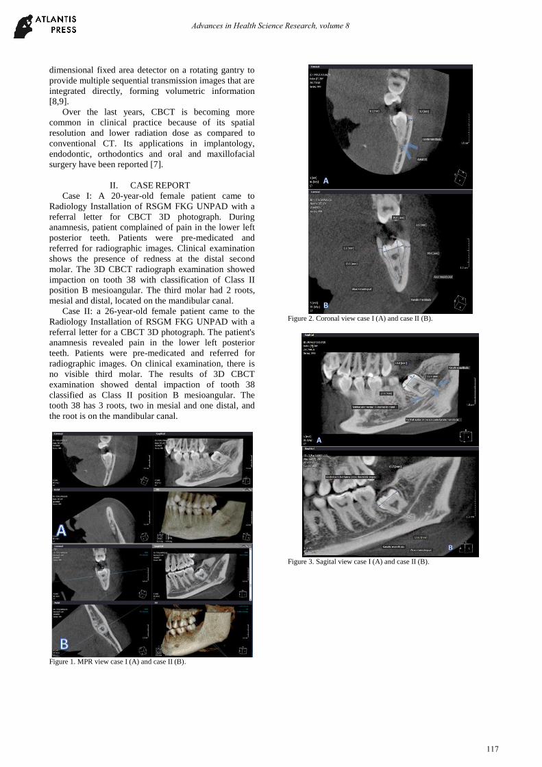
Figure 2. Coronal view case I (A) and case II (B).
Figure 3. Sagital view case I (A) and case II (B).
Advances in Health Science Research, volume 8
117
Figure 4. Axial view case I (B) and case II (B).
Figure 5. 3D and segmentation view case I (A) and case II (B).
III. DISCUSSION
Adequate investigation is necessary to obtain an
optimal treatment and action plan for the patient. In the
case of impaction, examining with CBCT 3D can
provide the information necessary to assist the clinician
while going to do the extraction on the tooth. CBCT 3D
can provide a sagittal, coronal and axial features (Figure
1). So the features of dental impaction condition can be
known more accurately. The coronal view can reveal
the distance of the crown to the bone of cortical bone in
lingual and buccal view, and its relationship of root
with mandibular canal (Figure 2). To assess the
visibility of the mandibular canal in buccolingual
direction, cross-sectional CBCT images can be used.
Cross-sectional images can be generated using different
slice thicknesses, interslice interval and angulations.
This, in turn, might affect the visibility of the MC as
shown in a few studies [10].
The sagittal view will provide features about the
position of impacted third molars, the length of the
crown to the apical at the mesial and distal roots, and
the abnormalities in the root form (Figure 3). The axial
view will provide information about mesial - distal and
buccal - lingual width, and the proximity of its root tip
to the mandible canal (Figure 4). Since CBCT images
can display the examined volume in all anatomical
planes, when an over projection of the mandibular canal
occurs in the traditional 2D images, it is expected that
the CBCT can reveal the exact relationship between the
third molar and the mandibular canal in cross-sectional
image sections. However, due to the high resolution and
low radiation dose in the case of CBCT, the use of
CBCT is recommended [6].
Cone Beam Computed Tomography (CBCT) can be
used as a technique of choice where three dimensional
view of mandibular third molar and its adjacent
anatomical structures are required. Hence, CBCT
contributes to optimal risk assessment and adequate
surgical planning, compared to panoramic radiography
[3].
The course of the mandibular canal is traced through
the mandibular ramus and body, starting from the
lingula on the lingual aspect of the ramus to the mental
foramen on the buccal aspect of the mandibular body.
In cross-sectional and coronal slices, the mandibular
canal is typically seen as an oval or round radiolucency
with corticated borders. Sometimes, the cortication may
be thin or imperceptible. The relationship of the canal to
the tooth roots should be assessed. This relationship
varies greatly among patients, especially in the molar
region, with the mandibular canal occupying a position
from close to the root apices to adjacent to the inferior
border of the mandible. Other variations include bifid
mandibular canals, with a reported frequency of about
15%. The mandibular canal exits to the buccal surface
of the mandible, via the mental foramen, usually at the
premolar region. There is significant variation in the
size, shape, and location of the mental foramen [4].
It is concluded that CBCT 3D can provide adequate
features in cases of mandibular third molar impaction as
the images can be obtained coronally, sagittally and
axially, so that the anatomy, position, and dental
relationships of the impacted teeth with the surrounding
area can be analyzed.
REFERENCES [1] S. Rahayu, “Odontektomi, tatalaksana gigi bungsu impaksi,” E-
Journal WIDYA Kesehatan dan Lingkungan, vol. 1, pp. 81-89, 2014.
Advances in Health Science Research, volume 8
118
[2] T.D. Sahetapy, S.P. Anindita, B. Hutagalung, “Prevalensi gigi
impaksi molar tiga partial erupted pada masyarakat desa
totabuan,” Jurnal e-GiGi, vol. 3, pp. 641-646, 2015.
[3] S. Singh, H. Rahman, R. Chandra, S. Tripathi, J. Jain, K.G.
Tarun, et al., “Assessment of inverted mandibular third molar impaction by 3d reconstruction - a rare case series,” Journal of
Dental and Medical Sciences, vol. 15, pp. 4-7, 2016.
[4] J.L. Peterson, Oral and maxillofacial surgery chapter 9, 4th ed., Mosby, 2012.
[5] A.D. McGowan. An atlas of minor oral surgery principle and
practice. 2nd ed. United Kingdom: Martin Dunitz, 1999. [6] L.H. Matzen, A. Wenzel, “Efficacy of CBCT for assessment of
impacted mandibular third molars: a review – based on a
hierarchical model of evidence,” Dentomaxillofacial Radiology,
vol. 44, 2015.
[7] M. Michele, C. Fulvia, B. Gabriele, “Classification of impacted
mandibular third molars on cone-beam CT images,” J. Clin.
Exp. Dent., vol. 7, pp. 224-31, 2015. [8] S.C. White, M.J. Pharoah. Oral Radiology Principles and
Interpretations. 7th ed., Canada: Mosby, 2014.
[9] E. Whaites. Essential dental radiography and radiology. 4th ed. Spain: Elsevier, 2007.
[10] M. Alkhader, J. Fadi, “Visibility of the mandibular canal on
crosssectional CBCT images at impacted mandibular third molar sites,” Biotechnology & Biotechnological Equipment,
vol. 30, pp. 578-584, 2016.
Advances in Health Science Research, volume 8
119


![Evaluation of Impacted Mandibular Third Molar using Panaromic Radiographs · 2015-11-24 · Third molar is the most frequently impacted tooth.[11] The prevalence of third molar impaction](https://static.documents.pub/doc/80x56/5eb53ec496df9411b42e942c/evaluation-of-impacted-mandibular-third-molar-using-panaromic-radiographs-2015-11-24.jpg)


![Case Report Coronectomy of Mandibular Third Molar: Four ......mandibular third molar extraction is lower in coronectomy compared to complete extraction surgery [3,4]. Nevertheless,](https://static.documents.pub/doc/80x56/60e1df1257eec93cc26c791e/case-report-coronectomy-of-mandibular-third-molar-four-mandibular-third.jpg)


