1Department of Clinical Hematology and Pediatric Oncology, Hospital August 20, 1953, University Hassan II, Casablanca, Morocco2Department of Cardiology, Ibn Rochd Hospital, University Hassan II, Casablanca, Morocco
Correspondence should be addressed to Bienvenu Houssou; [email protected]
Introduction. Venous thromboembolic disease is a common complication found in 8% of patients with acute myeloid leukemia.Thelocation at the right atrium is exceptional. These last fifty years, only 6 cases of thrombosis of the atrium in the diagnosis of acutemyeloid leukemia were published on PubMed search engine. Case Presentation. 35-year-old farmer, who had been admitted byemergency department for superior vena cava syndrome and had a hyperleukocytic AML with complex karyotype associated witha significant thrombosis of the right atrium, extended all along the superior vena cava. He has been treated by the 2011 AMLprotocolusing lowmolecular weight heparin and died from respiratory distress.Conclusions. If thrombosis is common in AML, the locationin right atrium is rare. Its management requires surgery that is sometimes difficult to achieve.
1. Introduction
Venous thromboembolism (VTE) is a frequent complicationfound in approximately 11% of patients with acute leukemia[1]. It is associated in 13.5% with the presence of a centralvenous catheter, or the steroids use, asparagine in acutelymphoblastic leukemia or inherited thrombophilic abnor-malities [2]. It is found in approximately 8% of acute myeloidleukemia (AML) patients [1] and sits in 73.3% of cases in thedeep veins of the upper limbs: axillary vein under subclavianand jugular veins and 16% in the deep veins of the lower limbs:femoral vein, superficial femoral, popliteal, and iliac veins [3].Pulmonary embolism represents 7.8% of the locations and ispotentially fatal [3]. Intracardiac thrombosis localizations inAML are rare and scarcely reported in the literature.
Using keywords thrombosis, atrium, and leukemia forresearch on PubMed there are only 6 cases of thrombosis ofthe right atrium which were published in the last fifty years.
We report a rare case of aAML revealed by a superior venacava syndrome secondary to thrombosis of the right atriumextended all along the superior vena cava.
2. Medical Observation
35-year-old man, active farmer, without past medical history,had been admitted to emergency department for superiorvena cava syndrome (SVCS). Complete blood count (CBC)had revealed hyperleukocytosis with peripheral blasts, whiteblood cell count: 116G/L with 95% blasts and platelets212G/L, and hemoglobin rate: 13.1 g/dL. Chest X-ray hadrevealed a pleural effusion syndrome of mean volume at leftside and of great abundance at right side, with a right basalpulmonary condensation syndrome. He had been sent tothe clinical hematology unit for treatment. The anamnesisrevealed that the symptoms had begun 10 days before theadmission by gradual swelling of the neck extended to theface with dyspnea all occurring in a context of impairedgeneral condition. Physical examination revealed an alteredcondition with WHO: 3, temperature: 37∘ 5C, blood pres-sure: 12/7 cmHg, SaO2 82% on room air, cardiac frequency:84 bpm, weight: 52 kg, height: 1.72m, a SVCS, a bilateral pleu-ral effusion syndrome, and right pulmonary condensationsyndrome. Bone marrow aspiration had concluded in 85%
Hindawi Publishing CorporationCase Reports in CardiologyVolume 2016, Article ID 6802429, 3 pageshttp://dx.doi.org/10.1155/2016/6802429
2 Case Reports in Cardiology
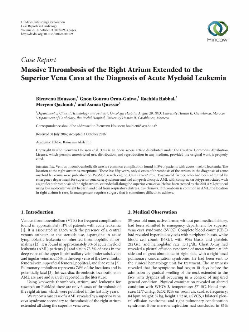
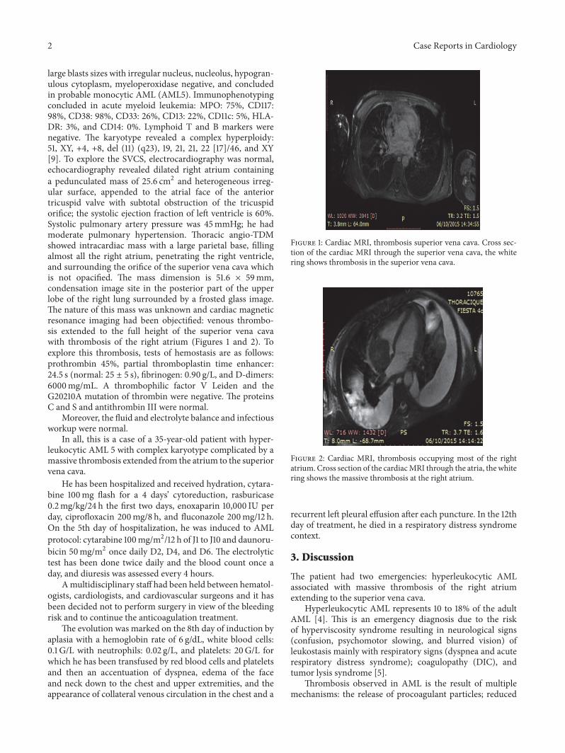
large blasts sizes with irregular nucleus, nucleolus, hypogran-ulous cytoplasm, myeloperoxidase negative, and concludedin probable monocytic AML (AML5). Immunophenotypingconcluded in acute myeloid leukemia: MPO: 75%, CD117:98%, CD38: 98%, CD33: 26%, CD13: 22%, CD11c: 5%, HLA-DR: 3%, and CD14: 0%. Lymphoid T and B markers werenegative. The karyotype revealed a complex hyperploidy:51, XY, +4, +8, del (11) (q23), 19, 21, 21, 22 [17]/46, and XY[9]. To explore the SVCS, electrocardiography was normal,echocardiography revealed dilated right atrium containinga pedunculated mass of 25.6 cm2 and heterogeneous irreg-ular surface, appended to the atrial face of the anteriortricuspid valve with subtotal obstruction of the tricuspidorifice; the systolic ejection fraction of left ventricle is 60%.Systolic pulmonary artery pressure was 45mmHg; he hadmoderate pulmonary hypertension. Thoracic angio-TDMshowed intracardiac mass with a large parietal base, fillingalmost all the right atrium, penetrating the right ventricle,and surrounding the orifice of the superior vena cava whichis not opacified. The mass dimension is 51.6 × 59mm,condensation image site in the posterior part of the upperlobe of the right lung surrounded by a frosted glass image.The nature of this mass was unknown and cardiac magneticresonance imaging had been objectified: venous thrombo-sis extended to the full height of the superior vena cavawith thrombosis of the right atrium (Figures 1 and 2). Toexplore this thrombosis, tests of hemostasis are as follows:prothrombin 45%, partial thromboplastin time enhancer:24.5 s (normal: 25 ± 5 s), fibrinogen: 0.90 g/L, and D-dimers:6000mg/mL. A thrombophilic factor V Leiden and theG20210A mutation of thrombin were negative. The proteinsC and S and antithrombin III were normal.
Moreover, the fluid and electrolyte balance and infectiousworkup were normal.
In all, this is a case of a 35-year-old patient with hyper-leukocytic AML 5 with complex karyotype complicated by amassive thrombosis extended from the atrium to the superiorvena cava.
He has been hospitalized and received hydration, cytara-bine 100mg flash for a 4 days’ cytoreduction, rasburicase0.2mg/kg/24 h the first two days, enoxaparin 10,000 IU perday, ciprofloxacin 200mg/8 h, and fluconazole 200mg/12 h.On the 5th day of hospitalization, he was induced to AMLprotocol: cytarabine 100mg/m2/12 h of J1 to J10 and daunoru-bicin 50mg/m2 once daily D2, D4, and D6. The electrolytictest has been done twice daily and the blood count once aday, and diuresis was assessed every 4 hours.
Amultidisciplinary staff had been held between hematol-ogists, cardiologists, and cardiovascular surgeons and it hasbeen decided not to perform surgery in view of the bleedingrisk and to continue the anticoagulation treatment.
The evolution was marked on the 8th day of induction byaplasia with a hemoglobin rate of 6 g/dL, white blood cells:0.1 G/L with neutrophils: 0.02 g/L, and platelets: 20G/L forwhich he has been transfused by red blood cells and plateletsand then an accentuation of dyspnea, edema of the faceand neck down to the chest and upper extremities, and theappearance of collateral venous circulation in the chest and a
Figure 1: Cardiac MRI, thrombosis superior vena cava. Cross sec-tion of the cardiac MRI through the superior vena cava, the whitering shows thrombosis in the superior vena cava.
Figure 2: Cardiac MRI, thrombosis occupying most of the rightatrium. Cross section of the cardiacMRI through the atria, the whitering shows the massive thrombosis at the right atrium.
recurrent left pleural effusion after each puncture. In the 12thday of treatment, he died in a respiratory distress syndromecontext.
3. Discussion
The patient had two emergencies: hyperleukocytic AMLassociated with massive thrombosis of the right atriumextending to the superior vena cava.
Hyperleukocytic AML represents 10 to 18% of the adultAML [4]. This is an emergency diagnosis due to the riskof hyperviscosity syndrome resulting in neurological signs(confusion, psychomotor slowing, and blurred vision) ofleukostasis mainly with respiratory signs (dyspnea and acuterespiratory distress syndrome); coagulopathy (DIC), andtumor lysis syndrome [5].
Thrombosis observed in AML is the result of multiplemechanisms: the release of procoagulant particles; reduced
Case Reports in Cardiology 3
levels of natural anticoagulants; the decrease in the fibri-nolytic activity; increased coagulation factors; platelet acti-vation; and cytokine-induced expression of tissue factor inendothelial cells [6]. Acute promyelocytic leukemia is themost common AML provider of VTE. The AML 5 is asso-ciated in 25% of patients with thrombosis.
Thrombosis in AML preferentially seats in the upperand lower limbs and the lungs [3]. Generally, the thrombusformation is less frequent in the right atrium compared withthe left atrium [7]. It occurs in the presence of implantedcatheters, particularly at the junction of the superior venacava and the right atrium and in patients with atrial fibril-lation or prothrombotic states [7]. Thoracic angio-TDM isuseful in diagnosis; magnetic resonance imaging can be usedin patients inwhich radio-contrast agents are contraindicated[8]. Thoracic angio-TDM could not afford to know thenature of the right atrial mass of the patient and it is themagnetic resonance imaging that made the diagnosis ofright atrial thrombosis. The treatment is not codified butseveral studies report a percutaneous stent implantation inpatients with severe respiratory distress symptoms beforethe etiological treatment improves prognosis [9]. Duringevolution, the observed venous circulations are due to thefact that, because of this thrombosis, the body uses the waysof substitutions made by the azygos system, lumbar veins,spinal plexus, and thyroid veins which result in appear-ance of edema of neck, chest, and limbs [1]. Pulmonaryhypertension could be an exaggerated cause of respiratorydistress.
4. Conclusions
If thrombosis is common inAML, the location in right atriumis rare, and one should think about it before a superiorvena cava syndrome. Its management requires surgery thatis sometimes difficult.
Written consent to publish this report was obtained from thepatient.
Competing Interests
The authors declare that they have no competing interests.
Authors’ Contributions
Bienvenu Houssou prepared the draft and Gnon GourouOrou-Guiwa, Rachida Habbal, Meryem Qachouh, andAsmaa Quessar all reviewed and contributed to the finalmanuscript.
References
[1] J. I. Weitz, R. Bauersachs, J. Beyer-Westendorf et al., “Two dosesof rivaroxaban versus aspirin for prevention of recurrent venousthromboembolism: rationale for and design of the einsteinchoice study,” Thrombosis and Haemostasis, vol. 114, no. 3, pp.645–650, 2015.
[2] J. Monika, C. Anna, P. Dariusy, and K. Mieczyslow, “Incidenceand risk factors for central venous catheter-related thrombosisin hematological patients,” Medical Oncology, vol. 31, pp. 772–778, 2014.
[3] K. Vu, N. V. Luong, J. Hubbard et al., “A retrospective study ofvenous thromboembolism in acute leukemia patients treated atthe University of Texas MD Anderson Cancer Center,” CancerMedicine, vol. 4, no. 1, pp. 27–35, 2015.
[4] L. Marbello, F. Ricci, A. M. Nosari et al., “Outcome ofhyperleukocytic adult acute myeloid leukaemia: a single-centerretrospective study and reviewof literature,”LeukemiaResearch,vol. 32, no. 8, pp. 1221–1227, 2008.
[5] G. J. Ventura, J. P. Hester, T. L. Smith, and M. J. Keating, “Acutemyeloblastic leukemia with hyperleukocytosis: risk factors forearly mortality in induction,” American Journal of Hematology,vol. 27, no. 1, pp. 34–37, 1988.
[6] A. Falanga and M. Marchetti, “Thrombosis in myeloprolifera-tive neoplasms,” Seminars in Thrombosis and Hemostasis, vol.40, no. 3, pp. 348–358, 2014.
[7] M. de Divitiis, H. Omran, R. Rabahieh et al., “Right atrialappendage thrombosis in atrial fibrillation: its frequency and itsclinical predictors,”TheAmerican Journal of Cardiology, vol. 84,no. 9, pp. 1023–1028, 1999.
[8] T.W. Rice, R.M. Rodriguez, andR.W. Light, “The superior venacava syndrome: clinical characteristics and evolving etiology,”Medicine, vol. 85, no. 1, pp. 37–42, 2006.
[9] R. Uberoi, “Quality assurance guidelines for superior vena cavastenting in malignant disease,” CardioVascular and Interven-tional Radiology, vol. 29, no. 3, pp. 319–322, 2006.