ABSTRACT Background. Thoracic Outlet Syndrome (TOS) involves compression of the brachial plexus, subclavius artery and vein. Many studies discuss efficacy of surgery and few dis- cuss conservative treatment. It is unknown what specific forms of conservative treatment are best. Objective. Describe conservative management for TOS using unique exercises. Case Description. A collegiate football player reported numbness/tingling down his right arm after a right brachial plexus stretch injury. Seven months later, he was diagnosed with recurrent cervical traction neuropraxia. Two months later, he reported bilateral symptoms and was diagnosed with functional TOS. The athlete began shoul- der strengthening (deltoid, middle trapezius, rhomboids, pectoralis major, latissimus dorsi, biceps, upper trapezius and rotator cuff) and stretching (pectoralis, scalene and upper trapezius) which failed to resolve his symptoms after four weeks. Surgical resection of bilateral first ribs and quitting football was recommended by four physi- cians. Unique therapeutic exercises developed by the Postural Restoration Institute™ were used to optimize res- piration/posture via muscle activation and inhibition. After six weeks, the athlete was asymptomatic and returned to football but still experienced paresthesia with contact. Additional exercises were prescribed and remain- ing symptoms were abolished. Outcomes. The Northwick Park Neck Pain Questionnaire was 55.5% at initial and 0% at four weeks and discharge. Discussion. Athlete did not demonstrate relief of symp- toms from shoulder stretching and strengthening. Intervention designed to optimize respiration/posture by repositioning the pelvis/trunk via specific muscle inhibi- tion and activation resulted in abolishing the athlete’s symptoms. Management that aims to optimize respiration via muscle inhibition, activation, and repositioning war- rants further research. Key words: thoracic outlet syndrome, postural restora- tion, respiration CORRESPONDENCE Kyndall L. Boyle, PT, PhD, OCS Program in Physical Therapy PO Box 15105 Flagstaff, AZ 86011-5105 e-mail: [email protected]ACKNOWLEDGEMENTS We would like to thank the Postural Restoration Institute™ for their creative development of the specific therapeutic exercises that were prescribed for the patient in this case report. CASE REPORT Bilateral Functional Thoracic Outlet Syndrome in a Collegiate Football Player Jason H. Robey, ATC a Kyndall L. Boyle, PT, PhD, OCS b a Appalachian State University Boone, NC b Northern Arizona University Flagstaff, AZ North American Journal of Sports Physical Therapy | Volume 4, Number 4 | November 2009 | Page 170 NAJSPT
Transcript
ABSTRACT
Background. Thoracic Outlet Syndrome (TOS) involvescompression of the brachial plexus, subclavius artery andvein. Many studies discuss efficacy of surgery and few dis-cuss conservative treatment. It is unknown what specificforms of conservative treatment are best.
Objective. Describe conservative management for TOSusing unique exercises.
Case Description. A collegiate football player reportednumbness/tingling down his right arm after a rightbrachial plexus stretch injury. Seven months later, he wasdiagnosed with recurrent cervical traction neuropraxia.Two months later, he reported bilateral symptoms and wasdiagnosed with functional TOS. The athlete began shoul-der strengthening (deltoid, middle trapezius, rhomboids,pectoralis major, latissimus dorsi, biceps, upper trapeziusand rotator cuff) and stretching (pectoralis, scalene andupper trapezius) which failed to resolve his symptomsafter four weeks. Surgical resection of bilateral first ribsand quitting football was recommended by four physi-cians. Unique therapeutic exercises developed by thePostural Restoration Institute™ were used to optimize res-piration/posture via muscle activation and inhibition.After six weeks, the athlete was asymptomatic andreturned to football but still experienced paresthesia withcontact. Additional exercises were prescribed and remain-ing symptoms were abolished.
Outcomes. The Northwick Park Neck Pain Questionnairewas 55.5% at initial and 0% at four weeks and discharge.
Discussion. Athlete did not demonstrate relief of symp-toms from shoulder stretching and strengthening.Intervention designed to optimize respiration/posture byrepositioning the pelvis/trunk via specific muscle inhibi-tion and activation resulted in abolishing the athlete’ssymptoms. Management that aims to optimize respirationvia muscle inhibition, activation, and repositioning war-rants further research.
CORRESPONDENCEKyndall L. Boyle, PT, PhD, OCSProgram in Physical Therapy PO Box 15105Flagstaff, AZ 86011-5105e-mail: [email protected]
ACKNOWLEDGEMENTSWe would like to thank the Postural Restoration Institute™for their creative development of the specific therapeuticexercises that were prescribed for the patient in this casereport.
CASE REPORT
Bilateral Functional Thoracic OutletSyndrome in a Collegiate Football Player Jason H. Robey, ATCa
Kyndall L. Boyle, PT, PhD, OCSb
a Appalachian State UniversityBoone, NC
b Northern Arizona UniversityFlagstaff, AZ
North American Journal of Sports Physical Therapy | Volume 4, Number 4 | November 2009 | Page 170
NA
JSP
T
INTRODUCTIONThoracic outlet syndrome (TOS), also known as neurovas-cular compression syndrome, consists of a group of dis-tinct disorders that affect the nerves or vascular structuresbetween the base of the neck and axilla.1,2 Specifically,these disorders result from positional compression of thesubclavian artery or vein, and the brachial plexus nervesin a variety of locations including the cervical spine (fromcervical rib), scalene interval, infraclavicular space orunder the pectoralis minor tendon.3 The brachial plexus isdivided into an upper plexus (median nerve distribution)and a lower plexus (ulnar nerve distribution).4 Upperplexus compression was initially described by Swank andSimeone4 with symptoms secondary to C5, C6, and C7nerve root compression. Sensory changes will primarilyoccur in the first three fingers, and associated muscleweakness or pain in the anterior chest, triceps, deltoids,and parascapular muscles, as well as down the outer fore-arm to the extensor muscles.4 Lower plexus irritationinvolves C8 and T1 nerve root compression. Sensorychanges primarily occur in the fourth and fifth fingers,with muscle weakness or pain from the rhomboid and thescapular muscles to the posterior axilla, down the ulnardistribution of the forearm, involving the elbow, wrist flex-ors, and the intrinsic muscles of the hand.4
Thoracic outlet syndrome disorders are complex, poorlydefined, and a diagnosis of exclusion that can cover a widerange of ailments each producing various signs and symp-toms arising from the upper extremity and the chest,neck, and head.2,5,6 An accurate diagnosis of TOS requiresa thorough history and physical examination. Several testsexist that may be used to assist in diagnosing TOS, includ-ing nerve conduction velocity (NCV), electromyography(EMG), radiographs, computed tomography (CT) scan,and magnetic resonance imaging (MRI).4 Authors haveattempted to study the thoracic outlet region by CT scanor MRI. One of the limitations is that the compromise ofthe neurovascular bundle is often positional and intermit-tent.2 In neurogenic TOS, electrophysiological testing isoften entirely normal. EMG can sometimes detect neuro-genic C8/T1 signs.2
Thoracic outlet syndrome can be a result of posturalalterations, hypertrophic muscles, muscle imbalances, anelevated first rib, presence of a cervical rib, and macrotrau-ma such as automobile accidents.5 These syndromes havebeen categorized into anatomical and functional TOS.5
Anatomical TOS includes congenital anomalies or trau-
matic osseous and soft tissue injury. Functional TOSincludes postural adaptations as a result of work or sportparticipation, respiratory changes, and psychological con-ditions.
The literature suggests that a variety of methods havebeen traditionally used to manage TOS.2,5,7 Conservativeinterventions focus on pain control,7,8 edema control, ver-bal posture education and ergonomics,7,9-14 relaxation,15
stretching, strengthening, and nerve gliding exercises.7,16,17
Additional interventions include moist heat,7,18,19 massage,20
acupuncture,20 cervical traction, manual joint mobiliza-tion,21 first rib mobilization,15,19 braces,22 and aerobic exer-cises.9 Exercises to manage TOS may focus on stretchingthe levator scapulae, lower trapezius, scalenes, and pec-toralis muscles,2,7,19,21 and strengthening the cervical exten-sors, scapular adductors, and shoulder retractor mus-cles.2,7,19,21 Additionally, cervical traction, isometric exercis-es (cervical spine and shoulder girdle), and manual jointmobilization (cervical-thoracic spine, sternoclavicularjoint, acromioclavicular joint and costothoracic joint) hasbeen described in the literature.21
Any of the described methods have been advocated toalter posture in some way.2,5,7 Authors of a recent literaturereview for TOS conclude that although conservative treat-ment may reduce symptoms and improve function, it isnot known if this approach is significantly better than notreatment or placebo.5 The most commonly recommend-ed interventions are strengthening and stretching of theshoulder girdle musculature.2,7,19,21 However, little agree-ment exists on which muscles need strengthening andwhich ones need lengthening.5 These types of exercises donot detail how they address functional TOS as a result ofrespiratory alterations and they do not aim to inhibit mus-cle.1,5,19
Postural Restoration is a holistic posture based approach topatient management that considers the influence of dys-functional respiration on posture and utilizes therapeuticexercises that activate or inhibit specific muscles and man-ual trunk techniques as needed in order to achieve opti-mal respiration and posture.23 Postural Restoration alsorecognizes patterns of postural asymmetries (similar toKendall’s24 right handed pattern) that are believed to bepresent in most people to varying degrees. Clinicians whouse Postural Restoration, therefore, often target interven-tion to correct the asymmetrical pattern.23,25 This patternis consistent with the pre-existent vertebral rotation in
North American Journal of Sports Physical Therapy | Volume 4, Number 4 | November 2009 | Page 171
“normal” individuals.26 The postural restoration methodol-ogy has been used to manage patients with sciatica andlow back pain,27 asthma,28 anterior knee pain,29,30 andtrochanteric bursitis.31 This approach has appeared to besuccessful in managing athletes with iliotibial band syn-drome,32 and patients with chronic pain33 and knee pain,34
however, the evidence for these three conditions remainsanecdotal. To date, no case studies describing the use ofPostural Restoration for a patient with TOS have been pub-lished. The purpose of this case is to describe manage-ment of a collegiate football player with bilateral function-al TOS who was first managed with traditional therapeuticexercises and then with unique Postural Restoration ther-apeutic exercises to address his faulty posture and subop-timal respiration.
CASE DESCRIPTIONThe athlete was a 22-year-old male collegiate football play-er (tight end). Social history included living with threeother football players in an apartment with parents livingan hour away. Other than his current condition, his gen-eral health including physical, psychological, and socialfunction was excellent. Athlete denied use of alcohol,tobacco, or any drugs. He had an unremarkable medicaland family history. The chief complaint for this footballplayer included sustaining a right brachial plexus injuryduring the fall football season. Approximately sevenmonths later, the athlete suffered multiple brachial plexusinjuries to his right neck during spring football practice.Cervical spine radiographs were taken and he was seen bythe team orthopedic surgeon. Radiographs revealed mild-moderate posterior osteophyte formation at C6- C7 greaterthan C5- C6 with elevation of the ribs. The physician diag-nosed the athlete with cervical traction neurapraxia andprescribed Ibuprofen and Vicodin.
Due to the athlete’s signs and symptoms progressivelybecoming worsened, he altered his posture to favor theinvolved side. This compensation in his posture potential-ly led to the left side becoming involved. He then begandeveloping radicular pain down his left arm and into hishand. Now his signs and symptoms consisted of bilateralnumbness, tingling, and weakness into his hands (mediannerve distribution), achy pain in shoulders, and a shootingsensation down both arms, pain at Erb’s point,7 tendernessto palpation over anterior chest and neck muscles, andtenderness and pain between his right scapula and verte-bral column at the level of T4. These findings were con-
sistent with someone who has sustained an upper plexuscompression injury.35
Postural observation revealed bilateral: hypertrophic/over-developed latissimus dorsi, pectoralis, upper trapez-ius and bicep muscles, anterior rib flares (ribs are in aposition of external rotation/elevation which is a com-bined position of chest expansion with pump handle andbucket handle motions),36 increased lumbar lordosis (over-active paraspinal muscles), left pelvic forward rotation,forward head posture, and over-active neck musculature.He reported that he was unable to sleep on either side andhad to sleep prone with his face buried in his hands. Theathlete had several positive tests including Adson’s,37
Allen’s, Military Brace and Roos.3,37-39
Two months later he was sent for a cervical spine MRI, abilateral brachial plexus MRI, a nerve conduction velocitytest, and an EMG. The cervical spine MRI was normal.The bilateral brachial plexus MRI revealed muscularhypertrophy in the neck and shoulders. All other struc-tures appeared normal. The nerve conduction velocitytest and EMG studies revealed ulnar nerve compression atthe elbow on the right. The test results were negative onthe left. It was interesting to note that the athlete did nothave any neurological symptoms in the ulnar distributionof his hand. An orthopaedic surgeon diagnosed him withbilateral functional TOS. The physician recommendedthat he should discontinue playing football. Multiplephysicians advised him to consider having surgery forresection of both first ribs as a last resort. The athletedecided to take a conservative approach to see if he couldbenefit from non-surgical management. He was managedby three different clinicians over the course of care andseen daily in the athletic training room. See Table 1 fordosage of interventions.
Intervention – Clinician 1The first clinician focused on traditional recommendedinterventions, primarily guided by information on a web-site for TOS including stretching and strengthening exer-cises.19 Specific interventions (done twice a day, sevendays a week) consisted of: moist heat for fifteen minutes(over the neck and both shoulders in sitting to prepare forstretching), self stretching exercises (three repetitions x 30seconds) for bilateral neck, shoulder, and chest muscles(scalenes, upper trapezius, pectoralis major muscles), ver-bal postural education (to keep head and shoulders back,and chin and chest up) and shoulder strengthening using
North American Journal of Sports Physical Therapy | Volume 4, Number 4 | November 2009 | Page 172
tubing for the rotator cuff (internal and external rotationmuscles), deltoid, pectoralis major, latissimus dorsi,supraspinatus, biceps, and upper trapezius muscles. Allstretches were performed three times for 30 seconds each,the exercises were done in three sets of fifteen each, andall treatment was done twice a day everyday for fourweeks. With this type of conservative treatment the ath-lete did not see much improvement and continued to pro-gressively get worse after each lifting and conditioningsession. After a month of conservative treatment, the ath-lete began to contemplate having surgery and ending hisfootball career. Because the athlete was not makingprogress, clinician one asked another clinician to take overhis case.
Intervention – Clinician 2During weeks 5-11, this new cli-nician changed the focus oftreatment by addressing the ath-lete’s specific impairments offaulty posture via activation andinhibition of specific muscles tochange position of bones andsoft tissue rather than throughverbal education alone. Faultyposture noted via visual observa-
tion included: over-developed latissimus dorsi, pectoralis,biceps, and upper trapezius muscles, over-active anteriorneck muscles, increased lumbar lordosis (anterior pelvictilt with left forward rotation), elevated ribs, downwardlyrotated scapula, and forward head posture. Exercises andmanual therapy techniques developed by the PosturalRestoration Institute™40 were utilized to optimize postureof the trunk/scapula and pelvis via activation, inhibition,and lengthening of specific muscles.
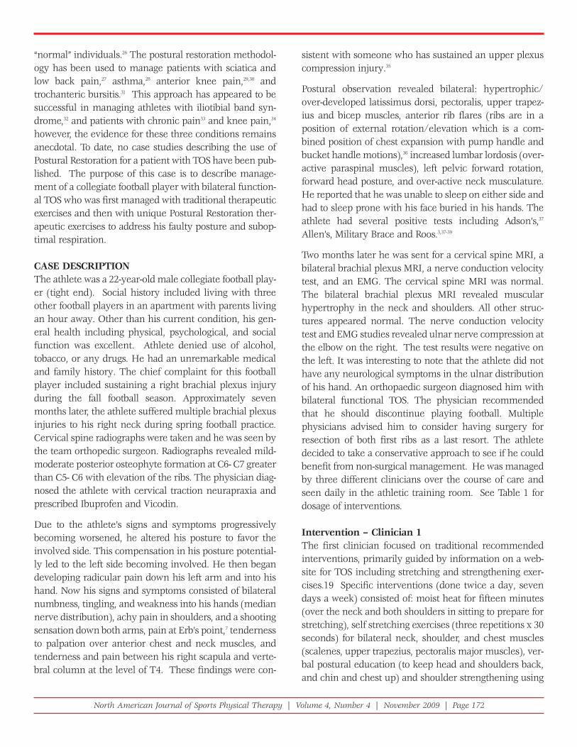
The initial exercises were prescribed in order to restore anormal pelvic and rib cage position (from left pelvic ante-rior tilt/forward rotation and elevated, externally rotated
ribs toward neutral) by doing a90/90 hip shift with hemibridgeand balloon (Figure 1). The exer-cise activated left hamstringmuscles which facilitate left hipextension (posterior pelvic tilt).Forced exhalation into the bal-loon should activate transversusthoracis (triangularis sterni),internal and external inter-costals, and abdominal obliquemuscles which facilitate depres-sion/internal rotation of the ribsand inhibition of paraspinalFigure 1. 90/90 hip shift exercise
North American Journal of Sports Physical Therapy | Volume 4, Number 4 | November 2009 | Page 173
muscles (lumbar flexion). Additionally, via the back pres-sure of the balloon, opening of the right apical chest wallis facilitated upon inhalation without using neck orparaspinal muscles. Depression of the ribs was thought toallow for a more optimal rest position of the scapula and,thereby, an optimal infraclavicular space to minimize theopportunity for compromise of the subclavius artery,vein, and brachial plexus nerves. This exercise was donetwice daily with two sets of 15 repetitions.
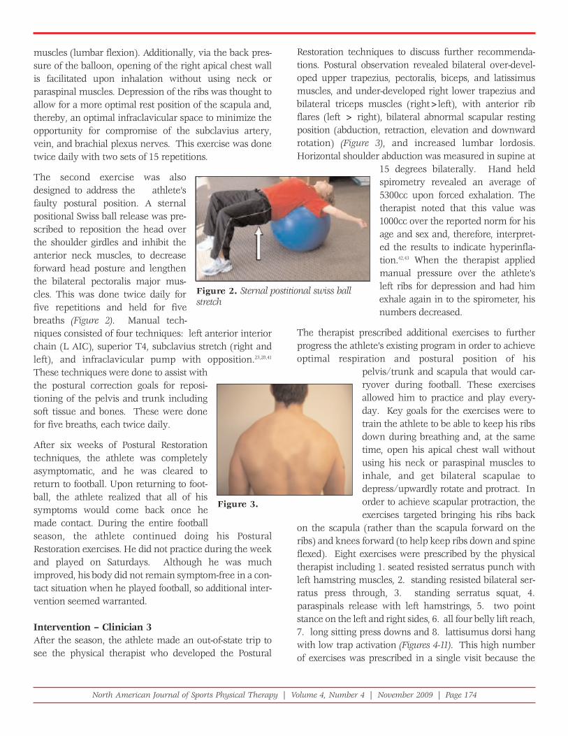
The second exercise was alsodesigned to address the athlete’sfaulty postural position. A sternalpositional Swiss ball release was pre-scribed to reposition the head overthe shoulder girdles and inhibit theanterior neck muscles, to decreaseforward head posture and lengthenthe bilateral pectoralis major mus-cles. This was done twice daily forfive repetitions and held for fivebreaths (Figure 2). Manual tech-niques consisted of four techniques: left anterior interiorchain (L AIC), superior T4, subclavius stretch (right andleft), and infraclavicular pump with opposition.23,28,41
These techniques were done to assist withthe postural correction goals for reposi-tioning of the pelvis and trunk includingsoft tissue and bones. These were donefor five breaths, each twice daily.
After six weeks of Postural Restorationtechniques, the athlete was completelyasymptomatic, and he was cleared toreturn to football. Upon returning to foot-ball, the athlete realized that all of hissymptoms would come back once hemade contact. During the entire footballseason, the athlete continued doing his PosturalRestoration exercises. He did not practice during the weekand played on Saturdays. Although he was muchimproved, his body did not remain symptom-free in a con-tact situation when he played football, so additional inter-vention seemed warranted.
Intervention – Clinician 3After the season, the athlete made an out-of-state trip tosee the physical therapist who developed the Postural
Restoration techniques to discuss further recommenda-tions. Postural observation revealed bilateral over-devel-oped upper trapezius, pectoralis, biceps, and latissimusmuscles, and under-developed right lower trapezius andbilateral triceps muscles (right>left), with anterior ribflares (left > right), bilateral abnormal scapular restingposition (abduction, retraction, elevation and downwardrotation) (Figure 3), and increased lumbar lordosis.Horizontal shoulder abduction was measured in supine at
15 degrees bilaterally. Hand heldspirometry revealed an average of5300cc upon forced exhalation. Thetherapist noted that this value was1000cc over the reported norm for hisage and sex and, therefore, interpret-ed the results to indicate hyperinfla-tion.42,43 When the therapist appliedmanual pressure over the athlete’sleft ribs for depression and had himexhale again in to the spirometer, hisnumbers decreased.
The therapist prescribed additional exercises to furtherprogress the athlete’s existing program in order to achieveoptimal respiration and postural position of his
pelvis/trunk and scapula that would car-ryover during football. These exercisesallowed him to practice and play every-day. Key goals for the exercises were totrain the athlete to be able to keep his ribsdown during breathing and, at the sametime, open his apical chest wall withoutusing his neck or paraspinal muscles toinhale, and get bilateral scapulae todepress/upwardly rotate and protract. Inorder to achieve scapular protraction, theexercises targeted bringing his ribs back
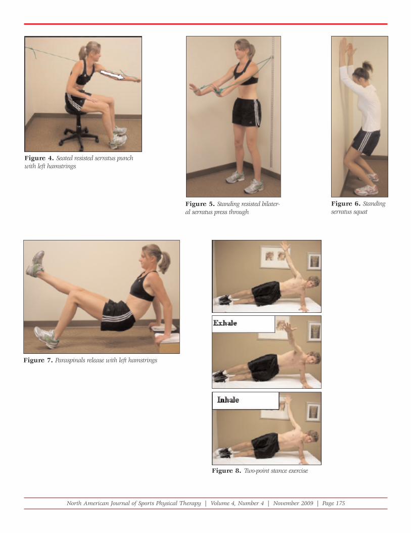
on the scapula (rather than the scapula forward on theribs) and knees forward (to help keep ribs down and spineflexed). Eight exercises were prescribed by the physicaltherapist including 1. seated resisted serratus punch withleft hamstring muscles, 2. standing resisted bilateral ser-ratus press through, 3. standing serratus squat, 4.paraspinals release with left hamstrings, 5. two pointstance on the left and right sides, 6. all four belly lift reach,7. long sitting press downs and 8. lattisumus dorsi hangwith low trap activation (Figures 4-11). This high numberof exercises was prescribed in a single visit because the
Figure 2. Sternal postitional swiss ballstretch
Figure 3.
North American Journal of Sports Physical Therapy | Volume 4, Number 4 | November 2009 | Page 174
Figure 4. Seated resisted serratus punchwith left hamstrings
Figure 5. Standing resisted bilater-al serratus press through
Figure 6. Standingserratus squat
Figure 7. Paraspinals release with left hamstrings
Figure 8. Two-point stance exercise
North American Journal of Sports Physical Therapy | Volume 4, Number 4 | November 2009 | Page 175
patient traveled by airplane tothe out-of-state appointment.
These exercises focused on neuromuscular re-education toalter or reposition the pelvis, ribs, and scapula to preventcompression of the brachial plexus nerves. After the ath-lete performed several repetitions of exercise one, the ther-apist rechecked his breathing (by visual observation of theathlete in supine), and noted that his upper trapezius mus-cle tone had noticeably decreased and he seemed to bebreathing only with his diaphragm. The athlete was awareof his altered breathing and he noted that he was almostshort of breath when not compensating with accessoryrespiratory muscles. The therapist then rechecked his hor-izontal shoulder abduction and it was 45 degrees, bilateral-ly. The therapist interpreted this increase in passive rangeof motion to be reflective of a decrease in tone/activity ofthe pectoralis and biceps musculature. A squat positionwas added to the second exercise to help train the quadri-ceps muscles since the therapist perceived the athleteusing his paraspinal muscles more than his quadricepsmuscles to run and push forward. After exercise numberthree, postural observation of the shoulder girdles revealednormal bilateral scapular rest position. This resting scapu-lar portion was interpreted as the athlete having his ribsdown (relatively depressed/internally rotated position)compared to his initial position of rib flares (anterior rib ele-vation/external rotation). This positioning allowed the ath-lete’s scapula to rest on the ribs and no longer required thescapula to be held up by upper trapezius muscles.
The athlete was instructed to work on the first three exer-cises until five repetitions of each could be performed eas-ily before progressing to the exercises in Figures 7-11. Withthe exercise in Figure 7, it was noted that the athlete’s rightupper extremity was weaker than the left. The exerciserequired lifting his right leg up to force his right upperextremity to work harder. The athlete was encouraged toachieve balance and work on the exercise until it was aseasy to do lifting his right leg as it was lifting his left leg.The exercise in Figure 8 was designed to teach the athleteto learn how to move his thorax on his scapulas. The exer-cises were instructed to be done on both the right and leftsides but with different breathing instructions during cer-tain phases of movement for each side to facilitate more airflow into the right chest wall rather than the left.
After several repetitions of each exercise were performed,the hand-held spirometer was used again and his valuesdropped to 4300cc. This was the reported norm for a 22-year-old male.43 The therapist attributed the drop in airexhaled into the spirometer as a result of taking away hiscompensatory strategies of using upper trapezius andparaspinal muscles and putting his ribs and diaphragm in abetter position so the diaphragm could be better used as theprimary muscle of respiration. When the ribs are elevatedand externally rotated, the diaphragm was more linear/flatand less surface area of the diaphragm was available for adome or zone of apposition (ZOA).44 The ZOA (area of thediaphragm from the most cephalad portion of the dome tothe inferior attachments on the sternum and ribs) is impor-
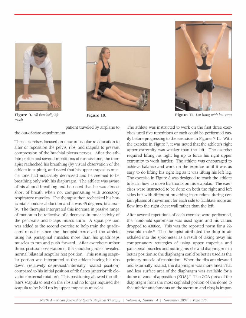
Figure 9. All four belly liftreach
Figure 10. Figure 11. Lat hang with low trap
North American Journal of Sports Physical Therapy | Volume 4, Number 4 | November 2009 | Page 176
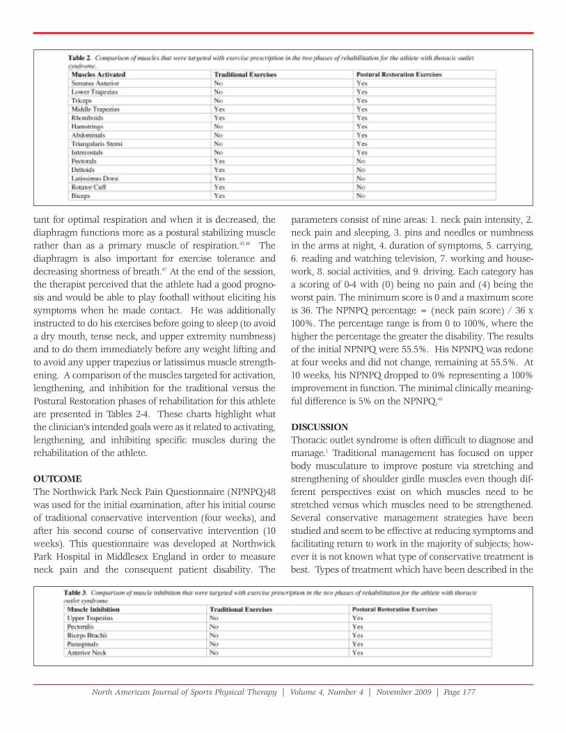
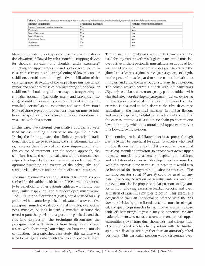
tant for optimal respiration and when it is decreased, thediaphragm functions more as a postural stabilizing musclerather than as a primary muscle of respiration.45,46 Thediaphragm is also important for exercise tolerance anddecreasing shortness of breath.47 At the end of the session,the therapist perceived that the athlete had a good progno-sis and would be able to play football without eliciting hissymptoms when he made contact. He was additionallyinstructed to do his exercises before going to sleep (to avoida dry mouth, tense neck, and upper extremity numbness)and to do them immediately before any weight lifting andto avoid any upper trapezius or latissimus muscle strength-ening. A comparison of the muscles targeted for activation,lengthening, and inhibition for the traditional versus thePostural Restoration phases of rehabilitation for this athleteare presented in Tables 2-4. These charts highlight whatthe clinician’s intended goals were as it related to activating,lengthening, and inhibiting specific muscles during therehabilitation of the athlete.
OUTCOME The Northwick Park Neck Pain Questionnaire (NPNPQ)48was used for the initial examination, after his initial courseof traditional conservative intervention (four weeks), andafter his second course of conservative intervention (10weeks). This questionnaire was developed at NorthwickPark Hospital in Middlesex England in order to measureneck pain and the consequent patient disability. The
parameters consist of nine areas: 1. neck pain intensity, 2.neck pain and sleeping, 3. pins and needles or numbnessin the arms at night, 4. duration of symptoms, 5. carrying,6. reading and watching television, 7. working and house-work, 8. social activities, and 9. driving. Each category hasa scoring of 0-4 with (0) being no pain and (4) being theworst pain. The minimum score is 0 and a maximum scoreis 36. The NPNPQ percentage = (neck pain score) / 36 x100%. The percentage range is from 0 to 100%, where thehigher the percentage the greater the disability. The resultsof the initial NPNPQ were 55.5%. His NPNPQ was redoneat four weeks and did not change, remaining at 55.5%. At10 weeks, his NPNPQ dropped to 0% representing a 100%improvement in function. The minimal clinically meaning-ful difference is 5% on the NPNPQ.49
DISCUSSIONThoracic outlet syndrome is often difficult to diagnose andmanage.1 Traditional management has focused on upperbody musculature to improve posture via stretching andstrengthening of shoulder girdle muscles even though dif-ferent perspectives exist on which muscles need to bestretched versus which muscles need to be strengthened.Several conservative management strategies have beenstudied and seem to be effective at reducing symptoms andfacilitating return to work in the majority of subjects; how-ever it is not known what type of conservative treatment isbest. Types of treatment which have been described in the
North American Journal of Sports Physical Therapy | Volume 4, Number 4 | November 2009 | Page 177
literature include upper trapezius muscle activation (shoul-der elevation) followed by relaxation;50 a strapping devicefor shoulder elevation and shoulder girdle exercises;22
stretching for upper trapezius and levator scapulae mus-cles; chin retraction and strengthening of lower scapularstabilizers; aerobic conditioning;9 active mobilization of thecervical spine; stretching of the upper trapezius, pectoralisminor, and scalenes muscles; strengthening of the scapularstabilizers;15 shoulder girdle massage; strengthening ofshoulder adduction (pectoralis major and latisimus mus-cles); shoulder extension (posterior deltoid and tricepsmuscles); cervical spine isometrics; and manual traction.5
None of these types of interventions focus on muscle inhi-bition or specifically correcting respiratory alterations, aswas used with this patient.
In this case, two different conservative approaches wereused by the treating clinicians to manage the athlete.During the first approach, the clinician prescribed tradi-tional shoulder girdle stretching and strengthening exercis-es, however the athlete did not show improvement afterthis course of treatment. For the second approach, theclinicians included non-manual exercises and manual tech-niques developed by the Postural Restoration Institute™40 tooptimize breathing and posture of the pelvis, ribs, andscapula via activation and inhibition of specific muscles.
The nine Postural Restoration Institute (PRI) exercises pre-scribed for this athlete with bilateral TOS, would potential-ly be beneficial to other patients/athletes with faulty pos-ture, faulty respiration, and over-developed musculature.The 90/90 hip shift exercise (Figure 1) could be used for anypatient with an anterior pelvic tilt, elevated ribs, over-activeparaspinal muscles, weak abdominal muscles, over-activeneck muscles, or long hamstring muscles. Because theexercise puts the pelvis into a posterior pelvic tilt and theribs into depression, the technique discourages theparaspinal and neck muscles from firing (inhibits) andassists with shortening hamstrings via hamstring musclecontraction. In a published case study, this exercise wasused to manage a female with sciatica and low back pain.51
The sternal postitional swiss ball stretch (Figure 2) could beused for any patient with weak gluteus maximus muscles,over-active or short pectoralis musculature, or acquired for-ward head posture. This exercise is designed to activate thegluteal muscles in a sagittal plane against gravity, to length-en the pectoral muscles, and to some extent the latisimusmuscles, and bring the head out of a forward head position.The seated resisted serratus punch with left hamstrings(Figure 4) could be used to manage any patient/athlete withelevated ribs, over-developed paraspinal muscles, excessivelumbar lordosis, and weak serratus anterior muscles. Theexercise is designed to help depress the ribs, discourageactivation of the paraspinal muscles via lumbar flexion,and may be especially helpful to individuals who run sincethe exercise mimics a closed kinetic chain position in onelower extremity while the contralateral upper extremity isin a forward swing position.
The standing resisted bilateral serratus press through(Figure 5) may be beneficial for patients/athletes who needlumbar flexion training (to inhibit over-active paraspinalmuscles), scapular depression (to oppose over-active uppertrapezius muscles and accessory respiratory breathing),and inhibition of over-active/developed pectoral muscles.With the exercise done in the squat position it would alsobe beneficial for strengthening quadriceps muscles. Thestanding serratus squat (Figure 6) could be used for anypatient needing activation of serratus anterior and lowtrapezius muscles for proper scapular position and dynam-ics without allowing excessive lumbar lordosis and over-activation of latissimus muscles to occur. This exercise isdesigned to train an individual to breathe with the ribsdown, pelvis back, spine flexed, latisimus muscles elongat-ed, and quadriceps muscles firing. The paraspinals releasewith left hamstrings (Figure 7) may be beneficial for anypatient/athlete who needs to strengthen one or both upperextremities (lower trapezius, rhomboids, and triceps mus-cles) in a closed kinetic chain position with the lumbarspine in a flexed position (rather than an anteriorly tiltedposition). This particular position would discourage over-
North American Journal of Sports Physical Therapy | Volume 4, Number 4 | November 2009 | Page 178
activation of the patient/athlete’s paraspinal muscles andencourage ribs to depress and abdominal muscles to workin a shortened (rather than lengthened) position.
The two-point stance exercise (Figure 8) could be pre-scribed for any higher level patient/client needing to acti-vate/strengthen core musculature, lower trapezius, rhom-boids and serratus anterior muscles and needing to length-en pectoral muscles and expand their apical chest wall.The all four belly lift reach (Figure 9) may be beneficial fora higher level patient/athlete to progress from the seatedresisted serratus punch (Figure 4) because the exercise issimilar (when they reach toward the floor) but in a fourpoint/quadruped position. The exercise likely requiresmore abdominal muscle activation, because the positionrequires the abdominal muscles to work/activate againstgravity. Lastly, the lat hang with low trap (Figure 11) couldbe prescribed for any patient/client who has the strengthand shoulder range of motion to do the exercise with prop-er form that may benefit from latissimus lengthening andlower trapezius muscle activation. The exercise is thoughtto be beneficial for patients who need to lengthen theirlatissimus muscles while maintaining a posterior tiltedpelvic position (rather than a pelvic anteriorly tilted andexcessively lumbar lordotic position). The exercise isdesigned to activate/strengthen the lower trapezius mus-cles too, which would oppose over-active/developed uppertrapezius muscles.
For the football player in this case study, rather than clini-cians two and three focusing on stretching the uppertrapezius, levator scapulae, scalene, and pectoralis muscles,the focus was on inhibiting these muscles. Rather thanfocusing on strengthening (rotator cuff, biceps, deltoids,pectoralis, latissimus, and upper trapezius muscles) thefocus was on identifying the muscles the patient was usingto breathe with (upper trapezius, paraspinal, and anteriorneck muscles), and the concomitant position the body wasin (rib elevation/external rotation, lumbar lordosis, scapu-lar elevation, retraction, and downward rotation) and whatmuscles were over-developed (upper trapezius, pectoral,and latissimus muscles). Specific exercise prescription wasthen recommended by the therapist to alter the athlete’spostural position and muscle activation patterns to opti-mize respiration.
Although mobilization of the first rib has been discussed inthe literature, altering the position of multiple ribs via acti-
vation of hamstring muscles has not been presented in theliterature. Activation of the hamstring muscles puts thepelvis in a posterior pelvic tilt helping the abdominal mus-cles obtain an optimal position/length to facilitate spinalflexion and to inhibit the paraspinal muscles to preventlumbar extension (lordosis). Serratus anterior muscle acti-vation to reposition the ribs posteriorly rather than to movethe scapula forward (protraction) has also not been dis-cussed in the literature. Triceps muscle activation to inhib-it biceps muscle and reposition the scapula in a relativelyabducted position is another novel concept. The PosturalRestoration techniques resulted in remarkable outcomes:the athlete was able to avoid surgery, return to full symp-tom-free football even during contact, and improved hisfunction by 100% based on the NPNPQ.
Further research is needed to help guide clinicians towardeffective interventions for patients with TOS. Case studiesmay be a good choice to establish efficacy of conservativemanagement for TOS because of the variation in impair-ments that patients have that may contribute to TOS. Acase study may be written in the near future by the authorafter having success treating three other patients who alsohave had TOS but other impairments as well. Furtherresearch is also warranted to explore the benefits of thera-peutic exercises to inhibit muscles rather than to stretchthem and to optimize respiration via specific muscle repo-sitioning techniques.
CONCLUSIONPostural Restoration management of a collegiate footballplayer with bilateral TOS using exercises (developed by thePRI) designed to optimize respiration/posture by reposi-tioning the pelvis/trunk via specific muscle inhibition andactivation resulted in abolishing the athlete’s symptomsand allowed him to participate in football without furthercomplications. Management that aims to optimize respira-tion via muscle inhibition, activation, and repositioningwarrants further research.
REFERENCES1. National Institute of Neurological Disorders and Stroke
(NINDS). Thoracic Outlet Syndrome Information Page, Available at: http:///www.ninds.nih.gov/disorders/thoracic/thoaracic.htm. Accessed March 16, 2007.
2. Dubuisson A. The Thoracic Outlet Syndrome, Available at: http://www.medschool.lsuhsc.edu/neurosurgery/nervecenter/tos.html. Accessed December 5, 2008.
North American Journal of Sports Physical Therapy | Volume 4, Number 4 | November 2009 | Page 179
3. Dutton M. Orthopaedic Examination, Evaluation, and Intervention. York, PA: The McGraw-Hill Companies; 2004:1083.
4. Urschel HC, Kourlis H. Thoracic outlet syndrome: A 50-year experience at Baylor University Medical Center. Proc (Bayl Univ Med Cent) 2007;20:125-135.
5. Vanti C, Natalini L, Romeo A, et al. Conservative treatment of thoracic outlet syndrome. Eura Medicophys. 2007;43:55-70.
6. Toso C, Robert J, Berney T, et al. Thoracic outlet syndrome: Influence of personal history and surgical technique on long-term results. Eur J Cardiothorac Surg.1999;16:44-47.
7. Crosby CA, Wehbe MA. Conservative treatment for thoracicoutlet syndrome. Hand Clin. 2004;20:43-49.
8. Walsh M. Therapist management of thoracic outlet syndrome. J Hand Ther. 1994;7:131-43.
9. Novak CB, Collins ED, Mackinnon SE. Outcome following conservative management of thoracic outlet syndrome. J Hand Surg. 1995;20:542-548.
10. Leffert R. Thoracic outlet syndrome. In: Tubiana R, ed. The Hand. Philadelphia: W.B. Saunders Co.; 1991.
11. Leffert R. Thoracic outlet syndrome. J Am Acad Orthop Surg. 1994;2:3173-3225.
12. Novak C. Conservative management of thoracic outlet syndrome. Semin Thorac Cardiovasc Surg. 1996;8:201-207.
13. Walsh M. Therapist’s management of brachial plexopathies.In: Hunter JM ME, Callahan AD, ed. Rehabilitation of the Hand and Upper Extremity. Philadelphia: Mosby, Inc.; 2002.
14. Tyson RR, Kaplan GF. Modern concepts of diagnosis and treatment of the thoracic outlet syndrome. Orthop Clin N Am. 1975;6:507-518.
15. Lindgren K. Conservative treatment of thoracic outlet syndrome: A 2-year follow-up. Arch Phys Med Rehabil.1997;78:373-378.
16. Walsh M. Rational and indications for the use of nerve mobilization and nerve gliding as a treatment approach. In:Hunter JM ME, Callahan AD., ed. Rehabilitation of the Handand Upper Extremity. Philadelphia: Mosby, Inc.; 2002.
17. Butler D. Adverse mechanical tension in the nervous system: A model for assessment and treatment. Aust J Physiotherapy. 1989;35:227-238.
18. Peet RM, Henriksen JD, Anderson TD,et al. Thoracic outlet syndrome: Evaluation of a therapeutic exercie program. Proc Mayo Clin. 1956;131:281-287.
19. Plone Foundation. Physical Therapy Corner: Thoracic Outlet Syndrome. Available at: http://www.nismat.org/ptcor/thoracicoutlet. Accessed March 16, 2007.
20. Peng J. 16 cases of scalenus syndrome treated by massageand acupoint-injection. J Tradit Chin Med. 1999;19A:218-220.
21. Boissonot P, Roubieu A. Criticisms of the Peet gymnastics. Proposal of a new exercise program for the patient. Rev Med Interne. 1999;20:500-5005.
22. Natatsuchi Y, Saitoh S, Hosaka M. Conservative treatment of thoracic outlet syndrome using an orthosis. J Hand Surg (Br). 1995;20:34-39.
23. Boyle K. Ethnography of the Postural Restoration Subculture: A Posture Based Approach to Patient/Client Management.Fort Lauderdale, FL: Nova Southeastern University; 2006.
24. Kendall FP, McCreary EK, Provance PG,et al. Muscles Testing and Function with Posture and Pain. Philadelphia: Lippincott Williams and Wilkins; 2005.
25. Boyle K. Postural Restoration. Musculoskeletal PhysiotherapyAustralia InTouch Magazine, 2007;13-15.
26. Kouwenhoven JM, Vinchen KL, Bartels LW, et al. Analysis of preexistent vertebral rotation in the normal spine. Spine.2006;31:1467-1472.
27. Boyle K. Conservative Management for patients with low back pain: A case series. J Ortho Sports Phys Ther. 2008; 38:A67.
28. Coughlin KJ, Hruska R, Masek J. Cough-variant asthma: Responsive to integrative management and Postural Restoration. Explore (NY). 2005;1:377-379.
29. Timm K. Randomized controlled trial of Protonics on patellar pain, position, and function. Med Sci Sports Exerc.1998;30:665-670.
30. Schneider F, Labs K, Wagoner S. Chronic patellofemoral pain syndrome: Alternatives for cases of therapyresistance. Knee Surg, Sports Traumatol, Arthrocs. 2001; 9:290-295.
31. Boyle K, Jansa S, Lauseng C, et al. Management of a woman diagnosed with trochanteric bursitis with the useof a Protonics® neuromuscular system. Journal on the Section of Women's Health. 2003;27:12-17.
32. Roers M. Hometown HeroesMeritCare Today Spotlight. Bemidji. 2006.
33. Helseth C. Therapeutic Technology Rids Patients of Chronic Pain. RiverView Health. Crookston, 2005.
34. Cleary J. Thanks to therapist, fencer wins silver. Str-Gazette. Elmira, 2005.
36. Oatis C. Structure and Function of the Bones and Joints of the Thoracic Spine. In: Oatis C, ed. Kinesiology The Mechanics and Pathomechanics of Human Movement.Philadelphia: Lippincott Williams and Wilkins; 2004.
North American Journal of Sports Physical Therapy | Volume 4, Number 3 | August 2009 | Page 180
37. Magee D. Shoulder. Orthopedic Physical Assessment. St Louis,MO: Saunders Elsevier; 2008.
38. Konin JG, Wiksten DL, Isear JA et al. Special Tests for Orthopedic Examination. Thorofare, NJ: SLACK Incorporated; 2006.
39. Starkey C, Ryan J. Evaluation of Orthopedic and Athletic Injuries. Philadelphia, PA: FA Davis Company; 2002.
40. Ebmeier J, Hruska R. Postural Restoration Institute Web site, Available at: http://www.posturalrestoration.com. Accessed March 17, 2008.
41. Hruska R. Postural Respiration: An Integrated Approach to Treatment of Patterned Thoraco-Abdominal Pathomechanics.Chandler, AZ: Postural Restoration Institute; 2007.
42. Sadowsky H. Pulmonary Diagnostic Tests and Procedures. Essentials of Cardiopulmonary Physical Therapy. Philadelphia, PA: Saunders Elsevier; 2001.
43. Hankinson JL, Odencrantz JR, Fedan KB. Spirometric reference values from a sample of the general U.S. population. Am J Respir Crit Care Med. 1999;159:179-187.
44. DeTroyer A, Estenne M. Respiratory Anatomy of the Respiratory Muscles. Clin Chest Med. 1988;9:175-193.
45. Hodges PW, Butler JE, McKenzie DK, et al. Contraction of the human diaphragm during rapid postural adjustments. J Physiol. 1997;505:539-548.
46. Hodges PW, Heijnen I, Gandevia SC. Postural activity of thediaphragm is reduced in humans when respiratory demand increases. J Physiol. 2001;537:999-1008.
47. Lando Y, Boiselle PM, Shade D, et al. Effect of lung volume reduction surgery of diaphragm length in severe chronic obstructive pulmonary disease. Am J Respir Crit Care Med.1999;159:796-805.
48. Leak AM, Cooper J, Dyer S, et al. The Northwick Park Neck Pain Questionnaire, devised to measure neck pain and disability. British J Rheumatol. 1994;33:469-474.
49. Moffett JK, Jackson DA, Richmond S, et al. Randomised trial of a brief physiotherapy intervention compared with usual physiotherapy for neck pain patients: Outcomes and patients' preference. BMJ. 2004;330:7482.
50. Kenny RA, Traynor GM; Withington D, et al. Thoracic outlet syndrome: A useful exercise treatment option. Am J Surg. 1993;165:282-283.
51. Boyle K, Demske J. Management of a female with chronic sciatica and low back pain: A case report. Physiother Theory Pract. 2009;25:44.
North American Journal of Sports Physical Therapy | Volume 4, Number 4 | November 2009 | Page 181