27
Identification Identification Gender Gender : Female : Female Birthday : Feb.20th, 1958 Birthday : Feb.20th, 1958 Age : 45 years old Age : 45 years old

IdentificationIdentification
GenderGender : Female: FemaleBirthday : Feb.20th, 1958Birthday : Feb.20th, 1958Age : 45 years oldAge : 45 years old

Chief ComplaintChief Complaint
Left thigh pain for two monthsLeft thigh pain for two months
Present IllnessPresent Illness
Left thigh pain for two monthsLeft thigh pain for two monthsThe pain is severer at medial part of the The pain is severer at medial part of the distal end of her left thighdistal end of her left thighThere was no trauma noted at the left There was no trauma noted at the left thighthigh
HistoryHistory
No previous traumatic history of legNo previous traumatic history of legNo systemic disease historyNo systemic disease historyNo previous surgery historyNo previous surgery historyNo admission historyNo admission historyNo family history of tumorNo family history of tumor
Physical ExaminationPhysical Examination
Left thigh : tenderness ; the pain is Left thigh : tenderness ; the pain is severer at medial part of left thighseverer at medial part of left thighSoft tissue swelling over the left medial Soft tissue swelling over the left medial thighthigh
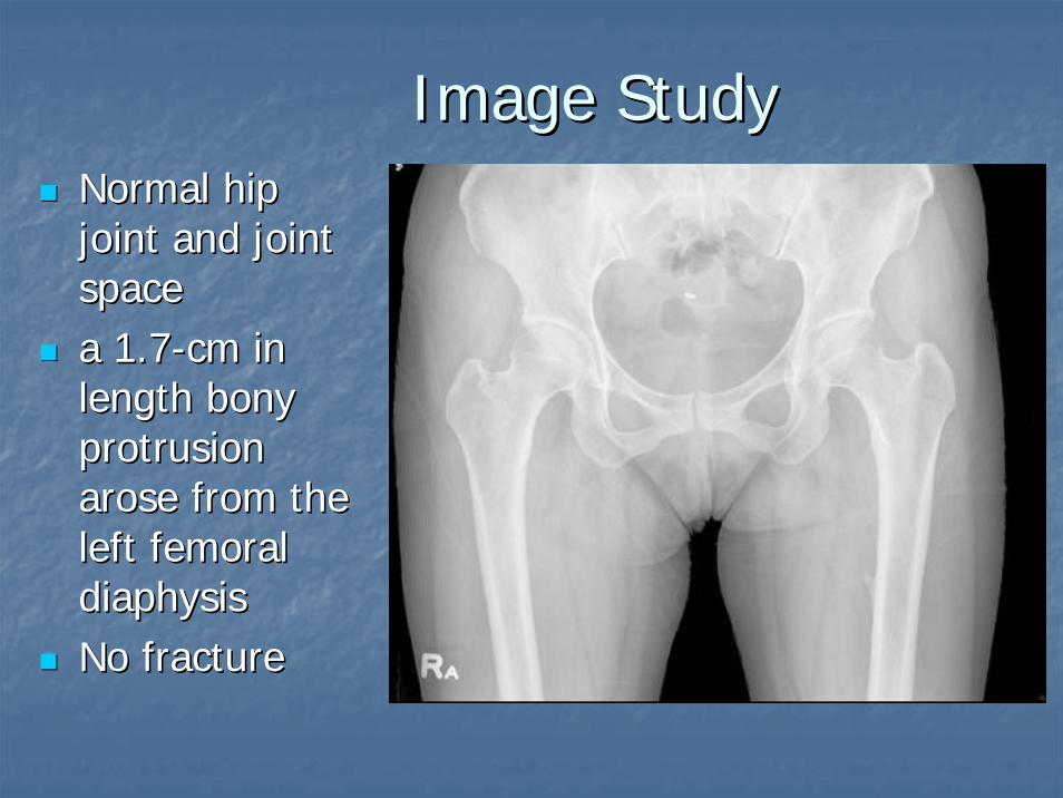
Image StudyImage StudyNormal hip Normal hip joint and joint joint and joint spacespacea 1.7a 1.7--cm in cm in length bony length bony protrusion protrusion arose from the arose from the left femoral left femoral diaphysisdiaphysisNo fractureNo fracture
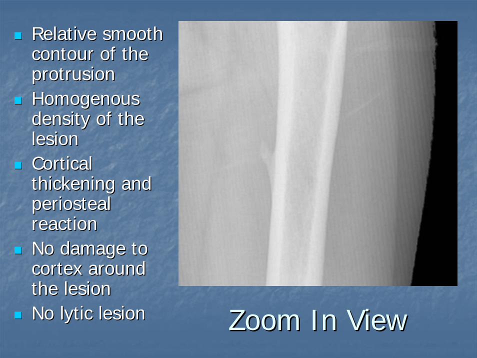
Zoom In ViewZoom In View
Relative smooth Relative smooth contour of the contour of the protrusionprotrusionHomogenous Homogenous density of the density of the lesionlesionCortical Cortical thickening and thickening and periostealperiostealreaction reaction No damage to No damage to cortex around cortex around the lesionthe lesionNo No lyticlytic lesionlesion
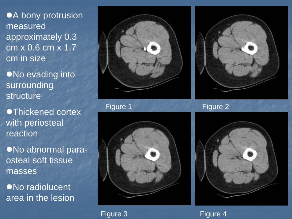
Figure 1 Figure 2
Figure 3 Figure 4
A bony protrusion measured approximately 0.3 cm x 0.6 cm x 1.7 cm in size
No evading into surrounding structure
Thickened cortex with periostealreaction
No abnormal para-osteal soft tissue masses
No radiolucent area in the lesion

Fig.1Fig.1

Fig.2Fig.2

Review of the LesionReview of the Lesion
Pain at medial aspect of left thighPain at medial aspect of left thighA bony protrusion at left femurA bony protrusion at left femurCortical thickening , Cortical thickening , periostealperiosteal reactionreactionSmooth contour, homogenous densitySmooth contour, homogenous densityNo bone destruction, no peripheral soft No bone destruction, no peripheral soft tissue mass or invasiontissue mass or invasionThe lesion is The lesion is radiopaqueradiopaque without without lyticlyticregionregion

Differential DiagnosisDifferential Diagnosis
A.A. OsteochondromaOsteochondromaB.B. PeriostealPeriosteal ChondromaChondromaC.C. ChondrosarcomaChondrosarcomaD.D. PeriostealPeriosteal OsteosarcomaOsteosarcomaE.E. ParostealParosteal OsteosarcomaOsteosarcomaF.F. OsteoidOsteoid OsteomaOsteomaG.G. Ossifying Ossifying FibromaFibroma

OsteochondromaOsteochondroma
Hard ,painless and fixed mass in the Hard ,painless and fixed mass in the metaphysealmetaphyseal regionregionMost commonly occurs in long bones Most commonly occurs in long bones including proximal & distal femur, including proximal & distal femur, proximal tibia, pelvis , or scapulaproximal tibia, pelvis , or scapulaAssociated symptoms due to tissue or Associated symptoms due to tissue or nerve irritation/compression .nerve irritation/compression .
PeriostealPeriosteal ChondromaChondroma
The cortex may be involved to a variable degree, The cortex may be involved to a variable degree, but the lesions do not involve the but the lesions do not involve the medullarymedullaryspace space No ring and arc figures seen in the ossified No ring and arc figures seen in the ossified matrix, and neither was there any trace of matrix, and neither was there any trace of trabeculartrabecular organization of the ossified material organization of the ossified material No No periostealperiosteal reaction reaction Amorphous character of the Amorphous character of the ossificossific material material within the lesion within the lesion

ChondrosarcomaChondrosarcoma
A A fusiformfusiform, lucent defect with scalloping of , lucent defect with scalloping of the inner cortex and the inner cortex and periostealperiosteal reaction on reaction on plain filmplain filmChondroidChondroid matrix mineralization of matrix mineralization of ““rings rings and arcsand arcs”” in 70%in 70%Extension into the soft tissue may be Extension into the soft tissue may be present as well as present as well as punctatepunctate or stippled or stippled calcification of the cartilage matrix calcification of the cartilage matrix

OsteosarcomaOsteosarcoma
As time goes on ,As time goes on ,osteosarcomaosteosarcoma pain increases .pain increases .OsteosarcomaOsteosarcoma calcifies from the center and calcifies from the center and continues to the periphery continues to the periphery Usually Usually extandsextands into soft tissue and metastasis into soft tissue and metastasis Night pain which awakes the patientNight pain which awakes the patientBone destruction, formation, Bone destruction, formation, periostealperiosteal reaction reaction and mineralized soft tissue mass are typical and mineralized soft tissue mass are typical features.features.CodmanCodman’’s Triangles Triangle

PeriostealPeriosteal OsteosarcomaOsteosarcoma
Often found on the anterior surface of the Often found on the anterior surface of the diaphysisdiaphysisA radiolucent, A radiolucent, fusiformfusiform mass attached to mass attached to the bone surface on the bone surface on plaimplaim filmfilmMay May creatcreat a crater on the cortex with a crater on the cortex with striated , radiating mineralizationstriated , radiating mineralization

ParostealParosteal OsteosarcomaOsteosarcoma
Found in the Found in the metaphysismetaphysis of long bones, of long bones, especially the posterior femur above the knee especially the posterior femur above the knee The lesion arises from the surface of the bone The lesion arises from the surface of the bone and has a tendency to encircle the bone and has a tendency to encircle the bone CT may show radiolucent zone of CT may show radiolucent zone of periosteumperiosteumand fibrous tissue that becomes trapped and fibrous tissue that becomes trapped between the encircling tumor and the cortexbetween the encircling tumor and the cortex

OsteoidOsteoid OsteomaOsteoma
A sharp round or oval lesion that is less A sharp round or oval lesion that is less than 2 cm in diameter than 2 cm in diameter A homogeneousA homogeneous dense center dense center A 1A 1--2 mm peripheral radiolucent zone2 mm peripheral radiolucent zoneA distinct clinical picture of dull pain that is A distinct clinical picture of dull pain that is worse at night and disappears within 20 to worse at night and disappears within 20 to 30 minutes of treatment with 30 minutes of treatment with NSAIDsNSAIDs
Ossifying Ossifying FibromaFibroma
It occurs during the first decade of life and It occurs during the first decade of life and presents clinically as a painless, enlarging mass.presents clinically as a painless, enlarging mass.The most common site in adults is the mandible, The most common site in adults is the mandible, followed by other long bones.followed by other long bones.It is a It is a lyticlytic lesion of bone and often causes lesion of bone and often causes anterioranterior--posterior bowing.posterior bowing.This wellThis well--circumscribed tumor has a multicircumscribed tumor has a multi--loculatedloculated appearance and causes distortion of appearance and causes distortion of the thin cortex.the thin cortex.

ImpressionImpression
OsteochondromaOsteochondroma

OsteochondromaOsteochondroma
Definition : A benign , Definition : A benign , chondrogenicchondrogenictumor of bone characterized by a tumor of bone characterized by a mass and pain.mass and pain.
Synonyms :Synonyms :1.1. OsteocartilaginousOsteocartilaginous exostosisexostosis2.2. OsteochondromatosisOsteochondromatosis3.3. DiaphysealDiaphyseal aclasisaclasis
EpidemiologyEpidemiology
It accounts for 20It accounts for 20--50% of benign bone 50% of benign bone tumors and 10tumors and 10--15% of all bone tumors.15% of all bone tumors.It can occur in any bone where cartilage It can occur in any bone where cartilage eventually forms bone. It occurs most eventually forms bone. It occurs most often at long bone. Distal femur and often at long bone. Distal femur and proximal tibia are the most common site.proximal tibia are the most common site.Peak incidence : 10Peak incidence : 10--20 years of age in 20 years of age in 80% of cases 80% of cases

PathogenesisPathogenesis
OsteochondromasOsteochondromas are most likely caused by are most likely caused by either a congenital defect or trauma of the either a congenital defect or trauma of the perichondriumperichondrium which results in the which results in the herniationherniation of of a fragment of the a fragment of the epiphysealepiphyseal growth plate growth plate through the through the periostealperiosteal bone cuff .bone cuff .The lesions occur only in bones that develop The lesions occur only in bones that develop from cartilage (from cartilage (endochondralendochondral ossification). ossification).
Symptoms And SignsSymptoms And Signs
Hard, painless, fixed massHard, painless, fixed massPain from pressure to nearby tissues or Pain from pressure to nearby tissues or nerve nerve Bone deformity may occur due to Bone deformity may occur due to undergrowth of the affected bones. undergrowth of the affected bones. ValgusValgus at knee, ankle, elbow, wrist.at knee, ankle, elbow, wrist.Limb length inequalityLimb length inequalityLowerLower--thanthan--normalnormal--height for age height for age

Imaging ProceduresImaging Procedures
A compact A compact pedunculatedpedunculated or sessile protuberance or sessile protuberance of bone. It is a wellof bone. It is a well--defined lesion projecting defined lesion projecting from the from the metaphysismetaphysis..Cortex and Cortex and spongiosaspongiosa are continuous with that are continuous with that of the affected bone of the affected bone Distinct and well demarcated external surface of Distinct and well demarcated external surface of the tumor the tumor No evading into surrounding tissueNo evading into surrounding tissueNo destruction to boneNo destruction to bone
TreatmentTreatment
Asymptomatic Asymptomatic osteochondromasosteochondromas : : Treatment is not necessary. Observing Treatment is not necessary. Observing only is suggested.only is suggested.Patients with pain or Patients with pain or neurologicneurologicsymptoms due to compression :symptoms due to compression :
1.1. Surgery : to remove the massSurgery : to remove the mass2.2. Medication : to control painMedication : to control pain


