Carotid axillary bypass in a patient with blockedsubclavian stents: a case reportTarig I Barakat*, Louise Kenny, Hazim Khout, Grace Timmons and Vish Bhattacharya
Abstract
Introduction: Surgical treatment of symptomatic occlusive lesions of the proximal subclavian artery is infrequentlynecessary. Carotid subclavian bypass has gained popularity and is now considered standard treatment whenstenting is not possible. Exposure of the subclavian artery and bypass grafting onto it is difficult, as the vessel isdelicate, thin-walled and located deep in the supraclavicular fossa. The thoracic duct and brachial plexus are inclose proximity to the left subclavian artery and are therefore susceptible to damage. Distal grafting to the axillaryartery instead of the subclavian artery has the potential of avoiding some of these risks. Infraclavicular exposure ofthe axillary artery is more straightforward. The vessel wall is thicker and is easier to handle. In this case report, wedescribe a patient with a left proximal subclavian occlusion which was stented twice and blocked on bothoccasions. The patient underwent a carotid axillary bypass, as grafting onto the subclavian artery was impossiblebecause of the two occluded metal stents.
Case presentation: A 56-year-old Caucasian woman, a heavy smoker, presented acutely with left arm numbnessand pain and blood pressure discrepancies in both arms. A diagnosis of subclavian stenosis was confirmed on thebasis of a computed tomographic scan and a magnetic resonance angiogram. The patient had undergonesubclavian artery stenting twice, and unfortunately the stents blocked on both occasions. The patient underwentcarotid axillary bypass surgery. She had an uneventful recovery and was able to return to a full, normal life.
Conclusion: Carotid axillary bypass appears to be a good alternative to carotid subclavian bypass in the treatmentof symptomatic proximal stenosis or occlusion of the subclavian artery.
IntroductionAlthough proximal subclavian artery disease is oftenasymptomatic, once ischemic or embolic complicationsoccur, surgery may be necessary. Transluminal therapyof lesions of subclavian, innominate and common caro-tid arteries by balloon angioplasty, with or withoutstenting, is an increasingly performed procedure, espe-cially in cases of stenosis.Although preliminary data for focal lesions are
encouraging, careful reporting of long-term results willbe the only way to determine whether these non-surgi-cal endoluminal procedures are sufficiently effective tobe offered as reasonable alternatives to the better-provensurgical reconstructions.The use of extrathoracic reconstruction for patients
with symptomatic proximal subclavian artery disease is
well-established. The carotid subclavian bypass is thecommonest surgical procedure in cases in which stent-ing is not possible.This procedure was first described by Diethrich et al.
in 1967 [1], and excellent long-term results have beendescribed in several case series [2-8]Exposure of the subclavian artery carries with it the
potential risk of damage to major lymphatic vessels andnerves. Exposure of the axillary artery using the infracla-vicular approach is technically easier. The artery iseasier to handle, and the wall of the vessel is thicker.However, there is a small risk of brachial plexusdamage.Criado [9] performed 26 carotid-axillary surgical pro-
cedures in 10 years, and he reported 96% graft patencyrate over four years. He used prosthetic Dacron andpolytetrafluoroethylene (PTFE) ringed grafts tunneledunder the clavicle. No shunting is needed unless the* Correspondence: [email protected]
Department of General Surgery, Queen Elizabeth Hospital, Sheriff Hill,Gateshead, Tyne & Wear, NE9 6SX, UK
Barakat et al. Journal of Medical Case Reports 2011, 5:237http://www.jmedicalcasereports.com/content/5/1/237 JOURNAL OF MEDICAL
patient has a significant internal carotid lesion. Thechance of distal embolization is minimal.
Case reportWe describe the case of a 56-year-old Caucasian womanwho presented acutely with left arm numbness and painlasting for nearly eight hours. She had had similar epi-sodes of numbness a few days previously, but these hadlasted for only five to 10 minutes each time.She smoked 20 cigarettes/day. Her heart rate was reg-
ular, although her blood pressure was lower in the leftarm than in the right arm (103/80 mmHg vs. 170/80mmHg, respectively). Her left arm looked pink but cool,with no palpable brachial, radial or ulnar pulse. Therewas decreased sensation over the forearm, though therewas no motor deficit.Her chest, cardiovascular and abdominal examinations
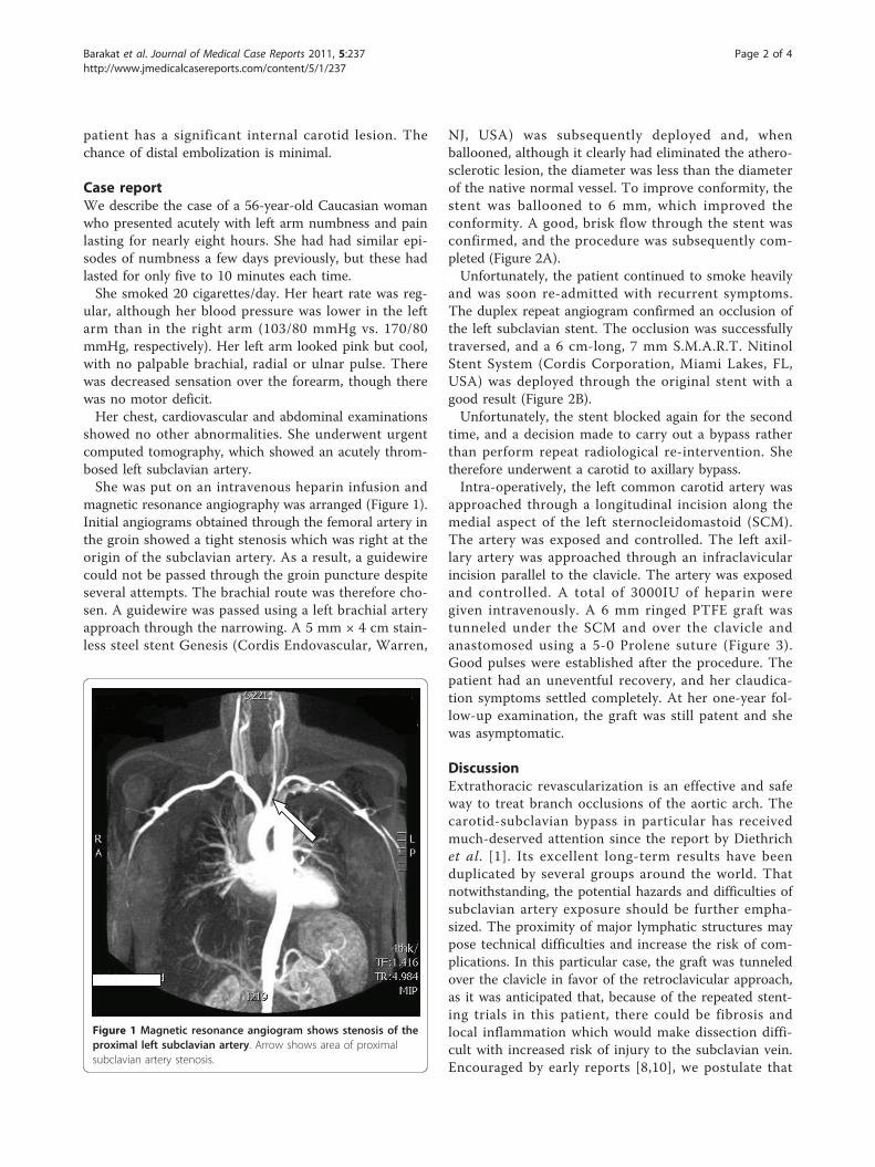
showed no other abnormalities. She underwent urgentcomputed tomography, which showed an acutely throm-bosed left subclavian artery.She was put on an intravenous heparin infusion and
magnetic resonance angiography was arranged (Figure 1).Initial angiograms obtained through the femoral artery inthe groin showed a tight stenosis which was right at theorigin of the subclavian artery. As a result, a guidewirecould not be passed through the groin puncture despiteseveral attempts. The brachial route was therefore cho-sen. A guidewire was passed using a left brachial arteryapproach through the narrowing. A 5 mm × 4 cm stain-less steel stent Genesis (Cordis Endovascular, Warren,
NJ, USA) was subsequently deployed and, whenballooned, although it clearly had eliminated the athero-sclerotic lesion, the diameter was less than the diameterof the native normal vessel. To improve conformity, thestent was ballooned to 6 mm, which improved theconformity. A good, brisk flow through the stent wasconfirmed, and the procedure was subsequently com-pleted (Figure 2A).Unfortunately, the patient continued to smoke heavily
and was soon re-admitted with recurrent symptoms.The duplex repeat angiogram confirmed an occlusion ofthe left subclavian stent. The occlusion was successfullytraversed, and a 6 cm-long, 7 mm S.M.A.R.T. NitinolStent System (Cordis Corporation, Miami Lakes, FL,USA) was deployed through the original stent with agood result (Figure 2B).Unfortunately, the stent blocked again for the second
time, and a decision made to carry out a bypass ratherthan perform repeat radiological re-intervention. Shetherefore underwent a carotid to axillary bypass.Intra-operatively, the left common carotid artery was
approached through a longitudinal incision along themedial aspect of the left sternocleidomastoid (SCM).The artery was exposed and controlled. The left axil-lary artery was approached through an infraclavicularincision parallel to the clavicle. The artery was exposedand controlled. A total of 3000IU of heparin weregiven intravenously. A 6 mm ringed PTFE graft wastunneled under the SCM and over the clavicle andanastomosed using a 5-0 Prolene suture (Figure 3).Good pulses were established after the procedure. Thepatient had an uneventful recovery, and her claudica-tion symptoms settled completely. At her one-year fol-low-up examination, the graft was still patent and shewas asymptomatic.
DiscussionExtrathoracic revascularization is an effective and safeway to treat branch occlusions of the aortic arch. Thecarotid-subclavian bypass in particular has receivedmuch-deserved attention since the report by Diethrichet al. [1]. Its excellent long-term results have beenduplicated by several groups around the world. Thatnotwithstanding, the potential hazards and difficulties ofsubclavian artery exposure should be further empha-sized. The proximity of major lymphatic structures maypose technical difficulties and increase the risk of com-plications. In this particular case, the graft was tunneledover the clavicle in favor of the retroclavicular approach,as it was anticipated that, because of the repeated stent-ing trials in this patient, there could be fibrosis andlocal inflammation which would make dissection diffi-cult with increased risk of injury to the subclavian vein.Encouraged by early reports [8,10], we postulate that
Figure 1 Magnetic resonance angiogram shows stenosis of theproximal left subclavian artery. Arrow shows area of proximalsubclavian artery stenosis.
Barakat et al. Journal of Medical Case Reports 2011, 5:237http://www.jmedicalcasereports.com/content/5/1/237
Page 2 of 4
use of the axillary artery as the distal anastomotic sitewould simplify the operation and avoid the risk of lym-phatic injury altogether.Carotid axillary bypass is a very good alternative to
carotid subclavian bypass. In this case specifically, thecarotid axillary bypass was favored because of the pre-sence of stents in the subclavian artery, which wouldhave made grafting very difficult.The risk of operation-related stroke is very minimal.
Shunting is unnecessary unless a critical internal carotid
lesion co-exists on the same side. Concomitant carotidendarterectomy at the donor graft site may be per-formed in patients with severe atheromatous plaques.With regard to radiology, attempted recanalization forsubclavian arteries is better approached from a brachialpuncture than from a groin puncture.
ConclusionCarotid axillary bypass is a very good alternative to car-otid subclavian bypass. It is safe and technically easier,
Figure 2 (A)The first stent was placed successfully. (B) The second Nitinol stent was placed within the first stent after 3 months.
Figure 3 Carotid axillary bypass using PTFE graft. Arrows show anastomosis sites.
Barakat et al. Journal of Medical Case Reports 2011, 5:237http://www.jmedicalcasereports.com/content/5/1/237
Page 3 of 4
and it provides equally good short- and long-termresults.
ConsentWritten informed consent was obtained from the patientfor publication of this case report and any accompany-ing images. A copy of the written consent is availablefor review by the editor in chief of this journal.
Authors’ contributionsTB was involved in the major parts of writing the paper and performing theliterature search, as well as being involved in performing the surgery and inthe patient’s pre-operative and post-operative care. LK and HK contributedto writing the manuscript and to the literature search. GT was involved inthe radiological procedures. VB was the responsible vascular surgeon andteam leader who set the management plan. All authors read the manuscriptand agreed to its contents.
Competing interestsThe authors declare that they have no competing interests.
Received: 7 February 2010 Accepted: 27 June 2011Published: 27 June 2011
References1. Diethrich EB, Garrett HE, Ameriso J, Crawford ES, el-Bayar M, De Bakey ME:
Occlusive disease of the common carotid and subclavian arteriestreated by carotid-subclavian bypass: analysis of 125 cases. Am J Surg1967, 114:800-808.
2. Ziomek S, Quiñones-Baldrich W, Bussutil RW, Baker JD, Machleder HI,Moore WS: The superiority of synthetic arterial grafts over autologousveins in carotid-subclavian bypass. J Vasc Surg 1986, 3:140-145.
3. Lord RS, Ehrenfeld WK: Carotid-subclavian bypass: a hemodynamic study.Surgery 1969, 66:521-526.
4. Moore WS, Malone JM, Goldstone J: Extrathoracic repair of branchocclusions of the aortic arch. Am J Surg 1976, 132:249-257.
5. Thompson BW, Read RC, Campbell GS: Operative correction of proximalblocks of the subclavian or innominate arteries. J Cardiovasc Surg (Torino)1980, 21:125-130.
7. AbuRahma AF, Robinson PA, Khan MZ, Khan JH, Boland JP: Brachiocephalicrevascularization: a comparison between carotid-subclavian arterybypass and axilloaxillary artery bypass. Surgery 1992, 112:84-91.
8. Lyons C, Galbraith G: Surgical treatment of atherosclerotic occlusion ofthe internal carotid artery. Ann Surg 1957, 146:487-498.
9. Criado FJ: Extrathoracic management of aortic arch syndrome. Br J Surg1982, 69(Suppl):S45-S51.
10. Lowell RC, Mills JL: Critical evaluation of axilloaxillary artery bypass forsurgical management of symptomatic subclavian and innominate arteryocclusive disease. Cardiovasc Surg 1993, 1:530-535.
doi:10.1186/1752-1947-5-237Cite this article as: Barakat et al.: Carotid axillary bypass in a patientwith blocked subclavian stents: a case report. Journal of Medical CaseReports 2011 5:237.
Submit your next manuscript to BioMed Centraland take full advantage of:
• Convenient online submission
• Thorough peer review
• No space constraints or color figure charges
• Immediate publication on acceptance
• Inclusion in PubMed, CAS, Scopus and Google Scholar
• Research which is freely available for redistribution
Submit your manuscript at www.biomedcentral.com/submit
Barakat et al. Journal of Medical Case Reports 2011, 5:237http://www.jmedicalcasereports.com/content/5/1/237