Journal of Indian Academy of Dental Specialist Researchers | Vol. 1 | Issue 2 | Jul-Dec 2014 70 Prosthetic rehabilitation in a partially edentulous patient with lost vertical dimension: A case report Arun Jaikumar R., Madhulika N. 1 , Pradeep Kumar R. 2 , Vijayalakshmi K. 3 Department of Prosthodoncs, Rajah Muah Dental College, Chidambaram, 1 Department of Oral Medicine and Radiology, Indira Gandhi Instute of Dental Science, Pondicherry, 2 Department of Oral and Maxillofacial Surgery, Rajah Muah Dental College, 3 Department of Orthodoncs, Adhiparasakthi Dental College, Melmaruvathur, Tamil Nadu, India to a successful outcome and patient satisfaction, in multidisciplinary cases. CASE REPORT A 35-year-old female reported with a chief complaint of several missing teeth, excessive wearing of teeth, reduced chewing efficiency, and discomfort due to over closure. Patient was in good general health, her medical and dental histories were not contradictory for dental treatment. Intraoral examination revealed partially edentulous maxillary and mandibular arches with reduced VD [Figure 1]. Full mouth rehabilitation of the mouth was planned to restore the function, esthetics, speech, and comfort of the patient. Patient was informed of the diagnosis, the treatment planning; and her consent was obtained. Due to the presence of decreased VD, an increase in VD was first considered. Diagnostic impression was taken with alginate. Maxillary cast was mounted on Hanau Wide-Vue Articulator, using facebow transfer and mandibular cast was mounted using centric relation (CR) record. Vertical dimension (VD) was increased by 5mm using an occlusal splint/bite raising splint which was fabricated using self-cure acrylic resin at the CR position [Figure 2]. INTRODUCTION The prosthetic rehabilitation of a patient with a functionally compromised dentition frequently involves a multidisciplinary approach. Successful integration of esthetics and function do not emerge by chance, but rather as a result of the meticulous development of clearly defined parameters and their subsequent incorporation into the design of the prosthesis. Appropriate case selection and careful treatment planning are critical ABSTRACT The stomatognathic system is a complicated structure, and patients usually adapt to their existing vertical dimension of occlusion (VDO). Therefore, prosthetic rehabilitation of decreased VDO should be considered only when dictated by esthetic or functional requirements. Successful rehabilitation of these patients may present us with a huge challenge of a lack of restorative space. Careful and comprehensive treatment planning is required for each individual patient. The important aspect is to find the most comfortable position for the patient. In this case a bite raising splint was used after the decision of increasing VD by anatomical landmark, facial and physiologic measurement. Once the compatibility of the new VD had been confirmed, interim fixed restoration and the permanent reconstruction was initiated. In this case report, a satisfactory clinical result was achieved by restoring the VD with an improvement in esthetics and function. Key words: Bite raising splint, prosthetic rehabilitation, stomatognathic system, vertical dimension of occlusion Case Report Address for correspondence: Dr. Arun R. Jaikumar, 2/1 A Pachaiyappan School Street, Opp to K. R. Medicals, Chidambaram - 608 001, Tamil Nadu, India. E-mail: [email protected]Access this article online Quick Response Code: Website: www.jiadsr.org DOI: 10.4103/2229-3019.148266
Transcript
Journal of Indian Academy of Dental Specialist Researchers | Vol. 1 | Issue 2 | Jul-Dec 201470
Prosthetic rehabilitation in a partially edentulous patient with lost vertical dimension: A case report
Arun Jaikumar R., Madhulika N.1, Pradeep Kumar R.2, Vijayalakshmi K.3
Department of Prosthodontics, Rajah Mutiah Dental College, Chidambaram, 1Department of Oral Medicine and Radiology, Indira Gandhi Institute of Dental Science, Pondicherry, 2Department of Oral and Maxillofacial Surgery, Rajah Mutiah Dental College, 3Department of
Orthodontics, Adhiparasakthi Dental College, Melmaruvathur, Tamil Nadu, India
to a successful outcome and patient satisfaction, in multidisciplinary cases.
CASE REPORT
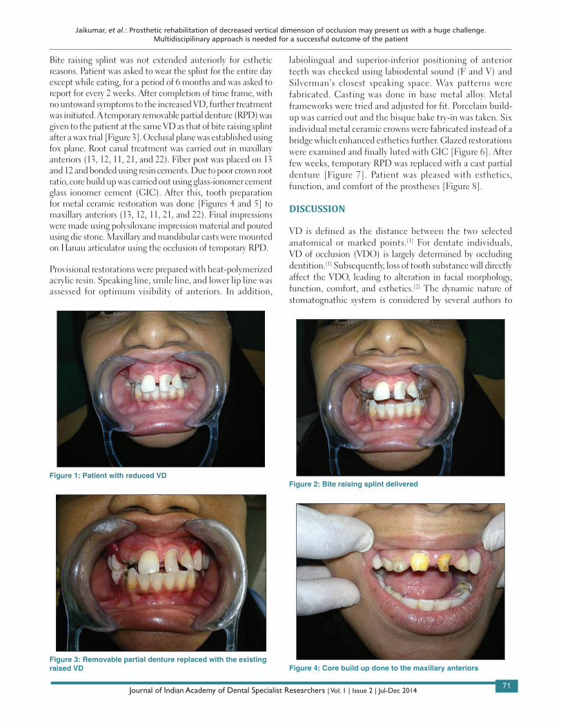
A 35-year-old female reported with a chief complaint of several missing teeth, excessive wearing of teeth, reduced chewing efficiency, and discomfort due to over closure. Patient was in good general health, her medical and dental histories were not contradictory for dental treatment. Intraoral examination revealed partially edentulous maxillary and mandibular arches with reduced VD [Figure 1]. Full mouth rehabilitation of the mouth was planned to restore the function, esthetics, speech, and comfort of the patient. Patient was informed of the diagnosis, the treatment planning; and her consent was obtained. Due to the presence of decreased VD, an increase in VD was first considered. Diagnostic impression was taken with alginate. Maxillary cast was mounted on Hanau Wide-Vue Articulator, using facebow transfer and mandibular cast was mounted using centric relation (CR) record. Vertical dimension (VD) was increased by 5mm using an occlusal splint/bite raising splint which was fabricated using self-cure acrylic resin at the CR position [Figure 2].
INTRODUCTION
The prosthetic rehabilitation of a patient with a functionally compromised dentition frequently involves a multidisciplinary approach. Successful integration of esthetics and function do not emerge by chance, but rather as a result of the meticulous development of clearly defined parameters and their subsequent incorporation into the design of the prosthesis. Appropriate case selection and careful treatment planning are critical
ABSTRACTThe stomatognathic system is a complicated structure, and patients usually adapt to their existing vertical dimension of occlusion (VDO). Therefore, prosthetic rehabilitation of decreased VDO should be considered only when dictated by esthetic or functional requirements. Successful rehabilitation of these patients may present us with a huge challenge of a lack of restorative space. Careful and comprehensive treatment planning is required for each individual patient. The important aspect is to find the most comfortable position for the patient. In this case a bite raising splint was used after the decision of increasing VD by anatomical landmark, facial and physiologic measurement. Once the compatibility of the new VD had been confirmed, interim fixed restoration and the permanent reconstruction was initiated. In this case report, a satisfactory clinical result was achieved by restoring the VD with an improvement in esthetics and function.
Address for correspondence: Dr. Arun R. Jaikumar, 2/1 A Pachaiyappan School Street, Opp to K. R. Medicals, Chidambaram - 608 001, Tamil Nadu, India. E-mail: [email protected]
Access this article online
Quick Response Code:Website: www.jiadsr.org
DOI: 10.4103/2229-3019.148266
Jaikumar, et al.: Prosthetic rehabilitation of decreased vertical dimension of occlusion may present us with a huge challenge. Multidiscipilinary approach is needed for a successful outcome of the patient
Journal of Indian Academy of Dental Specialist Researchers | Vol. 1 | Issue 2 | Jul-Dec 201471
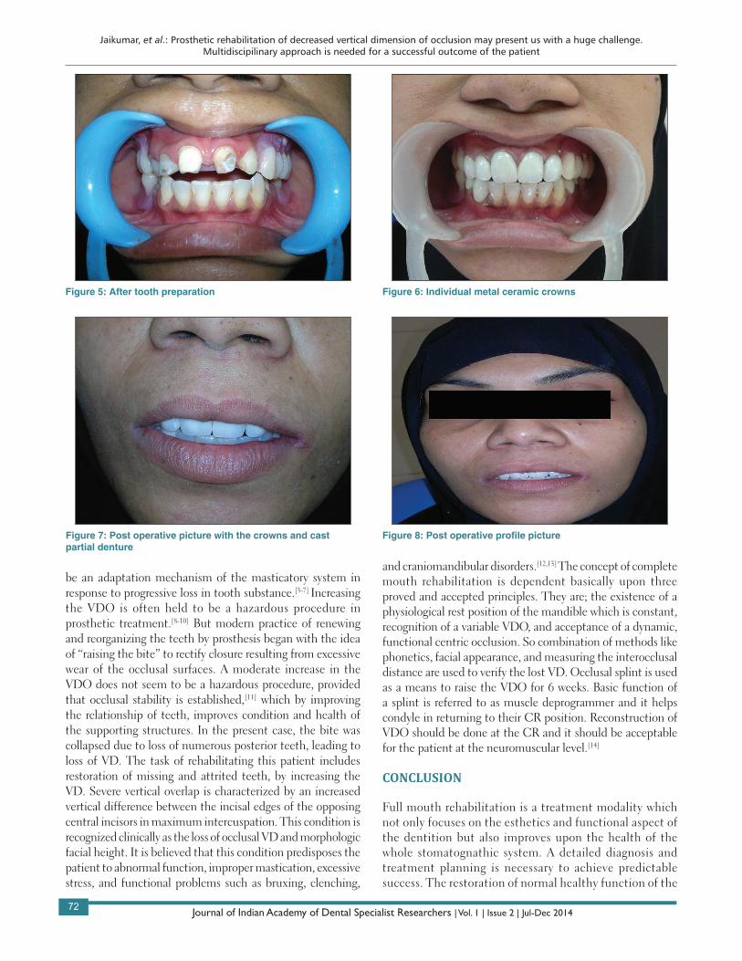
Bite raising splint was not extended anteriorly for esthetic reasons. Patient was asked to wear the splint for the entire day except while eating, for a period of 6 months and was asked to report for every 2 weeks. After completion of time frame, with no untoward symptoms to the increased VD, further treatment was initiated. A temporary removable partial denture (RPD) was given to the patient at the same VD as that of bite raising splint after a wax trial [Figure 3]. Occlusal plane was established using fox plane. Root canal treatment was carried out in maxillary anteriors (13, 12, 11, 21, and 22). Fiber post was placed on 13 and 12 and bonded using resin cements. Due to poor crown root ratio, core build up was carried out using glass-ionomer cement glass ionomer cement (GIC). After this, tooth preparation for metal ceramic restoration was done [Figures 4 and 5] to maxillary anteriors (13, 12, 11, 21, and 22). Final impressions were made using polysiloxane impression material and poured using die stone. Maxillary and mandibular casts were mounted on Hanau articulator using the occlusion of temporary RPD.
Provisional restorations were prepared with heat-polymerized acrylic resin. Speaking line, smile line, and lower lip line was assessed for optimum visibility of anteriors. In addition,
labiolingual and superior-inferior positioning of anterior teeth was checked using labiodental sound (F and V) and Silverman’s closest speaking space. Wax patterns were fabricated. Casting was done in base metal alloy. Metal frameworks were tried and adjusted for fit. Porcelain build-up was carried out and the bisque bake try-in was taken. Six individual metal ceramic crowns were fabricated instead of a bridge which enhanced esthetics further. Glazed restorations were examined and finally luted with GIC [Figure 6]. After few weeks, temporary RPD was replaced with a cast partial denture [Figure 7]. Patient was pleased with esthetics, function, and comfort of the prostheses [Figure 8].
DISCUSSION
VD is defined as the distance between the two selected anatomical or marked points.[1] For dentate individuals, VD of occlusion (VDO) is largely determined by occluding dentition.[1] Subsequently, loss of tooth substance will directly affect the VDO, leading to alteration in facial morphology, function, comfort, and esthetics.[2] The dynamic nature of stomatognathic system is considered by several authors to
Figure 1: Patient with reduced VD
Figure 3: Removable partial denture replaced with the existing raised VD
Figure 2: Bite raising splint delivered
Figure 4: Core build up done to the maxillary anteriors
Jaikumar, et al.: Prosthetic rehabilitation of decreased vertical dimension of occlusion may present us with a huge challenge. Multidiscipilinary approach is needed for a successful outcome of the patient
Journal of Indian Academy of Dental Specialist Researchers | Vol. 1 | Issue 2 | Jul-Dec 201472
be an adaptation mechanism of the masticatory system in response to progressive loss in tooth substance.[3-7] Increasing the VDO is often held to be a hazardous procedure in prosthetic treatment.[8-10] But modern practice of renewing and reorganizing the teeth by prosthesis began with the idea of “raising the bite” to rectify closure resulting from excessive wear of the occlusal surfaces. A moderate increase in the VDO does not seem to be a hazardous procedure, provided that occlusal stability is established,[11] which by improving the relationship of teeth, improves condition and health of the supporting structures. In the present case, the bite was collapsed due to loss of numerous posterior teeth, leading to loss of VD. The task of rehabilitating this patient includes restoration of missing and attrited teeth, by increasing the VD. Severe vertical overlap is characterized by an increased vertical difference between the incisal edges of the opposing central incisors in maximum intercuspation. This condition is recognized clinically as the loss of occlusal VD and morphologic facial height. It is believed that this condition predisposes the patient to abnormal function, improper mastication, excessive stress, and functional problems such as bruxing, clenching,
and craniomandibular disorders.[12,13] The concept of complete mouth rehabilitation is dependent basically upon three proved and accepted principles. They are; the existence of a physiological rest position of the mandible which is constant, recognition of a variable VDO, and acceptance of a dynamic, functional centric occlusion. So combination of methods like phonetics, facial appearance, and measuring the interocclusal distance are used to verify the lost VD. Occlusal splint is used as a means to raise the VDO for 6 weeks. Basic function of a splint is referred to as muscle deprogrammer and it helps condyle in returning to their CR position. Reconstruction of VDO should be done at the CR and it should be acceptable for the patient at the neuromuscular level.[14]
CONCLUSION
Full mouth rehabilitation is a treatment modality which not only focuses on the esthetics and functional aspect of the dentition but also improves upon the health of the whole stomatognathic system. A detailed diagnosis and treatment planning is necessary to achieve predictable success. The restoration of normal healthy function of the
Figure 5: After tooth preparation Figure 6: Individual metal ceramic crowns
Figure 7: Post operative picture with the crowns and cast partial denture
Figure 8: Post operative profile picture
Jaikumar, et al.: Prosthetic rehabilitation of decreased vertical dimension of occlusion may present us with a huge challenge. Multidiscipilinary approach is needed for a successful outcome of the patient
Journal of Indian Academy of Dental Specialist Researchers | Vol. 1 | Issue 2 | Jul-Dec 201473
How to cite this article: Jaikumar RA, Madhulika N, Kumar RP, Vijayalakshmi K. Prosthetic rehabilitation in a partially edentulous patient with lost vertical dimension: A case report. J Indian Acad Dent Spec Res 2014;1:70-3.Source of Support: Nil, Conflict of Interest: None declared
masticating apparatus is the ultimate aim of full mouth rehabilitation.
REFERENCES
1. The glossary of prosthodontic terms. J Prosthet Dent 2005;94:10-92.2. Turner KA, Misssirlian DM. Restoration of the extremely worn
dentition. J Prosthet Dent 1984;52:467-74.3. Berry DC, Poole DF. Attrition: Possible mechanism of compensation.
J Oral Rehabil 1976;3:201-6.4. Richards LC. Dental attrition and craniofacial morphology in two
Australian aboriginal populations. J Dent Res 1985;64:1311-5.5. Murphy T. Compensatory mechanism in facial height adjustment
to functional tooth attrition. Aust Dent 1959;4:312-23.6. Varrela J. Dimensional variation of craniofacial structures in relation
to changing masticatory — Functional demands. Eur J Orthod 1992;14:31-6.
7. Crothers A, Sandham A. Vertical height differences in subjects with severe dental wear. Eur J Orthod 1993;15:519-25.
8. Brecker C. Clinical procedures in occlusal rehabilitation.The C. V Mosby Co.; 1966. p. 203-5, 376.
9. Smith BH. Changes in occlusal face height with removable partial prostheses. J Prosthet Dent 1975;34:278-85.
10. Dawson PE. Evaluation diagnosis and treatment of occlusal problems. The C. V. Mosby Co. St. Louis; 1974. p. 275-85.
11. Carlson GE, Ingervall B, Kocak G. Effect of increasing vertical dimension on the masticatory system in subjects with natural teeth. J Prosthet Dent 1979;41:284-9.
12. Graber TM. Ortliodonlics, Principles and Practice. 3rd ed. Philadelphia: Saunders; 1972. p. 239-46.
13. Ricketts RM. A study of changes in lemporal mandibular relations associated wilh ihe treatment of Class II malocclusion (Angle). Am J Orthod 1952;38:918-33.
14. Bloom DR, Padayachy JN. Increasing occlusal vertical dimension--why, when and how. Br Dent J 2006;200:251-6.