Urethral stricture is a common urological pathology with a high recurrence rate after treatment. Urethral manipulations are amongits main causes. In this paper, urethral stricture developed secondary to urethral catheterization and was treated with cold-knifeinternal urethrotomy and the Otis urethrotomy procedure. During the follow-up period, severe ventral penile curvature preventingsexual intercourse developed due to fibrosis of the corpus spongiosum and tunica albuginea of the penis. This ventral penilecurvature was corrected with a separate operation using a tunica vaginalis flap harvested from the left scrotum.
1. Introduction
Urethral stricture results from fibrosis and scarring thatdevelop in the urethral mucosa and surrounding tissue. Itsetiology is mainly due to factors such as transurethral surg-eries, urethral catheterization, pelvic trauma, and hypospa-dias surgery [1].The treatment of urethral stricture using ure-throtomy is still a controversial issue in the current urologicalliterature [2]. Otis urethrotomy has been described as one ofthe internal urethrotomy techniques [3, 4]. Other therapeuticmodalities include urethral dilatation, scar excision, and end-to-end anastomosis, depending on the location and degree ofthe urethral stricture.
2. Case Report
A 58-year-old man, who had undergone a lobectomy due tolung cancer six months previously in a different center, wasreferred to our clinic with difficulty in urination. He also hada history of urethral catheterization to monitor urine outputduring the previous surgery. Uroflowmetry was performed,and the results were consistent with urethral stricture. Analmost complete 2 cm urethral stricture was detected at thelevel of the bulbomembranous urethra during urethroscopyunder regional anesthesia. The patient underwent cold-knife internal urethrotomy for the urethral stricture and
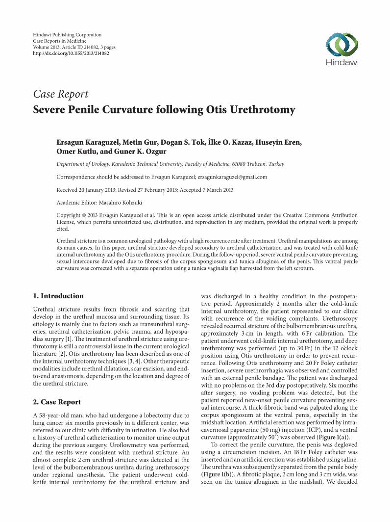
was discharged in a healthy condition in the postopera-tive period. Approximately 2 months after the cold-knifeinternal urethrotomy, the patient represented to our clinicwith recurrence of the voiding complaints. Urethroscopyrevealed recurred stricture of the bulbomembranous urethra,approximately 3 cm in length, with 6 Fr calibration. Thepatient underwent cold-knife internal urethrotomy, and deepurethrotomy was performed (up to 30 Fr) in the 12 o’clockposition using Otis urethrotomy in order to prevent recur-rence. Following Otis urethrotomy and 20 Fr Foley catheterinsertion, severe urethrorrhagia was observed and controlledwith an external penile bandage. The patient was dischargedwith no problems on the 3rd day postoperatively. Six monthsafter surgery, no voiding problem was detected, but thepatient reported new-onset penile curvature preventing sex-ual intercourse. A thick-fibrotic band was palpated along thecorpus spongiosum at the ventral penis, especially in themidshaft location. Artificial erection was performed by intra-cavernosal papaverine (50mg) injection (ICP), and a ventralcurvature (approximately 50∘) was observed (Figure 1(a)).
To correct the penile curvature, the penis was deglovedusing a circumcision incision. An 18 Fr Foley catheter wasinserted and an artificial erectionwas establishedusing saline.The urethra was subsequently separated from the penile body(Figure 1(b)). A fibrotic plaque, 2 cm long and 3 cmwide, wasseen on the tunica albuginea in the midshaft. We decided
2 Case Reports in Medicine
(a) (b) (c)
(d) (e)
Figure 1: (a) Preoperative appearance of the penile curvature, (b) intraoperative appearance of the penile curvature, (c) importation of theflap of tunica vaginalis, (white arrow), (d) closure of curvature area, which was excised by the flap of tunica vaginalis, (white arrow) and (e)appearance of the penis at 3rd week after the operation.
to perform plaque excision and to close the penile corporawith a tunica vaginalis flap. After the preparation of tunicavaginalis flap from the left testis, the fibrotic plaque wasexcised, and the corporeal body was patched with the flap,using watertight absorbable sutures (Figures 1(c) and 1(d)).Additionally, we performed dorsal penile plication usingnonabsorbable sutures for the residual curvature. At the 3rdweek postoperatively, the patient was reevaluated with penileerection induced with the use of oral phosphodiesteraseinhibitor (50mg sildenafil citrate) and visual sexual stimuli.The figure showed that the penile curvature was completelyresolved (Figure 1(e)).
3. Discussion
Urethral stricture is one of the most commonly encounteredpathologies in daily urology practice, especially in malepatients. This can be seen at any age and may be manifestedwith lower urinary tract symptoms or urinary tract infec-tions that severely impair quality of life. There are variousmodalities for treatment, depending on the localization,length, and etiology of the urethral stricture. One of themost important iatrogenic causes of the urethral strictureis urethral catheterization [5]. Excessive strain exerted onthe catheter along the urethra or inflation of the catheterballoon in the urethra by inexperienced health staff canlead to urethral stricture. In this case, urethral stricture wascaused by forced urethral catheterization during lobectomyperformed approximately six months before admission to
our clinic. The patient, who had no voiding problem beforethis urethral catheterization, gradually began to experiencedifficulty in urination during the postoperative period. Thepatient’s symptoms gradually worsened and he experienceddifficulties in urination, at which he applied to our clinic.
The literature contains few reports of penile curva-ture developing after prostatectomy, urethral dilatation, andtransurethral prostate and bladder tumor resection [6, 7].Kelami reported that some patients developed ventral devia-tion of the erect penis after transurethralmanipulations usingautophotography in which patients photographed them-selves [8]. This condition is known as urethral manipulationsyndrome (UMS, Kelami Syndrome). UMS is defined as“an acquired, mostly iatrogenic penile deformity caused byfibrotic changes of the corpus spongiosum” [9]. Inflammationmay occur in the corpus spongiosum as a result of traumaticcatheterization, recurrent urethral dilatations, cystoscopy, ortransurethral surgery. It has also been shown that, follow-ing traumatic catheterization procedures, inflammation maydevelop in the urethra, particularly due to the material usedin the urethral catheter [10]. Ventral penile curvature occursdue to the development of fibrosis in the corpus spongiosum.On the other hand, not all patients develop a spongiofibrosisthat may lead to penile deviation, even after several urethralmanipulations, and why this occurs in a very small numberof patients is unclear.
In this case as well, although the urethral stricturewas resolved after the surgical intervention, ventral penilecurvature occurred due to probable fibrotic process, whichdeveloped secondary to instrumentation and interventions
Case Reports in Medicine 3
in the urethra. We believe that penile curvature resultedfrom fibrosis that developed in the corpus spongiosumbut mostly in the tunica albuginea, especially secondary todeep Otis urethrotomy performed to prevent the recurrenceof the urethral stricture. During the surgical procedureperformed to correct the penile curvature, in addition tothe corpus spongiosum, the tunica albuginea also showedsigns of fibrosis. Urethrolysis alone was insufficient to correctthe penile curvature, and more complex surgery includingfibrotic plaque excision, flap, and penile plicationwas needed.Deep urethrotomy may exacerbate the fibrotic process anda faster development of fibrosis. Urethrorrhagia developingafter Otis urethrotomy was considered as evidence of deepurethrotomy. In addition, bleeding into periurethral tissuemay contribute to an excessive fibrotic process.
Consequently, the likelihood of serious ventral penilecurvature developing after Otis urethrotomy, one of themethods safely used for the treatment of urethral stricture,should always be kept in the mind, especially in the eventof excessive postprocedural bleeding. Great care is requiredin procedures to be performed blind, such as all forms ofurethral manipulation and particularly Otis urethrotomy,and potential urethral damage must be prevented. Urethraldamage developing as a result of procedures performed fortherapeutic procedures may lead to serious penile curvatureat later periods. Correct selection and proper planning ofmethod of treatment are very important in terms of avoidingall these potential complications.
Conflict of Interests
No conflict of interests was declared by the authors.
References
[1] N. Lumen, P. Hoebeke, P. Willemsen, B. De Troyer, R. Pieters,and W. Oosterlinck, “Etiology of urethral stricture disease inthe 21st century,” Journal of Urology, vol. 182, no. 3, pp. 983–987,2009.
[2] R. Santucci and L. Eisenberg, “Urethrotomy has a much lowersuccess rate than previously reported,” Journal of Urology, vol.183, no. 5, pp. 1859–1862, 2010.
[3] A. Schultz, H. Bay-Nielsen, T. Bilde et al., “Prevention ofurethral stricture formation after transurethral resection of theprostate: A controlled randomized study of Otis urethrotomyversus urethral dilation and the use of the polytetrafluoroethy-lene coated versus the uninsulated metal sheath,” Journal ofUrology, vol. 141, no. 1, pp. 73–75, 1989.
[4] H. H. Steenfos and N. Skovgaard, “The importance of internalurethrotomy a.m. Otis for the incidence of urethral stricturefollowing transurethral prostatectomy,” International Urologyand Nephrology, vol. 20, no. 1, pp. 55–59, 1988.
[5] C. Kashefi, K. Messer, R. Barden, C. Sexton, and J. K. Parsons,“Incidence and prevention of iatrogenic urethral injuries,”Journal of Urology, vol. 179, no. 6, pp. 2254–2258, 2008.
[6] W. Merkle, “Cause of deviation of the erectile penis afterurethral manipulations (Kelami syndrome)—demonstration ofultrasound findings and case reports,” Urologia Internationalis,vol. 45, no. 3, pp. 183–185, 1990.
[7] D. Yachia, “Acquired ventral penile curvature: spongiofibrosiscaused by urethralmanipulation,”British Journal ofUrology, vol.64, no. 6, pp. 629–631, 1989.
[8] A. Kelami, “Autophotography in evaluation of functional peniledisorders,” Urology, vol. 21, no. 6, pp. 628–629, 1983.
[9] A. Kelami, “Urethral manipulation syndrome. Description of anew syndrome,”Urologia Internationalis, vol. 39, no. 6, pp. 352–354, 1984.
[10] M. Talja, M. Ruutu, L. C. Andersson, and O. Alfthan, “Urinarycatheter structure and testing methods in relation to tissuetoxicity,” British Journal of Urology, vol. 58, no. 4, pp. 443–449,1986.