2
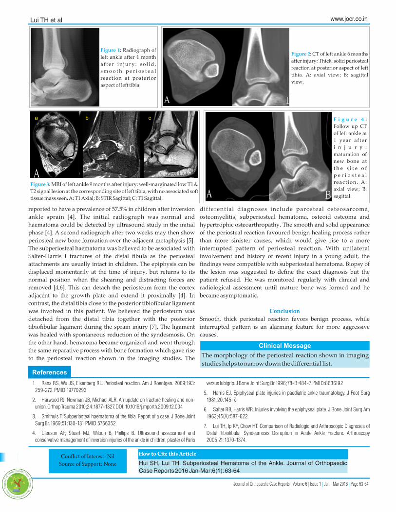
Author’s Photo Gallery 1 Department of Orthopaedics and Traumatology, North District Hospital 9 Po Kin Road, Sheung Shui, NT, Hong Kong SAR, China. Address of Correspondence Dr. Lui,Tun Hing, Department of Orthopaedics and Traumatology, Consultant, North District Hospital, 9 Po Kin Road, Sheung Shui, NT, Hong Kong SAR, China. Email: [email protected] Copyright © 2015 by Journal of Orthpaedic Case Reports Journal of Orthopaedic Case Reports | pISSN 2250-0685 | eISSN 2321-3817 | Available on www.jocr.co.in | doi:10.13107/jocr.2250-0685.380 This is an Open Access article distributed under the terms of the Creative Commons Attribution Non-Commercial License (http://creativecommons.org/licenses/by-nc/3.0) which permits unrestricted non-commercial use, distribution, and reproduction in any medium, provided the original work is properly cited. Abstract Journal of Orthopaedic Case Reports 2016 Jan-Mar: 6(1):Page 63-64 Case Report Introduction: Periosteal reaction has a long list of differential diagnoses ranging from trauma, infection, metabolic disease to malignancy. The morphology of periosteal reaction shown in imaging studies helps to narrow down the list of differential diagnoses. Case report: A 25 year old gentleman had an inversion injury to his left ankle. He complained of lateral ankle and posterior heel pain and swelling after the injury. Radiograph of his left ankle revealed solid, smooth periosteal reaction at posterior aspect of left distal tibia. MRI showed periosteal reaction at the corresponding site, which was better demonstrated in CT scan. Follow up MRI and CT showed maturation of the new bone formation at the site of periosteal reaction. Findings were compatible with subperiosteal hematoma formation from injury, which ossified with time. Conclusion: Smooth, thick periosteal reaction favours benign process, while interrupted pattern is an alarming feature for more aggressive causes. Keywords: subperiosteal haematoma; ankle; periosteal reaction. What to Learn from this Article? Subperiosteal haematoma of the distal tibia is one of the causes of ectopic ossification of the ankle after injury. S H Hui¹, T H Lui¹ Access this article online Website: www.jocr.co.in DOI: 2250-0685.380 Subperiosteal Hematoma of the Ankle Introduction Periosteal reaction has a long list of differential diagnoses, which ranges from benign process e.g. trauma, infection, metabolic disease to sinister causes e.g. malignancy [1]. Detailed history and physical examination are important keys to nail down the diagnosis. Moreover, the morphology of periosteal reaction shown in imaging studies helps to narrow down the differential list. We presented a case of subperiosteal haematoma of the ankle which was successfully diagnosed and monitored by imaging studies. Unnecessary invasive investigation and treatment e.g. excisional biopsy of the lesion was avoided. Case report A 25 year old gentleman had an inversion injury to his left ankle. He complained of lateral ankle and posterior heel pain and swelling after the injury. He was treated by a bonesetter and the lateral ankle pain subsided but he still complained of posterior heel pain. He attended our orthopaedic clinic 5 months after the injury. The posterior heel pain had improved by that time. Radiograph of his left ankle revealed solid, smooth periosteal reaction at posterior aspect of left distal tibia (Fig. 1). No definite fracture was detected. MRI showed periosteal reaction at the corresponding site, which was better demonstrated in CT scan (Fig. 2). Follow up MRI at 9 months after the injury (Fig. 3) and CT at 1 year after the injury (Fig. 4) showed maturation of the new bone formation at the site of periosteal reaction. Findings are compatible with subperiosteal hematoma formation from injury, which ossified with time. The patient became asymptomatic 9 months after the injury and there was no more local tenderness or swelling. Discussion Bone repair is divided into inflammatory phase, reparative phase, and remodelling phase. In case of fracture, the periosteum is torn and hematoma is formed across the fracture site in inflammatory phase [2]. If the periosteum is intact, hematoma will form under the periosteum [3]. Subperiosteal haematoma of distal fibula has been 63 Dr. SH Hui Dr. TH Lui