Objectives. To discuss the management of a squamous cell carcinoma in the presence of malignant otitis externa. Study Design.We present only the third reported case in the literature of a synchronous tumour with malignant otitis externa in the literature.Methods. A case report and review of malignant otitis externa and squamous cell carcinomas of the external auditory canal arediscussed. Results. A 66-year-old female is presented here with a 2-month history of a painful, discharging left ear refractoryto standard antibiotic therapy. Computerised tomography, magnetic resonance imaging, technetium 99m, and gallium citrateGa67 scans were consistent with malignant otitis externa. Biopsy in the operating theatre revealed a synchronous squamous cellcarcinomaof the external auditory canal. Primary resection of the tumour and surrounding tissueswas performedwith concomitanttreatment with intravenous antibiotics. Conclusions.This is only the third case to be reported in the literature and highlights severalimportant diagnostic and management issues of these two rare conditions. Both conditions may present in a similar manner onclinical assessment and radiological investigations. Aggressive management with surgical resection and treatment with appropriateintravenous antibiotics is necessary to give the best chance for cure.
1. Introduction
Malignant otitis externa (MOE) is a rare and potentially fatalinvasive infection of the skull base. It can arise as a result ofan infection of the external auditory canal “malignant otitisexterna,” the middle ear, and sinusitis and as a complica-tion of surgery of the skull base [1–3]. Although a varietyof organisms can cause MOE, the predominant organismresponsible for the infection is Pseudomonas aeruginosa,although fungal species such as Aspergillus may also beinvolved [3]. Squamous cell carcinomas (SCC) arise fromthe skin of the external auditory canal and are relativelyrare entities. They are most common in individuals in their6th decade of life and may be related to chronic infection,radiation exposure, and sun or cold exposure. Both squamouscell carcinomas of the external auditory canal and malignantotitis externa can present with a painful, discharging earwith granulation tissue involving the external canal refractoryto initial antimicrobial therapy [4]. This case highlights theimportance of a multidisciplinary team approach to both
the diagnosis and treatment of these rare pathologies andreinforces the importance of biopsy in the diagnosis of twodiseases, which are virtually indistinguishable on clinical,laboratory, or radiological grounds.
2. Materials and Methods
2.1. Case Report. A 66-year-old deaf lady presented witha 2-month history of left sided otalgia and discharge. Thisear pathology was refractory to standard treatment of otitisexterna, which included ear toilet with dry suction, paincontrol, and topical antibiotics. The initial trauma appearedto have been caused by cleaning the external canal with acotton wool bud. Physical examination revealed an inflamedleft external auditory canal with polypoid tissue obstruct-ing much of the canal at the approximate level of thebony cartilaginous junction. Examination of her cranialnerves was unremarkable. Ear aspirates revealed a heavygrowth of Pseudomonas aeruginosa and a moderate growthof Staphylococcus aureus. Her white cell count (WCC) and
2 Case Reports in Otolaryngology
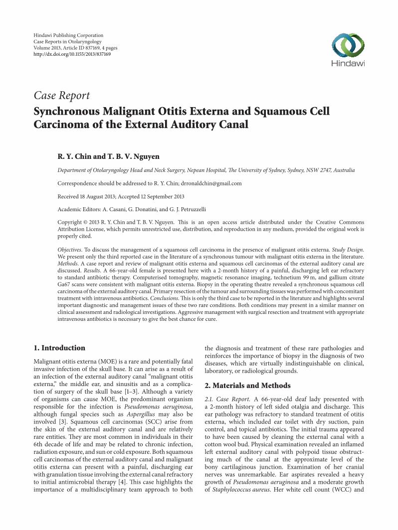
Figure 1: Axial T2weightedMRI image demonstrating the left sidedtumour mass.
C-reactive protein were elevated at 11.3 × 10³/𝜇L, and44.6mg/L respectively. She was not diabetic or immunosup-pressed. Computerised tomography (CT) scan showed softtissue density involving the external auditory canal with boneerosion of the tegmen consistent with inflammatory destruc-tion (Figure 1).Therewas also increased attenuation of the leftside mastoid antrum and air cells consistent with mastoiditis.Given these initial findings, a provisional diagnosis wasmade of malignant otitis externa arising from an infectionof the external auditory canal. Theatre time was allocatedfor her to have the external canal biopsied and toileted.Infectious disease and radiology were notified and she wascommenced on intravenous synthetic penicillin (ticarcillinclavulanate potassium) and a quinolone (Ciprofloxacin) viaa peripherally inserted intravenous catheter (PICC) line.An MRI scan and a technetium 99m (Tc99) and Galliumcitrate (Ga67) were organised. Both the Ga67 and Tc99 wereconsistent withmalignant otitis externa involving the petroustemporal bone and mastoid process.
Examination in theatre revealed granulation tissue andpolyps along the entire length of the external auditory canaland predominant growth of P. aeruginosa. Histopathologydemonstrated an inflamed stratified squamous epitheliumwith dysplasia and cellular atypia. Within this, there wereembedded irregular nests of atypical squamous cells infil-trating the underlying stroma, and a diagnosis of moderatelydifferentiated SCC was made.
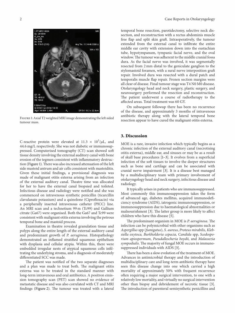
The patient was notified of the two separate diagnosesand a plan was made to treat both. The malignant otitisexterna was to be treated in the standard manner withlong-term intravenous and oral antibiotics. A positron emis-sion tomography scan (PET) scan showed no evidence ofmetastatic disease and was also correlated with CT and MRIfindings (Figure 2). The tumour was treated with a lateral
temporal bone resection, parotidectomy, selective neck dis-section, and reconstruction with a rectus abdominis musclefree flap and split skin graft. Intraoperatively, the tumourextended from the external canal to infiltrate the entiremiddle ear cavity with extension down into the eustachiantube, hypotympanum, tympanic facial nerve, and the ovalwindow.The tumour was adherent to themiddle cranial fossadura. As the facial nerve was involved, it was segmentallyresected from 2mm distal to the geniculate ganglion to thestylomastoid foramen, with a sural nerve interposition graftrepair. Involved dura was resected with a dural patch andtemporalis muscle flap repair. Frozen section margins wereall clear of disease. Final tumour stage was T4N0M0 disease.Otolaryngology head and neck surgery, plastic surgery, andneurosurgery performed the resection and reconstruction.The patient underwent a course of radiotherapy to theaffected areas. Total treatment was 60 GY.
On subsequent followup there has been no recurrenceof the disease, and approximately 3 months of intravenousantibiotic therapy along with the lateral temporal boneresection appear to have cured the malignant otitis externa.
3. Discussion
MOE is a rare, invasive infection which typically begins as achronic infection of the external auditory canal (necrotisingotitis externa), middle ear, and sinuses or may be as a resultof skull base procedures [1–3]. It evolves from a superficialinfection of the soft tissues to involve the deeper structuressuch as bone and cartilage and can be associated withcranial nerve impairment [3]. It is a disease best managedby a multidisciplinary team with primary involvement ofotolaryngology head and neck surgery, infectious disease andradiology.
It typically arises in patients who are immunosuppressed.Most commonly this immunosuppression takes the formof advanced age, diabetes mellitus, acquired immunodefi-ciency syndrome (AIDS), iatrogenic immunosuppression, orimmunosuppression due to haematological abnormalities ormalnourishment [3]. The latter group is more likely to affectchildren who have this disease [5].
The predominant organism in MOE is P. aeruginosa. Theinfection can be polymicrobial with other organisms such asAspergillus spp (fumigatus), S. aureus, Proteus mirabilis, Kleb-siella oxytoca, Burkholderia cepacia, Candida spp, Scedospo-rium apiospermum, Pseudallescheria boydii, and Malasseziasympodialis. Themajority of fungal MOE occurs in immuno-suppressed individuals with AIDS [3].
There has been a slow evolution of the treatment ofMOE.Advances in antimicrobial therapy and the introduction ofmultidisciplinary care and long-term antibiotic therapy haveseen this disease change into one which carried a highmortality of approximately 50% with frequent recurrenceoften requiring a major surgical intervention, to one with arelatively lowmortality, and virtually no surgical interventionother than biopsy and debridement of necrotic tissue [3].The introduction of parenteral semisynthetic penicillins and
Case Reports in Otolaryngology 3
Figure 2: SPECT scan demonstrating increased areas of uptake in the region of the left petrous temporal bone and external auditory canal.
the fluoroquinolones is largely responsible for the decreasedmortality of this disease [3].
Involvement of the ear and lateral skull base by squa-mous cell carcinoma is usually the result of a cutaneousneoplasm that originates from the skin of the pinna orthe external auditory canal. Ultraviolet light exposure orthermal injury (cold) and radiation exposure and chronicinfection are thought to predispose patients to this disease[4]. Rarely, squamous carcinomas can arise from the middleear from metaplastic middle ear mucosa and are associatedwith chronic otitis media and human papilloma virus [6].Treatment of squamous carcinomas of the external canalshould be aggressive because of the high rate of recurrence.Treatment with en-block resection, selective neck dissection,and radiotherapy is recommended in cases like the onepresented here, as recurrence rates and nodal metastasis arerelatively high.
The authors present only the third case in the literatureof a synchronous malignancy and malignant otitis externa[7, 8]. Both malignant otitis externa and squamous cellcarcinomas of the external auditory canals are rare entities,and it is even rarer to have both occurring at the same time.Both pathologies present in a remarkably similar manner—clinically, radiologically, and on laboratory investigations[3, 4].
Clinically, both conditions often present with a painful,discharging ear refractory to standard treatment regimes ofear toilet and antibiotic therapy. Both conditions may bepresent with cranial nerve palsies, trismus, and lympha-denopathy.
Radiologically, both conditions on CT andMRImay haveabnormalities of the external auditory canal, soft tissue, andfluid within the middle ear and mastoid cavity, eustachiantube, and parapharyngeal space with or without concomitantbony destruction [6]. There are no studies in the literaturewhich look specifically at Tc99 and Ga67 scanning in SCCof the external auditory canal, but it is conceivable that boththese investigations would be positive in the presence of anextensive neoplastic process with bone erosion and chronic
infection of the soft tissues without malignant otitis externabeing present.
This case and the other cases reported in the literature[7, 8] highlight the importance of ear toilet and biopsy inthe investigation and diagnosis of malignant otitis externawhen the cause is a chronic infection of the external auditorycanal (malignant or necrotizing otitis externa) refractory tostandard treatment regimes. Successful diagnosis and man-agement of both these pathologies involve amultidisciplinaryteam approach and a meticulous unification of a detailedclinical examination, laboratory investigations, appropriateradiology, and biopsy to create a complete clinical picture.
Conflict of Interests
The authors would like to state that there is no conflict ofinterests, financial or otherwise, in the preparation of thispaper.
References
[1] W. H. Slattery III and D. E. Brackmann, “Skull base osteomyeli-tis: malignant external otitis,” Otolaryngologic Clinics of NorthAmerica, vol. 29, no. 5, pp. 795–806, 1996.
[2] L. R. Grobman,W. Ganz, R. Casiano, and S. Goldberg, “Atypicalosteomyelitis of the skull base,” Laryngoscope, vol. 99, no. 7 I, pp.671–676, 1989.
[3] J. R. Grandis, B. F. Branstetter IV, and V. L. Yu, “The changingface ofmalignant (necrotising) external otitis: clinical, radiolog-ical, and anatomic correlations,” Lancet Infectious Diseases, vol.4, no. 1, pp. 34–39, 2004.
[4] S. A. Moody, B. E. Hirsch, and E. N. Myers, “Squamous cellcarcinoma of the external auditory canal: an evaluation of astaging system,” American Journal of Otology, vol. 21, no. 4, pp.582–588, 2000.
[5] J. Rubin and V. L. Yu, “Malignant external otitis: insight intopathogenesis, clinical manifestations, diagnosis, and therapy,”American Journal of Medicine, vol. 85, no. 3, pp. 391–398, 1988.
[6] G. Marioni, G. Altavilla, G. Busatto, S. Blandamura, C. DeFilippis, and A. Staffieri, “Detection of human papillomavirus
4 Case Reports in Otolaryngology
in temporal bone inverted papilloma by polymerase chainreaction,” Acta Oto-Laryngologica, vol. 123, no. 3, pp. 367–371,2003.
[7] J. R. Grandis, B. E. Hirsch, and V. L. Yu, “Simultaneous presen-tation of malignant external otitis and temporal bone cancer,”Archives of Otolaryngology, vol. 119, no. 6, pp. 687–689, 1993.
[8] K. F. Mattucci, M. Setzen, and P. Galantich, “Necrotizing otitisexterna occurring concurrently with epidermoid carcinoma,”Laryngoscope, vol. 96, no. 3, pp. 264–266, 1986.