GE Healthcare Case Studies How do you manage emergency patients with CT? Dr. Marius Wick MD, MSc. Karolinska University Hospital, Stockholm, Sweden Dr. Koenraad H. Nieboer MD Universitair Ziekenhuis Brussel, Brussels, Belgium
Transcript
GE Healthcare
Case Studies
How do you manage emergency patients with CT?
Dr. Marius Wick MD, MSc.Karolinska University Hospital, Stockholm, Sweden
Dr. Koenraad H. Nieboer MDUniversitair Ziekenhuis Brussel, Brussels, Belgium
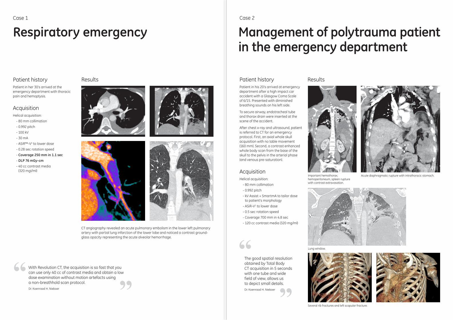
Patient historyPatient in his 20’s arrived at emergency department after a high impact car accident with a Glasgow Coma Scale of 6/15. Presented with diminished breathing sounds on his left side.
To secure airway, endotracheal tube and thorax drain were inserted at the scene of the accident.
After chest x-ray and ultrasound, patient is referred to CT for an emergency protocol. First, an axial whole skull acquisition with no table movement (160 mm). Second, a contrast enhanced whole body scan from the base of the skull to the pelvis in the arterial phase (and venous pre-saturation).
AcquisitionHelical acquisition:
- 80 mm collimation
- 0.992 pitch
- kV Assist + SmartmA to tailor dose to patient’s morphology
- ASiR-V1 to lower dose
- 0.5 sec rotation speed
- Coverage 700 mm in 4.8 sec
- 120 cc contrast media (320 mg/ml)
Results
Management of polytrauma patient in the emergency department
Case 2
The good spatial resolution obtained by Total Body CT acquisition in 5 seconds with one tube and wide field of view, allows us to depict small details.Dr. Koenraad H. Nieboer ”
“
Important hemothorax, hemoperitoneum, spleen rupture with contrast extravasation.
Acute diaphragmatic rupture with intrathoracic stomach.
Lung window.
Several rib fractures and left scapular fracture.
Patient historyPatient in her 30’s arrived at the emergency department with thoracic pain and hemoptysis.
AcquisitionHelical acquisition:
- 80 mm collimation
- 0.992 pitch
- 100 kV
- 30 mA
- ASiR™-V1 to lower dose
- 0.28 sec rotation speed
- Coverage 250 mm in 1.1 sec
- DLP 76 mGy-cm
- 40 cc contrast media (320 mgI/ml)
Results
CT angiography revealed an acute pulmonary embolism in the lower left pulmonary artery with partial lung infarction of the lower lobe and noticed a contrast ground-glass opacity representing the acute alveolar hemorrhage.
Respiratory emergencyCase 1
With Revolution CT, the acquisition is so fast that you can use only 40 cc of contrast media and obtain a low dose examination without motion artefacts using a non-breathhold scan protocol.Dr. Koenraad H. Nieboer ”
“
Patient historyYoung female on bicycle crashed into open car door. Patient found unconscious and referred to CT for a multi-trauma study. Exam included skull, facial bones/c-spine, whole body angiography followed by a venous phase abdomen.
AcquisitionWhole body angiography:
- Helical acquisition
- 80 mm collimation
- 0.992 pitch
- 100 kV
- 150 mA
- ASiR™-V 50%
- 0.5 sec rotation
- Coverage of 656 mm in 4 sec
- Soft kernel
- 120 cc of contrast media (350 mgI/ml)
- DLP 276.6 mGy-cm
Low-dose multi-trauma exam including whole body angiography
Results
Several fractures to pelvis, hips and transverse processes.
Whole body angiography.
Case 3
ConclusionThis case demonstrates the protocol optimization done at the Karolinska University Hospital where they have added a whole body angiography to their standard multi-trauma protocol after installing Revolution CT. The entire 4-series examination was performed with a DLP of only 1331 mGy-cm. By using ASiR-V, the dose of this entire study, with all 4 series, is even less than the previous protocol that didn’t include the whole body CT angiography. By adding a whole body angiography to the standard multi-trauma protocol, carotid dissections are found at an earlier stage even though the patient was scanned with arms up. As a result, the patient could start treatment earlier. A new neuro CT examination was performed 10 days later and no sign of infarction was found.
The patient had several injuries: dissection of right and left internal carotid, right being most severe, small pulmonary contusions, rib fractures, splenic contusion and several fractures to the hip and pelvis.
Right internal carotid dissection.
Thanks to the new protocol which was possible due to the low dose on Revolution CT, we were able to detect the two dissections early. Thanks to it, this young patient survived.Dr. Marius Wick MD, MSc. ”“
Imagination at work
About GE HealthcareGE Healthcare provides transformational medical technologies and services to meet the demand for increased access, enhanced quality and more affordable healthcare around the world.
GE Healthcare 3000 N. Grandview Blvd. Waukesha, WI 53188 U.S.A
gehealthcare.com
GE (NYSE: GE) works on things that matter - great people and technologies taking on tough challenges. From medical imaging, software & IT, patient monitoring and diagnostics to drug discovery, biopharmaceutical manufacturing technologies and performance improvement solutions, GE Healthcare helps medical professionals deliver great healthcare to their patients.
™ Trademark of General Electric Company. The clinical cases are displayed for educational purposes only and for the benefit of healthcare students and professionals1 In clinical practice, the use of ASiR-V may reduce CT patient dose depending on the clinical task, patient size, anatomical location and clinical practice. A consultation
with a radiologist and a physicist should be made to determine the appropriate dose to obtain diagnostic image quality for the particular clinical task.
Legal Mentions : The system is intended to produce cross-sectional images of the body by computer reconstruction of x-ray transmission projection data from thesame axial plane taken at different angles. The system has the capability to image whole organs in a single rotation. Whole organs include but are not limited to brain,heart, liver, kidney, pancreas, etc..The system may acquire data using Axial, Cine, Helical, Cardiac, and Gated CT scan techniques from patients of all ages. These images may be obtained either with orwithout contrast. This device may include signal analysis and display equipment, patient and equipment supports, components and accessories.Class: IIb – Manufacturer: GE Medical Systems LLC, USA – LNE/G-MED – NB 0459 – GMDN 37618.