25
Case Study 62 Kenneth Clark, MD
| Date post: | 23-Dec-2015 |
| Category: |
Documents |
| Upload: | neal-randall |
| View: | 217 times |
| Download: | 3 times |

Case Study 62Kenneth Clark, MD
Question 1
• This is a 32-year-old woman with progressive distortion of taste and smell. After seeing her primary care physician an MRI of the head was ordered.
• Describe the MRI findings.

Axial T1 Axial T1 + Contrast
Axial T2 Propeller Axial T2 FLAIR

Answer
• An ill-defined, minimally T1 hypointense, non-contrast lesion within the right thalamus. It shows moderate homogeneous signal intensity on T2 propeller and moderate T2 FLAIR signal as well.

Question 2
• What is the differential diagnosis?

Answer
• Low Grade Glioma• Inflammatory (viral)• Demyelinating Lesion• Acute Vascular Insult• Metabolic
Question 3
• A sterotactic biopsy of the lesion was performed and submitted for intra-operative examination. Describe the histologic findings. How would you report to the surgeon?
• Click here to see the smear

Answer
• The smear shows hypercellular white matter and scattered small vessels. The cells show moderate enlargement, nuclear contour irregularities/angulation and unevenly distributed chromatin. Many, but not all of the atypical cells appear to have processes. No mitotic figures are seen. Vague gliovascular structuring is also apparent around some vessels.
• A. Neoplastic• B. Glioma, grade deferred to permanent sections
Question 4
• The remainder of the biopsy is submitted for histologic evaluation. How would you describe the lesion?
• Click here to view the slide

Answer
• The tissue shows thalamic parenchyma with moderately increased cellularity due to the presence of an infiltrating population of atypical astrocytic cells. The cells show moderate enlargement with nuclear irregularities and hyperchromasia. There are 3 mitotic figures seen. No necrosis or endothelial proliferation is evident.
Question 5
• What is your differential diagnosis?

Answer
• Infiltrating glioma• Reactive process

Question 6
• What immunohistochemical stains would you order in order to better characterize this lesion and establish the diagnosis?
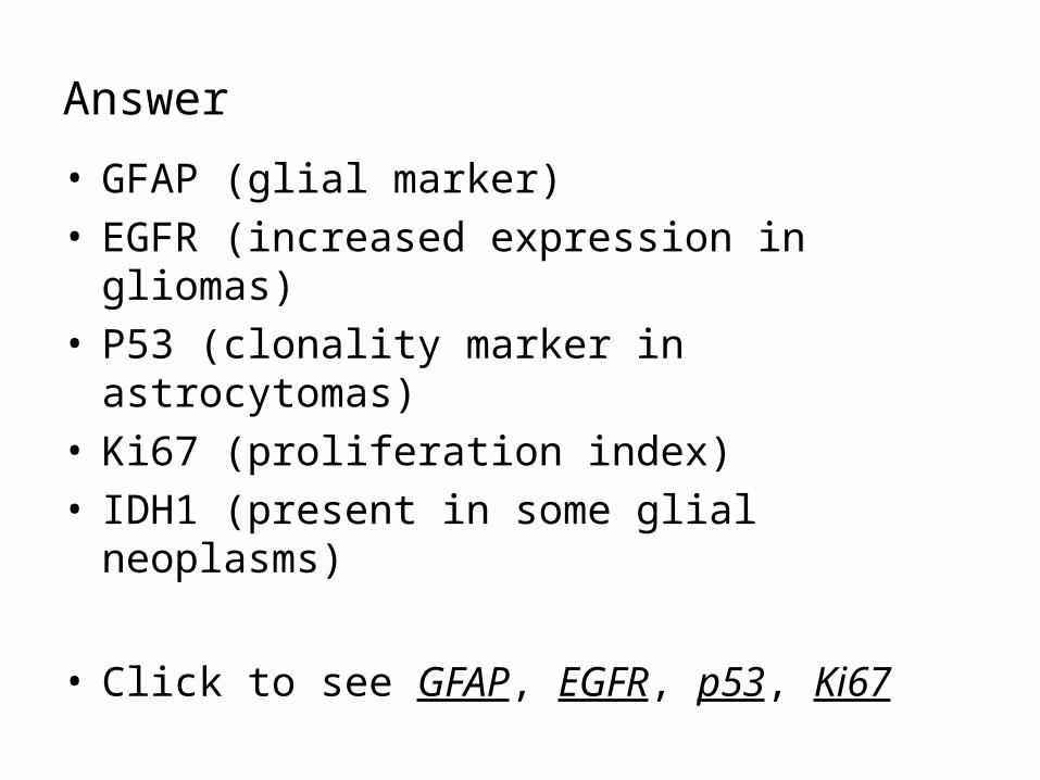
Answer
• GFAP (glial marker)• EGFR (increased expression in gliomas)• P53 (clonality marker in astrocytomas)• Ki67 (proliferation index)• IDH1 (present in some glial neoplasms)
• Click to see GFAP, EGFR, p53, Ki67
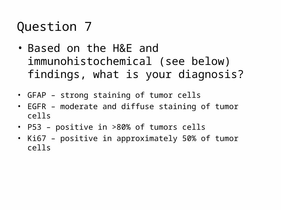
Question 7
• Based on the H&E and immunohistochemical (see below) findings, what is your diagnosis?
• GFAP – strong staining of tumor cells• EGFR – moderate and diffuse staining of tumor cells• P53 – positive in >80% of tumors cells• Ki67 – positive in approximately 50% of tumor cells
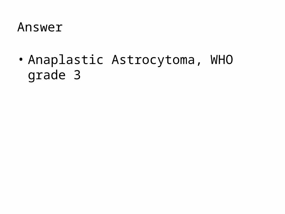
Answer
• Anaplastic Astrocytoma, WHO grade 3

Question 8
• How does the p53 mutation clonality help in establishing the diagnosis?
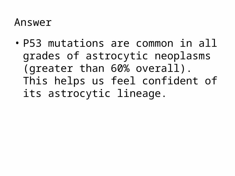
Answer
• P53 mutations are common in all grades of astrocytic neoplasms (greater than 60% overall). This helps us feel confident of its astrocytic lineage.
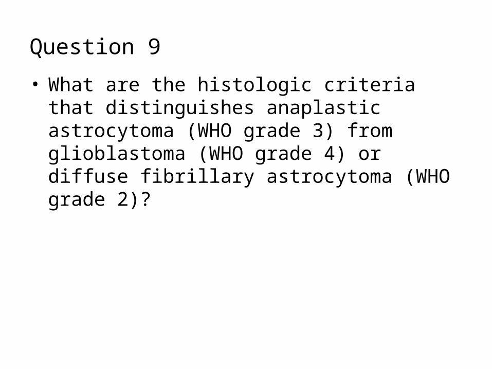
Question 9
• What are the histologic criteria that distinguishes anaplastic astrocytoma (WHO grade 3) from glioblastoma (WHO grade 4) or diffuse fibrillary astrocytoma (WHO grade 2)?
Answer
• Diffuse fibrillary astrocytoma (WHO 2)– Moderately increased cellularity with mild nuclear
atypia (hyperchromasia & enlargement), no mitoses, no necrosis or endothelial proliferation
• Anaplastic astrocytoma (WHO 3)– Higher cellularity than WHO astrocytomas, more
distinct nuclear atypia, MITOSES are most significant criteria (if single mitotic figures seen in small biopsies, Ki67 may be useful)
• Glioblastoma (WHO 4)– Obvious mitoses, palisading necrosis, endothelial
hyperplasia/proliferation, severe nuclear atypia, usually highly cellular
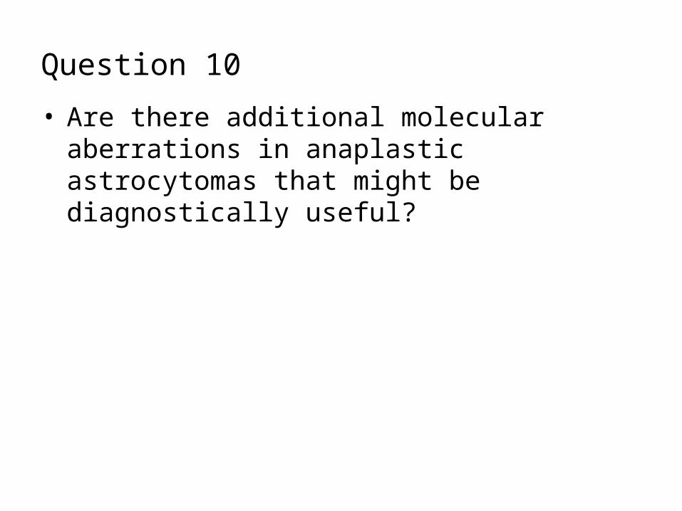
Question 10
• Are there additional molecular aberrations in anaplastic astrocytomas that might be diagnostically useful?

Answer
• 50-60% of anaplastic astrocytomas show chromosome 17p LOH.
• Up to 60% of anaplastic astrocytomas show 10q LOH.
• 19q LOH occurs in about 50% of anaplastic astrocytomas.
• EGFR amplification is seen in less than 10%
• This lesion was positive for 10q LOH, which is consistent with anaplastic astrocytoma
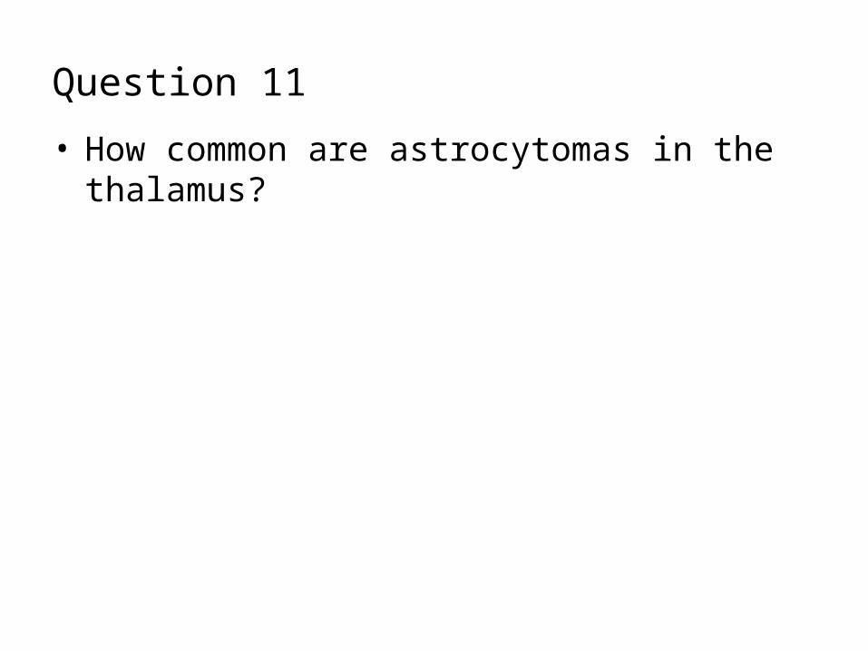
Question 11
• How common are astrocytomas in the thalamus?

Answer
• Thalamic astrocytomas are very rare. Thalamic tumors account for less than 1% of all intracranial neoplasms, with astrocytomas accounting for a fraction of those lesions. For this reason it is critical to strongly consider (and rule out) other processes that may be more common and less ominous than gliomas in the thalamus.
• The gross majority of thalamic astrocytomas are WHO grade 3 or 4. They most frequently occur in children, adolescents and young adults.
References
• Hendrikus G, et al. Infiltrative astrocytomas of the thalamus (1995). Neurosurg. 82:548–557
• Louis D, Ohgaki H, Wiestler O, Cavanee W. WHO Classification of Tumours of the Central Nervous System. IARC: Lyon 2007.


