19
Case Study: BPTI Markus Dittrich and Chalermpol Kanchanawarin 1

Case Study: BPTIMarkus Dittrich and Chalermpol Kanchanawarin
1

1 Introduction
Bovine pancreatic trypsin inhibitor (BPTI) is one of the smallest and simplestglobular proteins. BPTI’s function is the suppression of protein digestion,i.e., the breakdown of proteins into their peptide building blocks, by means ofinhibiting the action of the enzyme trypsin which is produced in the bovinepancreas. BPTI is a member of the serine protease family of inhibitors [1].A hallmark of this class of enzymes are the many conserved cysteine residuesthat form disulfide bonds stabilizing the proteins’ three-dimensional struc-tures. BPTI has a relatively broad specificity in that it can inhibit severalkinds of digestive enzymes [1].
BPTI is one of the most extensively studied globular proteins and hasbeen been investigated structurally by both x-ray crystallography and NMRspectroscopy. Furthermore, its protein folding pathway and dynamics havebeen investigated in great detail. Since BPTI forms complexes with sev-eral of the enzymes that it inhibits, it has also been the subject of studiesinvestigating protein-protein interaction and molecular recognition [1].
This case study is divided into three sections: First, the primary sequenceand the three-dimensional structure of BPTI will be introduced. The secondpart will examine the thermal stability of BPTI that is caused by its threeinternal disulfide bonds. Finally, in the last section, we study BPTI’s functionas trypsin inhibitor by investigating a trypsin-BPTI complex.
Figure 1 shows the files required for the Exercises in the present casestudy. The files can be found in the directory
BPTI/00-FORUSERS/supplementary-files/.
2 Sequence and structure of BPTI
BPTI is a single-chain polypeptide that contains 58 amino acid residues andhas a molecular mass of 6512 [1]. Atomic resolution X-ray crystal structuresof BPTI and several of its mutants have been available since 1970 [1, 3].
Fig. 2(a) shows the three dimensional conformation of BPTI and the im-age reveals that BPTI has a compact pear-shape with a maximum dimensionof about 30A. Topologically, the protein folds like a piece of string that isfolded upon itself twice as shown in Figure 2(b). Three disulfide bonds, D1,D2 and D3, link the protein’s secondary structure elements together.
2

The secondary structure of BPTI is shown in Fig. 3 along with its aminoacid sequence. Two α helical regions, H1 and H2, form a one and a halfturn 310-helix near the N-terminus and and a three turn α-helix near theC-terminus. The central part of the protein contains two β-strands, B1 andB2, that are connected by a turn, T1, and which form an anti-parallel β-hairpin loop. As already mentioned, six conserved cysteine residues in BPTIform three disulfide bonds that stabilize the three dimensional structure ofthe protein. Cys5 and Cys55 form the first disulfide bond D1 that joins theN- and C-termini of BPTI. Cys14 and Cys38 make the second disulfide bond
BPTI case study
Exercise 1 - Getting to know BPTI
Exercise 2 - Ramachandran plot of BPTI
Exercise 3 - Contact Map Analysisof BPTI
Exercise 4 - Multiple Sequence Align-ment of BPTI-like proteins
Exercise 5 - Thermal Stability of BPTI
bovine-BPTI-4PTI.pdb
bovine-BPTI-4PTI.pdb
bovine-BPTI-4PTI.pdbhuman-APPI-1AAP.pdbsea-anemone-1SHP.pdbsnake-venom-CI-1JC6.pdb
bpti-native-310K.dcdbpti-native-498K.dcdbpti-reduced-310K.dcdbpti-reduced-498K.dcdbpti-native-system.psfbpti-reduced-system.psf
bovine-BPTI-4PTI.pdb
trypsin-BPTI-2PTC.pdb
Exercise 6 - Trypsin-BPTI complex
Figure 1: Required files for exercises.
3
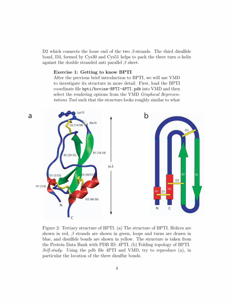
D2 which connects the loose end of the two β-strands. The third disulfidebond, D3, formed by Cys30 and Cys51 helps to pack the three turn α-helixagainst the double stranded anti parallel β sheet.
Exercise 1: Getting to know BPTIAfter the previous brief introduction to BPTI, we will use VMDto investigate its structure in more detail. First, load the BPTIcoordinate file bpti/bovine-BPTI-4PTI.pdb into VMD and thenselect the rendering options from the VMD Graphical Represen-tations Tool such that the structure looks roughly similar to what
H2 (48-56)
H1 (3-6)
B1 (18-24)B2 (29-35)
D2 (14/38)
D3 (30/51)D1 (5/55)
30 Ao
a b
N
C
N C
H1H2
B2 B1
D1
D2
D3
Lys15
Ala16
Figure 2: Tertiary structure of BPTI. (a) The structure of BPTI. Helices areshown in red, β strands are shown in green, loops and turns are drawn inblue, and disulfide bonds are shown in yellow. The structure is taken fromthe Protein Data Bank with PDB ID: 4PTI. (b) Folding topology of BPTI.Self-study: Using the pdb file 4PTI and VMD, try to reproduce (a), inparticular the location of the three disulfur bonds.
4

H1 (310 helix)
H2 (α-helix)
B1 (β−strand)
B2 (β−strand)
N-terminus
C-terminus
T1 (Turn)
Cys5
Cys14
Cys30
Cys38
Cys51
Cys55
B3
T2
D1
D2
D3
Figure 3: Primary sequence and secondary structure of BPTI. The diagramshows the amino acid sequence of BPTI (only odd numbered residues arelabeled explicitly). The secondary structures are represented using the fol-lowing color codes: α-helix = purple, 310 helix = pink, β-strand = yellow,turn = green, partial β-strand = gold, coil = white. Also shown are thethree disulfur bonds D1, D2, and D3 that link BPTI’s secondary structureelements. Self-study: Using the the pdb file 4PTI and VMD’s sequencebrowser try to identify the disulfur bonds and secondary structure elementsshown in Fig. 2 and, thereby, connect the primary, secondary, and tertiarystructure of BPTI.
is shown in Fig. 2 (a). Now use VMD to highlight the followingparts of the BPTI structure using the Sequence Viewer and the
5

Graphical Representations Tool :
• The one and a half turn 310 helix (Asp3 to Leu6) and thethree turn α-helix (Ala48 to Gly56).
• The two β-strands forming an anti-parallel β-sheet (Ile18 toAsn24 and Leu29 to Try35).
• The three disulfide bonds D1, D2, and D3 (Cys5/55, Cys14/38and Cys30/51).
• The reactive site which binds trypsin or other proteolyticenzymes, located between residues Lys15 and Ala16, nearthe disulfide bond D2 (Cys14/38).
More challenging, try to identify the following more detailed struc-tural features:
• Two β turns (Ala25 to Gly28 and Lys41 to Asn44).
• A partial third β-strand formed by Phe45 and Tyr21. Thekey observation to make here is the hydrogen-bonding pat-tern of the backbone oxygen and amine hydrogen atoms inβ-sheets.
• The hydrophobic core of BPTI consisting of Phe4, Cys5/55,Phe22, Tyr23, Cys30/51, Phe33, Tyr35 and Phe45. What isits significance in terms of the folding and overall stabilityof BPTI’s structure?
• The three hydrogen bonds formed by Asn43 and the aminehydrogen and carbonyl oxygen of Tyr23 and the carbonyloxygen of Gly7. What possible roles could these hydrogenbonds have?
A very convenient way to quickly obtain an overview over the secondarystructure elements present in a protein is to simultaneously plot the (φ,ψ)angle pairs of the peptide bond in a so called Ramachandran plot as shownin Fig. 4 for BPTI. Here, each pair of (φ,ψ) angles (corresponding to a singleamino acid residue) is plotted in a two dimensional plane spanned by theφ and ψ-axis whose values go from -180◦ to 180◦, respectively. The blueareas indicate allowed conformations and correspond to β-sheets in the fourthquadrant (labeled β in Fig. 4) and α-helices in the third quadrant (labeled
6

α in Fig. 4). The green areas represent partially allowed conformations. Allother pairs of (φ,ψ) angles are typically absent from protein structures. Asillustrated nicely in Fig. 4, BPTI is no exception to this rule, and most (φ,ψ)angles actually fall into the allowed regions.
-180 0 +180
+180
-180
0
Phi
Psi
α
β
L
(φ)
(ψ)
Type "resid 48 to 56"and press enter.
Figure 4: Ramachandran plot of BPTI. The figure shows the pairs of (φ,ψ)angles for all 58 amino acid residues of BPTI and was created using theRamachandran Plot Tool of VMD. The blue and green colored regions markedα, β and L indicate (φ,ψ) values corresponding to right handed α-helices, β-strands and left handed helices. Here, regions colored blue indicate the mostallowed angles and green the partially allowed angles.
Exercise 2: Ramachandran plot of BPTIAs a continuation of Exercise 1, we will now use VMD’s Ra-machandran Plot Tool to study BPTI’s structural properties inmore detail.
Again, the BPTI coordinate file bpti/bovine-BPTI-4PTI.pdb
will be used as starting point. Hence, load the structure intoVMD, open up the Ramachandran Plot Tool and consider thefollowing tasks:
• Type “resid 48 to 56” in the selection box. Do these residuesform an α-helix?
7

• Type “all” in the selection box. There are a few dots outsidethe blue and green regions. Find out what residues theycorrespond to by clicking on them and explain your findings!
In addition to a Ramachandran plot, a so called contact map consistingof a two dimensional plot of distances between the Cα atoms of any pair ofamino acid residues in a protein can provide important and useful informationregarding its the tertiary structure. Fig. 5 shows the contact map generatedfor BPTI, in which the pairwise Cα distances are displayed in a gray scaleranging from 0 to 10A. It is apparent, that the distance map immediatelyreveals the presence of the central β-sheet comprised of B1-B2.
Exercise 3: Contact Map Analysis of BPTIIn this exercise, we will use the Contact Map Tool of VMD to es-tablish the spatial arrangement of secondary structure elements inBPTI. To this end, load the BPTI structure file bpti/bovine-BPTI-4PTI.pdbinto VMD, open up the Contact Map Tool, and generate the map.With the help of the displayed contact map, identify the followingcontacts between parts of BPTI:
• Contact between β-strands B1 and B2 (c.f. Fig. 5).
• Contact between α-helix H1 and β-strand B2.
• Contact between the 310-helix and β-strand B1.
After having learned about the structural properties of BPTI, it is in-structive to study other members of the so called BPTI-like superfamily.The latter is characterized by the presence of three disulfide bonds stabiliz-ing their respective tertiary structures. All these proteins are structurallysimilar to BPTI but differ significantly in their amino acid sequences and,importantly, their physiological function.
Here, we consider three particular examples and compare them withBPTI. Our first example of a BPTI-like protein is the human BPTI-domainof the amyloid β-protein precursor [7] implicated in Alzheimer’s desease. Thesecond one is a chymotrypsin inhibitor isolated from snake venom [5]. Finally,we consider a BPTI-like protein taken from a Caribbean sea anemone [6].
First, we will examine the backbone conformation of bovine BPTI andthe other three BPTI-like proteins. They are depicted in Fig. 6(a) in tube
8

Residue number
Res
idu
e n
um
ber
B1-B2
Figure 5: Contact map of BPTI. Shown are the distances between two Cα
atoms of amino acids on BPTI in gray scale ranging from 0 to 10 A (blackto white).
representation. By inspection, it can be seen that the core region of all pro-teins are structurally very similar to that of BPTI. The main difference arethe lengths and the conformation of the N and C-termini, most apparentin the case of the C-terminus from the snake venom protein, which is sig-nificantly elongated. This structural similarity becomes clearer after align-ing all four protein structures using multiple sequence alignment. The soaligned structures are shown in Fig. 6(b) and reveal the proteins’ commoncore conformation and their variable termini. Fig. 6(c) makes this even moreapparent. It shows the same aligned structures but this time colored by Q-value which is a measure of structural similarity, blue and red indicating highand low similarity, respectively. Interestingly, this similarity comes despitethe fact that the underlying sequence identity among all four structures israther low as shown in Fig. 6(d), where blue and red indicate high and lowsequence similarity, respectively. Fig. 6(e) highlights the conserved residuesin the primary sequence that include the cysteines of the three conserveddisulfur bridges which are the hallmark of the BPTI-like proteins. Hence, asthis example clearly shows, different primary amino-acid sequences can giverise to very similar secondary and tertiary protein structures, a finding thathas emerged over and over again when examining members of structurallyclosely related protein families.
9

Cow Human Snake Sea anemone
Aligned Q-value Sequence Identity
CowHuman
SnakeSea anemone
a
b c d
e
Figure 6: Structural alignment of bovine BPTI and three BPTI-like proteins.Shown are the structures of BPTI and three BPTI-like proteins from the hu-man Alzheimer’s amyloid β protein precursor, snake venom and a Caribbeansea anemone in tube representation and colored by segment name (a), a struc-tural alignment of the four proteins colored by segment name (b), Q-value(c), and sequence identity (d), and the sequence alignment of BPTI and thethree BPTI-like proteins (e). Conserved amino acid residues are labeled inyellow.
Exercise 4: Multiple Sequence Alignment of BPTI-like
10

proteinsUsing VMD and its Multiple Sequence Alignment Tool [4] we willnow take a closer look at the three BPTI-like proteins and thencompare their 3D structures and amino acid sequences with thatof BPTI. First, load the coordinates of the three BPTI-like pro-teins by opening the following PDB files: human-APPI-1AAP.pdb,snake-venom-CI-1JC6.pdb and sea-anemone-1SHP.pdb and thenrender all the proteins similar to the way the look in Fig. 6(a)using VMD’s tube representation and coloring by segment name.
To allow for a better comparison of the conformational differ-ences between the four proteins, we align them using the struc-ture alignment option in the Multiple Sequence Alignment Toolas shown in Fig. 6 (b). Clearly, the 3D structures of the threeBPTI-like proteins align very well with that of BPTI. The 3Dstructure of the BPTI-like protein from snake venom seems todeviates most from that of BPTI. Now, try the following:
• Color the four proteins according to the Q value (see Fig. 6(c)) and find a part of one of the BPTI-like proteins (otherthan the N- or C- termini) that has its structure deviatemost from the other proteins. What species is that BPTI-like protein from?
• Color the four BPTI-like proteins according to sequenceidentity and identify the residues that are highly conserved.What kinds of amino acids are they and where are they in thestructures? Why do you think they are highly conserved?
• Use residue selection by Q value to find parts of the proteins’structures that are more than 80% conserved structurally.Where on the structures are they located?
• Use the multiple alignment tool to generate a phylogenetictree of the four proteins according to the Q value. Can youtell which BPTI-like protein is closest to BPTI in terms ofevolutionary history?
11

3 Structural and thermal stability of BPTI
Most globular proteins are stabilized by their hydrophobic core which isformed by the internal packing of non-polar amino acid side chains. BPTI isno exception and due to its rather small overall size it also has a relativelysmall hydrophobic core. Thus, extra stability to support its three dimensionalstructure is provided by the three internal disulfide bonds that were discussedpreviously.
It is, therefore, interesting to consider what would happen if any or severalof these disulfide bonds are being cleaved. This allows one to judge howimportant the disulfide bonds are for preserving BPTI’s three dimensionalstructure.
With all of its three disulfide bonds present, BPTI is one of the most stableglobular proteins. Below 100◦C, BPTI is quite inert to urea, a substance oftenused to denature proteins and only denatures under very acidic conditions.However, if a single one of the three disulfide bonds is reduced (cleaved),e.g. D2 (Cys14/Cys38), BPTI can be denatured quite easily. Fig. 7 showsthe percentage of native BPTI that is denatured at various temperaturescompared to reduced BPTI with a cleaved D2 disulfide bond. It can beseen that the native BPTI is quite stable as a function of temperature withonly 50% denatured at about 85◦C. On the other hand, when D2 is cleaved,BPTI becomes much less stable with 50% denatured at about 55◦C. Finally,experimentally it is found that when all the three disulfide bonds are removed,BPTI unfolds at room temperature.
Exercise 5: Thermal stability of BPTIWe will now have a closer look at the thermal stability of nativeand fully reduced BPTI (i.e., all three disulfur bonds are in thecleaved state) by investigating several computer simulation tra-jectories. The latter provide a dynamic view of the time evolutionof a biomolecular system. Four different trajectories of native andfully reduced BPTI immersed in water as solvent at two differenttemperatures will be considered. The respective lengths of thetrajectories are given in nano seconds (ns, 10−9s):
• T = 310K (0.7ns for the native system and 1.0ns for thereduced system) and
• T = 498K (2.3ns for the native system and 1.7ns for thereduced system).
12

20 40 60 80 1000%
50%
100%
Temperature, oC
Perc
ent
den
atu
red
Native
Cys(14/38) cleaved
Figure 7: Temperature denaturation of native and single disulfide reducedBPTI.
Fig. 8 shows structural views of the systems for native BPTI attimes t=0, t=0.7ns (T=310K)/t=2.3ns (T=498K) and reducedBPTI at times t=0, t=1.0ns (T=310K)/t=1.7ns (T=498K), re-spectively. To start the investigation, load the following fourtrajectory (dcd) files and protein structure (psf) files into VMD
• bpti-native-310K.dcd and bpti-native-system.psf
• bpti-native-498K.dcd and bpti-native-system.psf
• bpti-reduced-310K.dcd and bpti-reduced-system.psf
• bpti-reduced-498K.dcd and bpti-reduced-system.psf.
Note that each trajectory file shows trajectory frames only forevery 25 ps to reduce the file size. Now, looking at the trajecto-ries of native BPTI at T=310K and T=498K try to answer thefollowing question:
• From looking at the trajectories, is native BPTI stable atT=310K or T=498K?
Next, have a look at the trajectories of native BPTI and thereduced BPTI at T=498K and consider the following questions:
13

a
b
Native
Reduced
t = 0.7 ns t= 2.3 ns
T = 310 K T = 498 K
T = 310 K T = 498 K
t = 1.0 ns t = 1.7 ns
Figure 8: Structures of native and fully reduced BPTI. Shown are structuralsnapshots of the starting and final conformations of native and fully reducedBPTI at T=310K and T=498K. This figure is part of Exercise 5.
• Is reduced BPTI stable after being ”boiled” at T=498K fort=1.7ns? How about the native one, is it also stable atT=498K after t=2.3 ns?
• Fig. 9 shows two contact maps of the native and the re-duced BPTI system at T=498K after t=2.3ns and t=1.7 ns,
14

respectively. By comparing these two contact maps witheach other and with that of the BPTI’s crystal structure inFig. 5, can you tell whether the two systems are stable atT=498K and if not which parts are unfolding?
4 Trypsin-BPTI complex
This last section deals with BPTI and its interaction with other proteins inthe cellular environment. The human body, e.g., needs a constant supply ofamino acids for the repair and growth of its cells. The source of many ofthese amino acids are proteins that are ingested as parts of our diet. Theseproteins are then chopped up into their constituent amino acids by specializedenzymes in our digestive system. Trypsin is one such digestive enzyme [8]and is part of the digestive liquid that is produced by the pancreas.
Trypsin uses a special serine amino acid to cleave proteins specifically ata position near a lysine or arginine residue [8]. The cutting action of trypsinis so efficient that it has to be carefully controlled by inhibitor proteins suchas BPTI, to prevent the degradation of arbitrary proteins, particularly in thevery cells where trypsin is manufactured. BPTI binds to the serine site oftrypsin that is responsible for the cleavage reaction and, thereby, preventstrypsin from performing its function. Once the trypsin-BPTI complex ar-rives in the digestive tract, BPTI is released and trypsin begins to digest.BPTI’s ability to specifically inhibit trypsin is due to a special reactive site
Residue number
Res
idu
e n
um
ber
Residue number
Res
idu
e n
um
ber
a bNative (T = 498 K), t = 2.3 ns Reduced (T = 498 K), t = 1.7 ns
Figure 9: Contact maps of BPTI. Shown are the contact maps for the finalframe of the native and fully reduced BPTI simulations at a temperature ofT=498K. The figure is part of Exercise 5.
15

a b
Figure 10: Trypsin-BPTI complex. Show are the trypsin-BPTI complexcolored by segment BPTI (red) and trypsin (blue) (a), and the trypsin-BPTIcomplex colored by structure (b).
between residues Lys15 and Ala16 that can bind to trypsin without itselfbeing cleaved.
Fig. 10 shows the crystal structure of the trypsin-BPTI complex found inthe bovine pancreas [3]. The complex has a mushroom shape with trypsinforming the head of the mushroom and BPTI forming the stalk. The complexis stable and likely resembles other trypsin-protein complex intermediatesformed during protein cleavage by trypsin.
BPTI contacts trypsin using a loop region located between residues Pro13and Arg17. Upon docking to trypsin, this loop forms an anti parallel β-sheet structure with the cutting site of trypsin [1]. The cleavage activityof trypsin with proteins relies on the vicinity of the carbonyl carbon of thepeptide bond to be cleaved to the sidechain carbonyl of the special serineresidue, Ser195 [8]. To be more specific, trypsin uses a collection of three
16

D189S195
H57
D102K15
A16
Cutting site
Figure 11: Cutting site of trypsin. Shown is the contact region betweentrypsin and BPTI involving residues Ser195, His57 and Asp102. Theseresidues form a charge relay system and together with the negatively chargedAsp189 aid in selectively cleaving a peptide bond near the lysine of a protein,BPTI in this case.
amino acid residues, Ser195, His57 and Asp102, that form a charge relaysystem to efficiently cleave the peptide bond. Furthermore, the negativelycharged trypsin residue Asp189 ensures that proteins are selectively cut onlynear positively charged lysine or arginine residues due to its electrostaticinteraction. The peptide bond on BPTI that interacts with the cleavage siteon trypsin is located on the above mentioned loop region between residuesLys15 and Ala16. In contrast to other proteins, however, BPTI has evolvedsuch that the peptide bond between Lys15 and Ala16 is held far enough fromSer195 to avoid cleavage, thereby, allowing it to act as a trypsin inhibitor.This is shown in Fig. 11 and will be explored in more detail in Exercise 6.
Exercise 6: Trypsin-BPTI complexWe will now take a closer look at the contact region betweentrypsin and BPTI where proteolytic cleavage normally occurs.
17

First, load the coordinate file trypsin-bpti-2PTC.pdb that con-tains the trypsin-BPTI complex into VMD and render the systemsuch that it looks similar to the view shown in Fig. 10 (a).
• Can you identify the cutting site of trypsin (c.f. Fig. 11) atthe contact region between trypsin and BPTI using VMD’ssequence viewer or selection tool?
• By looking at the conformation of the charge relay systeminvolving Ser195, His57 and Asp102 can you determine howthis relay enhances the ability to cut the peptide bond?
References
[1] Ascenzi, P., Bocedi, A., Bolognesi, M., Spallarossa, A., Coletta, M.,De Cristofaro, R., Menegatti, E. (2003). The bovine basic pancreatictrypsin inhibitor (Kunitz inhibitor): a milestone protein., Curr. ProteinPeptide Sci. 4, 231-251.
[2] Protein Data Bank, http://www.rcsb.org/pdb/
[3] Marquart, M., Walter, J., Deisenhofer, J., Bode, W., Huber, R. (1983).The Geometry of the Reactive Site and of the Peptide Groups in Trypsin,Trypsinogen and its Complexes with Inhibitors Acta Crystallogr.,Sect.B39, 480.
[4] Stone, J., Wright, D., Eargle, J., Khalili, F., Villa, E.,Tajkhorshid, E., Dhaliwal, B., Luthey-Schulten, Z. (Dec.2004) Aquaporins with the VMD MultiSeq Tool (Tutorial),http://www.ks.uiuc.edu/Training/Tutorials/
[5] Chen, C., Hsu, C. H., Su, N. Y., Lin, Y. C., Chiou, S. H., Wu, S.H. (2001). Solution Structure of a Kunitz-Type Chymotrypsin InhibitorIsolated from the Elapid Snake Bungarus Fasciatus J. Biol. Chem. 276,45079-45087.
[6] Antuch, W., Berndt, K. D., Chavez, M. A., Delfin, J., Wuthrich, K.(1993). The NMR solution structure of a Kunitz-type proteinase in-hibitor from the sea anemone Stichodactyla helianthus. Eur. J. Biochem.212, 675-684.
18

[7] Hynes, T. R., Randal, M., Kennedy, L. A., Eigenbrot, C., Kossiakoff,A. A. (1990). X-ray crystal structure of the protease inhibitor domainof Alzheimer’s amyloid beta-protein precursor. Biochemistry 29, 10018-10022.
[8] Goodsell, D.S. (Oct. 2003). Molecule of the Month: Trypsin,http://www.rcsb.org/pdb/molecules/pdb46 1.html
19


![BPTI: Molecular Dynamics (300K) · 2012. 4. 26. · BPTI: Molecular Dynamics (300K) A model of Bacteriorhodopsin. BR photocycle. t [ps] C 13 – C 14 C 14 – C 15 ... Gautel et al.,](https://static.documents.pub/doc/80x56/608ac802a39724204f4df7ca/bpti-molecular-dynamics-300k-2012-4-26-bpti-molecular-dynamics-300k-a.jpg)


