Caspase-1 Inhibitors from an Extremophilic Fungus That TargetSpecific Leukemia Cell LinesAndrea A. Stierle,* Donald B. Stierle,* and Teri Girtsman
Department of Biomedical and Pharmaceutical Sciences, The University of Montana, Missoula, Montana 59812, United States
*S Supporting Information
ABSTRACT: Berkeley Pit Lake, Butte, Montana, is a 540 m deep abandonedopen-pit copper mine filled with over 140 billion liters of acidic, metal-sulfate-contaminated water. This harsh environment has yielded several micro-organisms that produce interesting biologically active compounds. Severalpolyketide metabolites including the new berkazaphilones A (1) and B (2) andoctadienoic acid derivatives berkedienoic acid (13) and berkedienolactone(15), as well as previously reported azaphilone 4, vermistatin (6),dihydrovermistatin (7), penisimplicissin (8), aldehyde 9, and methylparaconicacid (11), were isolated from a culture broth of Penicillium rubrum taken from adepth of 270 m. The structures of these compounds were deduced byinterpretation of spectroscopic data. The compounds were isolated either fortheir inhibition of the signal transduction enzyme caspase-1 or because of theirstructural similarity to these inhibitors. Selected compounds were furtherevaluated for their ability to inhibit interleukin-1β production by inflammasomes in induced THP-1 cells. Berkazaphilones B (2)and C (4) and vermistatin analogue penisimplicissin (8) exhibited selective activity against leukemia cancer cell lines in theNational Cancer Institute 60 human cell line assay.
The Berkeley Pit Lake system is one of the largestcontaminated sites in North America. The Pit itself is over
540 m deep with a surface area of 3.2 km2 and is continuallyfilling with metal-sulfate-rich, acidic water (pH 2.5), at a rate of10 million L/day. This represents roughly 140 billion L ofcontaminated water and constitutes an important componentof the largest EPA Superfund site in the United States.1
In 1995 we began to study the microbes inhabiting thewaters of this Pit Lake as if they were inhabitants of a new andexotic ecosystem. Over the past 15 years we have studied thesecondary metabolism of several microbes isolated from thewater and sediments of this ecosystem under a variety ofphysicochemical conditions to determine whether or not theyproduce metabolites with desirable bioactivity. This approachhas yielded interesting results.2−8
Bioactivity is currently assessed using 96-well plate assaysthat demonstrate the ability of crude extracts, column fractions,and pure compounds to inhibit specific signal transductionenzymes. We routinely target the enzymes matrix metal-loproteinase-3 (MMP-3), caspase-1, and caspase-3. Theseassays are proving to be effective tools for assessing thebioactivities of crude extracts and guiding isolation of pureenzyme inhibitors. The lead compounds presented in thisarticle were isolated because of their ability to inhibit caspase-1.After the structures of these compounds were elucidated,compounds with similar 1H NMR spectral characteristics werealso isolated to ascertain how subtle differences in structuremight affect biological activity.Caspase-1 was the first of a novel type of cysteine protease
responsible for converting interleukin-1β to its mature form in
monocytes. Caspase-1, also known as interleukin-1 convertingenzyme (ICE), is responsible for the activation of IL-1β and IL-18 from precursor molecules.9 Caspase-1 is activated uponbinding to the inflammasome, a multiprotein complex thatplays a key role in innate immunity by activating theproinflammatory pleiotropic cytokines interleukin 1-β and IL-18.9 There is a strong correlation between dysregulatedinflammasome activity and both inherited and acquiredinflammatory diseases.9
Several researchers have demonstrated that caspase-1inhibitors have shown promise in delaying the onset ofHuntington’s disease10 and amyotropic lateral sclerosis11 andin mitigating the effects of stroke12 and multiple sclerosis.13,14
All of these diseases exhibit autoimmune phenomena. Capsase-1 has also been implicated in the physiological production ofinterferon-gamma-inducing factor (IGIF). It therefore appearsto play a critical role in the regulation of multipleproinflammatory cytokines.15
The up-regulation of caspase-1 and concomitant chronicinflammation have been associated with a number of differentpathologies including the development of insulin resistance inobesity-related diabetes,16 degeneration of retinal capillariesassociated with diabetes and galactosemia,17 the demyelinationof neurons in multiple sclerosis,11,18 and the formation ofamyloid plaques in Alzheimer’s disease.19 High levels of
Special Issue: Special Issue in Honor of Gordon M. Cragg
caspase-1 and interleukin-1β have been found in certaincancers20 by many different researchers: acute myelogenousleukemia,21 melanoma,22,23 certain glioblastomas24,25 andpancreatic cancers,26−29 certain breast cancers,30 and humancancer xenografts,31 all of which may be exacerbated by chronicinflammation associated with activation of the inflammasome.Caspase-1 inhibitors have been proposed as potential
therapies for the above-mentioned cancers, as well asosteoarthritis and rheumatoid arthritis,32,33 Alzheimer’s dis-ease,19 amyotrophic lateral sclerosis,9 and brain and nervetrauma.34,35
Caspase-1 is also down-regulated in many solid tumorcancers, and activation of caspase-1 in prostate cancer andovarian cancer may be required for apoptotic breakdown oftumors. The development of new caspase-1 inhibitors will notonly provide potential chemotherapeutics but also provide toolsfor the investigation of the intricacies of signal transduction.One of the first microbes to be studied from the Pit Lake was
isolated from a depth of 270 m and was subsequently identifiedas Penicillium rubrum Stoll on the basis of rRNA sequencingalignment data (300 base pairs). The fungus was grown inacidified potato dextrose broth (pH 2.7) for 21 days as a stillculture. At time of harvest the mycelium was removed byfiltration and the broth was thoroughly extracted withchloroform. This organic extract was active in all three enzymeinhibition assays, although in this study we focused on thecaspase-1 inhibitors. Size exclusion chromatography (LH-20)followed by HPLC yielded the new berkazaphilones A and B (1and 2) as well as the new octadienoic acid derivativesberkedienoic acid (13) and berkedienolactone (15) and thepreviously reported azaphilone (4), vermistatin (6), dihydro-vermistatin (7), penisimplicissin (8), aldehyde 9, andmethylparaconic acid (11).
HREIMS of 1 gave a molecular formula of C13H16O3,corresponding to a molecule with six sites of unsaturation. The13C NMR spectrum showed seven sp2-hybridized carbons inthe molecule, suggesting the presence of three double bondsand one carbonyl carbon. A bicyclic ring system accommodatedthe two remaining sites of unsaturation. The UV spectrum(λmax 344 nm) indicated extended conjugation, and the IR
spectrum showed the presence of a dienone moiety (νmax 1644cm−1)36 that was supported by a carbon resonating at δC 198.2ppm in the 13C NMR spectrum. The DEPT spectrum showedthat 15 protons were attached to carbons. The strong OHstretch in the IR spectrum (νmax 3415 cm
−1) provided sufficientinformation to assign the remaining proton to the hydroxygroup. The 1H NMR (Table 1) and 1H−1H COSY spectraindicated the presence of two spin systems, CH3−CH−CHO−CH−CH2O and a terminal CHCH−CH3, as well as twoolefinic protons at δH 5.71 (d, J = 1.9 Hz) and 5.49 (s). TheHMBC spectrum provided correlations to connect the first spinsystem to ketone C-6 (δC 198.2). Mutually 3J-coupled methineH-7 (δH 2.41, J = 10.3, 6.6 Hz) and methyl doublet H3-12 (δH1.25, J = 6.6 Hz) showed strong HMBC correlations tocarbonyl C-6 and to oxygen-bearing C-8 (δC 74.2 ppm). H3-12also showed correlations to methine C-7 (δ 50.0 ppm). 1H−1HCOSY showed 3J-coupling between H-7 and H-8 (δH 3.43, J =10.3 Hz), between H-8 and methine H-8a (δH 2.84, J = 10.3Hz), and between H-8a and oxygen-bearing methylene H-1 (1α= δH 3.72, J = 13.2 Hz; 1β = δH 4.79, J = 5.4 Hz). MethyleneH-1β could be connected to the conjugated diene systemthrough three-bond HMBC correlations to oxygen-bearing C-3(δC 159.9) and C-4a (δC 150.5). HMBC correlations fromolefinic H-4 (δH 5.49) to C-5 (δC 118.1) established the dienebackbone, and from H-4 to C-9 (δC 125.4) connected the dienesystem to the terminal propylene moiety. H-4 also showed aHMBC correlation to C-8a (δC 40.9), which allowed theα,β,γ,δ-unsaturated ketone bicyclic ring system to beestablished. The configuration of the propylene olefin couldbe established as E on the basis of the magnitude of the 15.7 Hzcoupling between H-9 and H-10.The relative stereochemistry of 1 was established by
interpretation of 1H NMR and 1D NOE difference spectra.The broad triplet H-8 showed ax/ax coupling (J = 10.3 Hz) toboth H-7 and H-8a, which also exhibited strong ax/ax coupling(J = 13.2 Hz) to H-1α. In the NOE difference spectrumirradiation of H-8a enhanced the resonance of H-1β, supportingthe relative stereochemistry assignment as shown. These datawere used to generate the proposed structure for berkazaphi-lone A (1).Compound 1 belongs to the class of fungal metabolites
known as the azaphilones. A comprehensive review ofazaphilone analogues in 2010 listed over 170 compoundsfrom 23 different fungal genera.37 Most of the knownazaphilones are oxygenated at both C-7 and C-8 and oftenform orsellinic or chlorinated orsellinic acid esters. Of theazaphilone analogues reviewed, only one other compound,pseudohalonectrin, was not oxygenated at C-7.37
HREIMS established the molecular formula of compound 2as C21H22O7, which showed compound 2 to have eight morecarbons than 1 and five additional sites of unsaturation,indicative of an aromatic moiety. There were many similaritiesbetween the 13C and 1H NMR spectra of compounds 1 and 2(Table 1). Two obvious differences were due to thereplacement of the C-7 methine in compound 1 by aquaternary oxygen-bearing carbon (δC 89.3), leading to thereplacement of the H3-12 doublet of 1 with a singlet incompound 2 (δH 1.77). These data suggested that 2 had thesame carbon skeleton as 1 with a substituent at C-7. HMBCand 1H NMR spectral data provided the necessary informationto support this deduction. A terminal E-configured propylenemoiety, olefinic singlet H-4 (δH 5.51), and doublet H-5 (δH5.78 ppm) were present as in 1. 3J-coupling data again showed
Journal of Natural Products Article
dx.doi.org/10.1021/np200414c | J. Nat. Prod. 2012, 75, 344−350345
ax/ax interactions between H-8 and H-8a (J = 10.1 Hz) andbetween H-8a and H-1α (J = 13.5 Hz) and an ax/eq interactionbetween H-8a and H-1β (J = 5.3 Hz). Singlet H3-12 showedthree-bond correlations to ketone C-6 (δC 190.5) and methineC-8 (δC 74.6 ppm), as well as two-bond coupling to quaternaryC-7. H-1β showed a two-bond correlation to C-8a (δC 37.8)and three-bond correlations to C-3 (δC 160.8) and C-4a (δC152.5 ppm). Olefinic H-4 had three-bond correlations to C-8aand C-5 (δC 115.4), again establishing the bicyclic diene, and toC-9 (δC 125.2), connecting the ring system to the terminalpropylene moiety. H-4 also showed two-bond correlations toC-3 and C-4a. H-5 (δH 5.78) showed three-bond correlationsto C-4 (δC 102.4) and to C-7, supporting the location of theoxy substituent at C-7.Eight carbons, seven hydrogens, three oxygens, and five sites
of unsaturation remained to be assigned and attached to theoxygen at C-7. A carbon resonating at δC 172.0 (C-1′) and anIR absorbance of 1708 cm−1 showed the presence of anaromatic ester. The DEPT and 13C spectra showed thepresence of five quaternary carbons, including the carbonylcarbon, two methines, and a methyl carbon. Chemical shiftsindicated that all but the methyl were sp2-hybridized, consistentwith an aromatic ester, and that two carbons were oxygen-bearing. HMBC data showed two-bond correlations from H-4′(δH 6.25, J = 2.4 Hz) to both oxygen-bearing C-3′ (δC 166.0)and C-5′ (δC 161.4). H-6′ (δH 6.18, J = 2.4 Hz) showed a three-bond correlation to methyl C-8′ (δC 24.7), and H3-8′ (δH 2.28)showed three-bond correlations to both C-6′ (δC 101.5) and C-2′ (δC 104.7). These data established the substituent as anorsellinic acid moiety and were consistent with literature data.38
Acetylation of compound 2 resulted in triacetate 3. Theorsellinic acid moiety was diacetylated as expected, andchemical shift data were consistent with the literature.39
Acetylation of the hydroxyl group at C-8 induced a largedownfield shift of H-8 from δ 3.58 to δ 5.00 (d, J = 9.9 Hz) and
a downfield shift of H-8a from δH 2.82 to δH 3.34 ppm. H-8 was3J-coupled to H-8a and showed HMBC correlations to C-8a, C-1, C-7, and an acetate carbonyl. These data confirmed thestructure proposed for compound 2, berkazaphilone B, with theplacement of the orsellinic moiety at C-7.Compound 4 was isomeric with 2, with a molecular formula
of C21H22O7 established by HREIMS. The 13C NMR spectra of2 and 4 were very similar (see Table 1), but the 1H NMRspectrum had two distinct differences. Methylene proton H-1βshowed a marked upfield shift, and methine H-8 showed amarked downfield shift in compound 4 when compared to 2.These differences indicated that the orsellinic acid moiety wasat C-8 rather than C-7. HMBC correlations were observed fromH-8 (δH 5.29) to C-1′ (δC 170.6), confirming this deduction.Acetylation of 4 gave the expected diacetate 5. The proposedcompound has been previously reported, and the NMR andmass spectral data of compound 4, berkazaphilone C, comparedfavorably to the data reported for azaphilone Sch 725680.38
Unfortunately, the original authors did not provide the opticalrotation for their compound, so we cannot be sure if the twoare the same stereoisomer.38
Examination of mass spectral, NMR, and optical rotationdata indicated that the major cytotoxic compound in the extractwas the known fungal metabolite vermistatin (6), which waspreviously reported as a metabolite of Penicillium vermicula-tum.40 The NMR data of compounds 7 and 8 were similar tothose of vermistatin, indicating strong structural similarities.The HREIMS of compound 7 gave a molecular formula ofC18H18O6, with two more hydrogens than vermistatin. Indeed,the only major difference between the NMR spectra ofvermistatin 6 and compound 7 was the peaks associated withthe terminal propylene moiety. It was apparent from the NMRdata that it was reduced to an n-propyl moiety, designating 7 as14,15-dihydrovermistatin. Dihydrovermistatin was previouslyreported from broth cultures of Penicillium simplicissimum.41
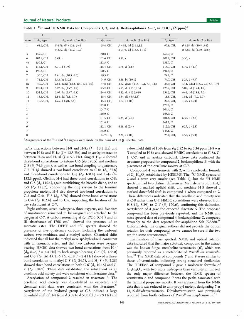
Table 1. 13C and 1H NMR Data for Compounds 1, 2, and 4, Berkazaphilones A−C, in CDCl3 (δ ppm)a
1 2 4
atom δC, type δH, mult. (J in Hz) δC, type δH mult. (J in Hz) δC, type δH mult. (J in Hz)
3 159.9, C 160.8, C 160.7, C4 102.8, CH 5.49, s 102.4, CH 5.51, s 102.8, CH 5.56, s4a 150.5, C 152.5, C 151.7, C5 118.1, CH 5.71, d (1.9) 115.4, CH 5.78, d (1.6) 115.7, CH 5.79, d (1.7)6 198.2, C 190.5, C 194.8, C7 50.0, CH 2.41, dq (10.3, 6.6) 89.3, C 74.1, C8 74.2, CH 3.43, bt (10.3) 74.6, CH 3.58, bt (10.1) 74.7, CH 5.29, d (9.9)8a 40.9, CH 2.84, dddd (13.2, 10.3, 5.4, 1.9) 37.8, CH 2.82, dddd (13.5, 10.1, 5.3, 1.6) 34.9, CH 3.44, dddd (13.0, 9.9, 5.4, 1.7)9 125.4, CH 5.87, dq (15.7, 1.7) 125.2, CH 5.85, dd (15.3,1.5) 125.2, CH 5.87, dd (15.4, 1.7)10 133.2, CH 6.40, dq (15.7, 6.8) 134.4, CH 6.45, dq (15.3,6.9) 134.5, CH 6.41, dd (15.4, 7.0)11 18.4, CH3 1.84, dd (6.8, 1.7) 18.4, CH3 1.84, dd (6.9,1.5) 18.5, CH3 1.84, dd, (7.0, 1.7)12 10.8, CH3 1.25, d (3H, 6.6) 15.4, CH3 1.77, s (3H) 20.4, CH3 1.38, s (3H)1′ 172.0, C 170.6, C2′ 104.7, C 104.4, C3′ 166.0, C 165.9, C4′ 101.5, CH 6.25, d (2.4) 101.6, CH 6.30, d (2.3)5′ 161.4, C 161.1, C6′ 112.1, CH 6.18, d (2.4) 112.0, CH 6.27, d (2.3)7′ 145.0, C 144.6, C8′ 24.7 CH3 2.28, s (3H) 25.0, CH3 2.58, s (3H)
aAssignments of the 13C and 1H signals were made on the basis of HSQC spectral data.
Journal of Natural Products Article
dx.doi.org/10.1021/np200414c | J. Nat. Prod. 2012, 75, 344−350346
The third vermistatin analogue, compound 8, had amolecular formula of C16H14O6, with two less carbons thaneither vermistatin (6) or dihydrovermistatin (7). In this case,the 1H NMR signals of both the terminal propylene and propylmoiety were absent and were replaced by a methyl singlet at δ2.44. This compound was previously reported as penis-implicissin from broth cultures of P. simplicissimum.41
Compounds 7 and 8 had the same sign and relative opticalrotations as vermistatin (6), suggesting that they had the sameconfiguration.40
The molecular formula of compound 9 was C13H16O5, asdetermined from HREIMS. 13C NMR and DEPT spectraindicated the presence of two carbonyl carbons: ketone C-8 atδC 208.8 (C) and aldehyde C-12 at δC 195.1 (CH). Sixadditional sp2-hybridized carbons indicated the presence of anaromatic moiety with two oxygen-bearing carbons resonating atδC 165.2 and 164.6 ppm. These assignments accommodated allsix sites of unsaturation associated with the molecular formula.Examination of the spectral data established the structure ofcompound 9 as shown, which was previously reported as ametabolite of Aspergillus versicolor.42 We prepared the Mosherester of 9 and found the same absolute configuration (R) asreported.43 The S stereoisomer of compound 9 has also beenreported from a Pseudobotrytis sp.44
EIMS established the molecular formula of compound 11 asC6H8O4, associated with three sites of unsaturation. Its infraredspectrum indicated the presence of two carbonyl moieties: thebroad −OH stretch (νmax 3027 cm−1) and carbonyl absorptionat 1716 cm−1 indicated the presence of a saturated carboxylicacid, while the carbonyl absorption at 1774 cm−1 indicated thepresence of a saturated γ-butyrolactone.45 The presence of theacid was confirmed by methylation of compound 11 withdiazomethane to yield the methyl ester, 12. Analysis of thespectral data established the structure of 11, which is the knowncompound α-methylparaconic acid.46
Compound 13 had a molecular formula of C11H16O3,established by the HRESIMS spectrum, with four sites ofunsaturation. The broad −OH stretch (νmax = 3021 cm−1) andthe carbonyl absorption at 1683 cm−1 in the infrared spectrumwere indicative of an α,β-unsaturated carboxylic acid.Methylation of 13 with diazomethane yielded the methylester 14, which had a molecular formula of C12H19O3, asdetermined by HREIMS.The 1H−1H COSY spectrum of 13 provided connectivity
data for two spin systems beginning with a terminal diene. The3J-coupling of the diene protons clearly correlated H-8a (δH5.20) and H-8b (δH 5.08) to H-7 (δH 6.28), H-7 to H-6 (δH6.20), H-6 to H-5 (δH 5.70), H-5 to oxygen-bearing methine H-4 (δH 4.31), and H-4 to methylene H2-3 (δH 2.57). The secondspin system consisted of a terminal propylidene moiety with 3J-coupling from H-9 (δH 7.02) to methylene H2-10 (δH 2.23) andfrom H-10 to the terminal methyl H3-11 (δH 1.05). TheHMBC spectrum provided the necessary information toconnect these two spin systems to the carbonyl carbon withthree-bond correlations from H-9 and H2-3 to C-1. H-9 alsoexhibited correlations to C-3 and C-10. Methylene H2-3provided a point of connectivity with three-bond correlations toC-1, C-9, and C-5 and two-bond correlations to flankingcarbons C-2 and C-4. H-4 also afforded nice bilateralconnectivity with three-bond correlations to C-2 and C-6 andtwo-bond correlations to C-3 and C-5. The chemical shift of H-9 indicated an E configuration for Δ2,9.47 The magnitude of thecoupling constant between olefinic H-5 and H-6 (J = 14.3 Hz)
also indicated an E configuration. These data generated thestructure proposed for compound 13, berkedienoic acid.Compound 15 had a molecular formula of C11H14O2
established by the HRESIMS [M + H]+ peak at m/z179.1082. The carbonyl absorption at 1751 cm−1 andaccompanying CC absorption at 1679 cm−1 in the infraredspectrum indicated the presence of an α,β-unsaturated γ-lactone.45 These data and the similarities in the NMR spectrasuggested that compound 15 was the γ-butyrolactone ofcompound 13. The 1H−1H COSY and the 1H NMR spectraprovided connectivity data for a single extended spin systembeginning with a terminal diene at one end. The 3J-coupling ofthe diene protons correlated H-8a (δH 5.28) and H-8b (δH5.18) to H-7 (δH 6.30), H-7 to H-6 (δH 6.30), H-6 to H-5 (δH5.69), H-5 to oxygen-bearing methine H-4 (δH 4.98), and H-4to methylene H2-3 (δH 2.58, 3.05 ppm).The terminal propylidene moiety produced 3J-coupling from
methyl H3-11 (δH 1.07 ppm) to methylene H2-10 (δH 2.17)and from H2-10 to olefinic H-9 (δH 6.72). The coupling patternfor methylene H2-10, however, was quite complex; the expectedpentet pattern was actually a pentet of triplets, and the 1H−1HCOSY spectrum showed connectivity to methylene H2-3, whichrequired five-bond coupling in our proposed structure. Thecoupling patterns for H2-3a and -3b were also complex, andboth were doublets of doublets of pentets. Bearing in mind thata pentet is actually a dddd with equivalent coupling constants,then these patterns become complex indeed. These datasuggest that these unusually complex patterns are the result ofallylic and homoallylic coupling. Studies on 2-butene andcompounds containing a butenyl moiety showed similarcorrelations.48
Homoallylic coupling data from both the 1H NMR and the1H−1H COSY spectra provided the information to connect thetwo ends of the molecule. The coupling patterns for thismolecule were complex but could be deconstructed to includehomoallylic coupling between the methylenes H2-10 and H2-3.The exocyclic double bond could be established as the E
isomer by chemical shift arguments. A series of E and Z isomersof α-alkylidene-γ-butyrolactones were synthesized, and thechemical shifts of the olefinic protons analogous to H-9 werecompared.49 In all cases the chemical shift of H-9 in the Zisomer approached δH 6.0 ppm, while in the E isomer it wascloser to δH 6.6 ppm.49 These data were used to generate thestructure proposed for compound 15, berkedienolactone. Aseries of similar lactones including the tricyclic compoundgallielalactone were isolated from an unidentified ascomy-cete.50,51 Unfortunately, the reported monocyclic lactones moststructurally similar to 15 were not isolated as pure compounds,so it was not possible to compare optical rotation data with thatof berkedienolactone (15).Compounds 1, 2, 4, and 6−9 were evaluated for their ability
to inhibit caspase-1 in vitro, and the most active compoundsand closely related analogues were evaluated for their ability toinhibit the production of interleukin 1-β in THP-1 cells (pro-monocytic leukemia cell line). THP-1 cells produce high levelsof IL-1β when induced with titanium nanowires and bacteriallipopolysaccharide (LPS). Caspase-1 inhibition was determinedin a fluorometric assay normalized to 1.00, where 0 is totalenzyme inhibition and 1.00 is lack of enzyme inhibition. Bothberkazaphilones B (2) and C (4) had IC100 values of 25 μMagainst caspase-1, while berkazaphilone A (1), penisimplicissin(8), and compound 9 were completely inhibitory at a
Journal of Natural Products Article
dx.doi.org/10.1021/np200414c | J. Nat. Prod. 2012, 75, 344−350347
concentration of 250 μM. Vermistatin (6) and dihydrovermis-tatin (7) were not inhibitory at the concentrations tested.Induced THP-1 cells were exposed to compounds 2, 4, and
6−8, and the concentrations of IL-1β postexposure weredetermined. All of the compounds tested inhibited theproduction of IL-1β in THP-1 cells at a concentration of 250μM. In dilution assays, however, only compounds 2 and 4inhibited the production of IL-1β. Compounds 2 and 4completely inhibited the production of IL-1β at concentrationsof 5 and 50 μM, respectively.Compounds 2, 4, 7, and 8 were tested in the National
Cancer Institute (NCI) antitumor screen against 60 human celllines.52 The compounds showed selective cytotoxicity towardleukemia cell lines only. Berkazaphilone B (2) exhibited a log10GI50 of −5.67 against cell line RPMI-8226, and berkazaphiloneC (4) exhibited a log10 GI50 of −6.42 against cell line SR. In thevermistatin family, penisimplicissin (8) exhibited a log10 GI50 of−6.70 against cell line CCRF-CEM and −5.83 against HL-60(TB), and dihydrovermistatin (7) was inactive at theconcentrations tested. Vermistatin (6) had been previouslytested by the NCI and was also inactive (SupportingInformation).The NCI Molecular Target database includes experiments
that determine relative RNA levels for nearly 10 000 humanclones, measured in microarray experiments for the NCI celllines. It was interesting to note that in several microarrayexperiments caspase-1 was upregulated almost exclusively indifferent leukemia cell lines.53−56
■ EXPERIMENTAL SECTIONGeneral Experimental Procedures. 1H and 13C NMR spectra
were run on a Bruker DPX-300. Chemical shifts were recorded withrespect to the deuterated solvent shift (CDCl3, δ 7.24 for the protonresonance and δ 77.0 for the carbon). IR spectra were recorded on aNicolet NEXUS 670 FT-IR spectrometer. Optical rotations weremeasured on a Perkin-Elmer 241 MC polarimeter using a 1 mL cell.Mass spectral data were provided by the Mass Spectrometry,Proteomics and Metabolomics Facility at Montana State Universityand the Mass Spectral Analysis Laboratory at the University ofMontana. All solvents used were spectral grade or distilled prior to use.Collection, Extraction, and Isolation Procedures. The
collection and isolation of the Berkeley Pit fungi have previouslybeen described.7,8 The fungus was identified as Penicillium rubrum byMicrobial Identification, Inc. The fungus was grown at roomtemperature in 26 × 300 mL of DIFCO potato dextrose broth(acidified to pH 2.7 with sulfuric acid) in 1 L Erlenmeyer flasks(shaken at 180 rpm for 6 days then still for 15 days). At time of harvest50 mL of MeOH was added per flask. The combined cultures (7.8 L)were filtered through cheesecloth to remove the mycelia mat for aseparate study. The filtrate from the combined cultures was extractedthree times with 1 L of CHCl3, and the extract was reduced in vacuo toan oil (1.14 g). This extract demonstrated inhibition of caspase-1 andMMP-3, antimicrobial activity against Staphylococcus aureus andEscherichia coli, and brine shrimp lethality.The CHCl3 extract was fractionated using a flash Si gel column
using hexanes, hexane/isopropyl alcohol (IPA) mixtures, to isopropylalcohol/MeOH mixtures. The 50% IPA/hexane fraction was furtherfractionated by preparative HPLC on a Rainin 21 mm preparative Sigel column with a hexanes/isopropyl alcohol gradient. The 10% IPAfraction was further fractionated on Si gel to yield the three azaphilonederivatives 1 (5.1 mg), 2 (35.1 mg), and 4 (7.1 mg). The fraction thateluted with 50% IPA/hexane yielded the three cytotoxic compounds 6(46.2 mg), 7 (4.0 mg), and 8 (18.0 mg). The 25% IPA/hexane fractionyielded aldehyde 9 (5.6 mg), lactone 11, berkedienoic acid 13 (1.5mg), and berkedienolactone 14 (2.0 mg)
Acetylation of Compound 2. Compound 2 (1.0 mg) wasdissolved in pyridine (50 μL) and Ac2O (50 μL) and stirred for 24 h.After that time the solvents were removed to give 3 as an oil (0.9 mg).
Berkazaphilone C (4): [α]25D +54.3 (c 0.0030 CHCl3);1H NMR
and 13C NMR (CDCl3) see Table 1; EIMS m/z 386; HREIMS m/z386.1365 [M]+ (calcd for C21H22O7, 386.1365).
Acetylation of Compound 4. Compound 4 (1.0 mg) wasdissolved in pyridine (50 μ L) and Ac2O (50 μL) and stirred for 24 h.After that time the solvents were removed to give 5 as an oil (0.9 mg).
Methylation of 13. Compound 13 (0.2 mg) was dissolved in Et2O(100 μL), and a solution of CH2N2 in Et2O was added dropwise untilthe yellow color persisted. The solution was stirred for 5 min, and thesolvent removed under a stream of N2 to yield the methyl ester 14 (0.2mg); HREIMS m/z [M + H]+ m/z 211.1325 (calcd for C12H19O3,211.1334).
dx.doi.org/10.1021/np200414c | J. Nat. Prod. 2012, 75, 344−350348
5), 125.1 (C, C-2), 75.7 (CH, C-4), 119.6 (CH2, C-8), 31.8 (CH2, C-3), 23.6 (CH2, C-10), 12.6 (CH3, C-11); HRESIMS [M + H]+ m/z179.1082 (calcd for C11H15O2, 179.1072).In Vitro THP-1 Assay. Human monocyte cell line THP-1 was
purchased from ATCC (#TIB-202). The cells were suspended at (2−4) × 105 viable cells/mL in RPMI media supplemented with 10% fetalbovine serum, 0.05 mM 2-mercaptoethanol, sodium pyruvate, and anantimycotic/antibiotic cocktail containing penicillin, streptomycin, andamphotericin B (Mediatech,VWR). The cells were differentiated intomacrophage-like cells by the phorbol ester PMA (1 μg/mL, Sigma) 24h prior to experimentation. The transformed cells were removed fromthe flask by scraping and centrifuged at 450g for 5 min. The resultingcell pellet was suspended at 1.0 × 10⁶ cells/mL and exposed tocaspase-1 inhibitors at concentrations described below (0.5−0.005%),LPS [20 ng/mL], and TiO2 nanowires (100 μg/mL). Experimentswere conducted in 96-well plates for 24 h in 37 °C water-jacketed CO2incubators (ThermoForma).Toxicity Assay. Cell viability was determined by MTS reagent
using the CellTiter96 assay (Promega), according to the manufac-turer’s protocol. The plate was read at 490 nm.Cytokine Assays. Human IL-1β DuoSet was obtained from R&D
Systems, and ELISA assays were performed according to themanufacturer’s protocol. The plate was read at 490 nm.
■ ASSOCIATED CONTENT*S Supporting Information1H NMR, 13C NMR, COSY, and HMBC spectra ofberkazaphilones A (1) and B (2), berkedienoic acid (13),and berkedienolactone (15); 1H NMR and 13C NMR ofberkazaphilone C (4); and NCI cell line data for compounds 2,4, and 8 are available free of charge via the Internet at http://pubs.acs.org.
NotesThe authors declare no competing financial interest.
■ ACKNOWLEDGMENTSWe thank Ms. B. Parker (University of Montana) for HRMSdata and our colleagues from the Department of Chemistry,Montana State University: Dr. S. Busse for assistance withNMR spectroscopy and Dr. L. J. Sears for mass spectral data.We thank the National Science Foundation grant 9506620 forproviding funding for NMR upgrades at the MSU facility andgrant CHE-9977213 for acquisition of a NMR spectrometer.We gratefully acknowledge NIH grants R01CA139159,P2 0RR16455 - 0 4 , a nd P20RR017670 (NCRR) ;5P30NS055022; and RC2ES018742.
■ DEDICATIONDedicated to Dr. Gordon M. Cragg, formerly Chief, NaturalProducts Branch, National Cancer Institute, Frederick, Mary-land, for his pioneering work on the development of naturalproduct anticancer agents.
■ REFERENCES(1) Montana Bureau of Mines and Geology, Berkeley Pit and ButteMine-Flooding Operable Unit website: http://www.mbmg.mtech.edu/env-berkeley.htm, accessed 5/16/11.(2) Stierle, A.; Stierle, D. Bioprospecting in the Berkeley Pit:Bioactive Metabolites from Acid Mine Waste Extremophiles. In
Bioactive Natural Products, Vol. 32; Atta-Ur-Rahman, Ed.; ElsevierScience Publishers: Amsterdam. 2005.(3) Stierle, A. A.; Stierle, D. B.; Parker, K.; Goldstein, E.; Bugni, T.;Baarson, C.; Gress, J.; Blake, D. J. Nat. Prod. 2003, 66, 1097−1100.(4) Stierle, D.; Stierle, A.; Hobbs, J. D.; Stokken, J.; Clardy, J. Org.Lett. 2004, 6, 1049−1052.(5) Stierle, A.; Stierle, D.; Kemp, K. J. Nat. Prod. 2004, 67, 1392−1395.(6) Stierle, A.; Stierle, D.; Kelley, K. J. Org. Chem. 2006, 71, 5357−5360.(7) Stierle, D. B.; Stierle, A. A.; Patacini, B. J. Nat. Prod. 2007, 70,1820−1823.(8) Stierle, A. A.; Stierle, D. B.; Patacini, B. J. Nat. Prod. 2008, 71,856−860.(9) Franchi, L; Eigenbrod, T.; Munoz-Planillo, R.; Nunez, G. Nat.Immunol. 2009, 10, 241−256.(10) Ona, V. O.; Li, M.; Vonsattel, J. P. G; Andrews, L. J.; Khan, S.Q.; Chung, W. M.; Frey, A. S.; Menon, A. S.; Li, X. J.; Stieg, P. E.;Yuan, J.; Penney, J. B.; Young, A. B.; Cha, J. H. J.; Friedlander, R. M.Nature 1999, 399, 263−267.(11) Li, M.; Ona, V. O.; Guegan, C.; Chen, M.; Jackson, V.; Andrews,L. J.; Olszewski, A. J.; Stieg, P. E.; Lee, J.; Przedborski, S.; Friedlander,R. M. Science 2000, 288, 335−339.(12) Rabuffetti, M.; Sciorati, C.; Tarozzo, G.; Clementi, E.; Manfredi,A. A.; Beltramo, M. . J. Neurosci. 2000, 20, 4398−4404.(13) Ming, X; Li, W; Maeda, Y.; Blumberg, B.; Raval, S.; Cook, S. D.;Dowling, P. C. J. Neurol. Sci. 2002, 197, 9−18.(14) Furlan, R.; Martino, G.; Galbiati, F.; Poliani, P. L.; Smiroldo, S.;Bergami, A.; Desina, G.; Comi, G.; Flavell, R.; Su, M. S.; Adorini, L. J.Immunol. 1999, 163, 2403−2409.(15) Franchi, L; Eigenbrod, T.; Munoz-Planillo, R.; Nunez, G. Nat.Immunol. 2009, 10, 241−256.(16) Stienstra, R.; Joosten, L..B.; Koenen, T.; van Tits, B.; vanDiepen, J. A.; van den Berg, S. A. A.; Rensen, P. C. N.; Voshol, P. J.;Fantuzzi, G.; Hijmans, A.; Kersten, S.; Mueller, M.; van den Berg, W.B.; van Rooijen, N.; Wabitsch, M.; Kullberg, B.-J.; van der Meer, J. W.M.; Kanneganti, T.; Tack, C. J.; Netea, M. G. Cell Metab. 2010, 12,593−605.(17) Vincent, J. A.; Mohr, S. Diabetes 2007, 56, 224−230.(18) Jha, S.; Srivastava, S. Y.; Brickey, W. J.; Iocca, H.; Toews, A.;Morrison, J. P.; Chen, V. S.; Gris, D.; Matsushima, G. K.; Jenny, P.-Y.;Ting, J. P.-Y. J. Neurosci. 2010, 30, 15811−15820.(19) Gamblin, T. C.; Chen, F.; Zambrano, A.; Abraha, A.; Lagalwar,S.; Guillozet, A. L.; Lu, M.; Fu, Y.; Garcia-Sierra, F.; LaPointe, N.;Miller, R.; Berry, R. W.; Binder, L. I.; Cryns, V. L. Proc. Nat. Acad. Sci.U. S. A. 2003, 100, 10032−10037.(20) Lewis, A. M.; Varghese, S.; Xu, H.; Alexander, H. R. J. Transl.Med. 2006, 4, 48−59.(21) Granot, T.; Milhas, D.; Carpentier, S.; Dagan, A.; Segui, B.; Gatt,S.; Levade, T. Leukemia 2006, 20, 392−399.(22) Okamoto, M.; Liu, W.; Luo, Y.; Tanaka, A.; Cai, X.; Norris, D.A.; Dinarello, C.; Fujita, M. J. Biol. Chem. 2010, 285, 6477−6488.(23) Voronov, E.; Shouval, D. S.; Krelin, Y.; Cagnano, E.;Benharroch, D.; Iwakura, Y.; Dinarello, C. A.; Apte, R. N. Proc. Natl.Acad. Sci. U. S. A. 2003, 100, 2645−2650.(24) Paugh, B. S.; Bryan, L.; Paugh, S. W.; Wilczynska, K. M.;Alvarez, S. M.; Singh, S. K.; Kapitonov, D.; Rokita, H.; Wright, S.;Griswold-Prenner, I.; Milstien, S.; Spiegel, S.; Kordula, T. J. Biol. Chem.2009, 284, 3408−3417.(25) Lu, T.; Tian, L.; Han, Y.; Vogelbaum, M.; Stark, G. R. Proc. Natl.Acad. Sci. U. S. A. 2007, 104, 4365−4370.(26) Schlosser, S.; Gansauge, F.; Ramadani, M.; Beger, H.-G.;Gansauge, S. FEBS Lett. 2001, 491, 104−108.(27) Muerkoster, S. S.; Lust, J.; Arlt, A.; Hasler, R.; Witt, M.; Sebens,T.; Schreiber, S.; Folsch, U. R.; Schafer, H. Oncogene 2006, 25, 3973−3981.(28) Muerkoster, S.; Wegehenkel, K.; Arlt, A.; Witt, M.; Sipos, B.;Kruse, M. L.; Sebens, T.; Kloppel, G.; Kalthoff, H.; Folsch, U. R.;Schafer, H. Cancer Res. 2004, 64, 1331−1337.
Journal of Natural Products Article
dx.doi.org/10.1021/np200414c | J. Nat. Prod. 2012, 75, 344−350349
(29) Arlt, A.; Vorndamm, J.; Muerkoster, S.; Yu, H.; Schmidt, W. E.;Folsch, U. R.; Schafer, H. Cancer Res. 2002, 62, 910−916.(30) Jin, L.; Yuan, R. Q.; Fuchs, A.; Yao, Y.; Joseph, A.; Schwall, R.;Schnitt, S. J.; Guida, A.; Hastings, H. M.; Andres, J.; Turkel, G.;Polverini, P. J.; Goldberg, I. D.; Rosen, E. M. Cancer 1997, 80, 421−434.(31) Elaraj, D. M.; Weinreich, D. M.; Varghese, S.; Puhlmann, M.;Hewitt, S. M.; Carroll, N. M.; Feldman, E. D.; Turner, E. M.;Alexander, H. R. Clin. Cancer Res. 2006, 12, 1088−1096.(32) Rudolphi, K.; Gerwin, N.; Verzijl, N.; van der Kraan, P.; van denBerg, W. Osteoarthritis Cartilage 2003, 11, 738−746.(33) Cascao, R.; Polido-Pereira, J.; Canhao, H.; Rodrigues, A. M.;Navalho, M.; Raquel, H.; Mourao, A. F.; Resende, C.; Fonseca, J. E.;Moita, L. F. J. Transl. Med. 2010, 8, 35.(34) Furlan, R.; Martino, G.; Galbiati, F.; Poliani, P. L.; Smiroldo, S.;Bergami, A.; Desina, G.; Comi, G.; Flavell, R.; Su, M. S.; Adorini, L. J.Immunol. 1999, 163, 2403−2409.(35) Rabuffetti, M.; Sciorati, C.; Tarozzo, G.; Clementi, E.; Manfredi,A. A.; Beltramo, M. J. Neurosci. 2000, 20, 4398−4404.(36) Reyes, F.; Arda, A.; Martın, R.; Fernandez, R.; Rueda, A.;Montalvo, D.; Gomez, C.; Jimenez, C.; Rodrıguez, J.; Sanchez-Puelles,J. J. Nat. Prod. 2004, 67, 1190−1192.(37) Osmanova, N.; Schultze, W.; Ayoub, N. Phytochem Rev. 2010, 9,315−342.(38) Yang, S.-W.; Chan, T.-M.; Terracciano, J.; Patel, R.; Patel, M.;Gullo, V.; Chu, M. J. Antibiot. 2006, 59, 720−723.(39) Schlorke, O.; Zeeck, A. Eur. J. Org. Chem. 2006, 1043−1049.(40) Fuska, J.; Uhrn, D.; Proksa, B.; Voticky, Z.; Ruppeldt, J. J.Antibiot. 1986, 39, 1605−1608.(41) Komai, S.; Hosoe, T.; Itabashi, T.; Nozawa, K.; Yaguchi, T.;Fukushima, K.; Kawai, K. Heterocycles 2005, 65, 2771−2776.(42) Arai, T.; Sano, H. Jpn. Kokai Tokkyo Koho 1994, 6.(43) Lee, Y. M; Li, H.; Hong, J.; Cho, H. Y.; Bae, K. S.; Kim, M. A.;Kim, D.-K.; Jung, J. H. Arch. Pharm. Res. 2010, 33, 231−235.(44) Yamaguchi, Y.; Masuma, R.; Kim, Y. P.; Uchida, R.; Tomoda,H.; Omura, S. Mycoscience 2004, 45, 9−16.(45) Pavia, D. L.; Lampman, G. M.; Kriz, G. S. Introduction toSpectroscopy, 3rd ed.; Harcourt College: Orlando, FL, 2001; p 53.(46) Forzato, C.; Furlan, G.; Nitti, P.; Pitacco, G.; Marchesan, D.;Coriani, S.; Valentin, E. Tetrahedron: Asymmetry 2005, 16, 3011−3023.(47) Jackman, L. M.; Sternhell, S. Applications of Nuclear MagneticResonance Spectroscopy in Organic Chemistry, 2nd ed.; Pergamon Press:Oxford, 1972; p 171.(48) Barfield, M.; Sternhell, S. J. Am. Chem. Soc. 1972, 94, 1905−1913.(49) Tanaka, K.; Uneme, H.; Yamagishi, N.; Tanikaga, R.; Kaji, A.Bull. Chem. Soc. Jpn. 1980, 53, 2910−2916.(50) Johansson, M.; Kopcke, B.; Anke, H.; Sterner, O. J. Antibiot.2002, 55, 104−106.(51) Johansson, M.; Sterner, O. Org. Lett. 2001, 3, 2843−2845.(52) Shoemaker, R. H. Nat. Rev. 2006, 6, 813−823.(53) http://dtp .nc i .n ih .gov/mtweb/target in fo?molt id=GC19100&moltnbr=9698, accessed 5/12/11.(54) http://dtp .nc i .n ih .gov/mtweb/target in fo?molt id=GC29740&moltnbr=29739, accessed 5/12/11.(55) http://dtp .nc i .n ih .gov/mtweb/target in fo?molt id=GC29740&moltnbr=29739, accessed 5/12/11.(56) http://dtp .nc i .n ih .gov/mtweb/target in fo?molt id=GC226538&moltnbr=131271, accessed 5/12/11.
■ NOTE ADDED IN PROOFBerkazaphilones B and C were also recently published as thepinophilins, J. Nat. Prod., Article ASAP, DOI: 10.1021/np200523b, submitted 6/22/2011.
Journal of Natural Products Article
dx.doi.org/10.1021/np200414c | J. Nat. Prod. 2012, 75, 344−350350