ORIGINAL RESEARCH ARTICLE published: 27 November 2013 doi: 10.3389/fcimb.2013.00083 Caspase-2 mediates a Brucella abortus RB51-induced hybrid cell death having features of apoptosis and pyroptosis Denise N. Bronner 1 , Mary X. D. O’Riordan 1 and Yongqun He 1,2,3 * 1 Department of Microbiology and Immunology, University of Michigan Medical School, Ann Arbor, MI, USA 2 Unit for Laboratory Animal Medicine, University of Michigan Medical School, Ann Arbor, MI, USA 3 Comprehensive Cancer Center, University of Michigan Medical School, Ann Arbor, MI, USA Edited by: Amal O. Amer, The Ohio State University, USA Reviewed by: Jianwu Pei, American Animal Health, Inc., USA Diego J. Comerci, Instituto de Investigaciones Biotecnologicas Dr. Rodolfo A. Ugalde, Argentina *Correspondence: Yongqun He, Unit for Laboratory Animal Medicine and Department of Microbiology and Immunology, University of Michigan Medical School, 018 Animal Research Facility, 1150 W. Medical Dr., Ann Arbor, MI 48109, USA e-mail: [email protected]Programmed cell death (PCD) can play a crucial role in tuning the immune response to microbial infection. Although PCD can occur in different forms, all are mediated by a family of proteases called caspases. Caspase-2 is the most conserved caspase, however, its function in cell death is ill-defined. Previously we demonstrated that live attenuated cattle vaccine strain Brucella abortus RB51 induces caspase-2-mediated and caspase-1-independent PCD of infected macrophages. We also discovered that rough attenuated B. suis strain VTRS1 induces a caspase-2-mediated and caspase-1-independent proinflammatory cell death in infected macrophages, which was tentatively coined “caspase-2-mediated pyroptosis” . However, the mechanism of caspase-2-mediated cell death pathway remained unclear. In this study, we found that caspase-2 mediated proinflammatory cell death of RB51-infected macrophages and regulated many genes in different PCD pathways. We show that the activation of proapoptotic caspases-3 and -8 was dependent upon caspase-2. Caspase-2 regulated mitochondrial cytochrome c release and TNFα production, both of which are known to activate caspase-3 and caspase-8, respectively. In addition to TNFα, RB51-induced caspase-1 and IL-1β production was also driven by caspase-2-mediated mitochondrial dysfunction. Interestingly, pore formation, a phenomenon commonly associated with caspase-1-mediated pyroptosis, occurred; however, unlike its role in S. typhimurium-induced pyroptosis, pore formation did not contribute to RB51-induced proinflammatory cell death. Our data suggest that caspase-2 acts as an initiator caspase that mediates a novel RB51-induced hybrid cell death that simulates but differs from typical non-proinflammatory apoptosis and caspase-1-mediated proinflammatory pyroptosis. The initiator role of the caspase-2-mediated cell death was also conserved in cellular stress-induced cell death of macrophages treated with etoposide, naphthalene, or anti-Fas. Caspase-2 also regulated caspase-3 and -8 activation, as well as cell death in macrophages treated with each of the three reagents. Taken together, our data has demonstrated that caspase-2 can play an important role in mediating a proinflammatory response and a hybrid cell death that demonstrates features of both apoptosis and pyroptosis. Keywords: proinflammatory, caspase-2, programmed cell death, mitochondrial dysfunction, macrophage, Brucella INTRODUCTION Programmed cell death (PCD) is a crucial process initiated by the host in response to cellular stress and microbial infections. PCD can occur in a variety of ways (Galluzzi et al., 2012). Apoptosis, pyroptosis, and necroptosis are three pathways of PCD that can occur during microbial infections with substantially different out- comes. Apoptosis is a silent PCD due to a lack of cytokine secre- tion (Elmore, 2007). Although the plasma membrane remains intact, apoptotic bodies can bleb off and be phagocytosed by other phagocytic cells in the surrounding area. Pyroptosis or “death by fire,” is inflammatory PCD typically mediated by caspase-1 (Bergsbaken et al., 2009). Caspase-1 processes proinflammatory cytokines (IL-1β and IL-18), and secretion of these cytokines requires pore formation in the plasma membrane, which leads to cell swelling and eventually lysis. Necroptosis is a newly iden- tified type of PCD that includes a proinflammatory response as well as loss of plasma membrane integrity (Vandenabeele et al., 2010). In contrast to apoptosis and pyroptosis, serine/threonine kinases, RIP1 and RIP3, mediate necroptosis. In addition, necrop- tosis leads to the release of intracellular contents; mostly damage associated molecular patterns (DAMPs). Many microbes can induce cell death during infection and dis- semination (Gao and Abu Kwaik, 2000). Avirulent Mycobacterium induces apoptosis in macrophages (Fratazzi et al., 1999). Frontiers in Cellular and Infection Microbiology www.frontiersin.org November 2013 | Volume 3 | Article 83 | 1 CELLULAR AND INFECTION MICROBIOLOG Y
Transcript
ORIGINAL RESEARCH ARTICLEpublished: 27 November 2013
doi: 10.3389/fcimb.2013.00083
Caspase-2 mediates a Brucella abortus RB51-inducedhybrid cell death having features of apoptosis andpyroptosisDenise N. Bronner1, Mary X. D. O’Riordan1 and Yongqun He1,2,3*
1 Department of Microbiology and Immunology, University of Michigan Medical School, Ann Arbor, MI, USA2 Unit for Laboratory Animal Medicine, University of Michigan Medical School, Ann Arbor, MI, USA3 Comprehensive Cancer Center, University of Michigan Medical School, Ann Arbor, MI, USA
Edited by:
Amal O. Amer, The Ohio StateUniversity, USA
Reviewed by:
Jianwu Pei, American AnimalHealth, Inc., USADiego J. Comerci, Instituto deInvestigaciones Biotecnologicas Dr.Rodolfo A. Ugalde, Argentina
*Correspondence:
Yongqun He, Unit for LaboratoryAnimal Medicine and Department ofMicrobiology and Immunology,University of Michigan MedicalSchool, 018 Animal ResearchFacility, 1150 W. Medical Dr.,Ann Arbor, MI 48109, USAe-mail: [email protected]
Programmed cell death (PCD) can play a crucial role in tuning the immune responseto microbial infection. Although PCD can occur in different forms, all are mediatedby a family of proteases called caspases. Caspase-2 is the most conserved caspase,however, its function in cell death is ill-defined. Previously we demonstrated that liveattenuated cattle vaccine strain Brucella abortus RB51 induces caspase-2-mediated andcaspase-1-independent PCD of infected macrophages. We also discovered that roughattenuated B. suis strain VTRS1 induces a caspase-2-mediated and caspase-1-independentproinflammatory cell death in infected macrophages, which was tentatively coined“caspase-2-mediated pyroptosis”. However, the mechanism of caspase-2-mediated celldeath pathway remained unclear. In this study, we found that caspase-2 mediatedproinflammatory cell death of RB51-infected macrophages and regulated many genes indifferent PCD pathways. We show that the activation of proapoptotic caspases-3 and -8was dependent upon caspase-2. Caspase-2 regulated mitochondrial cytochrome c releaseand TNFα production, both of which are known to activate caspase-3 and caspase-8,respectively. In addition to TNFα, RB51-induced caspase-1 and IL-1β production was alsodriven by caspase-2-mediated mitochondrial dysfunction. Interestingly, pore formation,a phenomenon commonly associated with caspase-1-mediated pyroptosis, occurred;however, unlike its role in S. typhimurium-induced pyroptosis, pore formation did notcontribute to RB51-induced proinflammatory cell death. Our data suggest that caspase-2acts as an initiator caspase that mediates a novel RB51-induced hybrid cell death thatsimulates but differs from typical non-proinflammatory apoptosis and caspase-1-mediatedproinflammatory pyroptosis. The initiator role of the caspase-2-mediated cell deathwas also conserved in cellular stress-induced cell death of macrophages treated withetoposide, naphthalene, or anti-Fas. Caspase-2 also regulated caspase-3 and -8 activation,as well as cell death in macrophages treated with each of the three reagents. Takentogether, our data has demonstrated that caspase-2 can play an important role in mediatinga proinflammatory response and a hybrid cell death that demonstrates features of bothapoptosis and pyroptosis.
INTRODUCTIONProgrammed cell death (PCD) is a crucial process initiated by thehost in response to cellular stress and microbial infections. PCDcan occur in a variety of ways (Galluzzi et al., 2012). Apoptosis,pyroptosis, and necroptosis are three pathways of PCD that canoccur during microbial infections with substantially different out-comes. Apoptosis is a silent PCD due to a lack of cytokine secre-tion (Elmore, 2007). Although the plasma membrane remainsintact, apoptotic bodies can bleb off and be phagocytosed by otherphagocytic cells in the surrounding area. Pyroptosis or “deathby fire,” is inflammatory PCD typically mediated by caspase-1(Bergsbaken et al., 2009). Caspase-1 processes proinflammatory
cytokines (IL-1β and IL-18), and secretion of these cytokinesrequires pore formation in the plasma membrane, which leadsto cell swelling and eventually lysis. Necroptosis is a newly iden-tified type of PCD that includes a proinflammatory response aswell as loss of plasma membrane integrity (Vandenabeele et al.,2010). In contrast to apoptosis and pyroptosis, serine/threoninekinases, RIP1 and RIP3, mediate necroptosis. In addition, necrop-tosis leads to the release of intracellular contents; mostly damageassociated molecular patterns (DAMPs).
Many microbes can induce cell death during infection and dis-semination (Gao and Abu Kwaik, 2000). Avirulent Mycobacteriuminduces apoptosis in macrophages (Fratazzi et al., 1999).
Frontiers in Cellular and Infection Microbiology www.frontiersin.org November 2013 | Volume 3 | Article 83 | 1
Bronner et al. Caspase-2-mediated proinflammatory cell death
Neighboring uninfected macrophages, upon phagocytosis, killedMycobacterium in apoptotic bodies released by Mycobacterium-infected macrophages. In addition, apoptotic blebs from bacte-rially infected cells induce a TH17 response. In contrast, Shigellaand Salmonella-induced pyroptosis leads to the release and expo-sure of bacteria to reactive oxygen species (ROS) and neutrophils(Hilbi et al., 1998; Brennan and Cookson, 2000; Miao et al., 2010).In addition to bacterial pathogens, parasitic and viral pathogenssuch as Trypanosoma cruzi and Vaccinia virus also have the abil-ity to induce apoptosis and necroptosis, respectively (Cho et al.,2009; Duaso et al., 2011). The outcomes of necroptosis are anincrease in cytokine secretion and leukocyte infiltration as wellas ROS production. As illustrated from previous studies, PCDcan play an important role in controlling microbial infections.Meanwhile, many pathogens can inhibit these PCD pathwaysin various approaches. For example, virulent wild type (WT)Brucella strains typically inhibit PCD of infected macrophages(Chen and He, 2009; Chen et al., 2011; Li and He, 2012).Elucidating the PCD mechanism induced or inhibited by suchpathogens is critical to uncovering mechanisms of pathogenesis,as well as protective immunity.
The main executors of the PCD process are caspases, which aredivided into two groups: initiators and effectors. Initiator caspasesactivate effector caspases via cleavage whereas effector caspasesinitiate cell death by cleaving various downstream apoptotic pro-teins. C. elegans has a single caspase, Ced-3, that mediates all celldeath. Of 13 caspases existing in mammalian systems, caspase-2 has the highest sequence homology with Ced-3 (Hengartner,1997; Geng et al., 2009). Caspase-2 plays important biologicalroles from oocyte development to aging control, and in interme-diary development stages including DNA damage repair, tumorprevention, and infection control (Guo et al., 2002; Ho et al.,2009; Shi et al., 2009; Bouchier-Hayes and Green, 2012). Caspase-2 can play different roles due to its unique domain structure,which resembles an initiator and effector caspase. It containsa caspase activation and recruitment domain (CARD) whichis required for auto-activation and binding to other molecules.Caspase-2 also contains a cleavage site (Hofmann et al., 1997)which resembles that of the effector caspase-3 (Talanian et al.,1997). These factors make the classification of caspase-2 diffi-cult. Caspase-2-deficient mice develop without an overt pheno-type although only mild apoptotic defects in oocyte and neurondevelopments were exhibited, suggesting that the function ofcaspase-2 is largely redundant for cellular homeostasis duringdevelopment (Bergeron et al., 1998). Caspase-2 has been shownto be instrumental in bacterial infections. Caspase-2 played arole in both caspase-1-dependent and -independent apoptosis ofmacrophages infected with Salmonella (Jesenberger et al., 2000).The various and often controversial roles of caspase-2 in differentorganisms and experimental conditions have been documentedand discussed (Troy and Ribe, 2008; Kitevska et al., 2009). Therole of caspase-2 in regulating cell death and the exact mechanismremain unclear.
We previously demonstrated that rough attenuated Brucellaabortus strain RB51 induces caspase-2-mediated, caspase-1-independent apoptotic and necrotic cell death (Chen andHe, 2009). As a licensed cattle vaccine strain, RB51 is able
to induce IFNγ and CD8+ T cell mediated cytotoxicity inmice (He et al., 2001). Unlike its virulent counterparts, RB51does not replicate in macrophages but it induces robustcaspase-2-mediated apoptotic and necrotic cell death (Chenand He, 2009). In addition, RB51 induces cell death indendritic cells (Li and He, 2012). However, the caspase-2-mediated RB51-induced cell death pathway is largely unknown.Previously, we showed that caspase-2 activation as well asdecrease of the mitochondrial membrane potential occurredin dying macrophages infected with RB51 (Chen and He,2009). These characteristics would suggest that apoptosis viathe mitochondria-driven intrinsic pathway was the cell deathmechanism. We also showed that rough attenuated B. suisstrain VTRS1 induces caspase-2-mediated proinflammatory celldeath, which we tentatively named “caspase-2-mediated pyrop-tosis” (Chen et al., 2011). It is likely that RB51 also inducesproinflammatory response that differs inherently from non-proinflammatory apoptosis. How RB51 induces cell deathremains unclear.
Here we investigated which PCD mechanism was responsi-ble for RB51-induced cell death in macrophages. We found thatRB51-infected macrophages exhibited mitochondrial dysfunc-tion, activation of the caspase cascade (caspase-3 and caspase-8),IL-1β and TNFα secretion, and pore formation in the plasmamembrane—all of which were dependent upon caspase-2. Inaddition to infection, we found that caspase-2 also mediatedcell death as well as caspase-3 and -8 activation in macrophagestreated with etoposide, naphthalene, and anti-Fas. These resultsillustrate that RB51-induced caspase-2-mediated macrophage celldeath is unique in that it exhibits characteristics of both non-proinflammatory apoptosis and caspase-1-mediated proinflam-matory pyroptosis.
RESULTSCASPASE-2 MEDIATES RB51-INDUCED CELL DEATH VIA INTRINSICAND EXTRINSIC APOPTOSIS PATHWAYSOur previous experiments using a chemical inhibitor andsiRNA demonstrated that caspase-2 mediates RB51-inducedmacrophage cell death (Chen and He, 2009). To confirm thecaspase-2 mediated cell death, WT and caspase-2 deficient(casp2−/−) bone-marrow derived macrophages (BMDMs) wereinfected with RB51, and cell death was assessed by AnnexinV/propidium iodide (PI) staining and lactate dehydrogenase(LDH) release. The RB51 infection was confirmed to inducestrong macrophage cell death as shown at 24 h post-infection(h.p.i., Figures S1A,B). In contrast, casp2−/− BMDMs exhibitedlittle or no cell death throughout infection and only reached 8.5 ±4.7% and 8.9 ± 5.1% at 24 h.p.i. in Annexin V/PI and LDHrelease assays, respectively. These results confirmed that RB51-induced cell death is mediated by caspase-2. We also assessedif there was any difference in terms of intracellular RB51 sur-vival inside infected WT or caspase-2 deficient macrophages. Ourstudy showed that the casp2−/− BMDMs were able to kill RB51just as efficiently as WT BMDM (without statistically significantdifference) (Figure S1C). Therefore, any differences in initiatingcell death were not likely due to a lack of infectivity or bacterialkilling in casp2−/− BMDMs.
Frontiers in Cellular and Infection Microbiology www.frontiersin.org November 2013 | Volume 3 | Article 83 | 2
Bronner et al. Caspase-2-mediated proinflammatory cell death
Table 1 | Cell death measurements in caspase-2 deficient and
caspase-3 and -8 inhibited macrophages.
Condition % Cell death (±SD)
Untreated 0.7 ± 0.9
WT BMDMs 84.7 ± 1.7
Casp2KO BMDMs 12.7 ± 2.5
Z-DEVD-FMK (Casp3 inhibitor) 57.3 ± 2.1
Z-IETD-FMK (Casp8 inhibitor) 65.7 ± 3.7
Z-DEVD-FMK + Z-IETD-FMK 36.3 ± 2.5
Percent ± SD of n ≥ 3 independent experiments. Cells were counted in ran-
domly selected fields of 100 cells. All conditions except untreated where in the
presence of RB51 (MOI 200).
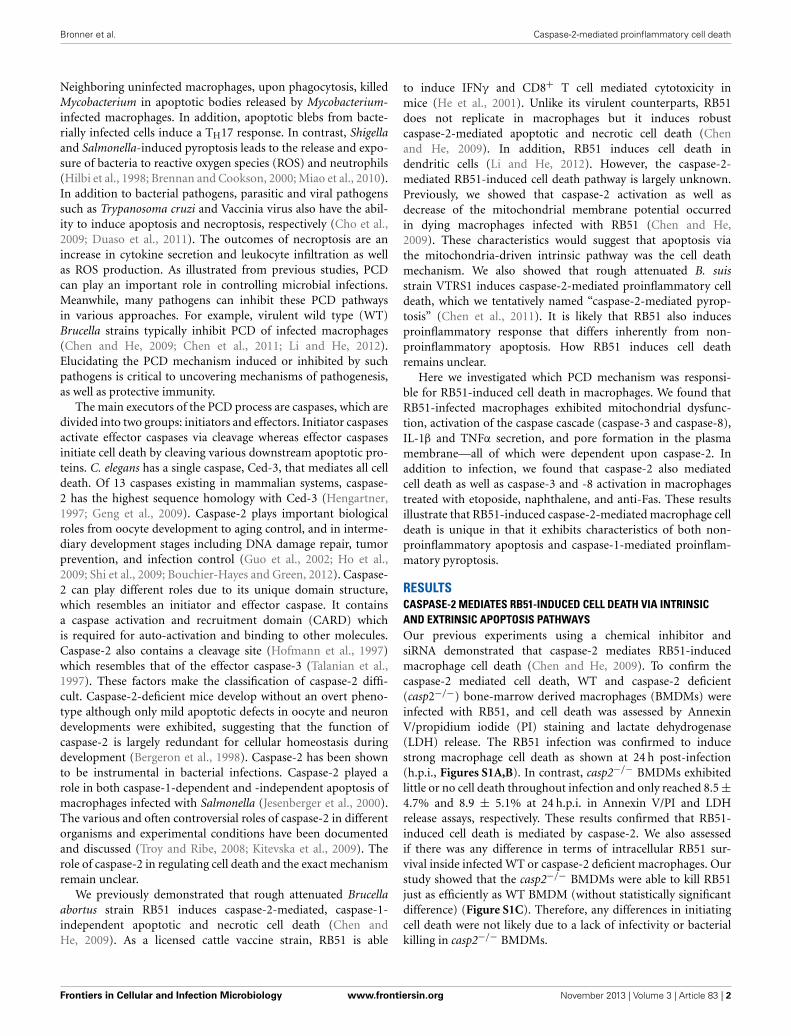
Since caspase-2 is required for RB51 induced cell death, weassessed which PCD pathway caspase-2 was mediating RB51-induced cell death. Inhibition of caspase-3 and/or caspase-8 activ-ity led to a decrease in cell death, however, it was not as significantas caspase-2 deficiency (Table 1). Inhibition of caspase-3 or -8 inRB51-infected macrophages resulted in 57.3 ± 2.1% and 65.7 ±3.7% cell death, respectively (P <0.001) (Table 1). Inhibition ofboth caspase-3 and -8 led to only 36.3 ± 2.5% cell death, suggest-ing the two types of inhibitions are likely additive but may alsobe synergistic. These data suggest that RB51-induced cell deathmay involve the apoptotic pathways associated with caspase-3 andcaspase-8.
Classically, caspase-3 and caspase-8 are linked to intrin-sic (intracellular signal driven) and extrinsic (death receptordriven) apoptotic pathways, respectively (Elmore, 2007). Bothpathways are linked to caspase-2 by mediating mitochondrialcytochrome c release and caspase-8 activation (Lin et al., 2004;Upton et al., 2008). To assess if caspase-2 regulated these path-ways during RB51 infection, we investigated caspase-3 andcaspase-8 activation in WT and casp2−/− BMDMs by mea-suring cleavage. The cleavage of both caspase-3 and caspase-8was abolished in RB51-infected casp2−/− BMDMs (Figure 1A).Inhibition of caspase-3 and -8 did not affect caspase-2 acti-vation (Figure S2). Previous studies illustrated that both theintrinsic and extrinsic cell death pathways can propagate sig-naling by inducing mitochondrial dysfunction, which eventuallyleads to cell death. Therefore, we investigated if caspase-2 medi-ated mitochondrial dysfunction in RB51-infected macrophages.Previously we observed that in RB51-infected macrophages,mitochondrial membrane potential decreased over time, suggest-ing that mitochondrial cytochrome c release (a marker of mito-chondrial dysfunction) occurs. We found that in WT BMDMs,cytochrome c release increased throughout infection, how-ever, in casp2−/− BMDMs cytochrome c release was abolished(Figure 1A).
To assess if mitochondrial dysfunction contributed to RB51-induced cell death, the cytochrome c release was blocked withcyclosporin A (CsA) in RB51-infected RAW264.7 (murine)macrophages. CsA prevents opening of the mitochondrial per-meability transition pore (MPTP), a pore responsible forthe release of mitochondrial contents such as cytochrome c(Handschumacher et al., 1984). In the presence of CsA, cell
FIGURE 1 | Caspase-2 drives both the intrinsic and extrinsic cell death
pathways. (A) caspase-3 and -8 cleavage (activation) as well ascytochrome c (cyto c) release in Live RB51-infected WT and casp2−/−BMDMs. β-actin served as a loading control. UT and ET represent untreatedand etoposide (25 μM, 6 hr treatment), respectively. Immunoblots arerepresentatives of n ≥ 3 independent experiments. (B) LDH release in LiveRB51-infected RAW264.7 macrophages in the presence of cyclosporin A(CsA; inhibitor of mitochondrial permeability transition pore, 10 μM) andAnti-TNFα (10 μ g/mL). Error bars represent mean ± SD of n ≥ 3independent experiments. ∗∗p < 0.001 and ∗∗∗p < 0.0001, Student’s t-test.n.s. = not significant. Immunoblots are representatives of n ≥ 3independent experiments.
death was significantly reduced (p < 0.0001) in RB51-infectedmacrophages (Figure 1B). Seeing that mitochondrial dysfunc-tion occurs and both caspase-3 and caspase-8 are activated,we explored if RB51-induced cell death was acting throughthe extrinsic pathway. Extrinsic or death receptor mediatedcell death can be activated by Fas ligand (FasL) and TNFα.Since live attenuated B. suis strain VTRS1 induces a proin-flammatory response (Chen et al., 2011), we investigated ifTNFα played a role in mediating RB51-induced cell death. Wetreated RAW264.7 macrophages with anti-TNFα and assessed celldeath via LDH release. Anti-TNFα treatment led to a decreasein LDH release when compared to untreated RB51-infectedRAW264.7 macrophages (Figure 1B, 85.5 ± 5.1% vs. 21.4 ±6.5%, respectively). These observations suggest that caspase-2 drives the extrinsic cell death pathway in RB51-infectedmacrophages, and the TNFα cytokine that triggers the extrin-sic cell death pathway is likely secreted from RB51-infectedmacrophages. It is also possible that the production of internal
Frontiers in Cellular and Infection Microbiology www.frontiersin.org November 2013 | Volume 3 | Article 83 | 3
Bronner et al. Caspase-2-mediated proinflammatory cell death
TNFα from a macrophage cell plays an important role of the PCDof the same macrophage cell.
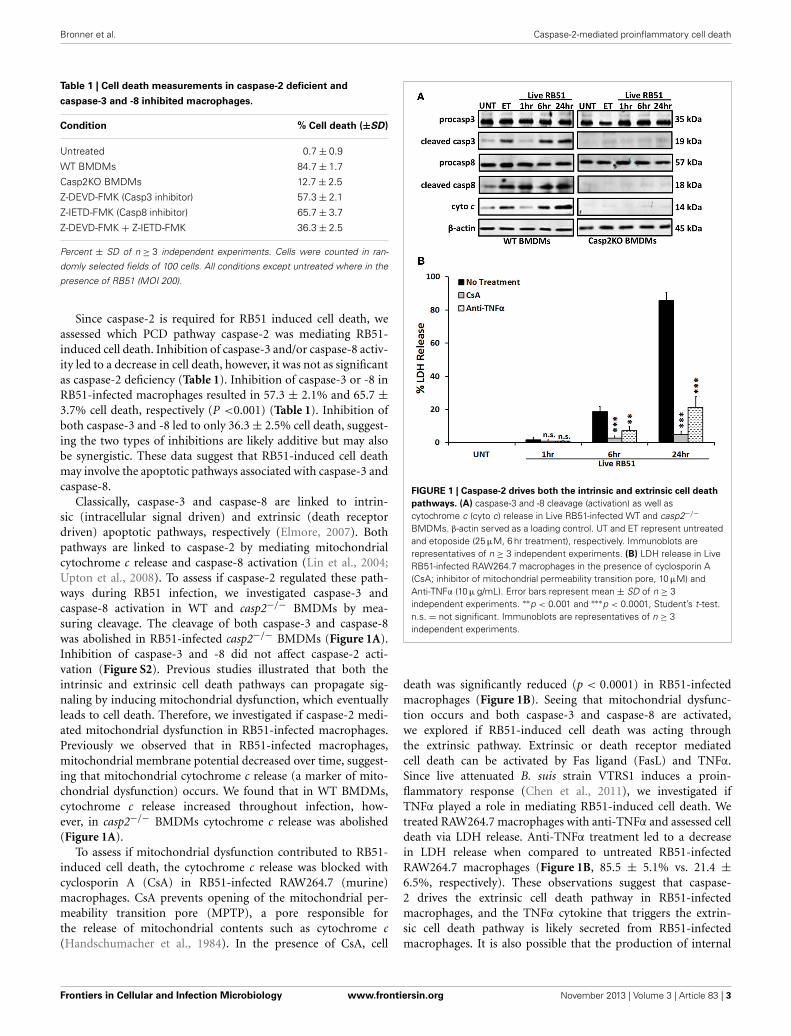
CASPASE-2 REGULATES CASPASE-1 ACTIVATION AND IL-1βPRODUCTION IN RB51-INFECTED MACROPHAGESStudies have illustrated that a proinflammatory response can bethe trigger or product of cell death (Elmore, 2007; Bergsbakenet al., 2009). In addition to trigger extrinsic apoptotic cell deathpathway, TNFα is also an important proinflammatory cytokine.Since TNFα played a role in RB51-induced cell death, we assessedif caspase-2 mediated TNFα production. Over time, TNFα levelsincreased in RB51-infected WT BMDMs. However, in casp2−/−BMDMs, TNFα was reduced to untreated levels (Figure 2A). Inaddition, CsA treatment led to a decrease in TNFα production.
Another cytokine associated with proinflammatory cell deathis IL-1β. During pyroptosis, caspase-1 processes IL-1β and aids
FIGURE 2 | Caspase-2 drives proinflammatory responses in
RB51-infected macrophages. (A) TNFα and (B) IL-1β levels in WT,casp2−/−, CsA (cyclosporin A, inhibitor of mitochondrial permeabilitytransition pore, 10 μM) and Z-WEHD-FMK (caspase-1 inhibitor, 20 μM)treated BMDMs. Error bars represent mean ± SD of n ≥ 3 independentexperiments. ‡, ∗p < 0.01 and ‡ ‡ ‡, ∗∗∗p < 0.0001, Student’s t-test. (∗, ∗∗∗)and (‡, ‡ ‡ ‡) represent comparison to untreated and WT + Live RB51,respectively. (C) caspase-1 cleavage (activation) in Live RB51-infected WTand casp2−/− BMDMs. UT represents untreated (negative control).Immunoblots are representatives of n ≥ 3 independent experiments.
in its secretion. Interestingly, caspase-2 contains a CARD domainand has been shown to mediate caspase-1 activation duringSalmonella infections (Jesenberger et al., 2000). Similar to TNFα,IL-1β levels increased above untreated levels starting at 6 h.p.i.,however, in casp2−/− and caspase-1 inhibited BMDMs, IL-1β production was abolished (Figure 2B). In the presence ofCsA, IL-1β levels were significantly reduced in RB51-infectedmacrophages. A decrease in IL-1β levels in casp2−/− BMDMssuggested that either caspase-2 regulates caspase-1 activationor caspase-2 is directly responsible for the processing of IL-1β. To evaluate these two possibilities, caspase-1 cleavage inRB51-infected WT and casp2−/− BMDMs was measured. RB51induced caspase-1 cleavage in WT BMDMs starting at 6 h.p.i.,however, cleaved caspase-1 was absent in casp2−/− BMDMs(Figure 2C). These data suggest that caspase-2, via mitochondrialdysfunction, mediates caspase-1 activation and IL-1β productionin RB51-infected macrophages.
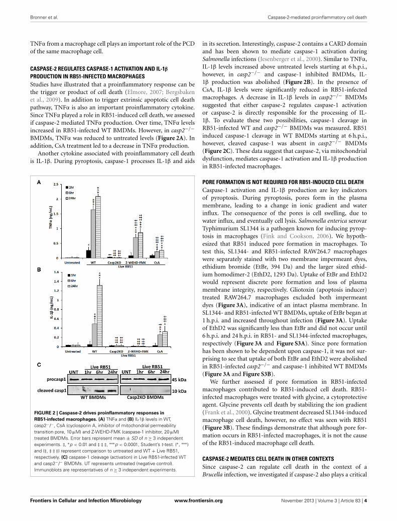
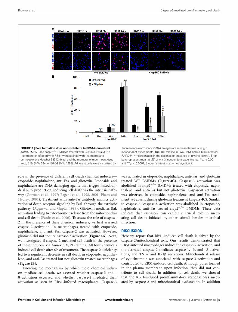
PORE FORMATION IS NOT REQUIRED FOR RB51-INDUCED CELL DEATHCaspase-1 activation and IL-1β production are key indicatorsof pyroptosis. During pyroptosis, pores form in the plasmamembrane, leading to a change in ionic gradient and waterinflux. The consequence of the pores is cell swelling, due towater influx, and eventually cell lysis. Salmonella enterica serovarTyphimurium SL1344 is a pathogen known for inducing pyrop-tosis in macrophages (Fink and Cookson, 2006). We hypoth-esized that RB51 induced pore formation in macrophages. Totest this, SL1344- and RB51-infected RAW264.7 macrophageswere separately stained with two membrane impermeant dyes,ethidium bromide (EtBr, 394 Da) and the larger sized ethid-ium homodimer-2 (EthD2, 1293 Da). Uptake of EtBr and EthD2would represent discrete pore formation and loss of plasmamembrane integrity, respectively. Gliotoxin (apoptosis inducer)treated RAW264.7 macrophages excluded both impermeantdyes (Figure 3A), indicative of an intact plasma membrane. InSL1344- and RB51-infected WT BMDMs, uptake of EtBr began at1 h.p.i. and increased throughout infection (Figure 3A). Uptakeof EthD2 was significantly less than EtBr and did not occur until6 h.p.i. and 24 h.p.i. in RB51- and SL1344-infected macrophages,respectively (Figure 3A and Figure S3A). Since pore formationhas been shown to be dependent upon caspase-1, it was not sur-prising to see that uptake of both EtBr and EthD2 were abolishedin RB51-infected casp2−/− and caspase-1 inhibited WT BMDMs(Figure 3A and Figure S3B).
We further assessed if pore formation in RB51-infectedmacrophages contributed to RB51-induced cell death. RB51-infected macrophages were treated with glycine, a cytoprotectiveagent. Glycine prevents cell death by stabilizing the ion gradient(Frank et al., 2000). Glycine treatment decreased SL1344-inducedmacrophage cell death, however, no effect was seen with RB51(Figure 3B). These findings demonstrate that although pore for-mation occurs in RB51-infected macrophages, it is not the causeof the RB51-induced macrophage cell death.
CASPASE-2 MEDIATES CELL DEATH IN OTHER CONTEXTSSince caspase-2 can regulate cell death in the context of aBrucella infection, we investigated if caspase-2 also plays a critical
Frontiers in Cellular and Infection Microbiology www.frontiersin.org November 2013 | Volume 3 | Article 83 | 4
Bronner et al. Caspase-2-mediated proinflammatory cell death
FIGURE 3 | Pore formation does not contribute to RB51-induced cell
death. (A) WT and casp2−/− BMDMs treated with Gliotoxin (10 μM, 6 htreatment) or infected with RB51 were stained with the membranepermeable dye Hoechst 33342 (blue) and the membrane impermeant dyes(red), EtBr (MW 394) or EthD2 (MW 1293). Adherent cells were visualized by
fluorescence microscopy (100x). Images are representatives of n ≥ 3independent experiments. (B) LDH release in Live RB51 and SL1344-infectedRAW264.7 macrophages in the absence or presence of glycine (5 mM). Errorbars represent mean ± SD of n ≥ 3 independent experiments. ∗∗p < 0.001and ∗∗∗p < 0.0001, Student’s t-test. n.s. = not significant.
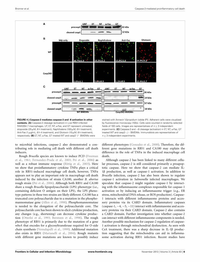
role in the presence of different cell death chemical inducers—etoposide, naphthalene, anti-Fas, and gliotoxin. Etoposide andnaphthalene are DNA damaging agents that trigger mitochon-drial ROS production, inducing cell death via the intrinsic path-way (Gorman et al., 1997; Bagchi et al., 1998, 2001; Pham andHedley, 2001). Treatment with anti-Fas antibody mimics acti-vation of death receptor signaling by FasL through the extrinsicpathway. (Aggarwal and Gupta, 1999). Gliotoxin mediates Bakactivation leading to cytochrome c release from the mitochondriaand cell death (Pardo et al., 2006). To assess the role of caspase-2 in the presence of these chemical inducers, we first assessedcaspase-2 activation. In macrophages treated with etoposide,naphthalene, and anti-Fas, caspase-2 was activated. However,gliotoxin did not induce caspase-2 activation (Figure 4A). Next,we investigated if caspase-2 mediated cell death in the presenceof these inducers via Annexin V/PI staining. All four chemicalsinduced cell death after 6 h of treatment. The caspase-2 deficiencyled to a significant decrease in cell death in etoposide, naphtha-lene, and anti-Fas treated but not gliotoxin treated macrophages(Figure 4B).
Knowing the mechanism by which these chemical induc-ers mediate cell death, we assessed whether caspase-3 and -8 activation occurred and whether caspase-2 mediated theiractivation as seen in RB51-infected macrophages. Caspase-3
was activated in etoposide, naphthalene, anti-Fas, and gliotoxintreated WT BMDMs (Figure 4C). Caspase-3 activation wasabolished in casp2−/− BMDMs treated with etoposide, naph-thalene, and anti-Fas but not gliotoxin. Caspase-8 activationwas observed in etoposide, naphthalene, and anti-Fas treat-ment yet absent during gliotoxin treatment (Figure 4C). Similarto caspase-3, caspase-8 activation was abolished in etoposide,naphthalene, anti-Fas treated casp2−/− BMDMs. These dataindicate that caspase-2 can exhibit a crucial role in medi-ating cell death initiated by other stimuli besides microbialinfection.
DISCUSSIONHere we report that RB51-induced cell death is driven by thecaspase-2/mitochondrial axis. Our results demonstrated thatRB51-infected macrophages induce the caspase-2 activation, andthe activated caspase-2 mediates caspase-1, -3, and -8 activa-tions, and TNFα and IL-1β secretions. Mitochondrial releaseof cytochrome c was associated with caspase-3 activation andcontributed to RB51-induced cell death. Although pores formedin the plasma membrane upon infection, they did not con-tribute to cell death. In addition to cell death, we showedthat the RB51-induced proinflammatory response was medi-ated by caspase-2 and mitochondrial dysfunction. In addition
Frontiers in Cellular and Infection Microbiology www.frontiersin.org November 2013 | Volume 3 | Article 83 | 5
Bronner et al. Caspase-2-mediated proinflammatory cell death
FIGURE 4 | Caspase-2 mediates caspase-3 and -8 activation in other
contexts. (A) Caspase-2 cleavage (activation) in Live RB51-infectedRAW264.7 macrophages. UT, ET, NT, α-Fas, and GT represent untreated,etoposide (25 μM, 6 h treatment), Naphthalene (100 μM, 6 h treatment),Anti-Fas (1 μg/mL, 6 h 4 treatment), and Gliotoxin (10 μM, 6 h treatment),respectively. (B) ET, NT, α-Fas, GT treated WT and casp2−/− BMDMs were
stained with Annexin V/propidium iodide (PI). Adherent cells were visualizedby fluorescence microscopy (100x). Cells were counted in randomly selectedfields of 100 cells. Images are representatives of n ≥ 3 independentexperiments. (C) Caspase-3 and −8 cleavage (activation) in ET, NT, α-Fas, GTtreated WT and casp2−/− BMDMs. Immunoblots are representatives ofn ≥ 3 independent experiments.
to microbial infections, caspase-2 also demonstrated a con-tributing role in mediating cell death with different cell deathinducers.
Rough Brucella species are known to induce PCD (Freemanet al., 1961; Fernandez-Prada et al., 2003; Pei et al., 2006) aswell as a robust immune response (Rittig et al., 2003). Herewe show that proinflammatory cytokine TNFα plays a criticalrole in RB51-induced macrophage cell death, however, TNFα
appears not to play an important role in macrophage cell deathinduced by the infection of strain CA180, another B. abortusrough strain (Pei et al., 2006). Although both RB51 and CA180share a rough Brucella lipopolysaccharide (LPS) phenotype (i.e.,containing deficient O antigen on their LPS), the LPS pheno-type patterns in these two strains are likely different. CA180 has atruncated core polysaccharide due to a mutation in the phospho-mannomutase gene (Allen et al., 1998). Phosphomannomutaseis needed to the elongation of the polysaccharide core. Thepolysaccharide core has been shown to elicit cytokine production,any changes (e.g., shortening) can decrease cytokine produc-tion (Otterlei et al., 1993; Berntzen et al., 1998). The roughphenotype of RB51 is primarily due to the mutation of a genewboA that encodes for a glycosyltransferase required for O-sidechain sysnthesis (Vemulapalli et al., 1999). Additional mutationalso exists in RB51 (Marianelli et al., 2004). Rough mutantswith different gene mutations are known to possibly induce
different phenotypes (Gonzalez et al., 2008). Therefore, the dif-ferent gene mutations in RB51 and CA180 may explain thedifference in the role of TNFα in the induced macrophage celldeath.
Although caspase-2 has been linked to many different cellu-lar processes, caspase-2 is still considered primarily a proapop-totic caspase. Here we show that caspase-2 can mediate IL-1β production, as well as caspase-1 activation. In addition toBrucella infection, caspase-2 has also been shown to regulatecaspase-1 activation in Salmonella infected macrophages. Wespeculate that caspase-2 might regulate caspase-1 by interact-ing with the inflammasome complexes responsible for caspase-1activation or by inducing an inflammasome trigger (e.g., ERstress, mitochondrial DNA release, or ROS production). Caspase-1 interacts with different inflammasome proteins and acces-sory proteins via its CARD domain. Inflammatory caspases(caspase-1, −4, −5, −11) interact with inflammasome and acces-sory proteins via their CARD domain. Caspase-2 also containsa CARD domain. Further investigation into whether caspase-2can interact with different inflammasome components is needed.Another possible mechanism for caspase-2 regulation of caspase-1 activation is through mitochondrial dysfunction. As seen withCsA treatment, there was a sharp decrease in IL-1β produc-tion suggesting that the mitochondria can aid in inflamma-some activation during RB51 infection. Recent studies have
Frontiers in Cellular and Infection Microbiology www.frontiersin.org November 2013 | Volume 3 | Article 83 | 6
Bronner et al. Caspase-2-mediated proinflammatory cell death
linked NLRP3 and AIM2 activation to mitochondrial dysfunc-tion (Nakahira et al., 2011; Misawa et al., 2013; Subramanianet al., 2013). It is possible that caspase-2-induced mitochon-drial dysfunction leads to release of mitochondrial dangerassociated molecular patterns (DAMPs). Mitochondrial DAMPs(e.g., mtDNA or cardiolipin exposure) can lead to NLRP3 andAIM2 inflammasome activation (Nakahira et al., 2011). Caspase-2 has been linked to mediating mitochondrial dysfunction—caspase-2 cleaves Bid leading to mitochondrial outer membranepore formation and eventually release of mitochondrial con-tent (e.g., cytochrome c). Whether caspase-2-mediated releaseof mitochondrial DAMPs is the mechanism by which theinflammasome and caspase-1 activation occurs remains to beelucidated.
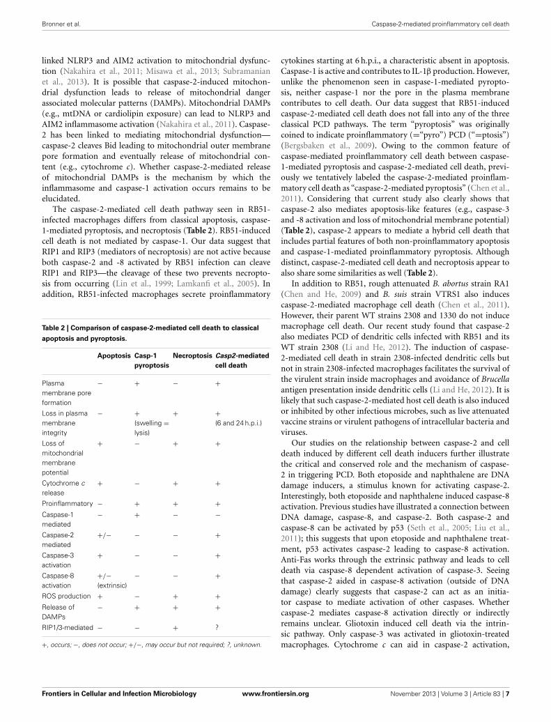
The caspase-2-mediated cell death pathway seen in RB51-infected macrophages differs from classical apoptosis, caspase-1-mediated pyroptosis, and necroptosis (Table 2). RB51-inducedcell death is not mediated by caspase-1. Our data suggest thatRIP1 and RIP3 (mediators of necroptosis) are not active becauseboth caspase-2 and -8 activated by RB51 infection can cleaveRIP1 and RIP3—the cleavage of these two prevents necropto-sis from occurring (Lin et al., 1999; Lamkanfi et al., 2005). Inaddition, RB51-infected macrophages secrete proinflammatory
Table 2 | Comparison of caspase-2-mediated cell death to classical
apoptosis and pyroptosis.
Apoptosis Casp-1
pyroptosis
Necroptosis Casp2-mediated
cell death
Plasmamembrane poreformation
− + − +
Loss in plasmamembraneintegrity
− +(swelling =lysis)
+ +(6 and 24 h.p.i.)
Loss ofmitochondrialmembranepotential
+ − + +
Cytochrome crelease
+ − + +
Proinflammatory − + + +Caspase-1mediated
− + − −
Caspase-2mediated
+/− − − +
Caspase-3activation
+ − − +
Caspase-8activation
+/−(extrinsic)
− − +
ROS production + − + +Release ofDAMPs
− + + +
RIP1/3-mediated − − + ?
+, occurs; −, does not occur; +/−, may occur but not required; ?, unknown.
cytokines starting at 6 h.p.i., a characteristic absent in apoptosis.Caspase-1 is active and contributes to IL-1β production. However,unlike the phenomenon seen in caspase-1-mediated pyropto-sis, neither caspase-1 nor the pore in the plasma membranecontributes to cell death. Our data suggest that RB51-inducedcaspase-2-mediated cell death does not fall into any of the threeclassical PCD pathways. The term “pyroptosis” was originallycoined to indicate proinflammatory (=“pyro”) PCD (“=ptosis”)(Bergsbaken et al., 2009). Owing to the common feature ofcaspase-mediated proinflammatory cell death between caspase-1-mediated pyroptosis and caspase-2-mediated cell death, previ-ously we tentatively labeled the caspase-2-mediated proinflam-matory cell death as “caspase-2-mediated pyroptosis” (Chen et al.,2011). Considering that current study also clearly shows thatcaspase-2 also mediates apoptosis-like features (e.g., caspase-3and -8 activation and loss of mitochondrial membrane potential)(Table 2), caspase-2 appears to mediate a hybrid cell death thatincludes partial features of both non-proinflammatory apoptosisand caspase-1-mediated proinflammatory pyroptosis. Althoughdistinct, caspase-2-mediated cell death and necroptosis appear toalso share some similarities as well (Table 2).
In addition to RB51, rough attenuated B. abortus strain RA1(Chen and He, 2009) and B. suis strain VTRS1 also inducescaspase-2-mediated macrophage cell death (Chen et al., 2011).However, their parent WT strains 2308 and 1330 do not inducemacrophage cell death. Our recent study found that caspase-2also mediates PCD of dendritic cells infected with RB51 and itsWT strain 2308 (Li and He, 2012). The induction of caspase-2-mediated cell death in strain 2308-infected dendritic cells butnot in strain 2308-infected macrophages facilitates the survival ofthe virulent strain inside macrophages and avoidance of Brucellaantigen presentation inside dendritic cells (Li and He, 2012). It islikely that such caspase-2-mediated host cell death is also inducedor inhibited by other infectious microbes, such as live attenuatedvaccine strains or virulent pathogens of intracellular bacteria andviruses.
Our studies on the relationship between caspase-2 and celldeath induced by different cell death inducers further illustratethe critical and conserved role and the mechanism of caspase-2 in triggering PCD. Both etoposide and naphthalene are DNAdamage inducers, a stimulus known for activating caspase-2.Interestingly, both etoposide and naphthalene induced caspase-8activation. Previous studies have illustrated a connection betweenDNA damage, caspase-8, and caspase-2. Both caspase-2 andcaspase-8 can be activated by p53 (Seth et al., 2005; Liu et al.,2011); this suggests that upon etoposide and naphthalene treat-ment, p53 activates caspase-2 leading to caspase-8 activation.Anti-Fas works through the extrinsic pathway and leads to celldeath via caspase-8 dependent activation of caspase-3. Seeingthat caspase-2 aided in caspase-8 activation (outside of DNAdamage) clearly suggests that caspase-2 can act as an initia-tor caspase to mediate activation of other caspases. Whethercaspase-2 mediates caspase-8 activation directly or indirectlyremains unclear. Gliotoxin induced cell death via the intrin-sic pathway. Only caspase-3 was activated in gliotoxin-treatedmacrophages. Cytochrome c can aid in caspase-2 activation,
Frontiers in Cellular and Infection Microbiology www.frontiersin.org November 2013 | Volume 3 | Article 83 | 7
Bronner et al. Caspase-2-mediated proinflammatory cell death
however, (Slee et al., 1999), in the context of gliotoxin intox-ication this was not the case because caspase-2 activation didnot occur. Gliotoxin acts on Bak and its downstream tar-get t-Bid [a known target cleaved by caspase-2, (Upton et al.,2008)]; therefore, it is possible that gliotoxin short-circuitsthe classical intrinsic pathway and does not require signalingcomponents upstream of the mitochondria. In the case of etopo-side and naphthalene, the data suggest that caspase-2 regulatescaspase-3 activation via the mitochondria. Caspase-3 is acti-vated via the apoptosome, a multiprotein complex dependentupon cytochrome c release. Caspase-2 was previously reportedto act upstream of caspase-8 during ceramide-induced mito-chondrial apoptosis in T cells (Lin et al., 2004). It appearsthat the caspase-2 regulation of caspase-3 and -8 can occurin different cell types with different treatments, so this typeof regulation is neither cell specific nor context specific. Theseobservations suggest that caspase-2 can play a critical role ininitiating PCD.
The caspase-2-mediated cell death pathway is likely criticalto microbial pathogenesis and host immunity. In the contextof RB51, cell death and the proinflammatory response mayhave synergistic effects on host immune responses. Cell deathmay result in the exposure of RB51 to a more hostile extra-cellular environment (as seen in pyroptosis and necroptosis).In addition, neighboring macrophages and dendritic cells mayrecognize processed RB51 antigens leading to cross priming ofCD8+ T cells (important for RB51-induced protective immu-nity). We recently showed that RB51 induced cell death inbone marrow derived dendritic cells and aided in maturation aswell as priming of T cells—all of which were dependent uponcaspase-2 (Li and He, 2012). These observations demonstrate theimportance for caspase-2, as well as cell death, in initiating theimmune response. Utilizing this PCD pathway may ensure thatthe host triggers a potent immune response. Prevention of thispathway may aid in enhancing survival of virulent Brucella inmacrophages. Our previous work suggested prevention—virulentstrain B. abortus S2308 did not induce caspase-2 activation norcytochrome c release in infected macrophages (Chen and He,2009).
Caspase-2 is also implicated in other processes (cancer reg-ulation and metabolism) and may take on a regulatory rolein these processes as well. We have made an original obser-vation that caspase-2 plays a non-redundant role in triggeringthe proinflammatory cell death of RB51-infected macrophagesand in macrophages treated with various drugs. After the evolu-tion of complex caspase-cascade cell death signaling pathways inadvanced animals, it is suggestive that the protein functions of thehighly conserved caspase-2 have been preserved during evolutionand serve as safeguards to regulate various cell death pathways.It is likely that intracellular pathogens with similar lifestyles toBrucella (e.g., Salmonella, Mycobacterium, Listeria, Francisella,and Legionella) may utilize caspase-2 during infection. Furtherunderstanding of caspase-2-mediated pyroptosis can aid in sup-plying a blueprint for effective brucellosis vaccines (both animalsand humans) as well as effective therapeutics against cancers andother diseases.
MATERIALS AND METHODSMICEThe caspase-2 knockout (Casp2KO) mice were originally gener-ated by Junying Yuan and kindly provided by Dr. Brian Hermanof the University of Texas Health Science Center at San Antoniowith Dr. Yuan’s consent (Bergeron et al., 1998). The deletioninactivates both the long and short form of caspase-2. The micewere backcrossed with C57BL/6 once in the Unit for LaboratoryAnimal Medicine at the University of Michigan Medical School,and then used as founders. Casp2KO and WT C57BL/6 (Jackson)mice with similar ages were applied in the experiments. TheUniversity Committee on Use and Care of Animals (UCUCA) atthe University of Michigan approved the protocol (#09695) to usemice for studies described here.
BACTERIAL STRAINS AND REAGENTSRAW264.7 macrophages and bone marrow derived macrophages(BMDMs) were infected with Brucella abortus strain RB51(from Dr. G. Schurig, Virginia Polytechnic Institute and StateUniversity) and Salmonella typhimurium SL1344 (from MaryO’Riordan, University of Michigan). The following inhibitorsand inducers were used: CsA (Sigma-Aldrich), etoposide (Sigma-Aldrich), naphthalene (Sigma-Aldrich), Anti-Fas (BioVision),gliotoxin (-Aldrich), glycine (Sigma-Aldrich), Z-WEHD-FMK(Caspase-1 inhibitor, R&D Systems), Z-DEVD-FMK (Caspase-3 inhibitor, R&D Systems), Z-IETD-FMK (Caspase-8 inhibitor,R&D Systems), and anti-TNFα (mouse specific, BioVision).
The following antibodies were used: anti-cytochrome c(cat#:4272S, Cell Signaling), anti-caspase-3 (cat#: 9662S, CellSignaling), anti-caspase-8 (cat#: 4927S, Cell Signaling), anti-caspase-2 (cat#: 3027-100, BioVision), anti-caspase-1 (cat#: sc-514, Santa Cruz), and anti-actin (cat#: MS1295P1, ThermoScientific).
CELL CULTURE AND INFECTIONBMDMs were isolated from WT and casp2−/− mice on aC57BL/6 background. Isolated BMDMs were differentiated inDMEM (GIBCO) supplemented with 20% heat-inactivated FBS(GIBCO), 1% L-glutamine (200 mM), 1% sodium pyruvate(100 mM), 0.1% β-mercaptoethanol (55 mM), and 30% L-929fibroblast conditioned medium. BMDMs were cultured in non-TC treated plates, fed fresh media on Day 3, and harvested onDay 6. BMDMs were maintained at 37◦C under 5% CO2.
Four million RAW264.7 macrophages and BMDMs wereseeded in 6 well plates 18 h prior to infection. The followingday, where indicated cells were pretreated with CsA (10 μM), Z-WEHD-FMK (20 μM), Z-DEVD-FMK (20 μM), Z-IETD-FMK(20 μM) and Anti-TNFα (10 μ g/mL) for 1 h prior to infection.Untreated and pretreated cells were infected with RB51 (MOI200) or SL1344 (MOI 25) for 30 min, after which the inoculumwas removed and cells were washed with PBS. Medium con-taining 50 μ g/ml of gentamicin was added to kill extracellularbacteria. To synchronize infection, cells were spun at 1200 rpmfor 3 min after adding inoculum. Cells were treated with etopo-side (25 μM), naphthalene (100 μM), anti-Fas (1 μ g/mL), andgliotoxin (10 μM) for 6 hr. At the indicated times, cells were lysed
Frontiers in Cellular and Infection Microbiology www.frontiersin.org November 2013 | Volume 3 | Article 83 | 8
Bronner et al. Caspase-2-mediated proinflammatory cell death
in buffer containing 1% NP-40 on ice for 15 min and spun at13,000 rpm for 15 min to pellet the insoluble fraction. Solublefractions were used for immunoblot assays.
IMMUNOBLOT ASSAYCytosolic extracts collected at various time points (1, 6, and24 h pi) were separated by SDS-PAGE, transferred to PVDFmembranes (Millipore), blocked with 5% milk in TBS-Tween20(TBS-T), and incubated overnight at 4◦C with primary anti-bodies stated above. Membranes were washed with TBS-T andincubated with secondary HRP conjugated to either goat anti-rabbit IgG (cat#: 12–348, Millipore) or goat anti-mouse IgG (cat#:1034-05, Southern Biotech) at room temperature for 1 h. Bandswere visualized using the ECL Western Blotting Substrate Kit(Pierce). Immunoblots in the figures are representative of n ≥ 3independent experiments.
CYTOKINE DETECTIONCulture supernatants were collected at different time points (1,6, and 24 h pi) from macrophages infected as described above.IL-1β and TNFα levels were determined by sandwich enzyme-linked immunosorbent assay (ELISA) according to manufac-turer’s instructions (BioLegend). A minimum of three technicalreplicates per experiment and three experimental replicates wereanalyzed for each condition.
CELL DEATH ASSAYRAW264.7 macrophages were seeded in 6 well plates at a con-centration 9.6 × 10⁴ per well and infected with RB51 as statedabove. Cells were stained with Annexin V and PI using theAnnexin V-FLUOS staining kit (Roche Diagnostics Corporation).Cells were washed with PBS and incubated with the fluorescentdyes for 15 min in the dark at room temperature. Fluorescencewas observed with a Nikon TK-2000-S microscope and pho-tographed with a RT Slide Spot digital camera and QCapturePro software. Uninfected macrophages served as negativecontrols.
ETHIDIUM BROMIDE (EtBr) AND ETHIDIUM HOMODIMER-2 (EthD2)STAININGMacrophages were grown in 6-well plates and infected asdescribed above. At different time points post-infection, cellswere washed with PBS (GIBCO) and stained with Hoechst 33342(5 μ g/mL) and either EtBr or EthD2 (25 μ g/mL) according to themanufacturer’s instructions. Cell were analyzed with a Nikon TK-2000-s microscope and photographed with a RT slide spot digitalcamera and QCapture Pro Software.
LACTATE DEHYDROGENASE (LDH) RELEASE ASSAYMacrophages were seeded in 96-well plates and infected withRB51 or SL1344 as stated above. Supernatants were ana-lyzed for the presence of LDH enzyme using the CytoTox-ONE™ Homogeneous Membrane Integrity Assay (Promega)as directed by the manufacturer’s instructions. Percentageof LDH release was calculated as 100 × [(ExperimentalLDH Release—Culture Medium Background)/(Maximum LDHRelease—Culture Medium Background)].
STATISTICAL ANALYSISAll p-values were generated between identified samples usingunpaired two-tailed Student’s t-tests and represent analysisof ≥3 replicates per condition. ‡, ∗p < 0.01 ‡ ‡, ∗p < 0.001and ‡ ‡ ‡, ∗∗∗p < 0.0001.
AUTHOR CONTRIBUTIONSDenise N. Bronner and Yongqun He designed the experi-ments. Denise N. Bronner performed the experiments. DeniseN. Bronner, Mary X. D. O’Riordan and Yongqun He ana-lyzed the data. Yongqun He and Mary X. D. O’Riordan con-tributed reagents and materials. Denise N. Bronner and YongqunHe wrote the paper. All authors edited and approved themanuscript.
ACKNOWLEDGMENTSDr. Xinna Li prepared bone marrow derived macrophages for thisstudy. We thank Drs. Michele S. Swanson, Victor J. DiRita, andGeorge W. Jourdian for their discussions. We acknowledge finan-cial support by the Rackham Spring/Summer Research grant (toYongqun He), Rackham Merit Fellowship (to Denise N. Bronner),startup and research bridging funds to Yongqun He from the Unitfor Laboratory Animal Medicine (ULAM) and the Endowmentfor Basic Science (EBS) in the University of Michigan MedicalSchool, and NIH R21AI101777 (MOR). The publication fee waspaid by the ULAM director (Dr. Robert Dysko)’s discretionaryfunding.
SUPPLEMENTARY METERIALThe Supplementary material for this article can befound online at: http://www.frontiersin.org/journal/10.3389/fcimb.2013.00083/abstract
Bronner et al. Caspase-2-mediated proinflammatory cell death
REFERENCESAggarwal, S., and Gupta, S. (1999). Increased activity of caspase 3 and
caspase 8 in anti-Fas-induced apoptosis in lymphocytes from ageinghumans. Clin. Exp. Immunol. 117, 285–290. doi: 10.1046/j.1365-2249.1999.00957.x
Allen, C. A., Adams, L. G., and Ficht, T. A. (1998). Transposon-derived Brucellaabortus rough mutants are attenuated and exhibit reduced intracellular survival.Infect. Immun. 66, 1008–1016.
Bagchi, M., Bagchi, D., Balmoori, J., Ye, X., and Stohs, S. J. (1998). Naphthalene-induced oxidative stress and DNA damage in cultured macrophage J774A.1cells. Free Radic. Biol. Med. 25, 137–143. doi: 10.1016/S0891-5849(98)00063-X
Bagchi, M., Balmoori, J., Ye, X., Bagchi, D., Ray, S. D., and Stohs, S. J. (2001).Protective effect of melatonin on naphthalene-induced oxidative stress andDNA damage in cultured macrophage J774A.1 cells. Mol. Cell. Biochem. 221,49–55. doi: 10.1023/A:1010946517651
Bergeron, L., Perez, G. I., Macdonald, G., Shi, L., Sun, Y., Jurisicova, A., et al. (1998).Defects in regulation of apoptosis in caspase-2-deficient mice. Genes Dev. 12,1304–1314. doi: 10.1101/gad.12.9.1304
Bergsbaken, T., Fink, S. L., and Cookson, B. T. (2009). Pyroptosis: host cell deathand inflammation. Nat. Rev. Microbiol. 7, 99–109. doi: 10.1038/nrmicro2070
Berntzen, G., Flo, T. H., Medvedev, A., Kilaas, L., Skjak-Braek, G., Sundan, A., et al.(1998). The tumor necrosis factor-inducing potency of lipopolysaccharide anduronic acid polymers is increased when they are covalently linked to particles.Clin. Diagn. Lab. Immunol. 5, 355–361.
Bouchier-Hayes, L., and Green, D. R. (2012). Caspase-2: the orphan caspase. CellDeath Differ. 19, 51–57. doi: 10.1038/cdd.2011.157
Brennan, M. A., and Cookson, B. T. (2000). Salmonella induces macrophagedeath by caspase-1-dependent necrosis. Mol. Microbiol. 38, 31–40. doi:10.1046/j.1365-2958.2000.02103.x
Chen, F., Ding, X., Ding, Y., Xiang, Z., Li, X., Ghosh, D., et al. (2011).Proinflammatory caspase-2-mediated macrophage cell death induced by arough attenuated Brucella suis strain. Infect. Immun. 79, 2460–2469. doi:10.1128/IAI.00050-11
Chen, F., and He, Y. (2009). Caspase-2 mediated apoptotic and necrotic murinemacrophage cell death induced by rough Brucella abortus. PLoS ONE 4:e6830.doi: 10.1371/journal.pone.0006830
Cho, Y. S., Challa, S., Moquin, D., Genga, R., Ray, T. D., Guildford, M., et al.(2009). Phosphorylation-driven assembly of the RIP1-RIP3 complex regulatesprogrammed necrosis and virus-induced inflammation. Cell 137, 1112–1123.doi: 10.1016/j.cell.2009.05.037
Duaso, J., Rojo, G., Jana, F., Galanti, N., Cabrera, G., Bosco, C., et al. (2011).Trypanosoma cruzi induces apoptosis in ex vivo infected human chorionic villi.Placenta 32, 356–361. doi: 10.1016/j.placenta.2011.02.005
Elmore, S. (2007). Apoptosis: a review of programmed cell death. Toxicol. Pathol.35, 495–516. doi: 10.1080/01926230701320337
Fernandez-Prada, C. M., Zelazowska, E. B., Nikolich, M., Hadfield, T. L., Roop, R.M. 2nd., Robertson, G. L. et al. (2003). Interactions between Brucella melitensisand human phagocytes: bacterial surface O-Polysaccharide inhibits phagocy-tosis, bacterial killing, and subsequent host cell apoptosis. Infect. Immun. 71,2110–2119. doi: 10.1128/IAI.71.4.2110-2119.2003
Fink, S. L., and Cookson, B. T. (2006). Caspase-1-dependent pore formation duringpyroptosis leads to osmotic lysis of infected host macrophages. Cell. Microbiol.8, 1812–1825. doi: 10.1111/j.1462-5822.2006.00751.x
Frank, A., Rauen, U., and de Groot, H. (2000). Protection by glycine against hypoxicinjury of rat hepatocytes: inhibition of ion fluxes through nonspecific leaks.J. Hepatol. 32, 58–66. doi: 10.1016/S0168-8278(00)80190-7
Fratazzi, C., Arbeit, R. D., Carini, C., Balcewicz-Sablinska, M. K., Keane, J.,Kornfeld, H., et al. (1999). Macrophage apoptosis in mycobacterial infections.J. Leukoc. Biol. 66, 763–764.
Freeman, B. A., Kross, D. J., and Circo, R. (1961). Host-parasite relationshipsin brucellosis. II. Destruction of macrophage cultures by Brucella of differentvirulence. J. Infect. Dis. 108, 333–338. doi: 10.1093/infdis/108.3.333
Galluzzi, L., Vitale, I., Abrams, J. M., Alnemri, E. S., Baehrecke, E. H., Blagosklonny,M. V., et al. (2012). Molecular definitions of cell death subroutines: recommen-dations of the Nomenclature Committee on Cell Death 2012. Cell Death Differ.19, 107–120. doi: 10.1038/cdd.2011.96
Gao, L., and Abu Kwaik, Y. (2000). Hijacking of apoptotic pathwaysby bacterialpathogens. Microbes Infect. 2, 1705–1719. doi: 10.1016/S1286-4579(00)01326-5
Geng, X., Zhou, Q. H., Kage-Nakadai, E., Shi, Y., Yan, N., Mitani, S., et al.(2009). Caenorhabditis elegans caspase homolog CSP-2 inhibits CED-3 autoac-tivation and apoptosis in germ cells. Cell Death Differ. 16, 1385–1394. doi:10.1038/cdd.2009.88
Gonzalez, D., Grillo, M. J., De Miguel, M. J., Ali, T., Arce-Gorvel, V., Delrue, R. M.,et al. (2008). Brucellosis vaccines: assessment of Brucella melitensis lipopolysac-charide rough mutants defective in core and O-polysaccharide synthesis andexport. PLoS ONE 3:e2760. doi: 10.1371/journal.pone.0002760
Gorman, A., McGowan, A., and Cotter, T. G. (1997). Role of peroxide andsuperoxide anion during tumour cell apoptosis. FEBS Lett. 404, 27–33. doi:10.1016/S0014-5793(97)00069-0
Guo, Y., Srinivasula, S. M., Druilhe, A., Fernandes-Alnemri, T., and Alnemri, E.S. (2002). Caspase-2 induces apoptosis by releasing proapoptotic proteins frommitochondria. J. Biol. Chem. 277, 13430–13437. doi: 10.1074/jbc.M108029200
Handschumacher, R. E., Harding, M. W., Rice, J., Drugge, R. J., and Speicher, D.W. (1984). Cyclophilin: a specific cytosolic binding protein for cyclosporin A.Science 226, 544–547. doi: 10.1126/science.6238408
He, Y., Vemulapalli, R., Zeytun, A., and Schurig, G. G. (2001). Induction of specificcytotoxic lymphocytes in mice vaccinated with Brucella abortus RB51. Infect.Immun. 69, 5502–5508. doi: 10.1128/IAI.69.9.5502-5508.2001
Hengartner, M. O. (1997). “Cell Death,” in C. elegans, 2nd Edn. New York, NY: ColdSpring Harbor Laboratory.
Hilbi, H., Moss, J. E., Hersh, D., Chen, Y., Arondel, J., Banerjee, S., et al. (1998).Shigella-induced apoptosis is dependent on caspase-1 which binds to IpaB.J. Biol. Chem. 273, 32895–32900. doi: 10.1074/jbc.273.49.32895
Ho, L. H., Taylor, R., Dorstyn, L., Cakouros, D., Bouillet, P., and Kumar, S. (2009).A tumor suppressor function for caspase-2. Proc. Natl. Acad. Sci. U.S.A. 106,5336–5341. doi: 10.1073/pnas.0811928106
Hofmann, K., Bucher, P., and Tschopp, J. (1997). The CARD domain: a new apop-totic signalling motif. Trends Biochem. Sci. 22, 155–156. doi: 10.1016/S0968-0004(97)01043-8
Jesenberger, V., Procyk, K. J., Yuan, J., Reipert, S., and Baccarini, M. (2000).Salmonella-induced caspase-2 activation in macrophages: a novel mecha-nism in pathogen-mediated apoptosis. J. Exp. Med. 192, 1035–1046. doi:10.1084/jem.192.7.1035
Kitevska, T., Spencer, D. M., and Hawkins, C. J. (2009). Caspase-2: controversialkiller or checkpoint controller? Apoptosis 14, 829–848. doi: 10.1007/s10495-009-0365-3
Lamkanfi, M., D’Hondt, K., Vande Walle, L., van Gurp, M., Denecker, G.,Demeulemeester, J., et al. (2005). A novel caspase-2 complex containingTRAF2 and RIP1. J. Biol. Chem. 280, 6923–6932. doi: 10.1074/jbc.M411180200
Li, X., and He, Y. (2012). Caspase-2-dependent dendritic cell death, maturation,and priming of T cells in response to Brucella abortus infection. PLoS ONE7:e43512. doi: 10.1371/journal.pone.0043512
Lin, C. F., Chen, C. L., Chang, W. T., Jan, M. S., Hsu, L. J., Wu, R. H., et al. (2004).Sequential caspase-2 and caspase-8 activation upstream of mitochondria dur-ing ceramideand etoposide-induced apoptosis. J. Biol. Chem. 279, 40755–40761.doi: 10.1074/jbc.M404726200
Lin, Y., Devin, A., Rodriguez, Y., and Liu, Z. G. (1999). Cleavage of the deathdomain kinase RIP by caspase-8 prompts TNF-induced apoptosis. Genes Dev.13, 2514–2526. doi: 10.1101/gad.13.19.2514
Liu, J., Uematsu, H., Tsuchida, N., and Ikeda, M. A. (2011). Essential role ofcaspase-8 in p53/p73-dependent apoptosis induced by etoposide in head andneck carcinoma cells. Mol. Cancer 10, 95. doi: 10.1186/1476-4598-10-95
Marianelli, C., Ciuchini, F., Tarantino, M., Pasquali, P., and Adone, R.(2004). Genetic bases of the rifampin resistance phenotype in Brucellaspp. J. Clin. Microbiol. 42, 5439–5443. doi: 10.1128/JCM.42.12.5439-5443.2004
Miao, E. A., Leaf, I. A., Treuting, P. M., Mao, D. P., Dors, M., Sarkar, A., et al. (2010).Caspase-1-induced pyroptosis is an innate immune effector mechanism againstintracellular bacteria. Nat. Immunol. 11, 1136–1142. doi: 10.1038/ni.1960
Misawa, T., Takahama, M., Kozaki, T., Lee, H., Zou, J., Saitoh, T., et al. (2013).Microtubule-driven spatial arrangement of mitochondria promotes activationof the NLRP3 inflammasome. Nat. Immunol. 14, 454–460. doi: 10.1038/ni.2550
Nakahira, K., Haspel, J. A., Rathinam, V. A., Lee, S. J., Dolinay, T., Lam, H. C.,et al. (2011). Autophagy proteins regulate innate immune responses by inhibit-ing the release of mitochondrial DNA mediated by the NALP3 inflammasome.Nat. Immunol. 12, 222–230. doi: 10.1038/ni.1980
Frontiers in Cellular and Infection Microbiology www.frontiersin.org November 2013 | Volume 3 | Article 83 | 10
Bronner et al. Caspase-2-mediated proinflammatory cell death
Otterlei, M., Sundan, A., Skjak-Braek, G., Ryan, L., Smidsrod, O., and Espevik,T. (1993). Similar mechanisms of action of defined polysaccharides andlipopolysaccharides: characterization of binding and tumor necrosis factoralpha induction. Infect. Immun. 61, 1917–1925.
Pardo, J., Urban, C., Galvez, E. M., Ekert, P. G., Muller, U., Kwon-Chung, J., et al.(2006). The mitochondrial protein Bak is pivotal for gliotoxin-induced apopto-sis and a critical host factor of Aspergillus fumigatus virulence in mice. J. CellBiol. 174, 509–519. doi: 10.1083/jcb.200604044
Pei, J., Turse, J. E., Wu, Q., and Ficht, T. A. (2006). Brucella abortus roughmutants induce macrophage oncosis that requires bacterial protein synthesisand direct interaction with the macrophage. Infect. Immun. 74, 2667–2675. doi:10.1128/IAI.74.5.2667-2675.2006
Pham, N. A., and Hedley, D. W. (2001). Respiratory chain-generated oxidativestress following treatment of leukemic blasts with DNA-damaging agents. Exp.Cell Res. 264, 345–352. doi: 10.1006/excr.2000.5148
Rittig, M. G., Kaufmann, A., Robins, A., Shaw, B., Sprenger, H., Gemsa, D., et al.(2003). Smooth and rough lipopolysaccharide phenotypes of Brucella inducedifferent intracellular trafficking and cytokine/chemokine release in humanmonocytes. J. Leukoc. Biol. 21, 21. doi: 10.1189/jlb.0103015
Seth, R., Yang, C., Kaushal, V., Shah, S. V., and Kaushal, G. P. (2005). p53-dependentcaspase-2 activation in mitochondrial release of apoptosis-inducing factor andits role in renal tubular epithelial cell injury. J. Biol. Chem. 280, 31230–31239.doi: 10.1074/jbc.M503305200
Shi, M., Vivian, C. J., Lee, K. J., Ge, C., Morotomi-Yano, K., Manzl, C.,et al. (2009). DNA-PKcs-PIDDosome: a nuclear caspase-2-activating com-plex with role in G2/M checkpoint maintenance. Cell 136, 508–520. doi:10.1016/j.cell.2008.12.021
Slee, E. A., Harte, M. T., Kluck, R. M., Wolf, B. B., Casiano, C. A., Newmeyer,D. D., et al. (1999). Ordering the cytochrome c-initiated caspase cas-cade: hierarchical activation of caspases-2, -3, -6, -7, -8, and -10 in acaspase-9-dependent manner. J. Cell Biol. 144, 281–292. doi: 10.1083/jcb.144.2.281
Subramanian, N., Natarajan, K., Clatworthy, M. R., Wang, Z., and Germain, R. N.(2013). The Adaptor MAVS Promotes NLRP3 Mitochondrial Localization andInflammasome Activation. Cell 153, 348–361. doi: 10.1016/j.cell.2013.02.054
Talanian, R. V., Quinlan, C., Trautz, S., Hackett, M. C., Mankovich, J. A., Banach,D., et al. (1997). Substrate specificities of caspase family proteases. J. Biol. Chem.272, 9677–9682. doi: 10.1074/jbc.272.15.9677
Troy, C. M., and Ribe, E. M. (2008). Caspase-2: vestigial remnant or masterregulator? Sci. Signal 1:pe42. doi: 10.1126/scisignal.138pe42
Upton, J. P., Austgen, K., Nishino, M., Coakley, K. M., Hagen, A., Han, D.,et al. (2008). Caspase-2 cleavage of BID is a critical apoptotic signal down-stream of endoplasmic reticulum stress. Mol. Cell. Biol. 28, 3943–3951. doi:10.1128/MCB.00013-08
Vandenabeele, P., Galluzzi, L., Vanden Berghe, T., and Kroemer, G. (2010).Molecular mechanisms of necroptosis: an ordered cellular explosion. Nat. Rev.Mol. Cell Biol. 11, 700–714. doi: 10.1038/nrm2970
Vemulapalli, R., McQuiston, J. R., Schurig, G. G., Sriranganathan, N., Halling, S.M., and Boyle, S. M. (1999). Identification of an IS711 element interruptingthe wboA gene of Brucella abortus vaccine strain RB51 and a PCR assay todistinguish strain RB51 from other Brucella species and strains. Clin. Diagn.Lab. Immunol. 6, 760–764.
Conflict of Interest Statement: The authors declare that the research was con-ducted in the absence of any commercial or financial relationships that could beconstrued as a potential conflict of interest.