Causes of mortality and pathological lesions observed post-mortem in red squirrels (Sciurus vulgaris) in Great Britain Simpson et al. Simpson et al. BMC Veterinary Research 2013, 9:229 http://www.biomedcentral.com/1746-6148/9/229
Transcript
Causes of mortality and pathological lesionsobserved post-mortem in red squirrels(Sciurus vulgaris) in Great BritainSimpson et al.
Simpson et al. BMC Veterinary Research 2013, 9:229http://www.biomedcentral.com/1746-6148/9/229
Simpson et al. BMC Veterinary Research 2013, 9:229http://www.biomedcentral.com/1746-6148/9/229
RESEARCH ARTICLE Open Access
Causes of mortality and pathological lesionsobserved post-mortem in red squirrels(Sciurus vulgaris) in Great BritainVictor R Simpson1*, Judith Hargreaves2, Helen M Butler3, Nicholas J Davison4,6 and David J Everest5
Abstract
Background: The red squirrel population in Great Britain has declined dramatically in recent decades, principallydue to squirrelpox. Concern exists that red squirrels may become extinct nationally and, as there has been limitedresearch in to diseases other than squirrelpox, this study aimed to identify additional causes of mortality.
Results: Post-mortem examinations on 163 red squirrels found dead on Isle of Wight (IoW) England, in Scotlandand at other locations in Great Britain showed that 41.7% (n = 68) were killed by road traffic and 9.2% (n = 15) bypredators, principally domestic cats and dogs. The overall male/female ratio was 1.08/1. Fleas were recorded on34.9% of IoW squirrels and on 43.8% of Scottish squirrels but sucking lice and ixodid ticks were only seen onScottish squirrels. Bacterial infections were significant, particularly in association with respiratory disease (n = 16); twosquirrels died of Bordetella bronchiseptica bronchopneumonia. Cases of fatal exudative dermatitis (n = 5) associatedwith a lukM-positive clone of Staphylococcus aureus occurred only on the IoW. Toxoplasmosis (n = 12) was alsoconfined to IoW where it was responsible for almost one tenth (9.5%) of all deaths. Hepatozoonosis was common,especially in IoW squirrels, but was not considered a primary cause of mortality. Hepatic capillariasis affected fourIoW squirrels and one from Scotland. Fungal infections included oral candidiasis, adiaspiromycosis and pulmonaryphaeohyphomycosis. Neoplastic conditions diagnosed were: pulmonary carcinoma, gastric spindle cell tumour,renal papillary adenoma and trichoepithelioma. Epidermal hyperplasia of unknown aetiology was seen in squirrelsshowing crusty lesions of the ear pinnae on IoW (n = 3) and Brownsea Island (n = 1), associated in two cases withcutaneous wart-like growths. Miscellaneous diagnoses included chylothorax, electrocution, intussusception,suspected cholecalciferol rodenticide poisoning and foetal death and mummification. No cases of squirrelpox werediagnosed.
Conclusions: Red squirrels in Britain suffer premature or unnatural mortality due to a number of conditions inaddition to squirrelpox, many of which result, directly or indirectly, from human activities: road traffic trauma, petpredation, toxoplasmosis, trap injuries, rodenticide poisoning and electrocution accounted for 61% of all recordedmortality in this study. Red squirrels are also affected by several diseases of unknown aetiology which merit furtherresearch.
* Correspondence: [email protected] Veterinary Investigation Centre, Chacewater, Truro, Cornwall TR4 8 PB, UKFull list of author information is available at the end of the article
Simpson et al. BMC Veterinary Research 2013, 9:229 Page 2 of 13http://www.biomedcentral.com/1746-6148/9/229
BackgroundHistorically, the red squirrel (Sciurus vulgaris) was acommon and widespread species in Great Britain butthe population has declined dramatically in recent de-cades. The principal reason for this is believed to bemortality caused by squirrelpox virus which is carriedasymptomatically by the introduced American greysquirrel (Sciurus carolinensis) [1,2]. Grey squirrels havecolonised most of mainland England and Wales and redsquirrels are now largely confined to north west Englandand Scotland with relict populations persisting on sev-eral islands which have been kept free of grey squirrels.Despite concerns that red squirrels may become extinctin Britain there has been only limited research intocauses of mortality other than squirrelpox and much ofthe published data relates only to specific conditions[3-7]. Keymer [8] in 1983 was the first to describe therange of diseases affecting red squirrels in Britain butthere were few comparable studies until 2010 whenLaRose et al. [9] conducted an extensive investigation into causes of red squirrel mortality in Scotland. The pur-pose of the present study was, firstly, to investigate thecauses of mortality and any associated pathological find-ings in red squirrels from geographically isolated popula-tions in Britain, and secondly, to consider the factorsthat might influence the occurrence of disease in thesepopulations. The results show that whilst trauma is themost commonly recorded cause of mortality, bacterialand parasitic infections are also important and that insome cases the presence or severity of infection variesmarkedly between geographic locations.
MethodsDuring the period 2002–2012 post-mortem examina-tions were carried out on 163 red squirrels found deador dying at various locations in the British Isles. Mosthad been found on or adjacent to roads or close to hu-man habitations. They were judged to be juvenile, sub-adult or adult on the basis of their dentition, bodydimensions and weight and on observer experience. Aset of 112 squirrels from the Isle of Wight (IoW) weresubjected to a basic post-mortem examination and tissuesamples, which typically included heart, lung, liver,spleen and kidney, were fixed in 10% neutral bufferedformalin. More detailed post-mortem examinations,which included microbiological examination where ap-propriate, were carried out on a further 14 squirrelsfrom the IoW and on 32 from Scotland, three fromAnglesey, Wales, and two from Cumbria, England. Aformalin-fixed ear pinna was submitted from a squirrelfound dead on Brownsea Island, England. Formalin-fixedtissue samples were embedded in paraffin wax, sectionedat 5 μm and stained by haematoxylin and eosin and, inselected cases, by special stains. Where gross lesions
suggestive of a bacterial infection were seen tissue sam-ples were inoculated onto 5 per cent sheep blood agarand MacConkey agar (Oxoid) and incubated aerobicallyat 37°C for 48 hr. Isolates were identified by their colo-nial morphology, Gram staining and their biochemicalproperties as determined using the appropriate analyticalprofile index (API-BioMerieux); suspect mycotic lesionswere sent to the Mycology Reference Laboratory, Bristol.Lesions of suspected viral aetiology were submitted toAnimal Health and Veterinary Laboratories Agency(AHVLA) Weybridge for examination by negative con-trast stain transmission electron microscopy (TEM) [10].Fresh spleen and faecal samples were screened for adeno-virus infection by TEM and polymerase chain reactionanalysis (PCR) [11]. Parasites were preserved in 70% etha-nol or 10% formalin; fleas and ticks were submitted to theRoyal Veterinary College and lice to the Natural HistoryMuseum for species identification. In selected casesformalin-fixed, wax-embedded tissue samples were sub-mitted to Moredun Research Institute for examination bythe immunoperoxidase test for Toxoplasma gondii antigen[12]. Statistical analysis was performed at AHVLA, BurySt Edmunds using Stata 12 (Stata corp, College Station,Texas). Proportions were compared pairwise between re-gions using Fisher’s exact test. Ratios were tested for parityassuming a binomial distribution. Kruskall Wallis test wasused to test median weights between sexes. p = 0.05 wasused for statistical significance. Weak evidence of a differ-ence was regarded as 0.1 < p < 0.05.
ResultsSimilar numbers of males (n = 84) and females (n = 78)were examined, a sex ratio of 1.08/1 (p = 0.695). The sexof one squirrel was not determined due to post-mortemdamage. Many squirrels had suffered some degree ofphysical trauma and this, together with variation in thestate of carcase preservation, meant that detailed grossand/or histopathological examination was not possiblein every case.Road traffic accidents were responsible for 41.7% (68 of
163) of mortality overall, with the figure for IoW (38.8%)similar to that for Scotland (43.8%). A diagnosis of roadtraffic death was based on a combination of case historyand characteristic gross lesions. Male squirrels were pos-sibly more likely to have died in road traffic accidents thanfemales with a male:female incident ratio, adjusted fortotal male and female group sizes, of 1.41 (p = 0.080).Fifteen squirrels (9.2%) had puncture wounds, haemor-rhage and fractures consistent with predation by domesticcats (n = 5), dogs (n = 6) and dogs or foxes (n = 4). Twosquirrels on the IoW had suffered multiple limb amputa-tions consistent with being caught in Fenn-type springtraps. Squirrel fatalities are summarised by geographic lo-cation, road traffic accidents and predation in Table 1.
Table 1 Numbers of squirrels examined per geographicalregion and the proportion killed by road traffic and bypredators
Comparisons between regions showed no significant differences.
Table 2 Principal ectoparasites found on red squirrels pergeographical region
Number + ve/total examined (% + ve)
IoW Scotland Anglesey Cumbria
Monopsyllus sciurorum 44/126(34.9)
14/32(43.8)
0/3 (0) 0/2 (0)
Neohaematopinus sciuri,Enderleinellus nitzschi
0/126(0†)
11*/32(34.4†)
0/3 (0) 0/2 (0)
Ixodes ricinus 0/126(0†)
8/32(25.0†)
0/3 (0) 0/2 (0)
* N. sciuri n = 10, E. nitzschi n = 1.† Significant difference between IoW and Scotland (p < 0.001).
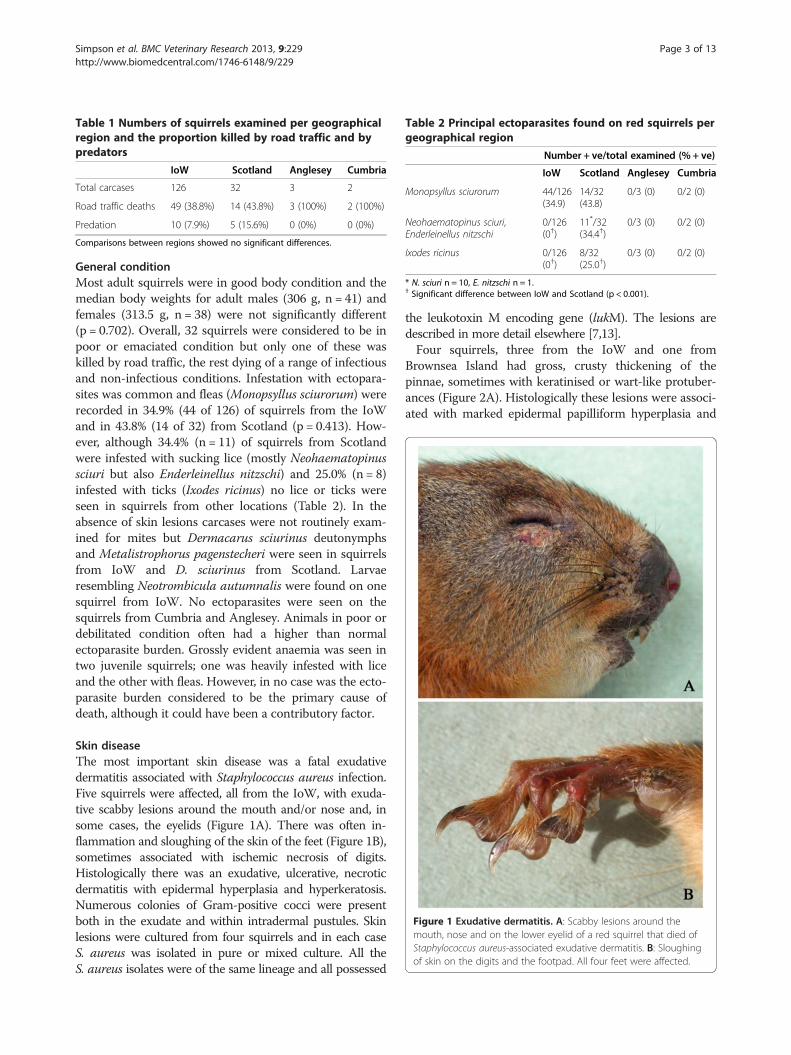
Figure 1 Exudative dermatitis. A: Scabby lesions around themouth, nose and on the lower eyelid of a red squirrel that died ofStaphylococcus aureus-associated exudative dermatitis. B: Sloughingof skin on the digits and the footpad. All four feet were affected.
Simpson et al. BMC Veterinary Research 2013, 9:229 Page 3 of 13http://www.biomedcentral.com/1746-6148/9/229
General conditionMost adult squirrels were in good body condition and themedian body weights for adult males (306 g, n = 41) andfemales (313.5 g, n = 38) were not significantly different(p = 0.702). Overall, 32 squirrels were considered to be inpoor or emaciated condition but only one of these waskilled by road traffic, the rest dying of a range of infectiousand non-infectious conditions. Infestation with ectopara-sites was common and fleas (Monopsyllus sciurorum) wererecorded in 34.9% (44 of 126) of squirrels from the IoWand in 43.8% (14 of 32) from Scotland (p = 0.413). How-ever, although 34.4% (n = 11) of squirrels from Scotlandwere infested with sucking lice (mostly Neohaematopinussciuri but also Enderleinellus nitzschi) and 25.0% (n = 8)infested with ticks (Ixodes ricinus) no lice or ticks wereseen in squirrels from other locations (Table 2). In theabsence of skin lesions carcases were not routinely exam-ined for mites but Dermacarus sciurinus deutonymphsand Metalistrophorus pagenstecheri were seen in squirrelsfrom IoW and D. sciurinus from Scotland. Larvaeresembling Neotrombicula autumnalis were found on onesquirrel from IoW. No ectoparasites were seen on thesquirrels from Cumbria and Anglesey. Animals in poor ordebilitated condition often had a higher than normalectoparasite burden. Grossly evident anaemia was seen intwo juvenile squirrels; one was heavily infested with liceand the other with fleas. However, in no case was the ecto-parasite burden considered to be the primary cause ofdeath, although it could have been a contributory factor.
Skin diseaseThe most important skin disease was a fatal exudativedermatitis associated with Staphylococcus aureus infection.Five squirrels were affected, all from the IoW, with exuda-tive scabby lesions around the mouth and/or nose and, insome cases, the eyelids (Figure 1A). There was often in-flammation and sloughing of the skin of the feet (Figure 1B),sometimes associated with ischemic necrosis of digits.Histologically there was an exudative, ulcerative, necroticdermatitis with epidermal hyperplasia and hyperkeratosis.Numerous colonies of Gram-positive cocci were presentboth in the exudate and within intradermal pustules. Skinlesions were cultured from four squirrels and in each caseS. aureus was isolated in pure or mixed culture. All theS. aureus isolates were of the same lineage and all possessed
the leukotoxin M encoding gene (lukM). The lesions aredescribed in more detail elsewhere [7,13].Four squirrels, three from the IoW and one from
Brownsea Island had gross, crusty thickening of thepinnae, sometimes with keratinised or wart-like protuber-ances (Figure 2A). Histologically these lesions were associ-ated with marked epidermal papilliform hyperplasia and
Figure 2 Crusty thickening of the pinna. A: Right pinna of a squirrelshowing gross irregular thickening and keratinised crusts. The other earwas similarly affected. B: Histological section of a similar case to thatseen in Figure 2A. There is epidermal hyperplasia, hyperkeratosis withrete ridges extending into the dermis and keratin-filled cysts. On theright margin there is also an ulcerated trichoepithelioma (arrow).Haematoxylin and eosin stain, bar = 5 mm.
Figure 3 Cutaneous wart–like lesions. A: A proliferative wart-likelesion on a digit. Several similar lesions were present elsewhere on thesame squirrel. B: Histological section of the lesion shown in Figure 3Ashowing keratinised papilliform proliferative projections of the epidermis.H & E stain, bar = 1 mm.
Simpson et al. BMC Veterinary Research 2013, 9:229 Page 4 of 13http://www.biomedcentral.com/1746-6148/9/229
orthokeratotic hyperkeratosis (Figure 2B). There wasvariable inflammatory cell infiltration of the dermis, noevidence of primary bacterial, fungal or parasite infectionsand no visible epidermal inclusions. However, in one casethere was serocellular crusting and pustule formation andin a second case there was an ulcerated trichoepithelioma(Figure 2B). The squirrel in the latter case died of apulmonary carcinoma but there was no apparent cause ofdeath for the other two. No details were available for thesquirrel from Brownsea with thickened ears but in twoIoW cases wart-like skin lesions were also observedelsewhere, notably on the digits (Figure 3A). One such
lesion examined histologically showed heavily keratinisedpapilliform proliferative projections of the epidermis butno significant inflammatory change and no evidence of aninfectious agent (Figure 3B).Other incidental skin lesions included a facial abscess
from which Yersinia enterocolitica was isolated and alarge ulcerated lesion over the scrotum associated withhistological evidence of dermal necrosis and a coccalbacterial infection.
Respiratory diseaseLungs from seven squirrels were examined bacterio-logically and 142 histologically. Excluding those due totrauma, pulmonary lesions of varying degree were ob-served in 50 squirrels (35.2%). Protozoal schizonts identi-fied morphologically as Hepatozoon sp. were frequentlyobserved and the infection was commonest and heavier insquirrels from the IoW compared with those fromScotland (p = 0.014). Comparison with Cumbria was notpossible due to the small number examined (n = 2) and noAnglesey cases were suitable for histological examination(Table 3). In most cases there was no significant inflam-matory response associated with the schizonts and there-fore their presence alone was not recorded as a lesion.
Table 3 Principal endoparasites identified histologicallyin red squirrels per geographical region
Capillaria hepatica 4/77 (5.2) 1/17 (5.9) u/s 0/1 (0)† IoW and Scotland (p = 0.014).* IoW and Scotland (p = 0.064).Specimens from Anglesey were unsuitable for examination (u/s).
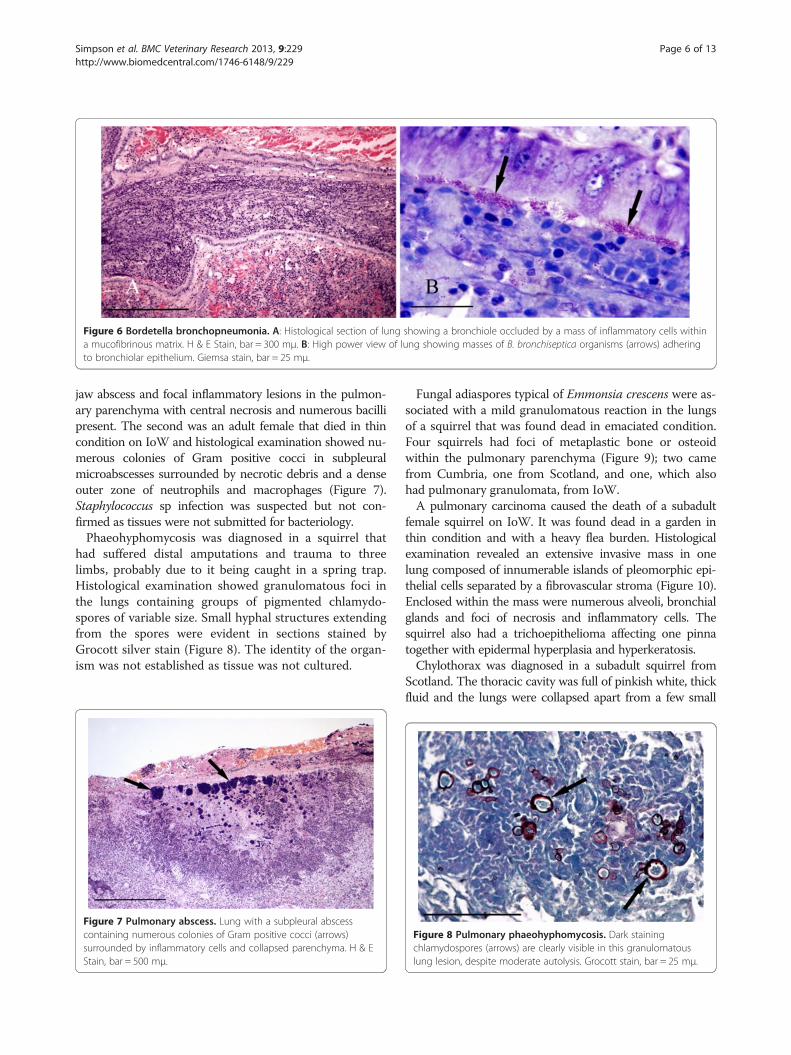
Figure 5 Bordetella bronchopneumonia. A Bordetella bronchisepticainfected lung showing multiple irregular foci of intense inflammationand consolidation.
Simpson et al. BMC Veterinary Research 2013, 9:229 Page 5 of 13http://www.biomedcentral.com/1746-6148/9/229
However, in four heavily infected squirrels there was thick-ening of alveolar walls and infiltration by lymphocytes andmacrophages. Heavy Hepatozoon infections were morecommon in squirrels suffering from other significantrespiratory diseases.Bacterial infections (n = 16) were important and included
cases of inhalation pneumonia (n = 4), bronchopneumonia(n = 6) and focal parenchymal abscessation (n = 2). In casesof inhalation pneumonia the predominant lesions were inthe apical lobes with focal inflammation and consolidationaround the bronchi and/or bronchioles, necrosis andinfiltration by neutrophils and macrophages. Within thelumen of the airway there was typically a mass of necroticcell debris, neutrophils, bacteria and foreign material thatappeared to be ingesta (Figure 4A); in some cases thisforeign material extended into the parenchyma (Figure 4B).One of the affected squirrels also had oral lesions whichmay have predisposed to inhalation of foreign materialand Staphylococcus aureus was isolated from both the orallesion and the lung.Bronchopneumonia due to Bordetella bronchiseptica
infection was confirmed in a squirrel that was observed todie in respiratory distress. Gross examination of lungsshowed large areas of intense inflammation and consolida-tion (Figure 5) from which B. bronchiseptica was isolated
Figure 4 Inhalation pneumonia. A: Histological section of lung showinga bronchiole. Within the mass there is an amorphous foreign body, possiblwithin and around the lesion. H & E Stain, bar = 200 mμ. B: A second examforeign bodies (arrows) surrounded by inflammatory cells and haemorrhag
in pure culture. Histologically many bronchi and bronchi-oles were seen to be occluded by masses of degenerateinflammatory cells together with numerous Gram negativecoccobacilli contained within a mucofibrinous matrix(Figure 6A). The surface of the lining epithelium wascovered in an almost continuous layer of coccobacilli(Figure 6B), large numbers of which were also presenttogether with mononuclear cells and proteinaceous fluidin the adjacent consolidated parenchyma. There was alsoa heavy Hepatozoon sp. infection. Almost identical lesionswere observed in a second case that died concurrently inthe same garden; similar lesions, although with heavyneutrophil infiltration, were seen in a third concurrentcase at the same location. Neither of these latter cases wasexamined bacteriologically.A squirrel with congested, collapsed and partly autolysed
lungs had intracytoplasmic inclusions in mononuclear cellsresembling those of Chlamydia sp. Two squirrels died ofpulmonary abscessation associated with bacterial infections.One was an adult female from Scotland which had a severe
a mass of necrotic cell debris and neutrophils occluding the lumen ofy inhaled ingesta (arrow). Numerous coccal bacteria were presentple of inhalation pneumonia shown at higher magnification withe within the pulmonary parenchyma. H & E Stain, bar = 100 mμ.
Figure 6 Bordetella bronchopneumonia. A: Histological section of lung showing a bronchiole occluded by a mass of inflammatory cells withina mucofibrinous matrix. H & E Stain, bar = 300 mμ. B: High power view of lung showing masses of B. bronchiseptica organisms (arrows) adheringto bronchiolar epithelium. Giemsa stain, bar = 25 mμ.
Simpson et al. BMC Veterinary Research 2013, 9:229 Page 6 of 13http://www.biomedcentral.com/1746-6148/9/229
jaw abscess and focal inflammatory lesions in the pulmon-ary parenchyma with central necrosis and numerous bacillipresent. The second was an adult female that died in thincondition on IoW and histological examination showed nu-merous colonies of Gram positive cocci in subpleuralmicroabscesses surrounded by necrotic debris and a denseouter zone of neutrophils and macrophages (Figure 7).Staphylococcus sp infection was suspected but not con-firmed as tissues were not submitted for bacteriology.Phaeohyphomycosis was diagnosed in a squirrel that
had suffered distal amputations and trauma to threelimbs, probably due to it being caught in a spring trap.Histological examination showed granulomatous foci inthe lungs containing groups of pigmented chlamydo-spores of variable size. Small hyphal structures extendingfrom the spores were evident in sections stained byGrocott silver stain (Figure 8). The identity of the organ-ism was not established as tissue was not cultured.
Figure 7 Pulmonary abscess. Lung with a subpleural abscesscontaining numerous colonies of Gram positive cocci (arrows)surrounded by inflammatory cells and collapsed parenchyma. H & EStain, bar = 500 mμ.
Fungal adiaspores typical of Emmonsia crescens were as-sociated with a mild granulomatous reaction in the lungsof a squirrel that was found dead in emaciated condition.Four squirrels had foci of metaplastic bone or osteoidwithin the pulmonary parenchyma (Figure 9); two camefrom Cumbria, one from Scotland, and one, which alsohad pulmonary granulomata, from IoW.A pulmonary carcinoma caused the death of a subadult
female squirrel on IoW. It was found dead in a garden inthin condition and with a heavy flea burden. Histologicalexamination revealed an extensive invasive mass in onelung composed of innumerable islands of pleomorphic epi-thelial cells separated by a fibrovascular stroma (Figure 10).Enclosed within the mass were numerous alveoli, bronchialglands and foci of necrosis and inflammatory cells. Thesquirrel also had a trichoepithelioma affecting one pinnatogether with epidermal hyperplasia and hyperkeratosis.Chylothorax was diagnosed in a subadult squirrel from
Scotland. The thoracic cavity was full of pinkish white, thickfluid and the lungs were collapsed apart from a few small
Figure 8 Pulmonary phaeohyphomycosis. Dark stainingchlamydospores (arrows) are clearly visible in this granulomatouslung lesion, despite moderate autolysis. Grocott stain, bar = 25 mμ.
Figure 9 Pulmonary metaplastic bone. Section of lung showing afocus of metaplastic bone in the parenchyma. H & E Stain, bar = 200 mμ.
Figure 11 A case of chylothorax. The thoracic cavity is filled withpink stained chyle and the lungs are collapsed.
Simpson et al. BMC Veterinary Research 2013, 9:229 Page 7 of 13http://www.biomedcentral.com/1746-6148/9/229
localised areas (Figure 11). Both jugular veins were greatlydistended. The squirrel had a purulent abscess circa 7 mmdiameter in the left caudal pharynx and the lower incisortooth on that side was markedly longer than that on theright. The thoracic fluid was confirmed to be chyle bydirect microscopic examination, examination of Giemsastained smears and by centrifugation. A light pure growthof Hafnia alvei was isolated from the fluid but thepharyngeal abscess produced a mixed growth of organisms.Extensive pulmonary haemorrhage was seen in an adult
squirrel that died of electrocution in Scotland (Figure 12).It was found at the base of an electricity utility pole andhad burn marks around the mouth and to the plantar sur-face of both hind feet and several large haemorrhages inthe lungs. Apart from burns and pulmonary haemorrhage,no other lesions were seen on either gross or histologicalexamination.A squirrel found dead in a water butt was thought to
have drowned. On histological examination of lung thealveoli appeared distended with acellular material con-taining various types of bacteria.
Figure 10 Pulmonary carcinoma. Carcinoma in lung composedmostly of polyhedral epithelial cells within a fibrovascular stroma.H & E Stain, bar = 200 mμ.
Hepatobiliary diseaseLesions of varying severity were present in approximately58.9% (56 of 95) of the livers that were suitable forhistological interpretation. The most frequent were small,scattered, foci of hepatocyte necrosis associated with lym-phocytes and mononuclear cells, often with a periportaldistribution. The aetiology of these lesions was not appar-ent but they were considered to be of limited pathologicalsignificance. More extensive pathological changes werepresent in 40% of livers (38 of 95), with almost half ofthese due to infection with Toxoplasma gondii (n = 12) orCapillaria hepatica (n = 5).In cases of toxoplasmosis there was typically widespread
multifocal, often periportal, hepatocyte necrosis withlymphocytic infiltration. Toxoplasma cysts were mostlypresent in hepatocytes around the margins of the lesions(Figure 13A). Affected squirrels often had similar lesions ofmultifocal necrosis in the spleen and in some cases pneu-monitis (Figure 13B) and focal cardiac myopathy. All thecases of toxoplasmosis occurred in squirrels from the IoWand, of the 77 livers from there that were suitable for histo-logical examination, 12 (15.6%) proved positive (Table 3).There was weak evidence for a difference in incidence ofT. gondii between IoW and Scotland (p = 0.064). Nine ofthe 12 affected squirrels were females. Tissues from eightcases were examined by immunohistochemistry and inseven the parasite cysts and/or tachyzoites labelled positive
Figure 12 Electrocution. Extensive pulmonary haemorrhage in asquirrel that was electrocuted after climbing a mains electricity utility pole.
Simpson et al. BMC Veterinary Research 2013, 9:229 Page 8 of 13http://www.biomedcentral.com/1746-6148/9/229
for T. gondii. In the eighth case cysts seen in lung and liverlesions labelled negative despite having typical morphology.Gross examination of livers affected by capillariasis
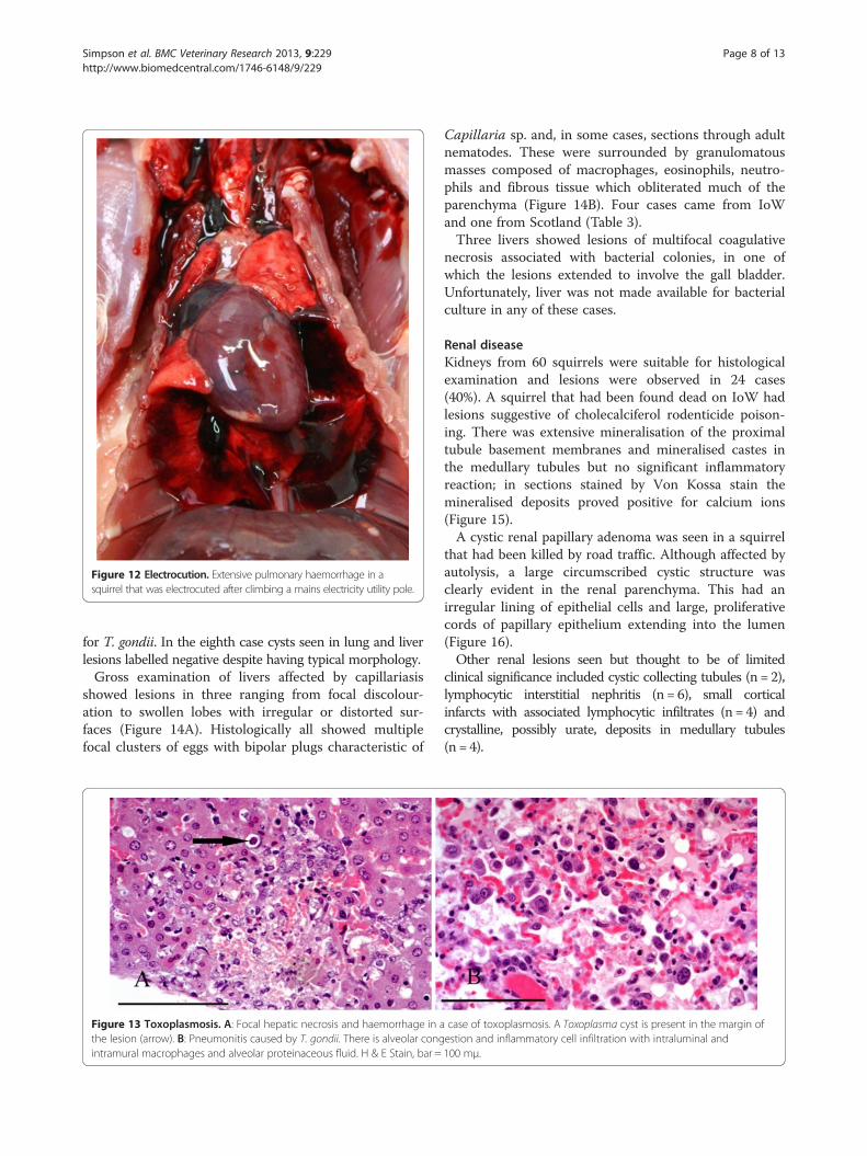
showed lesions in three ranging from focal discolour-ation to swollen lobes with irregular or distorted sur-faces (Figure 14A). Histologically all showed multiplefocal clusters of eggs with bipolar plugs characteristic of
Figure 13 Toxoplasmosis. A: Focal hepatic necrosis and haemorrhage in athe lesion (arrow). B: Pneumonitis caused by T. gondii. There is alveolar congintramural macrophages and alveolar proteinaceous fluid. H & E Stain, bar =
Capillaria sp. and, in some cases, sections through adultnematodes. These were surrounded by granulomatousmasses composed of macrophages, eosinophils, neutro-phils and fibrous tissue which obliterated much of theparenchyma (Figure 14B). Four cases came from IoWand one from Scotland (Table 3).Three livers showed lesions of multifocal coagulative
necrosis associated with bacterial colonies, in one ofwhich the lesions extended to involve the gall bladder.Unfortunately, liver was not made available for bacterialculture in any of these cases.
Renal diseaseKidneys from 60 squirrels were suitable for histologicalexamination and lesions were observed in 24 cases(40%). A squirrel that had been found dead on IoW hadlesions suggestive of cholecalciferol rodenticide poison-ing. There was extensive mineralisation of the proximaltubule basement membranes and mineralised castes inthe medullary tubules but no significant inflammatoryreaction; in sections stained by Von Kossa stain themineralised deposits proved positive for calcium ions(Figure 15).A cystic renal papillary adenoma was seen in a squirrel
that had been killed by road traffic. Although affected byautolysis, a large circumscribed cystic structure wasclearly evident in the renal parenchyma. This had anirregular lining of epithelial cells and large, proliferativecords of papillary epithelium extending into the lumen(Figure 16).Other renal lesions seen but thought to be of limited
clinical significance included cystic collecting tubules (n = 2),lymphocytic interstitial nephritis (n = 6), small corticalinfarcts with associated lymphocytic infiltrates (n = 4) andcrystalline, possibly urate, deposits in medullary tubules(n = 4).
case of toxoplasmosis. A Toxoplasma cyst is present in the margin ofestion and inflammatory cell infiltration with intraluminal and100 mμ.
Figure 14 Hepatic capillariasis. A: Lobe of liver affected by capillariasis showing an irregular surface and pale mottled areas. B: Histologicalsection of liver with clusters of Capillaria sp. eggs (arrows) enclosed in granulomatous reaction and fibrous tissue. H & E Stain, bar = 200 mμ.
Simpson et al. BMC Veterinary Research 2013, 9:229 Page 9 of 13http://www.biomedcentral.com/1746-6148/9/229
Cardiovascular diseaseThe heart was examined histologically in 128 of 155squirrels. Thirteen were unsuitable due to autolysis butlesions were seen in 27 of the remaining 115 cases. Thegreat majority of these squirrels came from the IoWwhere approximately 21% (20/97) had multiple smallfoci of lymphocytic infiltration and in some cases,myocyte necrosis. In two such cases tachyzoites typicalof T. gondii were observed in degenerating myocytesand toxoplasmosis was confirmed by examination ofother organs. The aetiology of the lesions in theremaining 17 cases was not clear but on examinationof other organs it was noted that toxoplasmosis wasconfirmed in four of the cases and suspected in afurther five. In several squirrels small ovoid structuresconsistent with Hepatozoon sp. gamonts were observedlying between myofibres in myocardium.
Figure 15 Suspected cholecalciferol poisoning. The brown stainingof the renal proximal collecting tubules basement membranes isindicative of widespread deposition of calcium and is consistent with adiagnosis of cholecalciferol poisoning. Von Kossa stain, bar = 100 mμ.
Splenic diseaseDuring post-mortem examination spleens were observedto vary greatly in size but no significant gross pathologicallesions were seen. Histologically, 35 of 45 spleens submit-ted were suitable for examination and lesions were seen in11 (31.4%). In four cases the changes were of a minornature, such as depletion of white pulp, but in seven therewere multiple foci of necrosis, predominantly in the redpulp, associated with Toxoplasma cysts. Three of sevenspleen samples screened by PCR for adenoviral DNAproved positive (Scotland 2, Anglesey 1) but none showedhistopathological lesions.
Alimentary tract diseaseOral lesions were a relatively important component ofalimentary tract disease. Two squirrels with unevenlyworn lower incisor teeth had unilateral osteomyelitis of alower mandible associated with a facial abscess in one caseand suspected bite wounds in the other. Penetration by aforeign body may have been responsible for a pharyngealabscess in a squirrel that died of chylothorax. Lingual andlaryngopharyngeal necrotising ulceration associated withS. aureus infection was considered to have predisposed toinhalation pneumonia in a juvenile squirrel on IoW andoropharyngeal/oesophageal candidiasis caused the deathof a juvenile from Scotland. (For further detail seeSimpson et al. [6]). Gastritis in a second juvenile fromScotland was associated histologically with a yeast infec-tion but although the organism was isolated it could notbe identified by API test. Coffee-ground haemorrhagesand pyloric ulcers, thought to be stress-related, were seenin three cases. Post-mortem change often precludeddetailed examination of the alimentary tract but 13 caseswere examined histologically. An adult road traffic victimfrom the IoW had a large subserosal swelling of thestomach wall and several circumscribed swellings up to
Figure 16 Renal papillary adenoma. A: Low magnification image of kidney showing proliferating cords of epithelial cells within a cysticstructure that is compressing the adjacent parenchyma. The tissue is markedly autolysed. H & E Stain, bar = 1 mm. B: Higher magnification imageof the same kidney section showing a proliferating, frond-like cord of the tumour. H & E Stain, bar =200 mμ.
Simpson et al. BMC Veterinary Research 2013, 9:229 Page 10 of 13http://www.biomedcentral.com/1746-6148/9/229
7 mm diameter in the adjacent mesentery. The lesion inthe stomach wall was a tumour composed of proliferatingbundles of spindle cells that effaced the smooth musclelayer (Figure 17). The swellings in the mesentery were alsocomposed of masses of spindle cells. The tumour distribu-tion suggested multicentric origin. Histological features areoften not predictive of malignant potential for gastro-intestinal spindle cell tumours and in the absence of immu-nohistochemistry further classification was not possible.The tumour(s) was therefore considered to be a spindle celltumour of uncertain histiogenesis and biological behaviour.Although signs of diarrhoea were occasionally observed
during post-mortem examination of squirrels on the IoW,none of the 51 squirrels examined post-mortem at theWildlife Veterinary Investigation Centre showed grossevidence of enteritis. Faecal samples from 33 squirrels(IoW 10, Scotland 20, Anglesey 3) were screened foradenovirus infection by TEM and all proved negative, des-pite spleen samples from three squirrels testing positive byPCR. Nematodes were sometimes observed during gross
Figure 17 Gastric spindle cell tumour. The smooth muscle layerof the stomach wall is effaced by proliferating bundles of spindlecells. H & E Stain, bar = 200 mμ.
and histological examination of intestine. Morphologicallythey resembled pin worms (Enterobius sp.) but, apart fromone case where they were identified as Rodentoxyurissciuri, none were submitted for species identification.Typically there was no apparent associated pathology butone juvenile with a heavy nematode infection died due toan intestinal intussusception. However, in a second case ofintussusception involving an adult there was no significantworm burden and the aetiology was obscure. Small tomoderate numbers of a variety of Eimeria sp. coccidialoocysts were frequently observed in wet preparations ofgut contents but without any associated pathology.
Miscellaneous diseaseA squirrel found dead on IoW had an enlarged, inflameduterus and histological examination revealed the remainsof a part-mummified foetus with trabecular bone evident.Unfortunately, it was too badly affected by autolysis forfurther examination. A second squirrel from IoW hadextensive adhesions involving the abdominal organs andan unidentified mass in the peritoneal cavity. Histologicalexamination of the mass showed it to be the necroticremains of what was judged to be a foetus, possibly theresult of an ectopic pregnancy.
DiscussionRoad traffic accident deaths were responsible for 41.7% ofmortality overall and there was no significant differencebetween locations. This result is consistent that obtainedLaRose et al. [9] in Scotland where 42.9% of mortality wascaused by traffic. A slightly lower figure of 36% wasrecorded in 36 squirrels found dead on the island of Jersey[14]. The impact of road traffic mortality on red squirrelpopulations is uncertain. On the IoW at least, squirrelnumbers appear to have been stable in recent years,although there was a suspected decrease during 2012(Butler H., unpublished data). Trauma due to attack by
Simpson et al. BMC Veterinary Research 2013, 9:229 Page 11 of 13http://www.biomedcentral.com/1746-6148/9/229
predators, principally cats and dogs, was 9.2%. Concernhas been expressed by Duff et al. [15] at the level of redsquirrel predation by pets and on the island of Jersey36% of squirrel mortality was attributed to attack bydomestic cats [14]. This problem is in large part due tosquirrels being attracted into gardens by people provid-ing supplementary food. Although this may be bene-ficial from a nutritional point of view [14] it alsoincreases the risk of predation.Gross and histopathological examination identified
pathological lesions associated with disease in 57 (35%)squirrels. The most important of these was toxoplasmosis.The susceptibility of red squirrels to this condition was firstreported by Coles [16] in 1914 but in recent years it has be-come increasingly recognised as a cause of mortality[5,17,18]. In this study it was notable that all the toxoplas-mosis cases were confined to the IoW where it wasestimated to have caused almost one tenth of all deaths.The practice of providing supplementary food for squirrelsis common on IoW and squirrels foraging in gardens are atincreased risk of ingesting food contaminated by cat faeces.The human population density in the IoW is relatively highwhereas at the other locations in the study, particularly innorth east Scotland, the environment is much more ruralwith a low human, and presumably cat, population density.A second point of note is that three quarters of thetoxoplasma-affected squirrels were females. This apparentsex bias does not appear to have been recorded previouslyand, whilst the reasons for it are not apparent, it could wellbe important as regards population recruitment.In the study of red squirrels in Scotland [9] squirrelpox
was the second most predominant cause of mortality(14.3%). However, squirrelpox does not occur on the IoWand was not seen in any of the squirrels submitted fromother locations in this study. This was not unexpected inthe case of squirrels submitted from Scotland as the major-ity came from an area believed to be free of squirrelpox; inaddition, any Scottish red squirrels showing pox-like lesionsare normally submitted direct to Edinburgh University fortheir pox screening program (http://www.red-squirrels.org.uk/surveillance.asp).Thirty two squirrels were in poor or emaciated condi-
tion and half of these were juveniles or subadults. How-ever, in no case was starvation diagnosed as the sole orprimary cause of death. In some cases a heavy ectoparasiteburden probably contributed to mortality but this wasconsidered secondary. This result is in contrast to thatobtained by LaRose et al. [9] in Scotland where starvationwas the most common cause of mortality in juveniles and,at 9.8%, the fourth most common cause of death overall.A possible explanation for this is the additional laboratoryprocedures carried out in the present study. Anaemiaassociated with severe louse infestations has been reportedas a likely cause of death in juvenile squirrels [9,15]. In this
study, although heavy louse infestations were seen in sixcases, in only one, a juvenile, was there obvious anaemia.It should be noted that whilst N. sciuri was the predomin-ant louse species, some squirrels were also infested withEnderleinellus nitzschi. Both species have been recordedpreviously in red and grey squirrels in Great Britain [19]but in most publications it would appear that the identifi-cation of N. sciuri is presumptive [8,9,11,20]. Neohaemato-pinus sciuri has a Holarctic distribution and Enderleinellusspecies are found worldwide [21]; it is therefore surprisingthat in this study lice were only found on squirrels fromScotland. The apparent absence of lice on squirrelssubmitted from Cumbria and Anglesey may be because sofew were examined from these locations but this does notexplain the apparent absence of sucking lice from the IoW.The authors accept that light burdens might have beenmissed during these post-mortem examinations but it ishighly unlikely that this would have happened with heavyinfestations. Similarly, ticks were only observed in squirrelsfrom Scotland although they have been observed historic-ally on IoW (H. Butler, unpublished data). In view of theapparent absence of both parasites from squirrels on IoWit is of interest that lice were not recorded, and ticks rarelyso, in a post-mortem study on red squirrels from the Islandof Jersey [T. Blackett, personal communication].Capillaria hepatica is capable of infecting a wide range
of species but is primarily a parasite of rodents. The lifecycle is dependent on the death of the host and digestionof the liver to release the eggs. Typically this occursthrough predation, cannibalism or necrophagy. A newhost only becomes infected by ingesting embryonated eggsthat have been excreted in the faeces of the predator orscavenger. As the highest prevalence of the parasite is nor-mally in commensal populations of brown rats (Rattusnorvegicus) [22] it is likely that rats are the main source ofinfection in squirrels. Rat infestations are a common prob-lem in gardens where people feed squirrels and birds. Inthis study it was difficult to assess the pathological signifi-cance of the lesions caused by C. hepatica. Four of the fiveinfected squirrels were in good condition and were killedby road traffic whilst the fifth was suffering from a severefacial abscess and mandibular osteomyelitis. However, inother studies [17, Simpson, V. unpublished data] mortalityhas been associated with severe parasite-induced liverlesions in both wild and captive red squirrels.Apart from T. gondii, the importance of protozoal infec-
tions in red squirrels is uncertain. Infection with Hepato-zoon sp. was common, especially on the IoW where athird of all squirrels were infected. Heavy Hepatozoonburdens were most commonly seen in squirrels sufferingother concurrent respiratory infections but it is unclearwhether a high Hepatozoon burden predisposes squirrelsto these infections or whether high burdens are a conse-quence of other infections or stress factors. There was no
Simpson et al. BMC Veterinary Research 2013, 9:229 Page 12 of 13http://www.biomedcentral.com/1746-6148/9/229
clear evidence that the parasite on its own was causingmortality. A similar situation possibly exists with coccidi-osis. Keymer [8] suggested that this is a common cause ofdeath in red squirrels but in the present study it wasapparent that, whilst Eimeria spp infection was common,there was no evidence that this was associated with dis-ease. Sainsbury and Gurnell [23] came to the same con-clusion in a study of red squirrels in Norfolk andSainsbury [20] suggests that, as in other mammalian spe-cies, disease in red squirrels due to coccidia is precipitatedby stressors or concomitant disease. Severe coccidiosis hasbeen described in squirrels dying of colonic intussuscep-tion [11] but there was no evidence of coccidiosis in thetwo cases of intussusception in the present study.An enteric adenovirus was first reported as a suspected
cause of enteritis and mortality in red squirrels translo-cated from Cumbria to Norfolk [3]. Since then there is in-creasing evidence that the virus is associated with fatalenteritis in wild and, particularly, in captive red squirrelsin England, Wales, Scotland and Northern Ireland[4,11,24,25]. However, although PCR analysis on spleensdemonstrated adenoviral DNA in three squirrels in thisstudy, and in four of seven squirrels from IoW examinedas part of another study (Everest and Butler, unpublisheddata), in no case was there evidence of associated path-ology. This suggests that in many cases adenovirus infec-tion in wild red squirrels is asymptomatic.Keymer [8] suggested that bacterial infections in red
squirrels are rare but in this study they were quite frequent,particularly those affecting the respiratory system wherehistological examination showed lesions associated withbacteria in approximately 11% of cases. However, in manycases the infection appeared to secondary, for example insquirrels dying of inhalation pneumonia. In the study byLaRose et al. [9] of red squirrel mortality in Scotland 7.3%of the deaths were attributed to pneumonia but as thisdiagnosis was based on gross pathology only it is not pos-sible to make a direct comparison with the present study.The most important bacterial infection in this study was
Staphylococcus aureus associated with fatal exudativedermatitis in IoW squirrels. This condition is also a majorcause of red squirrel mortality on the island of Jersey [7]and the S. aureus isolates from affected squirrels on bothislands are of the same lineage and all encode the lukMgene [13]. The pathogenesis of the condition is not under-stood but it is invariably fatal. Further studies into factorsthat may predispose squirrels to Staphylococcus-associatedfatal exudative dermatitis are in progress.Bordetella bronchiseptica is a common cause of respira-
tory disease in dogs and cats but is only occasionallyrecorded in wildlife. In most cases bordetellosis is seen asa secondary or opportunistic infection in stressed or com-promised animals, for example dogs and cats in boardingkennels or seals affected with phocine distemper [26-28].
In this study, acute, fatal bronchopneumonia due toB. bronchiseptica was diagnosed in two squirrels and wassuspected in a third. There was no evidence to suggestthat these were secondary infections although they didhave heavy Hepatozoon infections. The source of infectionwas not apparent.Neoplastic disease is uncommon in most British free-
living wild species but red squirrels appear to be unusuallysusceptible. Cases of soft tissue sarcoma and lymphomahave been recorded in squirrels in Scotland [9], two casesof lymphosarcoma in Wales [11] and pulmonary adenoma-tosis and lymphoma in Jersey [29]. Neoplasms seen thisstudy were pulmonary carcinoma, trichoepithelioma,gastric spindle cell tumour and renal papillary adenoma. Inaddition, four squirrels, three from IoW and one fromBrownsea, had unusual lesions of epidermal hyperplasia ofthe ear pinnae associated in two cases with cutaneous wart-like growths. None of the neoplastic lesions observed in thisstudy appear to have been recorded previously in redsquirrels and it was notable that all occurred in cases fromIoW. A preliminary investigation in to the possibility thatred squirrels, in common with other Rodentia [30] carryGammaretroviruses that are capable of acting as etiologicalagents for transmissible neoplasia indicated the presence ofone or more potentially infectious retroviruses [Tarlintonand Lucassen personal communication].
ConclusionAs the grey squirrel population continues to extend itsrange over Great Britain, the viability of the increasinglyisolated red squirrel populations becomes doubtful. Even inareas where squirrelpox does not currently occur red squir-rels suffer premature mortality from additional causes,many of which are due, directly or indirectly, to humanactivities. Road traffic trauma, pet predation, toxoplasmosis,trap injuries, rodenticide poisoning and electrocutionaccounted for 61% of all recorded mortality in this study.There is also evidence that infectious agents such as adeno-virus may be spread during translocation or reintroductionprogrammes [3,11]. The impact of human activities on thehealth and welfare of red squirrels in Great Britain meritsmore attention than it has received in the past. There isalso a need for continued disease surveillance. As this studyhas shown, red squirrels are affected by a wider range ofdiseases than previously described.
Competing interestsThe authors declare they have no competing interests.
Authors’ contributionsVS conceived, designed and coordinated the study, participated in the grossand histopathology and drafted the manuscript, HB participated in the designof the study and the gross pathology, JH participated in the histopathology,ND performed the bacteriology and DE performed the virology. All authorscontributed to writing the draft manuscript and all read and approved the finalmanuscript.
Simpson et al. BMC Veterinary Research 2013, 9:229 Page 13 of 13http://www.biomedcentral.com/1746-6148/9/229
AcknowledgementsThe authors gratefully acknowledge the support given by Trevor Whitbread,Richard Fox, Lucy Oldroyd, Sonja Rivers and technical staff at AbbeyVeterinary Services. They also thank Beverley Rule and Philip Booth at AHVLATruro, Mark Dagleish at Moredun Research Institute, Ian Mawhinney atAHVLA Bury St Edmunds, Ken Neil of Saving Scotland’s Red Squirrels, JohnMcGarry at Liverpool University, Andrew Borman at Mycology ReferenceLaboratory South West Health Protection Agency, Vince Smith at NaturalHistory Museum, Mark Fox and Sian Denney at Royal Veterinary College,Rachael Tarlinton at Nottingham University and Jane Simpson at WildlifeVeterinary Investigation Centre. Those parts of this study performed atAHVLA were funded under the Diseases of Wildlife Scheme. Additionalthanks go to two anonymous reviewers for their helpful comments andconstructive criticism.
Author details1Wildlife Veterinary Investigation Centre, Chacewater, Truro, Cornwall TR4 8 PB, UK.2Abbey Veterinary Services, 89 Queen Street, Devon, Newton Abbot TQ12 2BG,UK. 3PO Box 33, Ryde, Isle of Wight PO33 1BH, UK. 4Animal Health and VeterinaryLaboratories Agency Polwhele, Truro, Cornwall TR4 9AD, UK. 5Bio Imaging Unit,Animal Health and Veterinary Laboratories Agency Weybridge, New Haw, Surrey,Addlestone KT15 3NB, UK. 6Current address: Scottish Marine Animal StrandingScheme, SAC Veterinary Services, Drummondhill, Stratherrick Road, Inverness IV24JZ, Scotland, UK.
Received: 24 July 2013 Accepted: 9 November 2013Published: 16 November 2013
References1. Sainsbury AW, Nettleton P, Gilray J, Gurnell J: Grey squirrels have high
prevalence to a parapoxvirus associated with death in red squirrels.Anim Conserv 2000, 3:229–233.
2. Tomkins DM, Sainsbury AW, Nettleton P, Buxton D, Gurnell J: Parapoxcauses a deleterious disease in red squirrels associated with UKpopulation declines. Proc R Soc Lond B 2002, 269:529–533.
3. Sainsbury AW, Adair B, Graham D, Gurnell J, Cunningham AA, Benko M,Papp T: Isolation of a novel adeno virus associated with splenitis,diarrhoea, and mortality in translocated red squirrels, Sciurus vulgaris.Erkrankung der Zootiere 2001, 40:265–270.
4. Duff JP, Higgins R, Farrelly S: Enteric adenovirus infection in a red squirrel(Sciurus vulgaris). Vet Rec 2007, 160:384.
5. Simpson VR, Birtles RJ, Bown KJ, Panciera RJ, Butler H, Davison N:Hepatozoon species infection in wild red squirrels (Sciurus vulgaris) onthe Isle of Wight. Vet Rec 2006, 159:202–205.
6. Simpson VR, Davison J, Borman AM, Linton CJ, Everest D: Fatal candidiasisin a wild red squirrel (Sciurus vulgaris). Vet Rec 2009, 164:342–343.
7. Simpson VR, Hargreaves J, Everest DJ, Baker AS, Booth PA, Butler HM,Blackett T: Mortality in red squirrels (Sciurus vulgaris) associated withexudative dermatitis. Vet Rec 2010, 167:59–62. doi:10.1136/vr.b4887.
Milne EM: Epidemiological and postmortem findings in 262 redsquirrels (Sciurus vulgaris) in Scotland, 2005 to 2009. Vet Rec 2010,167:297–302.
10. Everest DJ, Stidworthy MF, Milne EM, Meredith AL, Chantrey J, Shuttleworth CM,Blackett T, Butler H, Wilkinson M, Sainsbury AW: Retrospective detection bynegative contrast electron microscopy of faecal viral particles in wild redsquirrels (Sciurus vulgaris) with suspected enteropathy in Great Britain.Vet Rec 2010, 167:1007–1010.
11. Everest DJ, Shuttleworth CM, Grierson SS, Duff JP, Jackson N, Litherland P,Kenward RE, Stidworthy MF: Systematic assessment of the impact ofadenovirus infection on a captive reintroduction project for red squirrels(Sciurus vulgaris). Vet Rec 2012. doi: 10.1136/vr.100617.
12. Kramer L, Buxton D: Aetiological Diagnosis. In Protozoal Abortion in FarmRuminants, Guidelines for Diagnosis and Control. Edited by Ortega-Mora LM,Gottstein B, Conraths FJ, Buxton D. Wallingford: CABI; 2007:132–136.
13. Simpson VR, Davison NJ, Kearns AM, Pichon B, Hudson LO, Koylass M, BlackettT, Butler H, Rasigade JP, Whatmore AM: Association of a lukM-positive cloneof Staphylococcus aureus with fatal exudative dermatitis in red squirrels(Sciurus vulgaris). Vet Microbiol 2013, 162:987–991.
14. Magris L, Gurnell J: Population ecology of the red squirrel(Sciurus vulgaris) in a fragmented woodland ecosystem on theIsland of Jersey, Channel Islands. J Zool 2002, 256:99–112. doi:10.1017/S0952836902000134.
15. Duff JP, Haley P, Wood R, Higgins RJ: Causes of red squirrel (Sciurus vulgaris)mortality in England. Vet Rec 2010, 167:461.
16. Coles AC: Blood parasites found in mammals, birds and fishes inEngland. Parasitology 1914, 7:17–61.
18. Jokelainen P, Nylund M: Acute fatal toxoplasmosis in three Eurasian redsquirrels (Sciurus vulgaris) caused by genotype II of Toxoplasma gondii.J Wildl Dis 2012, 48:454–457.
19. Blackmore D, Owen DG: Ectoparasites: the significance in British wildrodents. In Diseases in Free-living Wild Animals, Volume 24. Edited byMcDiarmid A, Symposia of the Zoological Society of London (1968).London: Academic; 1969:197–220.
20. Sainsbury AW: Squirrels. In BSAVA Manual of Wildlife Casualties. Edited byMullineaux E, Best R, Cooper JE. Gloucester, UK: British Small AnimalVeterinary Association; 2003:70.
21. Durden L: Ectoparasites. In Parasitic Diseases of Wild Mammals. 2nd edition.Edited by Samuel WM, Pybus MJ, Kocan AA. London: Iowa State UniversityPress; 2001:8.
22. Spratt DM, Singleton GR: Hepatic Capillariasis. In Parasitic diseases of WildMammals. 2nd edition. Edited by Samuel WM, Pybus MJ, Kocan AA.London: Iowa State University Press; 2001:367.
23. Sainsbury AW, Gurnell J: An investigation into the health and welfare ofred squirrels, Sciurus vulgaris, involved in re-introduction studies. Vet Rec1995, 137:367–370.
24. Everest DJ, Grierson SS, Meredith AL, Milne EM: Adenovirus in a redsquirrel (Sciurus vulgaris) from Scotland. Vet Rec 2010, 167:184.
25. Everest DJ, Griffin J, Warnock ND, Collins L, Dick J, Reid N, Scantlebury M,Marks N, Montgomery I: Adenovirus particles from a wild redsquirrel (Sciurus vulgaris) from Northern Ireland. Vet Rec 2012,170:188.
26. Datz C: Bordetella infections in dogs and cats: pathogenesis, clinicalsigns and diagnosis. Compend Cont Educ Pract Vet 2003, 25:896–901.
27. Baker J, Ross HM: The role of bacteria in phocine distemper. Sci TotalEnviron 1992, 115:9–14.
28. Munro R, Ross HM, Cornwell HJC, Gilmour J: Disease conditions affectingcommon seals (Phoca vitulina) around the coast of Scottish mainland,September-November 1988. Sci Total Environ 1992, 115:67–82.
29. Simpson VR, Blackett T, Butler HM, Borman AM, Hargreaves J, Davison NJ:Respiratory disease surveillance in geographically remote populations of redsquirrels (Sciurus vulgaris), Proceedings of the Joint 61st WDA/10th BiennialEWDA Conference “Convergence in Wildlife Health”. Lyon, France; 2012:46.
doi:10.1186/1746-6148-9-229Cite this article as: Simpson et al.: Causes of mortality and pathologicallesions observed post-mortem in red squirrels (Sciurus vulgaris) in GreatBritain. BMC Veterinary Research 2013 9:229.
Submit your next manuscript to BioMed Centraland take full advantage of:
• Convenient online submission
• Thorough peer review
• No space constraints or color figure charges
• Immediate publication on acceptance
• Inclusion in PubMed, CAS, Scopus and Google Scholar
• Research which is freely available for redistribution
Submit your manuscript at www.biomedcentral.com/submit