Page 1
SHODH SANGAM - A RKDF University Journal of Science and Engineering
Volume 01, No. 01, 2018
28
Hematological Genetic Disorders: A Review
Ramdas Malakar,* S.N. Malviya and C.B.S. Dangi
Department of Biotechnology, RKDF University Bhopal (M.P.) India
Corresponding author*: C.B.S. Dangi Department of Biotechnology RKDF University Bhopal
(M.P.) India;
e-mail:- [email protected] , [email protected]
ABSTRACT: Thalassaemia major requires regular blood transfusions to maintain an adequate
supply of haemoglobin and sustain life. As a result of multiple transfusions, organs become
severely overloaded with iron and a specific treatment is needed to manage this condition.
Thalassaemias can be cured by a successful bone-marrow transplant, however this procedure is
expensive and not readily available in most settings. Recently, gene therapy has been
successfully applied to a patient with thalassaemia. The most cost-effective strategy for reducing
the burden of haemoglobin disorders is to complement disease management with prevention
programmes. In expensive and reliable blood tests can identify couples at risk for having affected
children. This screening is especially opportune before marriage or pregnancy, allowing couples
to discuss the health of their family. Subsequent genetic counseling informs trait carriers of risks
that the condition may be passed along to their children, the treatment needed, if affected by a
haemoglobin disorder, and the possible options for the couple. Prenatal screening of genetic
diseases raises specific ethical, legal and social issues that require appropriate consideration.
Key word: Thalassaemias, sickle cell disease
1. INTRODUCTION
Thalassaemia and Sickle Cell Anaemia (SCA) is a hereditary blood disorder characterized by
red blood cells that assume an abnormal, rigid, sickle shape. It is a genetic defect in the
synthesis of hemoglobin and is the best-known hemoglobinopathy in man (Kato GJ, 2007). It
Page 2
SHODH SANGAM - A RKDF University Journal of Science and Engineering
Volume 01, No. 01, 2018
29
occurs as a result of the substitution of glutamic by valine at position 6 of the amino-acid
sequence, which leads to the formation of defective hemoglobin molecules and causes sickling
of the red blood cells (Ballas SK, 2002). Even though thalassaemia is present from birth, but
symptoms are rare before the age of three to six months because of the high level of fetal
hemoglobin (HbF) which is present at birth. It is of interest to note that cells are less prone to
sickling in individuals who retain a high level of HbF. The HbF inhibits the polymerization of
the HbS owing to its high oxygen affinity. The HbF (α2 γ2 ) dissociates to a dimer, which when
combined with HbS, gives a tetramer that does not form a polymer, symptoms of thalassaemia
are almost completely eliminated with HbF levels above 25%; however, any increment in HbF
level was observed to improve the overall survival (Atweh GF, 2001), (Bank A,2006). The
production of HbF is normally switched off soon after birth in favor of production of adult-type
HbA.
2. REVIEW OF LITERATURE
2.1 Facts about haemoglobin disorders
It is estimated that each year over 300000 babies with severe forms of these diseases are born
worldwide, the majority in low and middle income countries. Approximately 5% of the
world’s populations are healthy carriers of a gene for sickle-cell disease or thalassaemia. The
percentage of people who are carriers of the gene is as high as 25% in some regions. These
conditions are most prevalent in tropical regions; however population migration has spread
these diseases to most countries. Thalassaemias are the most common in Asia, the
Mediterranean basin, and the Middle East. Sickle-cell disease predominates in Africa
2.2 What causes haemoglobin disorders
Haemoglobin disorders are inherited from parents in much the same way as blood type, hair
colour and texture, eye colour and other physical traits. Sickle-cell disease and severe forms
of thalassaemia (thalassaemia major) can occur only when both parents are carriers of trait
genes for the particular condition. A child who inherits two of the same trait genes one from
each parent will be born with the disease. However, a child of two carriers has only a 25%
chance of receiving two trait genes and developing the disease, and a 50% chance of being a
carrier. Most carriers lead completely normal, healthy lives.
2.3 What are haemoglobin disorders
Page 3
SHODH SANGAM - A RKDF University Journal of Science and Engineering
Volume 01, No. 01, 2018
30
Haemoglobin disorders are inherited blood diseases that affect how oxygen is carried in the
body. Hemoglobinopathy is a kind of genetic defect that results in abnormal structure of one of
the globin chains of the hemoglobin molecule. Haemoglobinopathies are inherited single gene
disorders caused by genetic mutations that result in abnormal, disfunctional hemoglobin
molecules or lower levels of normal haemoglobin molecules in red blood cells. (Old, 2003;
Sylvie Langlois et al., 2008; Rekha Vij et al. 2010).
The patients with different haemoglobinopathies suffer from various complications such as
growth retardation (De Sanctis, 2002; Zemel et al. 2007) impaired immune status, severe
anemia, endocrine complications (Rund, 2005; Abdelrazik et al. 2007) spinal deformities, nerve
compression, fractures, severe osteoporosis and painful vasoclusive episode (Hasanato, 2006).
The cumulative effects of various factors like trace elements, vitamins and growth hormones are
involved in various haemoglobinopathies. Trace elements like copper and zinc play a vital role
in preventing the oxidative stresses in human (Hennig et al. 1999). Haemoglobin disorders fall
into two main categories: sickle-cell disease and thalassaemias.
2.4 Sickle cell anaemia (sca)
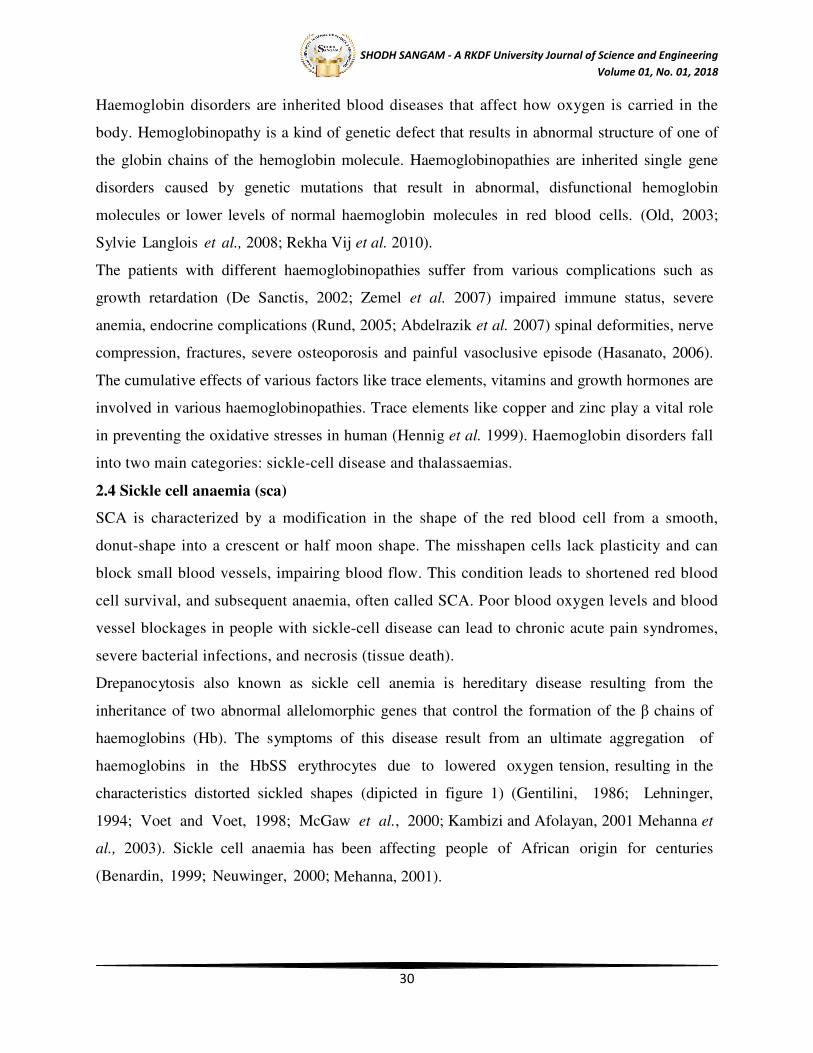
SCA is characterized by a modification in the shape of the red blood cell from a smooth,
donut-shape into a crescent or half moon shape. The misshapen cells lack plasticity and can
block small blood vessels, impairing blood flow. This condition leads to shortened red blood
cell survival, and subsequent anaemia, often called SCA. Poor blood oxygen levels and blood
vessel blockages in people with sickle-cell disease can lead to chronic acute pain syndromes,
severe bacterial infections, and necrosis (tissue death).
Drepanocytosis also known as sickle cell anemia is hereditary disease resulting from the
inheritance of two abnormal allelomorphic genes that control the formation of the β chains of
haemoglobins (Hb). The symptoms of this disease result from an ultimate aggregation of
haemoglobins in the HbSS erythrocytes due to lowered oxygen tension, resulting in the
characteristics distorted sickled shapes (dipicted in figure 1) (Gentilini, 1986; Lehninger,
1994; Voet and Voet, 1998; McGaw et al., 2000; Kambizi and Afolayan, 2001 Mehanna et
al., 2003). Sickle cell anaemia has been affecting people of African origin for centuries
(Benardin, 1999; Neuwinger, 2000; Mehanna, 2001).
Page 4
SHODH SANGAM - A RKDF University Journal of Science and Engineering
Volume 01, No. 01, 2018
31
Figure 1: Normal Red blood cells & Sickle cells
2.5 Thalassaemia
Thalassaemias are also inherited blood disorders. People with thalassaemia are not able to
make enough haemoglobin, which is found in red blood cells. When there is not enough
haemoglobin in the red blood cells, oxygen cannot get to all parts of the body. Organs then
become starved for oxygen and are unable to function properly. The term thalassaemia is
derived from the Greek, thalassa (sea) and haima (blood) (Thein, 2005). Thalassaemia
syndromes are a family of genetic blood disorders characterized by an imbalance in the synthesis
of globin chains, which may result in the absence or reduction in production of adult
hemoglobin (Clegg, 1996; Weatherall, 1997). About 4.5% all human beings carry a gene
for thalassaemias (Angastiniotis et al., 1998; Weatherall et al., 2001). It is prevalent across the
world. Affected population estimated rates are: Europe 0.9%; Asia 4.1%; Africa 13.3%; Oceania
(including Australia, New Zealand, Papua New Guinea and Fiji) 1.3%; and the Americas 2%
(WHO 1994). Cooley and Lee in 1925 first described a form of severe anemia occurring
in childhood associated with splenomegaly and characteristic bone changes. The condition was
called Cooley anemia but later named Thalassaemia (Whipple et al. 1993). It may be
defined as genetic blood disease that affect the body's ability to produce a protein in the
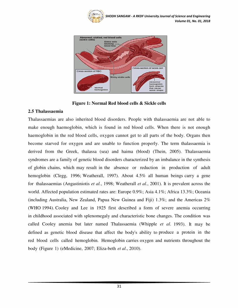
red blood cells called hemoglobin. Hemoglobin carries oxygen and nutrients throughout the
body (Figure 1) (eMedicine, 2007; Eliza-beth et al., 2010).
Page 5
SHODH SANGAM - A RKDF University Journal of Science and Engineering
Volume 01, No. 01, 2018
32
Figure 2: Function of Haemoglobin Molecule
People with mild variety of thalassaemia usually do not present any symptom. In
more severe cases of thalassaemia, symptoms may include:
• Weakness,
• Pale skin or jaundice,
• Dark urine,
• Fatigue,
• Lightheadedness,
• Rapid heartbeat,
• Abnormal facial bones and poor growth,
• Protruding abdomen with enlarged spleen or liver.
Thalassaemic patient require blood transfusions, Due to repeated blood transfusions, many
patients with β-thalassaemia may be infected with either hepatitis C virus (HCV) or hepatitis B
virus (HBV) (Aach et al. 1991). Cure may be possible with stem cell transplantation. However,
This form of treatment is possible only for a minority group of patients who have a suitable
HLA-matched donor (Fucharoen et al., 2007).
Thalassaemia is named with reference to the affected globin chain: α-thalassaemia involves the-
chain and β-thalassaemia the β-chain (Ford et al. 2008). In α- thalassaemia, there are four
severities are involved: Hydrops fetalis (four gene deletion), Haemoglobin H (Hb H) disease
(three gene deletion), α-thalassaemia minor (two gene deletion); Slient carrier (one gene
deletion). While β-thalassaemia have been classified into three clinical categories: β-
Page 6
SHODH SANGAM - A RKDF University Journal of Science and Engineering
Volume 01, No. 01, 2018
33
thalassaemia major (two gene mutations); β-thalassaemia intermediate (two gene mutations) and
β-thalassaemia minor (one or two gene mutations) (Bunn et al. 1986; Dheeraj et al. 1999).
Types of Thalassemia
There are two major types of thalassaemia, alpha and beta, which are named for the two
protein chains that make up normal haemoglobin. Alpha and beta thalassaemia have both mild
and severe forms.
2.5.1 Alpha-Thalassaemia
Alpha-thalassaemia is a common hereditary condition caused by deletions or point mutations in
one or both alpha-globin genes, located on chromosome 16 (Weatherall, 1997; Sen et al.,
2004; Higgs et al. 2009). With this disorder, the failed genes are almost invariably lost from
the cell. The genes that are most commonly associated with alpha thalassaemia are HBA1
and HBA2 (Galanello et al., 2008). α-Thalassaemia refers to a deletion of both α globin genes
on the same chromosome, while α+-thalassaemia refers to a deletion of a single α globin gene,
leaving the other α globin gene on that chromosome intact ( Leung et al. 2005). Alpha
thalassaemia is one of the most common single gene disorders, affecting 5% of the world’s
population (Chui et al. 1998).
Hemoglobin H (HbH) disease, the clinically significant intermediate form of alpha-
thalassaemia, is characterized by mild to moderate (sometime severe) microcytic, hypo chromic
hemolytic anemia, jaundice, hepatosplenomegaly, and occasionally mild thalassaemia like
bone modifications. Most commonly HbH disease results from deletion or dysfunction of 3 of
4 alpha-globin genes, and rarely from deletions in the upstream regulatory region (Viprakasit et
al. 2003; Fucharoen et al. 2009).
The severity of alpha thalassaemia depends on the number of defective genes:
2.5.1.1 Silent carrier
With one defective gene, the body still makes hemoglobin. Therefore, the person will not have
any symptom and can lead a normal and healthy life.
2.5.1.2 Alpha Thalassaemia minor
The loss of two normal genes causes the red blood cells to be smaller than usual. Except for
possible mild anemia, patients remain in good health.
2.5.1.3 Haemoglobin h disease
Page 7
SHODH SANGAM - A RKDF University Journal of Science and Engineering
Volume 01, No. 01, 2018
34
Hemoglobin made from only one gene does not carry oxygen properly. Patients with
hemoglobin H disease can suffer from severe anemia.
2.5.1.4 Alpha thalassaemia major
With all four genes failing to produce the alpha chain, the body has a significant loss of
hemoglobin which results in a severe form of anemia (DeMaeyer,et al., 1985; Bunn et al.
1986; Kliegman, et al., 2009).
2.5.2 Beta-thalassaemia
Beta-thalassaemia syndromes are a group of hereditary blood disorders caused by mutations in
the ß- globin genes on chromosome 11 and characterized by reduced or absent beta globin chain
synthesis, resulting in reduced Hb in red blood cells (RBC), decreased RBC production and
anemia (Leung et al. 2005; Galanello et al.2010). The geographic distribution of this disease has
made it a worldwide health problem with a high frequency in Africa, India, Southeast Asia and
the Mediterranean area (Pearson et al. 1996).>400,000 new borns affected per year
worldwide (Angastiniotis et al., 1998).
It is a hereditary anemia caused by more than 200 point mutations and rarely by deletions
(Higgs et al. 2001) and characterized by absent to decreased synthesis of β- globin chains
resulting in imbalance between α- and β- chains and ensuing ineffective erythropoeisis and
hemolysis (Schrier, 1997; Olivieri, 1999). The only curative treatment for this disaese
currently available is allogeneic bone marrow transplantation (BMT), which involves
significant risks such as graft vs. host disease and engraftment failure (Rivella et al.2003).
Three classes of beta-thalassaemia have been recognized clinically:
• Beta-thalassaemia major,
• Beta-thalassaemia intermediate,
• Beta-thalassaemia minor (Thein, 2004; Galanello and Origa, 2010).
2.5.2.1 Thalassaemia major
Thalassaemia major, also known as Cooley’s anemia and Mediterranean anemia, is the most
severe form of beta-thalassaemia, with mutations of both HBB alleles’ results in severely
impaired beta-globin chain production (Rund et al., 2005). Three of the general allele
combinations are responsible for this thalassaemia phenotype- B/B
, B /B+
, and sometimes
Page 8
SHODH SANGAM - A RKDF University Journal of Science and Engineering
Volume 01, No. 01, 2018
35
B+
/B+
(Thein, 2004). In thalassaemia major, the excess unpaired alpha-globin chains aggregate
to form inclusion bodies. These chain inclusion bodies damage RBC membrane, leading to
intravascular hemolysis (Cao., 2005). This form of beta thalassaemia presents within the first
two years of life and, with proper treatment affected individuals can live five decades or
more (Wonke., 2001).
2.5.2.2 Thalassaemia intermediate
This condition is milder than thalassaemia major due to inheritance of a HBB mutation
associated with reduced beta-globin chain production (Cao, 2005). Patients with beta-
thalassaemia intermediate have mild to moderate anemia and in most cases do not require blood
transfusion (Taher et al. 2006). Common clinical features include splenic enlargement due to
entrapment of damaged RBCs, with risk of iron overload due in part to increased intestinal
absorption. Although thalassaemia intermediate can be associated with poor growth and bone
abnormalities, it presents later in life and rarely affects longevity (Bunn et al. 1984). A rare
variant form called “Silent beta thalassaemia” results from a mild imbalance of globin chain
synthesis due to reduced beta-globin synthesis, leading to thalassaemia intermediate. Silent
beta- thalassaemia mutations are found mainly in the regulatory regions, HBB promoter and 5’
and 3’ UTRs. The most common silent mutation is the nt-101C>T (c.-151C>T) transition in
HBB (Gonzalez- Redondo et al. 1989).
2.5.2.3 Thalassaemia minor
Thalassaemia minor is most common form of beta thalassaemia, and is also known as the
‘thalassaemia trait’, in which affected individuals are asymptomatic (Rund et al. 2005).
These patients are typically heterozygous for beta thalassaemia since they carry one
normal HBB allele and one thalassaemia allele-either Bº or B+
(Thein, 2004).
Asymptomatic patients are usually detected through routine hematologic testing (Bunn, 1984).
The primary caution for individuals with thalassaemia minor is a potential risk of having
children affected with more serious thalassaemia if their partner is also a carrier of
thalassaemia minor (Thein, 2004).
2.6 How can haemoglobin disorders be reduced
Haemoglobin disorders can be effectively reduced through a strategic balance of disease
management and prevention programmes.
Page 9
SHODH SANGAM - A RKDF University Journal of Science and Engineering
Volume 01, No. 01, 2018
36
Sickle-cell disease can be managed by simple procedures including:
• high fluid intake
• healthy diet
• folic acid supplementation
• pain medication
• vaccination and antibiotics for the prevention and treatment of infections
• a number of other therapeutic measures
Thalassaemia major requires regular blood transfusions to maintain an adequate supply of
haemoglobin and sustain life. As a result of multiple transfusions, organs become severely
overloaded with iron and a specific treatment is needed to manage this condition. Thalassaemias
can be cured by a successful bone-marrow transplant, however this procedure is expensive and
not readily available in most settings. Recently, gene therapy has been successfully applied to a
patient with thalassaemia.
The most cost-effective strategy for reducing the burden of haemoglobin disorders is to
complement disease management with prevention programmes. Inexpensive and reliable blood
tests can identify couples at risk for having affected children. This screening is especially
opportune before marriage or pregnancy, allowing couples to discuss the health of their family.
Subsequent genetic counselling informs trait carriers of risks that the condition may be passed
along to their children, the treatment needed, if affected by a haemoglobin disorder, and the
possible options for the couple. Prenatal screening of genetic diseases raises specific ethical,
legal and social issues that require appropriate consideration.
2.7 Epidemiology
2.7.1 Worldwide status
Thalassaemia is one of the most common genetic blood disorders in the world, affecting
approximately 240 million people worldwide who are heterozygous for β-thalessemia and
approximately 200,000 affected homozygotes are born annually (Cao et al., 2005). About 5%
of the world population is affected by it. Its prevalence is more around the Mediterranean
Sea i.e. countries like Greece, Italy, Turkey and North African countries. It is also seen in
Saudi Arabia, Iran, Afghanistan, Pakistan (Ahmed et.al., 2010) India (Agarwal, Mehta., 1982)
and south East Asian countries like Thailand (Riewpaiboon et al., 2010) and Indonesia
(illustrate in table1). The prevalence is highest in Italy, Greece and Cyprus (Hazza and Jamal,
Page 10
SHODH SANGAM - A RKDF University Journal of Science and Engineering
Volume 01, No. 01, 2018
37
2006). In worldwide 4.83 percent of the world’s population carry globin variants, including
1.67 percent of the population who are heterozygous for alpha-thalassaemia and beta–
thalassaemia (Angastiniotis, 1998).
Figure4. The Worldwide Geographical distribution of Haemoglobinopathies.
http://upload.wikimedia.org/wikipedia/commons/2/26/Red_Blood_Cell_abnormalities.png
Table:- 1 distribution of thalassaemia in world
SN COUNTRY THALASSAEMIA
TYPE
DISTRIBUTION
(%)
YEAR REFERENCES
1. Turkey β-thalassaemia 2.10% 1971 Cavdar et al
2. Cyprus β-thalassaemia 14% 1998 Flint et al.
3. Sardinia β-thalassaemia 10.3% 1998 Flint et al.
4. Southern China α- thalassaemia 5% 2003 Westwood et al.
5. India β-thalassaemia trait 3.5% 2004 Shah
6. Northern Europe β-thalassaemia 1.5% 2005 Vichinsky
7. Asia α- thalassaemia 3-14% 2005,1998 Chui et al
8. Northern thailand α- thalassaemia 4.6% 2005,1998 Chui et al
9. Hong kong α- thalassaemia 4.1% 2005,1998 Chui et al
10. China α- thalassaemia 4.1% 2005,1998 Chui et al
2.7.2 Asia
The thalassemia belt stretches across African continent, Mediterranean regions, Middle
East, Indian subcontinent, South east Asia, Thailand (Riewpaiboon et al., 2010), Cambodia,
Page 11
SHODH SANGAM - A RKDF University Journal of Science and Engineering
Volume 01, No. 01, 2018
38
Laos, Vietnam, Malaysia, Singapore, Southern China. The prevalence of Thalassemia and
Falciparum Malaria are similar, suggesting the hypothesis that nature developed genetic
mutation to overcome mortality and morbidity of malaria (Dallaman, 1987; Seshadri 1990;
Gomber et al., 1998; NFHS, 2000). Thalassaemia is more prominent among the Malays and
Chinese, whereas the Indians are less affected (Tan et al., 2006). The frequency of the genes is
20% for α- thalassaemia (3-5%, α°, α", 16%), 3- 50% for Hb E, 3-4% for β-thalassaemia, and
1-
4% for Hb Constant Spring (George, Khuziah, 1984). In Pakistan, the estimated carrier rate
is 5-7%, with 9.8 million carriers in the total population (Ahmed et al., 2010).
2.7.3 India
In India prevalence of the β-thalassaemia trait is about 3.5%, Sindhis and Punjabis are known to
carry the β-thalassaemia gene more commonly than other Indian populations (Shah, 2004). β-
thalassaemia is detected in almost every Indian population, however, it is seen with highest
frequency in north-west and Far East. Sindhis, Gujaratis, Bengabhlis, Punjabis and Muslims
account for most of β-thalassaemia. Carrier state for β-thalassaemia in India varies from 1-17%
with an average of 3.2% (Agarwal, Mehta., 1982). The frequency of β-thalassaemia, varies from
1· 0% to 6· 0% and 0% to 9· 5% in different districts of Maharasthra and Gujarat. The rate of
homozygosity per 1000 births annually was 0·28 in Maharashtra and 0·39 in Gujarat (Colah et
al., 2010). Table2 illustrate the distribution of thalassaemia in various community of India.
Table-2. Distribution of Thalassaemia in various community of India
S.N. COMMUNITY THALASSAEMIA
TYPE
DISTRIBUTION YEAR REFERENCES
1. Sindhi β-thalassaemia trait 3.5% 2004 Shah
2. Khandyat β-thalassaemia 30.3% 2005 Balgir
3. Brahmin β-thalassaemia 21.1% 2005 Balgir
4. Karan β-thalassaemia 9.2% 2005 Balgir
5. Teli β-thalassaemia 8.5% 2005 Balgir
6. Gauda β-thalassaemia 5.6% 2005 Balgir
7. Muslim β-thalassaemia 2.1% 2005 Balgir
Page 12
SHODH SANGAM - A RKDF University Journal of Science and Engineering
Volume 01, No. 01, 2018
39
8. Chasa β-thalassaemia 0.2% 2005 Balgir
9. Kshatriya β-thalassaemia 0.3% 2005 Balgir
10. Agharia β-thalassaemia 0.2% 2005 Balgir
11. Pana β-thalassaemia 0.2% 2005 Balgir
12. Haddi β-thalassaemia 0.2% 2005 Balgir
13. Gonda β-thalassaemia 0.2% 2005 Balgir
14. Dhoba β-thalassaemia 0.2% 2005 Balgir
15. Bhulia/Tanti β-thalassaemia 0.2% 2005 Balgir
16. Domb β-thalassaemia 0.3% 2005 Balgir
17. Kurmi β-thalassaemia 2.1% 2005 Balgir
18. Barber β-thalassaemia 3.5 2005 Balgir
19. Punjabi β-thalassaemia trait 3.5% 2004 Shah
2.7.4 Madhya Pradesh
Abnormal haemoglobin among Sindhi community of Jabalpur city is shown in table 3. The
prevalence of β- Thalassaemia trait was 20.7%. Comparatively it was higher than all other
populations of this area (Pande et al., 1999). In Bhopal region the overall prevalence of β-
thal trait in the study population was 9.59% [95% confidence interval (95% CI) 8.78-10.4%].
The prevalence of β-thal trait varied across the states of origin and within the state of Madhya
Pradesh (Chatterjee et al., 2010).
Table-3. Prevalence of Beta Thalassaemia in Madhya Pradesh.
S.N. STATE DISEASE PERCENTAGE YEA
R
REFERENCES
1. Jabalpur -Thalassaemia trait 20.7 1999 Pande et.al.
2. Jabalpur -Thal major 0.2 1999 Pande et.al.
3. Bhopal -Thalassaemia trait 9.59 2010 Chatterjee et al.
2.8 Worldwide study
Page 13
SHODH SANGAM - A RKDF University Journal of Science and Engineering
Volume 01, No. 01, 2018
40
The red cell shows increased osmotic resistance due to mild form of jaundice (Rietti., 1925). The
first definitive evidence that Cooley’s anemia is genetically determined (Angelini., 1937). The
work on genetic transmission of thalassaemia was seminal, named mild form of anemia
‘Thalassaemia Minor’ & severe type ‘Thalassaemia Major’ (Valentine et al., 1948). So, picture
may clear that Cooley’s anemia is homozygous state for recessive or partially dominant
mendelian gene & heterozygous by extremely mild anemia with osmotic resistance red cells.
Thalassaemia might result from a defect in Hb A synthesis with persistent production of Hb F
(Rich., 1952).
Globin consists of two identical half molecule each made of two different peptide chains, α & β
(Ingram., 1956). There are two major classes of disease α & β-thalassaemia, in the same way as
there are two major types of structural hemoglobin variants i.e. abnormal α & β chains. (Ingram.,
1959). The presence of large, ragged inclusion bodies in the red cell precursor of patient with β-
thalassaemia that precipitate α chains was first clue for destruction of red cell and their
precursors.(Fessas.,1963). The free α chains that are produced in β-thalassaemia are unstable and
rapidly precipitate to become associated with red cell membrane (Bargellesiet al., 1968).
The first successful identification of a single nucleotide base substitution, G--A, at nucleotide
110 of the first intervening sequence (IVS 1) of β-globin gene, causing β+-thalassaemia by gene
sequencing (Spritz et al., 1981; Westwayet al., 1981). A major breakthrough in isolating
mapping and transcription in vitro of β- globin gene (Jackson et al., 1981). The expression of βE-
globin genes introduced into HeLa cells & revealed two abnormalities of RNA processing,
excision of intervening sequence-1 (IVS-1) & alternative splicing into exon-1 at cryptic donor
sequence within which the codon 26 nucleotide substitutions resides. These results demonstrated
that a disturbance in the expression of βE-globin gene attributed solely to the exon mutation - a
notable mechanism for gene mutation (Orkinet al., 1982).
3. CONCLUSION
In the present study which is conducted on subject suffering with genetic disorder like
haemoglobinopathies major imposes highly clinical, psychological burden on the patients and
economical burden their family and hence this article briefly describes the epidemiology, types,
clinical features, diagnosis and management of the β-thalassemia. From the information known
so for said it can be well that the Thalassemia is a dangerous disorder which is spreading
Page 14
SHODH SANGAM - A RKDF University Journal of Science and Engineering
Volume 01, No. 01, 2018
41
worldwide important point to be considered that about 10% people in india are affected by it and
the cases may increase as it is a hereditary disorder. Every year about 15,000 infants are born
with haemoglobinopathies in India. Nearly 28 mutations are reported in beta Thalassemia Indian
population of which eight accounts for 95% of the cases.
So, it is important to take into consideration about this disorder as it may prove deadly one. The
marked increase in survival, to the fifth decade of life, of patients with well-managed β-
thalassemia in developed countries represents one of the most dramatic alterations in morbidity
and mortality associated with a genetic disease but in our county 75 years after the fascinating
initial description of “peculiar bone changes” and other signs and symptoms of the disorder, the
β-thalassemias have emerged as a huge public health problem worldwide. They remain a
therapeutic challenge for the next millennium.
4. REFERENCES
� A.M.Hazza’a and G AL-Jamal. Radiograpic featuers of the jaws and teeth in
thalassaemia major Dentomaxillofacial Radiology.2006; 35 (4) 283-288;
PMID: 16798927
� Abdelrazik N., Ghanem H., Failure of puberty in Egyptian beta thalassemic
patients: experience in north east region – Dakahlia province. Hematology. 12: 449-
56 (2007). PMID: 17852439
� Aessopos A, Kati M, Farmakis D. Heart disease in thalassemia intermedia: a
review of the underlying pathophysiology. Haematologica 2007; 92(5): 658-65. PMID:
17488690
� Ahmed S, Saleem M, Modell B, Petrou M. Screening extended families for
genetic hemoglobin disorders in Pakistan. N Engl J Med 2010; 347: 1162-68
PMID: 12374877.
� Alayash A.I., Dafallah A., Al-Quorain A.A., Omer A.H.S., Wilson M.T., Zinc and
Copper Status in Patients with Sickle Cell Anemia. Acta Haematol. 77: 2 (1987).
PMID: 3111146
� Angastiniotis M, Modell B. Global epidemiology of hemoglobin disorders. Ann N Y
Acad Sci 1998;850:251-9 PMID: 9668547
Page 15
SHODH SANGAM - A RKDF University Journal of Science and Engineering
Volume 01, No. 01, 2018
42
� Angastiniotis M.,Modell B Global epidemiology of haemoglobin disorders. Ann N Y
Acad Sci.1998;850:251-69. PMID: 9668547
� Ataga KI, Cappellini MD, Rachmilewitz EA. Beta-thalassaemiaand sickle cell
anaemia as paradigms of hypercoagulability. Br JHaematol 2007; 139(1): 3-13. PMID:
17854302
� Azizi F, Hatami H, Janghorbani M. Epidemiology and control of common
diseases in Iran. Tehran; Khosravi Publisher. 2004; Pp: 254-62.
� Azubuike, C.J. and Nkanginieme, K.E.O. Hemoglobinopathies. In Pediatrics and Child
health in a tropical Region. Publishers, African Educational Services, Owerri, (1999).
194-213.
� Balgir RS. The burden of haemoglobinopathies in India and the challenges ahead.
Current Science 2000; 79:1536-1547.
� Bashir N.A., Serum zinc and copper levels in sickle cell anaemia and beta-
thalassaemia in North Jordan. Ann Trop Paediatr. 15(4): 291-3 (1995). PMID: 8687204
� Bashir NA. Serum zinc and copper levels in sickle cell anemia and beta-
thalassemia in north Jordan. Ann Trop Paediatr. 1995; 15(4):291-3.
� Bridges K. Chelators for Iron Overload. Information Center for Sickle Cell and
Thalassemic Disorders 1999 [cited 16/11/2007; Available from: http:
//sickle.bwh.harvard.edu/chelators.html
� Bunn HF FB. Hemoglobin: Molecular, Genetic and Clinical Aspects W.B.
Saunders Company 1984.
� C. K. Phebus, B. J. Maciak, M. F. Gloninger, and H. S. Paul, “Zinc status of
children with sickle cell disease: relationship to poor growth,” American Journal of
Hematology, , 1988. vol. 29, no. 2, 67–73.
� Cao A GR. Beta-Thalassemia. Gene Reviews 2005 [cited 16/11/2007; Available from:
www.genetest.org.
� Cavallo E., Gerber M., Marubini E., Richardoson S., Barbieri A., Costa A.,
Pecarli A., Pujol H., Zinc and copper in breast cancer, a joint study in nothern Italy
and southern France. Cancer. 67: 738-745 (1991). PMID: 1985767
Page 16
SHODH SANGAM - A RKDF University Journal of Science and Engineering
Volume 01, No. 01, 2018
43
� Cengiz B, Soylemez F, Ozturk E, Cavdar AO. Serum zinc, selenium, copper, and lead
levels in women with second-trimester induced abortion resulting from neural tube
defects: a preliminary study. Biol Trace Elem Res 2004; 97:225-235. PMID: 14997023
� Chan AC, Chow CK, Chiu D., Interaction of antioxidants and their implication in
genetic anemia. Proc Soc Exp Biol Med. 222: 274- 82 (1999). PMID: 10601886
� Chatterjee N, Mishra A, Soni R, Kulkarni H, Mamtani M, Shrivasatava
M.Bayesian estimates of the prevalence of β-thalassemia trait in voluntary blood
donors of central India: a survey. 2010;34(6):548-60. PMID: 21077762.
� Chui DH, Fucharoen S, Chan V. Hemoglobin H disease: not necessarily a benign
disorder. Blood. 2003;101(3):791-800 PMID: 12393486
� Chui DH, Waye JS. Hydrops fetalis caused by alphathalassemia: an emerging health
care problem. Blood. 1998; 91:2213-2222. PMID: 9516118
� De Sanctis V., Growth and puberty and its management in thalassaemia. Horm
(2002). 58: 72- 9.
� De Sanctis V., Wonke B., Aetiology of growth retardation in Thalassemia major.
In Growth in thalassemia, Roma. Mediprint. 39 (1994).
� Dheeraj shah, Panna Choudhury, A.P.Dubey Current Trends in management of the
beta thalassaemias. Indian pediatrics. 1999;36:1229-1242. PMID: 10745364
� Eldor A, Rachmilewitz EA. The hypercoagulable state in thalassemia. Blood
2002; 99(1): 36-43. PMID: 11756150.
� Fuchs G.J., Tienboon P., Linpisarn S., Nimsakul S., Leelapat P., Tovanabutra S.,
Tubtong V, DeWier M, Suskind RM., Nutritional factors and thalassaemia major. Arch
Dis Child. 74: 224-7 (1996). PMID: 8787427
� Gomber S, Kumar S, Ruia U, Gupta P, Agarwal KN, Sharma S. Prevalence and
etiology of nutritional anaemias in early childhood in an urban slum. Indian J Med Res
1998; 107 : 269 -73.
� Gray N.T., Bartlett J.M., Kolasa K.M., Marcuard S.P., Holbrook C.T., Horner R.D,
Nutritional status and dietary intake of children with sickle cell anemia. Am J Pediatr
Hematol Oncol.14: 57-61 (1992). PMID: 1550264
� Hasanato R.M., Zinc and antioxidant vitamin deficiency in patients with severe sickle
cell anemia. Ann Saudi Med. 26: 17-21 (2006). PMID: 16521870
Page 17
SHODH SANGAM - A RKDF University Journal of Science and Engineering
Volume 01, No. 01, 2018
44
� Higgs DR, Thein SL, Woods WG. The molecular pathology of the thalassaemias. In:
Weatherall DJ, Clegg B, eds. The thalassaemia syndromes. 4th ed. Oxford, England:
Blackwell Science, 2001:133-91.
� Higgs DR, Weatherall DJ. The alpha thalassaemias. Cell Mol Life Sci. 2009;
66(7):1154-1162. PMID: 19020805
� Kliegman R, Behrman RE, Jenson HB & Stanton BF (2009) [beta]-thalassemia Trait
as a Protective Factor Against Alzheimer Disease, July/September 2009 - Volume 23 -
Issue 3 - pp 186-187.
� Kwan E.Y., Lee A.C., Li A.M., Tam S.C., Chan C.F., Lau Y.L, Low LC., A cross-
sectional study of growth, puberty and endocrine function in patients with thalassaemia
major in Hong Kong. J Paediatr Child Health. (1995); 31: 83-7.
� Leung, TN, Lau TK, Chung TKH. Thalassaemia screening in pregnancy. Curr
Opin Obstet Gynecol 2005;17:129–34 PMID:15758603
� Mehanna, A.S., 2001. Sickle cell anemia and antisickling agent then and now.
Curr . Med, Chem., 8 92): 79-88.
� Olivieri N. (1999) The b-thalassemias. New England Journal of Medicine 341,
99-109. PMID:10395635
� Reed J.D., Redding-Lallinger R., Orringer E.P., Nutrition and sickle cell disease.
Am J Hematol. 24: 441-55 (1987). PMID: 3551592
� Rekha Vij and Roberto F. Machado, Pulmonary Complications of
Hemoglobinopathies Chest 2010;138;973-983 PMID: 20923801
� Sylvie Langlois, MD, Jason C. Ford, MD,David Chitayat, MD: Carrier Screening
for Thalassemia and Hemoglobinopathies in Canada, No. 218, October 2008.
� Tabatabei M, Kamkar M, Habibzadeh MR. Metabolic and endocrine
complications in beta-thalassemia major; a multicenter study in Tehran.
Boshehr Med J. 2003;5(1):72-73. (In Persian)
� Taher A, Isma'eel H, Cappellini MD. Thalassemia intermedia: revisited. Blood cells,
molecules & diseases. 2006; 37(1): 12-20. PMID: 16737833
� Tan JA, Chin PS, Wong YC, Tan KL, Chan LL, George E. Characterisation and
confirmation of rare beta-thalassaemia mutations in the Malay, Chinese and Indian
ethnic groups in Malaysia. 2006 Oct;38(5):437-41PMID:17008283
Page 18
SHODH SANGAM - A RKDF University Journal of Science and Engineering
Volume 01, No. 01, 2018
45
� Thein SL. Genetic insights into the clinical diversity of beta thalassaemia. Br J
Haematol 2004 ; 124(3): 264-74. PMID: 14717773
� Vento S, Cainelli F, Cesario F. Infections and thalassaemia. The Lancet Infectious
Diseases 2006; 6(4): 226-33. PMID: 16554247
� Vichinsky EP: Changing patterns of thalassemia worldwide. Ann N Y Acad Sci 2005,
1054:18-24.
� Weatherall DJ. Fortnightly review: The thalassaemias. BMJ 1997;314(7095):1675 PMID:
9193293
� Weatherall DJ, Clegg JB . Thalassemia–a global public health problem. Nature Med
1996 PMID: 8705845
� Whipple, G.H. and brad ford, W.C. Mediterrian disease Thalassaemia
(rrythroblastic anemia of cooley. Associated pigment abnormalties simulating
haemoglobinopathies J.Paediatric. 1993;9:279.
� Wonke B. Clinical management of beta-thalassemia major. Semin Hematol 2001; 38(4):
350-9. PMID: 11605170
� Zemel B.S., Kawchak D.A, Ohene-Frempong K., Schall J.I., Stallings V.A.,
Effects of delayed pubertal development, nutritional status, and disease severity on
longitudinal patterns of growth failure in children with sickle cell disease. Pediatr Res.
61: 607-13 (2007). PMID: 17413865.