CCR9 is a homing receptor for plasmacytoid dendriticcells to the small intestineMeike Wendland*, Niklas Czeloth*, Nicolas Mach†, Bernard Malissen‡, Elisabeth Kremmer§, Oliver Pabst*,and Reinhold Forster*¶
*Institute of Immunology, Hannover Medical School, 30625 Hannover, Germany; †Oncology Division, Geneva University Hospital, 1211 Geneva, Switzerland;‡Centre d’Immunologie de Marseille-Luminy, Institut National de la Sante et de la Recherche Medicale, U631, Centre National de la Recherche Scientifique,UMR6102, 13288 Marseille Cedex 9, France; and §GSF National Research Center for Environment and Health, Institute of Molecular Immunology,81377 Munich, Germany
Edited by Dan R. Littman, New York University Medical Center, New York, NY, and approved February 23, 2007 (received for review October 17, 2006)
Small intestine plasmacytoid dendritic cells (pDC) are poorly char-acterized. Here, we demonstrate that intestinal pDC show thecharacteristic plasma cell-like morphology, and are recognized byantibodies against B220, Ly6c, 120G8, and PDCA-1, markers that aretypically expressed by pDC. Furthermore, intestinal pDC carry highlevels of CCR9 and are largely absent in the intestine, but not inlung, liver, or secondary lymphoid organs of CCR9-deficient ani-mals. Competitive adoptive transfers reveal that CCR9-deficientpDC are impaired in homing to the small intestine after i.v. transfer.In a model of cholera toxin-induced gut inflammation, pDC arerecruited to the intestine in WT but not CCR9-deficient animals.Furthermore, after oral application of a Toll-like receptor (TLR) 7/8ligand, myeloid DC of the lamina propria are rapidly mobilized inWT but not in CCR9-deficient animals. Mobilization of myeloid DCcan be completely rescued by adoptively transferred WT pDC toCCR9-deficient mice before oral challenge. Together, our datareveal an essential role for CCR9 in the homing of pDC to theintestine under homeostatic and inflammatory conditions anddemonstrate an important role for intestinal pDC for the rapidmobilization of lamina propria DC.
Among the different dendritic cell (DC) subsets described, apopulation of cells has been identified possessing a distinct
morphology and secreting large amounts of type I IFN after viralinfection (1) or triggering through Toll-like receptors (TLR) 7 or9. This subpopulation has gained much attention recently,because it is believed that these cells link innate and adaptiveimmunity (2). Based on their morphology, some have termedthese cells plasmacytoid DC (pDC; ref. 3), whereas others havereferred to them as natural IFN-producing cells (IPC; ref. 4). Inmice, pDC are CD11cintB220�Ly6C� (3) and, after activation,up-regulate MHC class II and costimulatory molecules (4). pDCare continuously produced in the bone marrow (BM), andfms-like tyrosine kinase3 ligand (Flt3L) has been identified as animportant growth and differentiation factor for these cells (5, 6).Some have suggested that pDC, like naive B and T cells, mayconstitutively migrate from blood to noninflamed lymphoidorgans via high endothelial venules (3, 7), whereas others haveproposed that circulating pDC are preferentially recruited toinflamed lymph nodes (8, 9). In this model, L- and E-selectinmediate rolling of pDC on inflamed endothelium whereas firmattachment of pDC to the vessel wall is mediated by �1 and �2integrins. pDC express both inflammatory and homeostaticchemokine receptors: CXCR3, CCR2, and CCR5, which all bindinflammatory chemokines, and CXCR4 and CCR7, which bindthe constitutive chemokines CXCL12 and CCL19/CCL21, re-spectively. Although each of these chemokine receptors iscapable of mediating chemotactic response of pDC in vitro, thereis evidence that only CXCR3 (8–10) or CCR5 (7) is able to fulfillthis task at the inflamed lymph node (LN) vessel in vivo. In
contrast to the scenario described for inflamed LN there iscurrently virtually no information available regarding the role ofchemokines in homing of pDC to nonlymphoid tissues such asmucosal tissues.
In the present study, we reveal a role for intestinal pDC in therapid mobilization of lamina propria (LP) myeloid DC and showthat the chemokine receptor CCR9 controls the migration ofpDC to the small intestine under both steady-state and inflam-matory conditions.
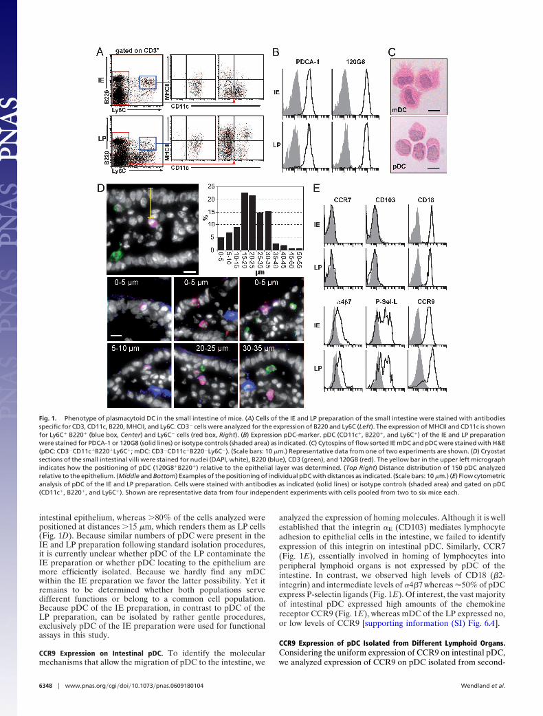
ResultsCharacterization of Plasmacytoid Dendritic Cells of the Small Intes-tine. We applied standard procedures to isolate immune cellslocated in the epithelium (intraepithelial, IE) and the LP fromthe intestine. In both cell preparations we found a distinctpopulation of CD11c�B220�Ly6C� cells that accounted for upto 1% of all cells. In contrast to pDC, myeloid (m)DC(CD11c�MHCII�CD3�B220�Ly6C�) were present only at verylow numbers in the IE preparation (Fig. 1A). Both pDC of theLP and IE preparation showed low levels of surface MHC classII expression (Fig. 1 A). Further analysis revealed thatCD11c�B220�Ly6C� cells of both preparations uniformly ex-press the pDC markers PDCA1 as well as 120G8 (Fig. 1B).Cytospins from sorted pDC (CD11c�B220�Ly6C�) and myeloidDC (mDC) of the IE preparation revealed a round and smooth,plasma cell-like morphology of pDC, whereas mDC showed thecharacteristic dendrites (Fig. 1C). To further characterize thelocalization of pDC within the intestine we applied anti-B220,anti-120G8, and anti-CD3 mAb in immunohistology. Micro-graphs were randomly taken from sections and, as depictedin Fig. 1D, the positioning of 120G8�B220�CD3� cells wasdetermined relative to epithelial cells by using image analysis(analySIS; Olympus, Hamburg, Germany). Evaluating the posi-tioning of �150 pDC we observed that 4.9% of these cells wereclearly located within the epithelial cell layer whereas another6.7% were situated within a distance of 5–10 �m from the apicaltip of the epithelial cells (Fig. 1D). These data demonstrate thata certain amount of the pDC locate within or close to the
Author contributions: O.P. and R.F. contributed equally to this work; O.P. and R.F. designedresearch; M.W. and N.C. performed research; N.M., B.M., and E.K. contributed new re-agents/analytic tools; M.W. and N.C. analyzed data; and R.F. wrote the paper.
¶To whom correspondence should be addressed at: Institute of Immunology, HannoverMedical School, Carl Neuberg Strasse 1, 30625 Hannover, Germany. E-mail:[email protected].
This article contains supporting information online at www.pnas.org/cgi/content/full/0609180104/DC1.
intestinal epithelium, whereas �80% of the cells analyzed werepositioned at distances �15 �m, which renders them as LP cells(Fig. 1D). Because similar numbers of pDC were present in theIE and LP preparation following standard isolation procedures,it is currently unclear whether pDC of the LP contaminate theIE preparation or whether pDC locating to the epithelium aremore efficiently isolated. Because we hardly find any mDCwithin the IE preparation we favor the latter possibility. Yet itremains to be determined whether both populations servedifferent functions or belong to a common cell population.Because pDC of the IE preparation, in contrast to pDC of theLP preparation, can be isolated by rather gentle procedures,exclusively pDC of the IE preparation were used for functionalassays in this study.
CCR9 Expression on Intestinal pDC. To identify the molecularmechanisms that allow the migration of pDC to the intestine, we
analyzed the expression of homing molecules. Although it is wellestablished that the integrin �E (CD103) mediates lymphocyteadhesion to epithelial cells in the intestine, we failed to identifyexpression of this integrin on intestinal pDC. Similarly, CCR7(Fig. 1E), essentially involved in homing of lymphocytes intoperipheral lymphoid organs is not expressed by pDC of theintestine. In contrast, we observed high levels of CD18 (�2-integrin) and intermediate levels of �4�7 whereas �50% of pDCexpress P-selectin ligands (Fig. 1E). Of interest, the vast majorityof intestinal pDC expressed high amounts of the chemokinereceptor CCR9 (Fig. 1E), whereas mDC of the LP expressed no,or low levels of CCR9 [supporting information (SI) Fig. 6A].
CCR9 Expression of pDC Isolated from Different Lymphoid Organs.Considering the uniform expression of CCR9 on intestinal pDC,we analyzed expression of CCR9 on pDC isolated from second-
Fig. 1. Phenotype of plasmacytoid DC in the small intestine of mice. (A) Cells of the IE and LP preparation of the small intestine were stained with antibodiesspecific for CD3, CD11c, B220, MHCII, and Ly6C. CD3� cells were analyzed for the expression of B220 and Ly6C (Left). The expression of MHCII and CD11c is shownfor Ly6C� B220� (blue box, Center) and Ly6C� cells (red box, Right). (B) Expression pDC-marker. pDC (CD11c�, B220�, and Ly6C�) of the IE and LP preparationwere stained for PDCA-1 or 120G8 (solid lines) or isotype controls (shaded area) as indicated. (C) Cytospins of flow sorted IE mDC and pDC were stained with H&E(pDC: CD3�CD11c�B220�Ly6C�; mDC: CD3�CD11c�B220�Ly6C�). (Scale bars: 10 �m.) Representative data from one of two experiments are shown. (D) Cryostatsections of the small intestinal villi were stained for nuclei (DAPI, white), B220 (blue), CD3 (green), and 120G8 (red). The yellow bar in the upper left micrographindicates how the positioning of pDC (120G8�B220�) relative to the epithelial layer was determined. (Top Right) Distance distribution of 150 pDC analyzedrelative to the epithelium. (Middle and Bottom) Examples of the positioning of individual pDC with distances as indicated. (Scale bars: 10 �m.) (E) Flow cytometricanalysis of pDC of the IE and LP preparation. Cells were stained with antibodies as indicated (solid lines) or isotype controls (shaded area) and gated on pDC(CD11c�, B220�, and Ly6C�). Shown are representative data from four independent experiments with cells pooled from two to six mice each.
6348 � www.pnas.org�cgi�doi�10.1073�pnas.0609180104 Wendland et al.
ary lymphoid organs including spleen, skin-draining LN, mes-enteric LN, and Peyer’s patches (PP; Fig. 2A) and usedCD4�CD8� thymocytes, known to express high levels of CCR9,as a positive control (11); Fig. 2B). Interestingly, only a fractionof pDC present in any of these organs expressed CCR9 (Fig. 2 A),whereas �95% of pDC isolated from the BM carried thisreceptor (Fig. 2C). Most BM pDC also express CCR5 andCXCR3 whereas CCR2 is present on only roughly 25% of thecells. CXCR4, known to retain cells to the BM, is only veryweakly expressed (Fig. 2C). Together, these data demonstratethat pDC are equipped with various chemokine receptors beforebeing released from the BM. This feature presumably allowshoming not only to places of inflammation but also to thenoninflamed intestine.
Chemotaxis Analysis of Flt3L-Expanded pDC. Based on the strongexpression of CCR9 on BM and intestinal pDC, we compared themigration capacity of pDC and mDC toward the chemokineCCL25/TECK, which is the sole known ligand for this receptor(12). The frequency of pDC varies between different mousestrains and it is well known that, for example, C57BL/6 (B6) miceharbor much less pDC than 129SV mice (13). However, irre-spective of the genetic background the number of pDC that canbe isolated from mouse tissues is insufficient to conduct standardin vitro chemotaxis assays. To overcome this limitation weexpanded the DC population in vivo by implanting a Flt3-L-secreting tumor cell line (B16-FL) in B6 mice for 14 days (14).
During this time period, the percentage of CD11c� cellspresent in the spleen increased from 3% to 30–35% (data notshown). Eighty percent of the in vivo expanded pDC expressedCCR9, with levels very similar to those present on BM pDC(Figs. 2C and Fig. 4C) whereas in vivo expanded mDC expressedonly small amounts of this receptor (SI Fig. 6B). Spleen pDCwere enriched by CD11c� MACS-sorting, resulting in 95%purity for CD11c� cells that contained �15% Ly6C�B220�
pDC. In vitro transwell migration assays revealed strong chemo-tactic response of pDC to CCL25, as well as to CXCL9, a ligandfor CXCR3 and to CXCL12, which is a ligand for CXCR4. Onlya weak response was observed toward CCL19, which serves as aligand for CCR7. mDC showed little chemotactic responsetoward CCL25 and CXCL9 and moderate response towardCXCL12 and CCL19 (Fig. 3A).
Reduced Numbers of pDC in the Small Intestine of CCR9-DeficientMice. Based on these observations, we characterized the distri-bution of pDC in CCR9-deficient mice. We found similarpercentages of pDC in lung and liver (SI Fig. 7) as well as ininguinal and mesenteric lymph nodes whereas the number ofsplenic pDC was slightly increased in CCR9�/� mice. In contrast,we observed a �90% decrease of intestinal and a 50% reductionof PP pDC (Fig. 3B).
pDC Preferentially Migrate to the Small Intestine. Based on thefindings described so far, it seemed likely that pDC requireCCR9 for homing to the small intestine. To prove this hypoth-esis, we adoptively transferred cells from B6 and CCR9�/�
donors that carried a Flt3-L-secreting tumor for 14 days. Underthe influence of Flt3L pDC expanded to a similar extent in B6and CCR9�/� mice (Fig. 4A and SI Fig. 8) and were indistin-guishable regarding the expression of CCR2, CCR5, and CXCR3whereas CCR9 was only detected on pDC derived from B6 butnot CCR9-deficient donors (Fig. 4C). Without further purifica-tion, splenocytes of these donors were labeled with 5(6)-carboxyfluorescein diacetate N-succinimidyl ester (CFSE) and5(6)-carboxytetramethylrhodamine N-succinimidyl ester(TAMRA) respectively. A mixture of WT and CCR9-deficientcells, adjusted to contain equal numbers of pDC, was i.v.transferred in B6 recipients. After 18 h of transfer, we firstanalyzed the composition of adoptively transferred WT cellspresent in the IE as well as LP preparation and noticed that 54%and 25% of all cells recovered from the IE and LP fractionrespectively were pDC (Fig. 4B). These findings demonstratethat among the diverse cell populations transferred pDC homemost efficiently to the intestine. These competitive transfers alsorevealed that CCR9-deficient pDC are largely impaired in theircapacity to home to the intestine as reflected by the lowmigration ratio of CCR9-deficient vs. WT pDC found in the IEand LP preparation (Fig. 4D). In contrast, CCR9-deficient andB6 pDC migrated with similar efficiency to peripheral andmesenteric lymph nodes (Fig. 4D). The nature of the cell labelinghad no impact on these experiments because interchanging thedyes between B6 and CCR9�/� cells yielded identical results(data not shown). Although these data clearly demonstrate thatpDC require CCR9 for efficient homing to the gut, it should bementioned that the trafficking properties of pDC from Flt3-
Fig. 2. Expression of CCR9 on pDC. (A) Cells were isolated from different lymphoid organs as indicated. pDC were addressed as CD11c�B220�Ly6C� and analyzedfor the expression of CCR9 (solid line). (B) CD4�CD8� thymocytes served as a positive control. (C) Expression of chemokine receptors (solid lines) on pDC isolatedfrom the BM (shaded area: isotype control). SPL, spleen; ALN, axillary LN; MLN, mesenteric LN; PP, Peyer’s patches; Thy, thymus). Representative data from fourindependent experiments with cells pooled from two to six mice each (A and B) or from three mice (C).
Wendland et al. PNAS � April 10, 2007 � vol. 104 � no. 15 � 6349
ligand treated mice might be different from those present underphysiologic situations.
pDC Are Recruited to the Inflamed Intestine. Because it is knownthat pDC play an important role in anti viral immunity (4, 10)we speculated that pDC might be recruited to the intestineduring inflammatory processes to strengthen first line defense.Cholera toxin (CT) is known to induce intestinal inflammationwhen administered orally and it has been reported that within 2 hafter oral application the number of mDC transiently recruitedto the intestine increases 4-fold (15). We observed that, inaddition to mDC, pDC numbers also increased 3-fold whenfeeding B6 mice with 10 �g of CT (Fig. 4E). Of interest, theidentical experimental setup never resulted in any increase ofpDC neither in the IE nor the LP preparation of CCR9-deficientmice after 1, 2, or 3 h of CT-treatment (Fig. 4E and data notshown). The specific requirement for CCR9 on pDC for theirrecruitment to the intestine during inflammatory processes was
Fig. 4. CCR9-dependent homing of pDC to the small intestine. (A–D and F)DC were expanded in vivo by treating B6 and CCR9-deficient mice withFlt3L-secreting tumor cells. (A) Splenocytes of B6 donors were analyzed for thepresence of pDC (B220�Ly6C�). (B) Nonpurified WT Flt3L-expanded spleno-cytes were labeled with CFSE and i.v. injected into recipients. The occurrenceof donor pDC in the recipient’s IE (Upper) and LP (Lower) preparation wasanalyzed 18 h later (gate on CFSE� cells). (A and B) Data shown are represen-tative for five animals of two independent experiments. (C)CD11�B220�Ly6C� pDC in Flt3L-expanded splenocytes of B6 (Upper) andCCR9-deficient mice (Lower) were stained for the expression of differentchemokine receptors as indicated. (D) Splenocytes isolated from Flt3L-treatedB6 or CCR9-deficient mice were labeled with CFSE and TAMRA, respectively.Cells were adjusted to equal numbers of pDC and injected at a ratio of 1:1 inB6 recipients. After 18 h, recipients were killed, and the ratio of donor B6 andCCR9-deficient pDC was analyzed in the recipient’s IE and LP preparation ofthe intestine and the inguinal (ILN) and mesenteric (MLN) lymph node (mean� SD; n � 5–9 recipients). (E) WT (�/�) and CCR9-deficient (�/�) mice received10 �g of CT or saline orally. After 1 h, animals were killed, and the number ofpDC present in the small intestine IE preparation was determined. Circlesrepresent individual mice; bars are mean values. Similar results were obtainedin two additional experiments. (F) CFSE-labeled splenocytes isolated fromFlt3L-treated B6 and TAMRA-labeled splenocytes isolated from Flt3L-treatedCCR9-deficient mice were adoptively transferred to CCR9-deficient mice at aratio of 1:1. After 18 h, recipients were gavaged orally with 10 �g of CT. Onehour later, mice were killed, and the number of labeled WT (�/�) and CCR9�/�
(�/�) pDC isolated from the IE and LP preparation was analyzed. Circlesrepresent individual mice; bars are mean values.
Fig. 3. Chemotactic response of pDC toward the CCR9 ligand CCL25. (A) DCwere expanded in vivo by treating B6 mice with Flt3L-secreting tumor cells for14 days. Chemotactic activity of splenic pDC and mDC toward differentconcentrations of CCL25, CXCL9, CXCL12, and CCL19 was analyzed (opencolumns, mDC; black columns, pDC; mean � SD; n � 4 independent experi-ments with pooled cells from two or three mice each). (B) Lack of intestinalpDC in CCR9-deficient mice. Shown are the percentage (Left) and number(Right) of pDC isolated from the inguinal LN (ILN), mesenteric LN (MLN), spleen(SPL), PP, and the IE and the LP preparation of the small intestine from B6 andCCR9-deficient mice. Circles represent data of individual mice (n � 3); barsshow mean values. Similar results were obtained in four additional experi-ments using mice on a mixed genetic background (BALB/c 129SV; n � 20 miceper genotype).
6350 � www.pnas.org�cgi�doi�10.1073�pnas.0609180104 Wendland et al.
confirmed by adoptive transfer of WT and CCR9-deficient pDCto CCR9-deficient recipients followed by application of CT asdescribed above. Whereas adoptively transferred WT pDC wereamply present in the IE and LP preparation, CCR9-deficientpDC were almost completely excluded from these compartments(Fig. 4F). Together, these results show that during inflammatoryevents pDC can be recruited to the intestinal mucosa and thatthis mechanism relies to a large extend on CCR9.
A Role for Intestinal pDC for the Rapid Mobilization of LP Myeloid DC.It has been shown recently that oral application of the TLR7/8ligand resiquimod (R848) results in the rapid mobilization of LPDC and that TNF�, possibly released by pDC, is involved in thisprocess (16). We thus speculated that intestinal pDC might bethe source of TNF� that potentially triggers the mobilization ofneighboring mDC. To test this hypothesis, we in vitro stimulatedB6 pDC of the IE and LP preparation for 16 h with R848. Indeed,pDC secreted considerable amounts of TNF� but failed toproduce any detectable quantities of IL-2, IL-4, IL-5, or IFN�(Fig. 5A). We then analyzed the mobilization of LP mDC in vivoafter oral application of R848. Whereas untreated B6 andCCR9�/� mice did not differ regarding the presence of intestinalmDC (Fig. 5B), within 2 h oral R848 induced the mobilizationof �60% of mDC in WT but only 10.8% in CCR9�/� mice.Importantly, once CCR9-deficient mice i.v. received splenicpDC of Flt3L-treated WT donors 16 h prior oral application ofR848, this deficiency in intestinal mDC mobilization could becompletely rescued (Fig. 5C). These experiments show that aCCR9-dependent homing of pDC to the intestine is involved inthe rapid mobilization of intestinal mDC after oral applicationof a TLR7/8 ligand. Because it has been shown by others in therat model that application of LPS also induces mobilization of LPmDC (17), we applied 50 �g of LPS i.p. to WT and CCR9-deficient animals. Of interest, under these experimental condi-tions we failed to observe any difference between WT andCCR9-deficient animals regarding the mobilization of LP mDC(SI Fig. 9).
DiscussionThe CCR9 ligand, CCL25, is expressed by epithelial cells of thesmall intestine and has been suggested to target immune cells tothe intestinal epithelium (18). The present study supports theidea that this chemokine attracts defined populations of immunecells to the small intestine. Agace and colleagues (19) further
demonstrated that CD8��� T cells, activated within the mes-enteric LN, selectively home to the small intestinal mucosa andthat this homing depends on CCR9. Data from our group suggesta similar mechanism for plasma cells (11). Results provided heredemonstrate that CCR9-deficient mice possess reduced numbersof pDC in the small intestine under steady-state conditions, anobservation that correlates well with the impaired recruitment ofCCR9-deficient pDC to this organ under inflammatory condi-tions. In accordance with the hypothesis that CCR9 is requiredfor pDC gut homing is our finding that pDC derived fromCCR9-deficient donors are impaired in homing to the intestineonce adoptively transferred to WT recipients.
In addition to targeting immune cells to the epithelium,CCL25 also mediates T cell entrance into the LP across intestinalvenules (20). A similar mechanism might allow homing of pDCto the small intestine. Therefore, it seems possible that CCR9recruits pDC into the LP and, in addition, targets a fraction ofthese cells to the epithelium. Apart from CCR9, it is currentlyunclear, which adhesion molecules are involved in pDC homingto the intestine. Our data would suggest that �4�7 integrin, aswell as P-selectin, might also be involved in this process.
This study also reveals a previously undescribed function forintestinal pDC. We show that after oral application of a TLR7/8ligand, intestinal pDC are required for the rapid mobilization ofLP mDC, a mechanism that might involve the release of TNF�from this cell population. Although it is currently unclear howimpaired mobilization might affect immunity to pathogens, itseems conceivable that the rapid mobilization of LP DC to themesenteric LN favors the fast onset of adaptive immunity.Interestingly, pDC mediated mobilization of mDC seems re-stricted to distinct TLR ligands because LPS-activity bypassesthe need of pDC for successful emigration of mDC from thesmall intestine. This finding corroborates the concept thatpathogens may bias immune responses already at the early stageof their entry into the body because it is known that immatureDC primed under distinct cytokine environment such as TNF�cause a shift to the subsequent Thelper1/Thelper2 answer. Further-more, it is also tempting to speculate that pDC might help toenforce the armed battery of IE lymphocytes residing at thefrontline of mucosal immune defense. In particular intestinalpDC might supplement mucosal protection against viral attack.However, these scenarios still await experimental approval en-compassing animal models for inflammatory bowel disease andviral infections.
Fig. 5. Rapid mobilization of LP mDC relies on the intestinal pDC. (A) Cytokine bead array profile from the supernatant of sorted pDC of IE (Left) and LP (Right)preparation after 16 h in vitro stimulation in the absence (�R848) or presence (�R848) of R848. Control, cell culture medium. (B) Percentage of LP mDC(CD45�CD11c�MHCIIhigh) of untreated mice (mean �SD, n � 6 per group). (C) WT (open column) or CCR9�/� mice (black column) were orally gavaged with 10�g of R848. After 2 h, the number of mDC (CD45�CD11c�MHCIIhigh) present in the LP of the small intestine was determined and expressed as percentage ofuntreated WT control. Gray column, CCR9�/� mice that i.v. received MACS-purified B6 pDC 16 h prior R848 treatment (mean � SD; n � 6–11 mice per group; ns,not significant; **, P � 0.01; ***, P � 0.001).
Wendland et al. PNAS � April 10, 2007 � vol. 104 � no. 15 � 6351
Materials and MethodsMice. Animals were bred under specific pathogen-free condi-tions. CCR9-deficient mice, either on a mixed genetic (BALB/c � 129SV) or a C57BL/6 background (backcrosses for 5 or 9generations) have been described elsewhere (21). Most of theexperiments described in this manuscript were performed onboth genetic backgrounds yielding identical results. Data de-picted derive from experiments performed with mice on aC57BL/6 background except those depicted in Fig. 4E. Allanimal experiments were conducted in accordance with localand institutional guidelines.
Flow Cytometry. Immune cells of the intestine were isolated from6- to 8-week-old mice as recently described in detail (22). Cellswere stained with the following antibodies: Ly6C-FITC, �4�7-biotin, CD103-biotin, B220-PerCP, CD11c-PE (all from BDBioscience), CD4-PE, CD62L-PE, CD45-APC, CD18-FITC(Caltag), P-selectin-ligand (R & D Systems), CCR7-biotin(eBioscience), 120G8, (Vector Laboratories), PDCA-1-APC(Dianova). Anti CD3-Cy5 (clone 17A2) and anti CD8 (cloneCD8.2) were grown in our laboratories. Anti CCR2 and CCR5mAb were kindly provided by Matthias Mack (University ofRegensburg, Regensburg, Germany) (23). The rat anti-mouseCCR9 mAb (clone 7E7) was produced in our lab and has beendescribed (11).
Immunohistology and Cytospins. Immunhistological analysis of thesmall intestine of mice was done on 8-�m cryosections asdescribed (11, 22). pDC (CD3�CD11c�B220�Ly6C�) and mDC(CD3�CD11c�B220�Ly6C�) of the IE preparation were sortedby flow cytometry (FACSAria, BD Biosciences). Acetone-fixedcytospins were prepared from sorted cells.
In Vivo Generation of pDC and in Vitro Migration Assay. B6 andCCR9-deficient mice received s.c. 5 � 105 to 1 � 106 B16-FLcells, a murine melanoma tumor cell line engineered to stablyproduce murine Flt3-L (14). After 14 days, animals were killed.Flt3L-expanded, CD11c� MACS-sorted splenocytes (1 � 106),containing �15% pDC, were resuspended in 100 �l of RPMImedium 1640 and loaded into collagen-coated transwells (Corn-ing BV; 5 �m pore size) that were placed in 24-well platescontaining 400 �l medium or medium supplemented with var-ious concentrations of CCL25, CXCL9, CCL19, or CXCL12 (R& D systems). After 3 h of incubation at 37°C, the migrated cellswere collected, counted, and stained with mAb to determine by
flow cytometry the number of migrated pDC and mDC. Theratio of the number of pDC that migrated in the presence ofchemokine vs. the number of cells that migrated to PBS controlwas calculated and is given as the migration index.
Adoptive Transfer of Labeled Cells. Splenocytes from B16-FLtumor-carrying B6 or CCR9-deficient mice were labeled withTAMRA (red fluorescent) or CFSE (green fluorescent) or viceversa. Cell populations were adjusted to contain equal numbersof pDC. For adoptive transfers, 106 pDC for both colors were i.v.injected into the tail vein of recipients. After 18 h, recipientswere killed and cells were isolated from the intestine as well asfrom mesenteric and peripheral lymph nodes.
MACS-Purification of pDC. Splenocytes from B16-FL tumor-bearing mice were negative sorted for CD3 and CD19. In asubsequent step B220� cells were enriched.
In Vivo Mobilization of Cells. Ten micrograms of CT (Sigma) in 300�l of carbonic buffer (0.1 M NaHCO3) or 10 �g of R848 in 300�l of PBS were orally administered by gavage. Fifty microgramsof LPS were i.p. injected in 150 �l of PBS. One hour to 3 h afterthe application of CT, mice were killed, and the number ofintestinal pDC was determined. Mice that received R848 werekilled 2 h after the application of this drug, and the number ofmDC of the LP was determined. Mice that received LPS werekilled 12 h later, and the number of LP mDC was determined.Some of the R848-treated CCR9�/� mice received 2–4 � 106
MACS-purified WT pDC i.v. 16 h prior R848 application.
In Vitro Stimulation of pDC. 106 MACS-purified pDC were culturedin 200 �l of RPMI medium 1640/10% FCS for 14 h in the absenceor presence of CpG2216 (16.5 �g/ml) or R848 (2 �g/ml). mDC(CD11c�MHCII�) of the LP preparation were sorted by flowcytometry (MoFlo, Dako-Cytomation) and activated withCpG2216 as describe above. Supernatants were collected and theamount of IFN-� determined by ELISA (Hycult). Interleukinsand TNF� were detected by cytokine bead arrays (BD).
We thank Matthias Mack for providing CCR2 and CCR5 mAb andGunter Bernhardt for valuable suggestions on the manuscript. Thegeneration and characterization of CCR9-deficient mice was supportedby the Association pour la Recherche sur le Cancer and MUGEN. Thiswork was supported by Deutsche Forschungsgemeinschaft GrantSFB621-A1 (to R.F.).
1. Siegal FP, Kadowaki N, Shodell M, Fitzgerald-Bocarsly PA, Shah K, Ho S,Antonenko S, Liu YJ (1999) Science 284:1835–1837.
2. Iwasaki A, Medzhitov R (2004) Nat Immunol 5:987–995.3. Nakano H, Yanagita M, Gunn MD (2001) J Exp Med 194:1171–1178.4. Asselin-Paturel C, Boonstra A, Dalod M, Durand I, Yessaad N, Dezutter-
Dambuyant C, Vicari A, O’Garra A, Biron C, Briere F, Trinchieri G (2001) NatImmunol 2:1144–1150.
5. Maraskovsky E, Brasel K, Teepe M, Roux ER, Lyman SD, Shortman K,McKenna HJ (1996) J Exp Med 184:1953–1962.
6. Gilliet M, Boonstra A, Paturel C, Antonenko S, Xu XL, Trinchieri G, O’GarraA, Liu YJ (2002) J Exp Med 195:953–958.
7. Diacovo TG, Blasius AL, Mak TW, Cella M, Colonna M (2005) J Exp Med202:687–696.
8. Yoneyama H, Matsuno K, Zhang Y, Nishiwaki T, Kitabatake M, Ueha S,Narumi S, Morikawa S, Ezaki T, Lu B, et al. (2004) Int Immunol 16:915–928.
9. Kohrgruber N, Groger M, Meraner P, Kriehuber E, Petzelbauer P, Brandt S,Stingl G, Rot A, Maurer D (2004) J Immunol 173:6592–6602.
10. Cella M, Jarrossay D, Facchetti F, Alebardi O, Nakajima H, Lanzavecchia A,Colonna M (1999) Nat Med 5:919–923.
11. Pabst O, Ohl L, Wendland M, Wurbel MA, Kremmer E, Malissen B, ForsterR (2004) J Exp Med 199:411–416.
12. Zabel BA, Agace WW, Campbell JJ, Heath HM, Parent D, Roberts AI, EbertEC, Kassam N, Qin S, Zovko M, et al. (1999) J Exp Med 190:1241–1256.