CD11b regulates obesity-induced insulin resistance via limiting alternative activation and proliferation of adipose tissue macrophages Chunxing Zheng a , Qian Yang a , Chunliang Xu a , Peishun Shou a , Jianchang Cao a , Menghui Jiang a , Qing Chen a , Gang Cao a , Yanyan Han a , Fengying Li a , Wei Cao a , Liying Zhang b , Li Zhang c , Yufang Shi a,b,d,1 , and Ying Wang a,1 a Key Laboratory of Stem Cell Biology, Institute of Health Sciences, Shanghai Institutes for Biological Sciences, Chinese Academy of Sciences/Shanghai Jiao Tong University School of Medicine, Shanghai 200031, China; b The First Affiliated Hospital of Soochow University, Institutes for Translational Medicine, Soochow University, Suzhou 215006, China; c Center for Vascular and Inflammatory Diseases, Department of Physiology, University of Maryland School of Medicine, Baltimore, MD 21201; and d Child Health Institute of New Jersey, Rutgers-Robert Wood Johnson Medical School, New Brunswick, NJ 08901 Edited by Ruslan Medzhitov, Yale University School of Medicine, New Haven, CT, and approved November 24, 2015 (received for review January 8, 2015) Obesity-associated inflammation is accompanied by the accumula- tion of adipose tissue macrophages (ATMs), which is believed to predispose obese individuals to insulin resistance. CD11b (integrin α M ) is highly expressed on monocytes and macrophages and is crit- ical for their migration and function. We found here that high-fat diet–induced insulin resistance was significantly reduced in CD11b- deficient mice. Interestingly, the recruitment of monocytes to adipose tissue is impaired when CD11b is deficient, although the cellularity of ATMs in CD11b-deficient mice is higher than that in wild-type mice. We further found that the increase in ATMs is caused mainly by their vig- orous proliferation in the absence of CD11b. Moreover, the prolifera- tion and alternative activation of ATMs are regulated by the IL-4/STAT6 axis, which is inhibited by CD11b through the activity of phosphatase SHP-1. Thus, CD11b plays a critical role in obesity-induced insulin resis- tance by limiting the proliferation and alternative activation of ATMs. integrin CD11b | macrophage proliferation | alternative activation | obesity | insulin resistance O besity is associated with chronic inflammation characterized by progressive accumulation of immune cells in adipose tissue. Cytokines secreted by these immune cells, such as TNF-α, have been demonstrated to augment adipose tissue inflammation and consequentially induce insulin resistance (1, 2). As one of the major cell types that contribute to the proinflammatory response, macrophages are central players in obesity-related inflammation (3, 4). This heterogeneous cell population possesses broad plasticity that can be influenced by the local microenvironment. Changes in macrophage properties play distinct roles in regulating inflamma- tion and also metabolic responses (5, 6). Classically activated mac- rophages (CAMs) secrete proinflammatory cytokines such as TNF-α, IL-6, and IL-1 (7). The accumulation of CAMs in adipose tissue exacerbates the development of obesity and subsequent tissue inflammation and insulin resistance. On the other hand, resident macrophages in adipose tissue of lean mice display a phenotype of alternatively activated macrophages (AAMs) with high expression of IL-10, Ym1/chitinase3-like3, and arginase1 (8). It has been reported that AAMs driven by IL-4 and IL-13 could improve insulin sensitivity (9, 10). Thus, differential accumulation of these two macrophage populations could result in distinct metabolic states and could influence the progression of insulin resistance. Traditionally, the accumulation of macrophages is considered as the migration of monocytes to inflammatory sites and subsequent differentiation into macrophages (5). One of the molecules that control monocytes immigration is integrin α M (CD11b), which combines with integrin β 2 (CD18) to form MAC-1 (integrin α M β 2 ). MAC-1 is well known for its role in regulating leukocyte trans- migration through endothelial cells (11, 12). The function of CD11b is dependent on a cascade of inside– out and outside–in activation signaling processes. The activation of tyrosine kinases, especially members of the Src family, is indispensable for the outside–in sig- naling of different integrins (13). In addition to its role in regulating leukocyte adhesion and migration, CD11b has been reported to modulate various aspects of immune responses. Our previous studies showed that CD11b on antigen-presenting cells plays a critical role in promoting oral tolerance by inhibiting Th17 differentiation (14). Moreover, CD11b also has been shown to suppress Toll-like receptor– initiated signals in macrophages and thus to protect animals from endotoxic shock (15). Interestingly, activation of CD11b im- pedes the accumulation of lipid in macrophages and the formation of foam cells in the presence of IL-13 (16). Its negative regulation of the immune response also can be observed in B cells, because CD11b has been shown to inhibit the autoreactive B-cell response in systemic lupus erythematous (17). Importantly, CD11b is found to be involved in regulating body fat deposition (18); however, the effect of CD11b in obesity-induced insulin resistance has not been reported. In the present study, we show that CD11b is important for the influx of monocytes to adipose tissue during the development of obesity. Surprisingly, the accumulation of macrophages in the adipose tissue of CD11b-deficient mice is significantly increased compared with that in wild-type mice. We further demonstrated that CD11b de- ficiency promotes in situ proliferation of adipose tissue macrophages (ATMs), a process mediated by the IL-4/STAT6 signaling pathway. The infiltrated ATMs in CD11b-deficient mice phenotypically re- semble AAMs. Depletion of these ATMs reversed the reduction of Significance Obesity is associated with long-term low-grade inflammation characterized by the accumulation of adipose tissue macrophages (ATMs). One important molecule that regulates the migration of monocytes/macrophages is CD11b (integrin α M ). Here we show an unexpected role of CD11b in modulating the IL-4/STAT6 sig- naling in macrophages, thereby limiting IL-4/STAT6–mediated proliferation and alternative activation of ATMs. In the absence of CD11b, there is an increase in ATM in situ proliferation and an enhancement of alternatively polarized phenotypes. Importantly, the alternatively activated ATMs attenuate obesity-related in- sulin resistance in CD11b-deficient mice. These results reveal a previously unidentified physiological function of CD11b, which could be a therapeutic target for insulin resistance. Author contributions: C.Z., Y.S., and Y.W. designed research; C.Z., Q.Y., J.C., M.J., Q.C., G.C., Y.H., F.L., W.C., and Liying Zhang performed research; C.Z., Q.Y., C.X., P.S., Li Zhang, and Y.W. analyzed data; and C.Z., Y.S., and Y.W. wrote the paper. The authors declare no conflict of interest. This article is a PNAS Direct Submission. Freely available online through the PNAS open access option. 1 To whom correspondence may be addressed. Email: [email protected] or yingwang@ sibs.ac.cn. This article contains supporting information online at www.pnas.org/lookup/suppl/doi:10. 1073/pnas.1500396113/-/DCSupplemental. www.pnas.org/cgi/doi/10.1073/pnas.1500396113 PNAS | Published online December 15, 2015 | E7239–E7248 IMMUNOLOGY AND INFLAMMATION PNAS PLUS
Transcript
CD11b regulates obesity-induced insulin resistance vialimiting alternative activation and proliferation ofadipose tissue macrophagesChunxing Zhenga, Qian Yanga, Chunliang Xua, Peishun Shoua, Jianchang Caoa, Menghui Jianga, Qing Chena, Gang Caoa,Yanyan Hana, Fengying Lia, Wei Caoa, Liying Zhangb, Li Zhangc, Yufang Shia,b,d,1, and Ying Wanga,1
aKey Laboratory of Stem Cell Biology, Institute of Health Sciences, Shanghai Institutes for Biological Sciences, Chinese Academy of Sciences/Shanghai JiaoTong University School of Medicine, Shanghai 200031, China; bThe First Affiliated Hospital of Soochow University, Institutes for Translational Medicine,Soochow University, Suzhou 215006, China; cCenter for Vascular and Inflammatory Diseases, Department of Physiology, University of Maryland School ofMedicine, Baltimore, MD 21201; and dChild Health Institute of New Jersey, Rutgers-Robert Wood Johnson Medical School, New Brunswick, NJ 08901
Edited by Ruslan Medzhitov, Yale University School of Medicine, New Haven, CT, and approved November 24, 2015 (received for review January 8, 2015)
Obesity-associated inflammation is accompanied by the accumula-tion of adipose tissue macrophages (ATMs), which is believed topredispose obese individuals to insulin resistance. CD11b (integrinαM) is highly expressed on monocytes and macrophages and is crit-ical for their migration and function. We found here that high-fatdiet–induced insulin resistance was significantly reduced in CD11b-deficient mice. Interestingly, the recruitment of monocytes to adiposetissue is impaired when CD11b is deficient, although the cellularity ofATMs in CD11b-deficient mice is higher than that in wild-type mice. Wefurther found that the increase in ATMs is caused mainly by their vig-orous proliferation in the absence of CD11b. Moreover, the prolifera-tion and alternative activation of ATMs are regulated by the IL-4/STAT6axis, which is inhibited by CD11b through the activity of phosphataseSHP-1. Thus, CD11b plays a critical role in obesity-induced insulin resis-tance by limiting the proliferation and alternative activation of ATMs.
Obesity is associated with chronic inflammation characterizedby progressive accumulation of immune cells in adipose
tissue. Cytokines secreted by these immune cells, such as TNF-α,have been demonstrated to augment adipose tissue inflammationand consequentially induce insulin resistance (1, 2). As one of themajor cell types that contribute to the proinflammatory response,macrophages are central players in obesity-related inflammation (3,4). This heterogeneous cell population possesses broad plasticitythat can be influenced by the local microenvironment. Changes inmacrophage properties play distinct roles in regulating inflamma-tion and also metabolic responses (5, 6). Classically activated mac-rophages (CAMs) secrete proinflammatory cytokines such asTNF-α, IL-6, and IL-1 (7). The accumulation of CAMs in adiposetissue exacerbates the development of obesity and subsequent tissueinflammation and insulin resistance. On the other hand, residentmacrophages in adipose tissue of lean mice display a phenotype ofalternatively activated macrophages (AAMs) with high expressionof IL-10, Ym1/chitinase3-like3, and arginase1 (8). It has beenreported that AAMs driven by IL-4 and IL-13 could improve insulinsensitivity (9, 10). Thus, differential accumulation of these twomacrophage populations could result in distinct metabolic statesand could influence the progression of insulin resistance.Traditionally, the accumulation of macrophages is considered as
the migration of monocytes to inflammatory sites and subsequentdifferentiation into macrophages (5). One of the molecules thatcontrol monocytes immigration is integrin αM (CD11b), whichcombines with integrin β2 (CD18) to form MAC-1 (integrin αMβ2).MAC-1 is well known for its role in regulating leukocyte trans-migration through endothelial cells (11, 12). The function of CD11bis dependent on a cascade of inside–out and outside–in activationsignaling processes. The activation of tyrosine kinases, especiallymembers of the Src family, is indispensable for the outside–in sig-
naling of different integrins (13). In addition to its role in regulatingleukocyte adhesion and migration, CD11b has been reported tomodulate various aspects of immune responses. Our previous studiesshowed that CD11b on antigen-presenting cells plays a critical role inpromoting oral tolerance by inhibiting Th17 differentiation (14).Moreover, CD11b also has been shown to suppress Toll-likereceptor–initiated signals in macrophages and thus to protect animalsfrom endotoxic shock (15). Interestingly, activation of CD11b im-pedes the accumulation of lipid in macrophages and the formationof foam cells in the presence of IL-13 (16). Its negative regulation ofthe immune response also can be observed in B cells, becauseCD11b has been shown to inhibit the autoreactive B-cell response insystemic lupus erythematous (17). Importantly, CD11b is found to beinvolved in regulating body fat deposition (18); however, the effect ofCD11b in obesity-induced insulin resistance has not been reported.In the present study, we show that CD11b is important for the influxof monocytes to adipose tissue during the development of obesity.Surprisingly, the accumulation of macrophages in the adipose tissueof CD11b-deficient mice is significantly increased compared withthat in wild-type mice. We further demonstrated that CD11b de-ficiency promotes in situ proliferation of adipose tissue macrophages(ATMs), a process mediated by the IL-4/STAT6 signaling pathway.The infiltrated ATMs in CD11b-deficient mice phenotypically re-semble AAMs. Depletion of these ATMs reversed the reduction of
Significance
Obesity is associated with long-term low-grade inflammationcharacterized by the accumulation of adipose tissuemacrophages(ATMs). One important molecule that regulates the migration ofmonocytes/macrophages is CD11b (integrin αM). Here we showan unexpected role of CD11b in modulating the IL-4/STAT6 sig-naling in macrophages, thereby limiting IL-4/STAT6–mediatedproliferation and alternative activation of ATMs. In the absenceof CD11b, there is an increase in ATM in situ proliferation and anenhancement of alternatively polarized phenotypes. Importantly,the alternatively activated ATMs attenuate obesity-related in-sulin resistance in CD11b-deficient mice. These results reveal apreviously unidentified physiological function of CD11b, whichcould be a therapeutic target for insulin resistance.
Author contributions: C.Z., Y.S., and Y.W. designed research; C.Z., Q.Y., J.C., M.J., Q.C., G.C.,Y.H., F.L., W.C., and Liying Zhang performed research; C.Z., Q.Y., C.X., P.S., Li Zhang, and Y.W.analyzed data; and C.Z., Y.S., and Y.W. wrote the paper.
The authors declare no conflict of interest.
This article is a PNAS Direct Submission.
Freely available online through the PNAS open access option.1To whom correspondence may be addressed. Email: [email protected] or [email protected].
This article contains supporting information online at www.pnas.org/lookup/suppl/doi:10.1073/pnas.1500396113/-/DCSupplemental.
www.pnas.org/cgi/doi/10.1073/pnas.1500396113 PNAS | Published online December 15, 2015 | E7239–E7248
insulin resistance. Thus, our studies revealed a previously un-identified role of CD11b in regulating macrophage cellularityand function in adipose tissue and insulin resistance.
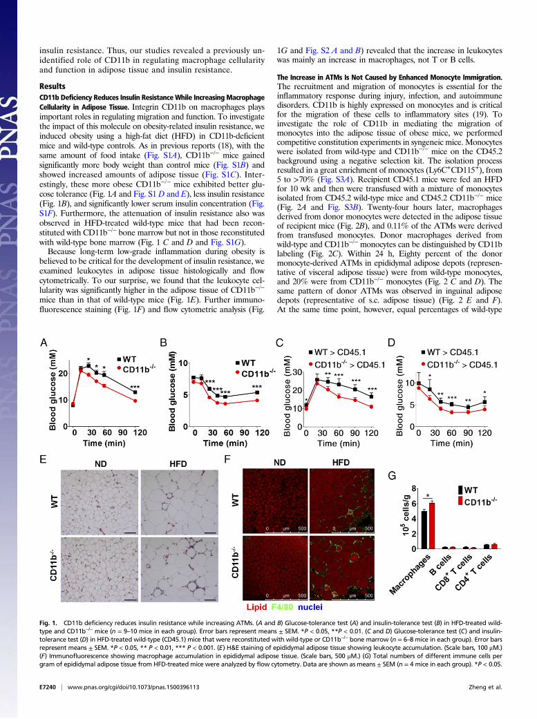
ResultsCD11b Deficiency Reduces Insulin Resistance While Increasing MacrophageCellularity in Adipose Tissue. Integrin CD11b on macrophages playsimportant roles in regulating migration and function. To investigatethe impact of this molecule on obesity-related insulin resistance, weinduced obesity using a high-fat diet (HFD) in CD11b-deficientmice and wild-type controls. As in previous reports (18), with thesame amount of food intake (Fig. S1A), CD11b−/− mice gainedsignificantly more body weight than control mice (Fig. S1B) andshowed increased amounts of adipose tissue (Fig. S1C). Inter-estingly, these more obese CD11b−/− mice exhibited better glu-cose tolerance (Fig. 1A and Fig. S1 D and E), less insulin resistance(Fig. 1B), and significantly lower serum insulin concentration (Fig.S1F). Furthermore, the attenuation of insulin resistance also wasobserved in HFD-treated wild-type mice that had been recon-stituted with CD11b−/− bone marrow but not in those reconstitutedwith wild-type bone marrow (Fig. 1 C and D and Fig. S1G).Because long-term low-grade inflammation during obesity is
believed to be critical for the development of insulin resistance, weexamined leukocytes in adipose tissue histologically and flowcytometrically. To our surprise, we found that the leukocyte cel-lularity was significantly higher in the adipose tissue of CD11b−/−
mice than in that of wild-type mice (Fig. 1E). Further immuno-fluorescence staining (Fig. 1F) and flow cytometric analysis (Fig.
1G and Fig. S2 A and B) revealed that the increase in leukocyteswas mainly an increase in macrophages, not T or B cells.
The Increase in ATMs Is Not Caused by Enhanced Monocyte Immigration.The recruitment and migration of monocytes is essential for theinflammatory response during injury, infection, and autoimmunedisorders. CD11b is highly expressed on monocytes and is criticalfor the migration of these cells to inflammatory sites (19). Toinvestigate the role of CD11b in mediating the migration ofmonocytes into the adipose tissue of obese mice, we performedcompetitive constitution experiments in syngeneic mice. Monocyteswere isolated from wild-type and CD11b−/− mice on the CD45.2background using a negative selection kit. The isolation processresulted in a great enrichment of monocytes (Ly6C+CD115+), from5 to >70% (Fig. S3A). Recipient CD45.1 mice were fed an HFDfor 10 wk and then were transfused with a mixture of monocytesisolated from CD45.2 wild-type mice and CD45.2 CD11b−/− mice(Fig. 2A and Fig. S3B). Twenty-four hours later, macrophagesderived from donor monocytes were detected in the adipose tissueof recipient mice (Fig. 2B), and 0.11% of the ATMs were derivedfrom transfused monocytes. Donor macrophages derived fromwild-type and CD11b−/− monocytes can be distinguished by CD11blabeling (Fig. 2C). Within 24 h, Eighty percent of the donormonocyte-derived ATMs in epididymal adipose depots (represen-tative of visceral adipose tissue) were from wild-type monocytes,and 20% were from CD11b−/− monocytes (Fig. 2 C and D). Thesame pattern of donor ATMs was observed in inguinal adiposedepots (representative of s.c. adipose tissue) (Fig. 2 E and F).At the same time point, however, equal percentages of wild-type
Fig. 1. CD11b deficiency reduces insulin resistance while increasing ATMs. (A and B) Glucose-tolerance test (A) and insulin-tolerance test (B) in HFD-treated wild-type and CD11b−/− mice (n = 9–10 mice in each group). Error bars represent means ± SEM. *P < 0.05, **P < 0.01. (C and D) Glucose-tolerance test (C) and insulin-tolerance test (D) in HFD-treated wild-type (CD45.1) mice that were reconstituted with wild-type or CD11b−/− bone marrow (n = 6–8 mice in each group). Error barsrepresent means ± SEM. *P < 0.05, ** P < 0.01, *** P < 0.001. (E) H&E staining of epididymal adipose tissue showing leukocyte accumulation. (Scale bars, 100 μM.)(F) Immunofluorescence showing macrophage accumulation in epididymal adipose tissue. (Scale bars, 500 μM.) (G) Total numbers of different immune cells pergram of epididymal adipose tissue from HFD-treated mice were analyzed by flow cytometry. Data are shown as means ± SEM (n = 4 mice in each group). *P < 0.05.
E7240 | www.pnas.org/cgi/doi/10.1073/pnas.1500396113 Zheng et al.
donor monocytes and CD11b−/− donor monocytes were found inthe peripheral blood of recipient mice (Fig. S3 C and D), excludingthe possibility that the observed difference in the recruitment toadipose tissue is caused by differential survival or clearance ofmonocytes from the two types of mice. These results demonstratedthat CD11b deficiency greatly compromises the ability of mono-cytes to influx into the adipose tissue of obese mice.The observation of less monocyte immigration but more ATM
accumulation contradicts the general notion that the influx ofmonocytes determines the degree of macrophage accumulation.Two possible reasons might explain the paradoxical enhancement ofATM accumulation when the immigration of monocytes is actuallyreduced. One is that the life span of macrophages is prolonged byCD11b deficiency. In fact, it has been reported that CD11b en-gagement promotes the proapoptotic signals and that CD11b/CD18promoted phagocytosis-induced apoptosis of neutrophils (20, 21). Todetermine whether there is a decrease in cell death in CD11b−/−
ATMs, we performed a TUNEL assay. We found that cell death ofATMs from both wild-type and CD11b−/− mice is negligible, and wedid not find any difference in the level of cell death in ATMs fromwild-type and CD11b−/− mice (Fig. S4). The other possible so-lution for the increase in ATMs in CD11b−/− mice is enhancedproliferation.
Increased ATM Proliferation in Obese CD11b-Deficient Mice. Previousstudies demonstrated that the accumulation of tissue macrophagesduring inflammation can result from the expansion of the residentpopulation (22), suggesting that enhanced in situ proliferation of
macrophages can lead to the observed increase in ATM accu-mulation in CD11b−/− mice. To examine whether ATMs couldproliferate locally, we treated mice receiving either a normal dietor an HFD with 5-ethynyl-2′-deoxyuridine (EdU) for 3 h and thenanalyzed macrophages with EdU incorporation in the epididymaladipose tissue by flow cytometry. EdU-incorporated ATMs wereincreased after HFD treatment (Fig. S5A). It is worth noting thatthere are no detectable EdU-incorporated monocytes in blood(Fig. S2C and Fig. S5B), suggesting a special property of ATMsand excluding the possibility that the EdU-incorporated ATMs inadipose tissue were from circulation. We also checked Ki67, ahallmark of cells in the cell cycle, and found that Ki67+ macro-phages from epididymal adipose tissue were greatly increased inHFD-treated mice (Fig. S5C). These experiments showed that anHFD could induce ATM proliferation in wild-type mice.We hypothesized that CD11b deficiency could lead to a further
increase in the proliferation of macrophages in the adipose tissuein mice on an HFD. This increase accounts for the increasedATM accumulation in obese CD11b−/− mice. By comparing Ki67+
ATMs in wild-type mice and CD11b−/− mice on an HFD, wefound a dramatic increase in Ki67+ macrophages in the adiposetissue of CD11b−/− mice (Fig. 3 A and B). We also analyzed Ki67expression in ATMs from HFD-treated CD11b−/− mice and theirheterozygous littermates and found a significant increase in Ki67expression in CD11b−/− mice (Fig. S5D). To determine in situwhether the Ki67+ macrophages are indeed in adipose tissue, wecarried out whole-mount staining on epididymal adipose tissue andlocalized those Ki67+ macrophages in the crown-like-structures
Fig. 2. CD11b deficiency impairs the migration of monocytes to adipose tissue. (A) Schematic representation of competitive monocyte constitution. Twomillion monocytes isolated from the bone marrow of CD45.2 wild-type mice were mixed with 2 × 10⁶ monocytes from CD45.2 CD11b−/− mice and wereinjected i.v. into HFD-treated CD45.1 mice. Twenty-four hours later the stromal cell fraction was analyzed for CD45, F4/80, and CD11b. (B) Detection of ATMsderived from donor monocytes by flow cytometry based on F4/80 and CD45.2. (C and E) Proportion of ATMs derived from wild-type and CD11b−/− monocytesin epididymal adipose tissue (C) and inguinal adipose tissue (E). Recipient mice were injected with only CD11b−/− monocytes as a gating control (Left) or withwild-type and CD11b−/− monocytes (Right). (D and F) Summarizing the proportion of ATMs derived from wild-type and CD11b−/− monocytes in epididymaladipose tissue (D) and inguinal adipose tissue (F) in mice of four independent experiments. Data are shown as mean ± SEM; **P < 0.01, paired t-test.
Zheng et al. PNAS | Published online December 15, 2015 | E7241
(CLSs) formed by accumulated macrophages. We found thatKi67+ macrophages are increased in the CLSs of adipose tissuefrom CD11b−/− mice (Fig. 3C). We also performed the EdUincorporation assay on HFD-treated wild-type and CD11b−/−
mice and observed significantly augmented EdU incorporationin CD11b−/− ATMs (Fig. 3 D and E). Those EdU-incorporatedATMs are sporadic in adipose tissue and exhibit CLS localizationin close proximity to other macrophages (Fig. 3F). Thus, CD11bplays a negative role in regulating macrophage proliferation in theadipose tissue of obese mice.
ATM Proliferation Depends on the IL-4/STAT6 Signaling Axis. Severalcytokines, including IL-4, macrophage colony-stimulating factor(M-CSF), and monocyte chemotactic protein 1 (MCP-1), havebeen shown to modulate the proliferation of monocytic cells. TheTh2 cytokine IL-4 has been shown to drive local proliferation ofmacrophages in helminth-induced inflammation responses (22).Interestingly, we found the IL-4 receptor alpha (IL-4Rα) is
expressed on ATMs (Fig. S6A). To test whether ATM proliferationcould be caused by IL-4, we i.p. injected recombinant IL-4 (23) intolean mice and performed the EdU incorporation assay and Ki67staining 48 h later. We found that the EdU-incorporated ATMs(Fig. 4A) and Ki67+ ATMs (Fig. 4B) are induced dramatically byIL-4 treatment. It should be noted that the incorporation of suchthymidine analog was not detectable in blood monocytes (22).Macrophage differentiation and proliferation also can be regulatedby M-CSF in native or inflammatory status (24–26). We examinedthe expression of colony-stimulating factor 1 receptor (CSF-1R), aspecific M-CSF receptor, on cells of the monocytic lineage andfound that it was high on peritoneal macrophages as well asmonocytes in adipose tissue but not on ATMs of either lean orobese mice (Fig. S6 A and B). Thus, the proliferation of ATMs isunlikely to be related to M-CSF. A recent report suggested thatMCP-1 regulates the local proliferation of ATMs (27). To examinefurther the effect of M-CSF and MCP-1 on ATM proliferation, wei.p. injected M-CSF and MCP-1 at different concentrations into
Fig. 3. CD11b deficiency promotes in situ proliferation of ATMs during obesity. (A) Flow cytometric analysis of Ki67 expression in ATMs from epididymaladipose tissue of wild-type and CD11b−/− mice on an HFD. ATMs were gated on CD45+F4/80hiSiglec-F− as shown in Fig. S2A. (B) ATM proliferation as detectedby Ki67 expression in all mice from two independent experiments. Data are shown as means ± SEM. **P < 0.01. (C) Immunofluorescence staining for Ki67+
ATMs in epididymal adipose tissue of wild-type and CD11b−/− mice on an HFD. (Scale bars, 100 μM.) (D) Flow cytometric analysis of EdU incorporation in ATMsfrom epididymal adipose tissue of wild-type and CD11b−/− mice on an HFD. Mice were pulsed with 10 μg EdU per gram of body weight for 3 h. (E) ATMproliferation as detected by EdU in all mice from two independent experiments. Data are shown as mean ± SEM. **P < 0.01. (F) Immunofluorescence stainingfor EdU-incorporated ATMs in the epididymal adipose tissue of wild-type and CD11b−/− mice on an HFD. (Scale bars, 100 μM.)
E7242 | www.pnas.org/cgi/doi/10.1073/pnas.1500396113 Zheng et al.
lean mice. Forty-eight hours later, myeloid cells in the peripheralblood were significantly induced by either cytokine (Fig. S6C),possibly because of recruitment and maturation (28, 29). However,we did not find significant increase when the EdU incorporationassay was performed on ATMs, (Fig. S6D). These results indicatethat IL-4, but not MCP-1 or M-CSF, could be the cytokine thatleads to ATM proliferation.Upon binding to IL-4, IL-4R recruits STAT6 and IRS2 to
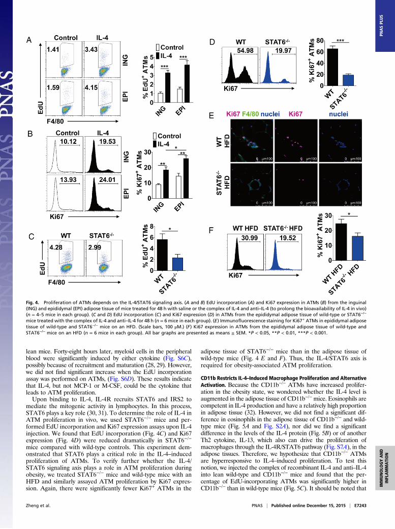
mediate the mitogenic activity in lymphocytes. In this process,STAT6 plays a key role (30, 31). To determine the role of IL-4 inATM proliferation in vivo, we used STAT6−/− mice and per-formed EdU incorporation and Ki67 expression assays upon IL-4injection. We found that EdU incorporation (Fig. 4C) and Ki67expression (Fig. 4D) were reduced dramatically in STAT6−/−
mice compared with wild-type controls. This experiment dem-onstrated that STAT6 plays a critical role in the IL-4–inducedproliferation of ATMs. To verify further whether the IL-4/STAT6 signaling axis plays a role in ATM proliferation duringobesity, we treated STAT6−/− mice and wild-type mice with anHFD and similarly assayed ATM proliferation by Ki67 expres-sion. Again, there were significantly fewer Ki67+ ATMs in the
adipose tissue of STAT6−/− mice than in the adipose tissue ofwild-type mice (Fig. 4 E and F). Thus, the IL-4/STAT6 axis isrequired for obesity-associated ATM proliferation.
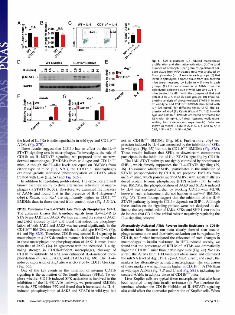
CD11b Restricts IL-4–Induced Macrophage Proliferation and AlternativeActivation. Because the CD11b−/− ATMs have increased prolifer-ation in the obesity state, we wondered whether the IL-4 level isaugmented in the adipose tissue of CD11b−/−mice. Eosinophils arecompetent in IL-4 production and have a relatively high proportionin adipose tissue (32). However, we did not find a significant dif-ference in eosinophils in the adipose tissue of CD11b−/− and wild-type mice (Fig. 5A and Fig. S2A), nor did we find a significantdifference in the levels of the IL-4 protein (Fig. 5B) or of anotherTh2 cytokine, IL-13, which also can drive the proliferation ofmacrophages through the IL-4R/STAT6 pathway (Fig. S7A), in theadipose tissues. Therefore, we hypothesize that CD11b−/− ATMsare hyperresponsive to IL-4–induced proliferation. To test thisnotion, we injected the complex of recombinant IL-4 and anti–IL-4into lean wild-type and CD11b−/− mice and found that the per-centage of EdU-incorporating ATMs was significantly higher inCD11b−/− than in wild-type mice (Fig. 5C). It should be noted that
Fig. 4. Proliferation of ATMs depends on the IL-4/STAT6 signaling axis. (A and B) EdU incorporation (A) and Ki67 expression in ATMs (B) from the inguinal(ING) and epididymal (EPI) adipose tissue of mice treated for 48 h with saline or the complex of IL-4 and anti–IL-4 (to prolong the bioavailability of IL-4 in vivo)(n = 4–5 mice in each group). (C and D) EdU incorporation (C) and Ki67 expression (D) in ATMs from the epididymal adipose tissue of wild-type or STAT6−/−
mice treated with the complex of IL-4 and anti–IL-4 for 48 h (n = 6 mice in each group). (E) Immunofluorescence staining for Ki67+ ATMs in epididymal adiposetissue of wild-type and STAT6−/− mice on an HFD. (Scale bars, 100 μM.) (F) Ki67 expression in ATMs from the epididymal adipose tissue of wild-type andSTAT6−/− mice on an HFD (n = 6 mice in each group). All bar graphs are presented as means ± SEM. *P < 0.05, **P < 0.01, ***P < 0.001.
Zheng et al. PNAS | Published online December 15, 2015 | E7243
the level of IL-4Rα is indistinguishable in wild-type and CD11b−/−
ATMs (Fig. S7B).These results suggest that CD11b has an effect on the IL-4/
STAT6 signaling axis in macrophages. To investigate the role ofCD11b on IL-4/STAT6 signaling, we prepared bone marrow-derived macrophages (BMDMs) from wild-type and CD11b−/−
mice. Although the IL-4Rα levels are equal on BMDMs fromeither type of mice (Fig. S7C), the CD11b−/− macrophagesexhibited greatly increased phosphorylation of STAT6 whentreated with IL-4 (Fig. 5D and Fig. S7D).In addition to regulating proliferation, Th2 cytokines are well
known for their ability to drive alternative activation of macro-phages via STAT6 (6, 33). Therefore, we examined the markersof AAMs and found that in the presence of IL-4 Arginase I(Arg1), Retnla, and Ym1 are significantly higher in CD11b−/−
BMDMs than in those derived from control mice (Fig. 5 E–G).
CD11b Constrains the IL-4/STAT6 Axis Through Phosphatase SHP-1.The upstream kinases that transduce signals from IL-4–IL-4R toSTAT6 are JAK1 and JAK3. We thus examined the status of JAK1and JAK3 induced by IL-4 and found that indeed the phosphory-lation of both JAK1 and JAK3 was increased significantly in theCD11b−/− BMDMs compared with that in wild-type BMDMs (Fig.6A and Fig. S7D). Therefore, CD11b may control IL-4 signaling inmacrophages in a JAK-dependent manner. It should be noted thatin these macrophages the phosphorylation of JAK1 is much lowerthan that of JAK3 (34). In agreement with the increased IL-4 sig-naling strength in CD11b-deficient macrophages, blockage ofCD11b by antibody, M1/70, also enhanced IL-4–induced phos-phorylation of JAK1, JAK3, and STAT6 (Fig. 6B). The IL-4–induced expression of Arg1 also was elevated by CD11b blockage(Fig. 6C).One of the key events in the initiation of integrin CD11b
signaling is the activation of Src family kinases (SFKs). To ex-plore whether CD11b-induced SFK activation is involved in theinhibition of the IL-4/STAT6 pathway, we pretreated BMDMswith the SFK inhibitor PP2 and found that it increased the IL-4–induced phosphorylation of JAK3 and STAT6 in wild-type but
not in CD11b−/− BMDMs (Fig. 6D). Furthermore, Arg1 ex-pression induced by IL-4 was increased by the inhibition of SFKsin wild-type (Fig. 6E) but not in CD11b−/− BMDMs (Fig. S7E).These results indicate that SFKs or downstream componentsparticipate in the inhibition of IL-4/STAT6 signaling by CD11b.The JAK–STAT pathways are tightly controlled by phosphatase
SHP-1, which directly suppresses the IL-4–STAT6 signaling (35,36). To examine whether SHP-1 participates in the inhibition ofSTAT6 phosphorylation by CD11b, we prepared BMDMs frommev/mev mice, which possess mutated SHP-1 with substantially re-duced protein tyrosine phosphatase (PTP) activity (37). In wild-type BMDMs, the phosphorylation of JAK3 and STAT6 inducedby IL-4 was increased further by blocking CD11b with M1/70;however, such enhancement did not happen in mev/mev BMDMs(Fig. 6F). These findings suggest that the inhibition of the IL-4/STAT6 pathway by integrin CD11b depends on SHP-1. Althoughthese studies on the signaling process were not designed to de-lineate the sequential order of JAKs, SFKs, and SHP-1, our resultsdo indicate that CD11b has critical roles in negatively regulating theIL-4 signaling process.
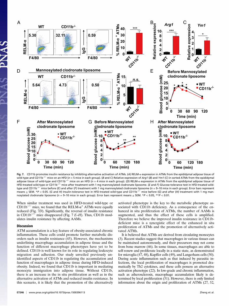
Alternatively Activated ATMs Reverse Insulin Resistance in CD11b-Deficient Mice. Because our data clearly showed that macro-phage accumulation and alternative activation can be regulated byCD11b, we further investigated the relevance of such changes inmacrophages to insulin resistance. In HFD-induced obesity, wefound that the percentage of RELM-α+ ATMs was dramaticallyhigher in CD11b−/− mice than in wild-type mice (Fig. 7A). We alsosorted the ATMs from HFD-induced obese mice and examinedthe mRNA level of Arg1, Ym1, Ppard, Gas6, Lyve1, and Pdgfc, themarkers for alternatively activated macrophages. The expressionof these markers was significantly higher in CD11b−/− ATMs thanin wild-type ATMs (Fig. 7 B and C and Fig. S8A), indicating in-creased AAMs in adipose tissue of CD11b−/− mice.Liver Kupffer cells are typical tissue macrophages that also have
been reported to regulate insulin resistance (9). We therefore de-termined whether the CD11b inhibition of IL-4/STAT6 signalingalso could affect the alternative polarization of Kupffer cells. When
Fig. 5. CD11b restrains IL-4–induced macrophageproliferation and alternative activation. (A) The totalnumber of eosinophils per gram of epididymal adi-pose tissue from HFD-treated mice was analyzed byflow cytometry (n = 4 mice in each group). (B) IL-4levels in epididymal adipose tissue from HFD-treatedmice were measured by ELISA (n = 5 mice in eachgroup). (C) EdU incorporation in ATMs from theepididymal adipose tissue of wild-type and CD11b−/−
mice treated for 48 h with the complex of IL-4 andanti–IL-4 (n = 5 mice in each group). (D) Immuno-blotting analysis of phosphorylated STAT6 in lysatesof wild-type and CD11b−/− BMDMs stimulated withIL-4 (20 ng/mL) for different times. (E–G) The ex-pression of Arg1 (E), Retnla (F), and Ym1 (G) in wild-type and CD11b−/− BMDMs untreated or treated for12 h with 10 ng/mL IL-4 (four repeated wells repre-senting two independent experiments). Data areshown as means ± SEM in A, B, C, E, F, and G. *P <0.05, **P < 0.01, ***P < 0.001.
E7244 | www.pnas.org/cgi/doi/10.1073/pnas.1500396113 Zheng et al.
the expression of ARG-1 in Kupffer cells was analyzed by flowcytometry, we could not detect significant difference between wild-type and CD11b-deficient mice (Fig. S8B). We also examined theexpression of several other markers of AAMs (Clec10a,Clec7a, Il1rn,Jag1, Mrc1, Pdcd1lg, Ppard, Retnla, and Ym1) in liver and found nosignificant difference between wild-type and CD11b-deficient mice(Fig. S8C). It has been reported that the expression of CD11b onKupffer cells is very low (38), and we also found that the level ofCD11b is more than sevenfold lower on Kupffer cells than on ATMs(Fig. S8D). Because of its low level of expression on Kupffer cells,CD11b has much less effect on Kupffer cells than on ATMs.CD11b also is expressed on eosinophils, neutrophils, and natural
killer (NK) cells. The infiltration of those cells into adipose tissuehas also relevance to obesity-related insulin resistance (32, 39, 40).Specifically, eosinophils improve insulin resistance by secreting IL-4that promotes alternative activation of ATMs, neutrophils mediateinsulin resistance by producing elastase, and NK cells contribute toinsulin resistance by releasing IFN-γ (32, 39, 40). We thereforeexamined these cells in epididymal adipose tissue. We did not findany difference in the number of these cells per gram of adiposetissue from wild-type and CD11b−/− mice (Fig. 5A and Figs. S2Aand S8 E and F), nor did we find difference in the expression ofIL-4, neutrophil elastase (Elane), or IFN-γ (Fig. S8 G–I).Gut microbiota have been shown to be involved in the devel-
opment of obesity and metabolic abnormalities (41). It also hasbeen reported that there is an increase in the abundance of bac-teria belonging to the phylum Firmicutes and a decrease in theabundance of bacteria in the phylum Bacteroidetes in obese hu-mans and mice (42). To investigate whether microbiota play a rolein the development of metabolic abnormalities associated withCD11b status, we examined the abundance of fecal bacteria in
these two phyla in CD11b-deficient and wild-type mice on anHFD using quantitative PCR (qPCR)-based analysis of bacterial16S rRNA genes. Interestingly, we did not find any significantdifferences between CD11b-deficient and wild-type mice. Wefurther analyzed in more detail the fecal bacteria that have beenreported to be associated with obesity and blood glucose changes.Again, we did not find significant differences between CD11b-deficient and wild-type in the abundance of these bacteria (Fig.S8J). Therefore, the reduced insulin resistance in CD11b-deficientmice is not a consequence of altered gut microbiota.We further investigated whether the increase in the proliferation
and alternative activation of ATMs is associated with the reductionin insulin resistance in the paradigm of bone marrow reconstitution.As revealed in our immunohistochemical staining, significantly moreKi67+ macrophages and RELM-α+macrophages existed in theadipose tissue of CD45.1 mice reconstituted with CD11b−/− bonemarrow than in the adipose tissue of CD45.1 mice reconstitutedwith wild-type bone marrow (Fig. S9 A–D), indicating an increase inthe proliferation and alternative activation of CD11b−/− ATMs.Alternatively activated ATMs could alleviate insulin resistance
by attenuating the inflammation as well as by improving themetabolic function of adipose tissue (9, 10, 43). To verify the roleof alternatively activated ATMs on improved insulin resistance inCD11b−/− mice, we selectively depleted these macrophages by i.p.injection of mannosylated clodronate liposome, which would bespecifically incorporated by AAMs in the facilitation of mannosereceptors and would induce apoptosis of these cells (44). We didnot find a reduction in Kupffer cells after 4 d of treatment withmannosylated clodronate liposome, whereas the RELM-α+ ATMswere greatly decreased in both the inguinal adipose tissue and theepididymal adipose tissue of HFD-treated mice (Fig. S9 E and F).
Fig. 6. Integrin CD11b inhibits the IL-4/STAT6 signaling axis through SHP-1. (A) Immunoblotting analysis of phosphorylated JAK1, JAK3, and STAT6 in lysates ofwild-type and CD11b−/− BMDMs stimulated with 20 ng/mL IL-4 for 30 min or not stimulated (Control). (B) Immunoblotting analysis of phosphorylated JAK1, JAK3,and STAT6 in lysates of BMDMs. Wild-type BMDMs were pretreated with CD11b-blocking antibody (M1/70) or isotype (rat IgG2b) and were stimulated with 20 ng/mLIL-4 for 0–2 h. (C) The expression of Arg1 in BMDMs. Wild-type BMDMs were pretreated with M1/70 or IgG2b and were stimulated with 10 ng/mL IL-4 for 12 h orwere not stimulated. (D) Immunoblotting analysis of phosphorylated JAK3 and STAT6 in lysates of wild-type and CD11b−/− BMDMs. BMDMs were stimulated with20 ng/mL IL-4 for 30 min after pretreatment with PP2 (0.5 μM or 10 μM for 1 h) or solvent (Control). (E) The expression of Arg1 in BMDMs. Wild-type BMDMs werestimulated with 10 ng/mL IL-4 for 12 h after pretreatment with PP2 (10 μM for 1 h) or solvent (Control). (F) Immunoblotting analysis of phosphorylated JAK3 andSTAT6 in wild-type andmev/mev BMDMs. BMDMswere pretreated withM1/70 or IgG2b before being stimulated for 30min with 20 ng/mL IL-4. Data in C and E areshown as means ± SEM of three or four replicates representing two independent experiments. *P < 0.05, **P < 0.01.
Zheng et al. PNAS | Published online December 15, 2015 | E7245
When similar treatment was used in HFD-treated wild-type orCD11b−/− mice, we found that the RELM-α+ ATMs were equallyreduced (Fig. 7D). Significantly, the reversal of insulin resistancein CD11b−/− mice disappeared (Fig. 7 E–H). Thus, CD11b mod-ulates insulin resistance by affecting AAMs.
DiscussionATM accumulation is a key feature of obesity-associated chronicinflammation. These cells could promote further metabolic dis-orders such as insulin resistance (45). However, the mechanismsunderlining macrophage accumulation in adipose tissue and thefunction of different macrophage phenotypes have yet to bedefined. CD11b is well known for its role in regulating leukocytemigration and adhesion. Our study unveiled previously un-identified aspects of CD11b in regulating the accumulation andfunction of macrophages in adipose tissue during HFD-inducedobesity. Indeed, we found that CD11b is important in mediatingmonocyte immigration into adipose tissue. Without CD11b,there is an increase in the in situ proliferation as well as in thealternative activation of ATMs and reduced insulin resistance. Inthis scenario, it is likely that the promotion of the alternatively
activated phenotype is the key to the metabolic phenotype as-sociated with CD11b deficiency. As a consequence of the en-hanced in situ proliferation of ATMs, the number of AAMs isaugmented, and thus the effect of these cells is amplified.Therefore we believe the improved insulin resistance in CD11b-deficient mice is a synergistic effect of the enhanced in situproliferation of ATMs and the promotion of alternatively acti-vated ATMs.It is believed that ATMs are derived from circulating monocytes
(3). Recent studies suggest that macrophages in various tissues canbe maintained autonomously, and their precursors may not comefrom bone marrow (46). In some tissues, macrophages are able toself-renew and proliferate locally in a static state, as demonstratedfor microglia (47, 48), Kupffer cells (49), and Langerhans cells (50).During acute inflammation such as that induced by parasitic in-fections, the local proliferation of macrophages is promoted dra-matically by Th2 cytokines, and these cells possess an alternativeactivation phenotype (22). In low-grade and chronic inflammation,such as atherosclerosis, macrophage accumulation likely is de-termined by local proliferation (51). However, there is only limitedinformation about the origin and proliferation of ATMs (27, 52,
Fig. 7. CD11b promotes insulin resistance by inhibiting alternative activation of ATMs. (A) RELM-α expression in ATMs from the epididymal adipose tissue ofwild-type and CD11b−/− mice on an HFD (n = 5 mice in each group). (B and C) Relative expression of Arg1 (B) and Ym1 (C) in sorted ATMs from the epididymaladipose tissue of wild-type and CD11b−/− mice on an HFD (n = 4 mice in each group). (D) RELM-α expression in ATMs from the epididymal adipose tissue ofHFD-treated wild-type or CD11b−/− mice after treatment with 1 mg mannosylated clodronate liposome. (E and F) Glucose-tolerance test in HFD-treated wild-type and CD11b−/− mice before (E) and after (F) treatment with 1 mg mannosylated clodronate liposome (n = 9–10 mice in each group). Error bars representmeans ± SEM. *P < 0.05. (G and H) Insulin-tolerance test in HFD-treated wild-type and CD11b−/− mice before (G) and after (H) treatment with 1 mg man-nosylated clodronate liposome (n = 9–10 mice in each group). Error bars represent means ± SEM. *P < 0.05, **P < 0.01.
E7246 | www.pnas.org/cgi/doi/10.1073/pnas.1500396113 Zheng et al.
53). We systemically examined the immigration and proliferationof ATMs in wild-type and CD11b-deficient mice. Our studydemonstrated that in situ macrophage proliferation contributesgreatly to ATM accumulation, especially in the absence of CD11b.In CD11b-deficient mice, we found that, although there is afourfold reduction in the immigration of blood monocytes intoadipose tissue, the ATM accumulation is much higher. Indeed, wefound much stronger proliferation of macrophages in the adiposetissue of CD11b-deficient mice, as demonstrated by the EdU in-corporation assay and the analysis of Ki67 expression.Various factors, such as M-CSF and Th2 cytokines, have been
reported to regulate macrophage proliferation (22, 24). Instead,upon the addition of exogenous IL-4, the proliferation of ATMscould be boosted dramatically, a process that depends on STAT6signaling. In support of the role of an IL-4–induced signalingprocess, the obesity-associated macrophage proliferation wasgreatly decreased in STAT6-deficient mice. It has been reportedthat several cell types in the adipose tissue, e.g. Th2 cells (54),eosinophils (32), and adipocytes themselves (10), are capable ofproducing IL-4. It has been reported that the initiation of obesityis concomitant with an evoked Th2 immune response in adiposetissue characterized by increased IL-4 expression and alternativeactivation of macrophages (55). Interestingly, the IL-4 level also isincreased in obese patients (56). Although the aim of this in-vestigation was not to identify the cell types that produce IL-4, wedid find that IL-4 signaling is required and that in vivo adminis-tration of IL-4 could promote further ATM proliferation.The mitogenic property of IL-4 often is investigated in the context
of IL-4R expression. We found that CD11b deficiency promotes IL-4–induced ATM proliferation; i.e., CD11b could provide a negativeeffect on the mitogenic signal of IL-4. Although the exact mechanismof the CD11b-regulated IL-4 signaling process remains to be eluci-dated, we did show that members of Src family kinases and SHP-1are indispensable for this effect. The activation of members of Srcfamily kinases, one of the earliest events in integrin signaling, hasdual effects on the signal transduction of immune receptors, phos-phorylating both immunoreceptor tyrosine-based activation motifs(ITAMs) and immunoreceptor tyrosine-based inhibitory motifs(ITIMs) (57). Interestingly, IL-4Rα possesses an ITIM domain onthe C terminal that has been reported to recruit SHP-1, which in turndown-regulates the IL-4–initiated signaling process (58). Based onthis information, we propose the following model: Signals fromCD11b activate members of Src family kinases, and those kinasesfurther phosphorylate the ITIM domain on IL-4Rα, leading to therecruitment of SHP-1 to suppress IL-4/STAT6 signaling. Althoughthis model seems to explain our data logically, the exact hierarchy ofthese molecules requires more detailed investigation.In summary, our study demonstrates that integrin CD11b nega-
tively regulates IL-4/STAT6–induced proliferation of ATMs and thepolarization of the alternatively activated phenotype. This effect ofCD11b likely depends on a complex signaling process involvingmembers of the Src family kinases and SHP-1. This function ofCD11b not only provides insights into the physiological function ofintegrins but also has important implications for designing newtherapeutic strategies for obesity-related insulin resistance.
Materials and MethodsAnimal Experiments. C57BL/6 and BALB/c mice were purchased from theShanghai Laboratory Animal Center of the Chinese Academy of Sciences.CD11b−/−mice were backcrossed with the C57BL/6 mice as described previously(14). STAT6−/− (C.129S2-Stat6tm1Gru/J) and mev/mev (C57BL/6J-Ptpn6me-v/J) micewere from Jackson Laboratory. CD45.1 C57BL/6 mice were kindly provided byYanyun Zhang of the Institute of Health Sciences of the Chinese Academy ofSciences in Shanghai, China. All experiments were approved by the In-stitutional Animal Care and Use Committee of the Institute of Health Sciences,Shanghai Institutes for Biological Sciences of Chinese Academy of Sciences.
HFD-induced obesity was induced by feeding male mice with a diet con-taining 60 kcal% fat (D12492; Research Diets) starting at age 5 wk. The controlgroup was fed with a normal chow diet. All experimental analyses were per-
formed after mice were fed an HFD for 8–10 wk. Glucose-tolerance tests werecarried out with i.p. injection of 1 g glucose per kilogram of body weight, andinsulin-tolerance tests were carried out with i.p. injection of 0.75 U insulin perkilogram of body weight. The serum insulin level was measured by a multipleximmunoassay using the Bio-Plex technology (Bio-Rad). To compare the mi-gration capacity of monocytes, CD11b−/− and wild-type monocytes were iso-lated from bone marrow with a negative selection kit (Stemcell Technologies),and either CD11b−/− and wild-type monocytes in a 1:1 ratio or CD11b−/−
monocytes alone were i.v. injected into HFD-treated CD45.1 mice. The EdUincorporation assay was carried out with the Click-iT EdU assay kit (LifeTechnologies). Mice were i.p. injected with 10 μg EdU per gram of body weightand were killed 3 h later. Adipose tissue was collected, and the Click-iT reactionwas performed and analyzed flow cytometrically and immunohistologicallyaccording to the manufacturer’s instruction. To examine the role of MCP-1,M-CSF, and IL-4 in ATMs proliferation, 0.1 or 1 μg MCP-1 (Life Technologies),1 or 10 μg mouse M-CSF (R&D Systems), a complex of 5 μg IL-4 (PeproTech) and25 μg IL-4 antibody (clone 11B11; Harlan Bioproducts for Science), or PBS wasi.p. injected into mice, and ATMs analysis was performed 48 h later. For de-pletion of AAMs, HFD-treated mice were i.p. injected with 1 mgmannosylatedclodronate liposome (Encapsula NanoSciences). The glucose-tolerance test andanalysis of ATMs were carried out between day 4 and day 8.
Flow Cytometric Analysis of Cellular Proteins. Adipose tissues were cut intosmall pieces, rinsedwith PBS, and digestedwith 2mg/mL collagenase I (Sigma-Aldrich) for 1 h. The cell suspensions were filtered through 70-μm sieves andcentrifuged to separate adipocytes. The cell pallets were resuspended andsubjected to erythrocyte lysis. The cell suspension then was preincubatedwith anti-CD16/CD32 (eBioscience) to block Fc receptors before surfacemolecule staining. The antibodies used to detect surface proteins includingCD3e, CD4, CD8a, CD19, NK1.1, F4/80, CD45, CD11b, CD45.1, CD45.2, Ki67,and Ly6C were from eBioscience, and those for CD115, Ly6G, and Siglec-Fwere from BioLegend. Anti–IL-4Rα was from R&D Systems.
For intracellular RELM-α staining in ATMs, GolgiPlug (BD Biosciences) wasadded to collagenase in a 1:1,000 ratio before digestion. After staining ofsurface molecules, intracellular staining was performed with fixation andpermeabilization kits according to the manufacturer’s instructions (eBio-science). Rabbit anti–RELM-α (Abcam) and Alexa Fluor 647-conjugateddonkey anti-rabbit IgG (Life Technologies) were used.
Immunohistology. Adipose tissues were cut into small pieces and fixed in 4%paraformaldehyde for 24 h at 4 °C before whole-mount staining. Briefly, thespecimen was permeabilized with 1% Triton X-100, blocked with 1% BSA and3% FBS in PBS, then stained with Alexa Fluor 488-conjugated rat anti-F4/80mAbs (eBioscience) and Alexa Fluor 647-conjugated rat anti-Ki67 mAbs (eBio-science). The nuclei were counterstained with Hoechst 33342 (Life Technolo-gies). The adipocytes were counterstained with BODIPY 558/568 C12 (LifeTechnologies). The TUNEL assay was performed on paraffin-embedded adiposetissue sections with an in situ cell-death detection kit (Roche Applied Science).
BMDMs. Bone marrow cells were isolated from femurs and tibias of CD11b−/−
and wild-type mice and then were cultured in DMEM/F12 medium with theaddition of 10% (vol/vol) FBS and 20% (vol/vol) L929 conditioned mediumfor 7 d. For CD11b blockage, BMDMs were pretreated with either 20 μg/mLCD11b antibodies M1/70 (eBioscience) or 20 μg/mL rat IgG2b (eBioscience) atroom temperature for 1 h before plating. For inhibition of SFKs, BMDMswere treated with DMSO or PP2 (at a concentration of 0.5 μM or 10 μM) for1 h and were stimulated with IL-4 (PeproTech) 1 h later.
Immunoblotting Assay. Cells were lysed with radioimmunoprecipitation assay(RIPA) lysis buffer (Millipore) supplemented with protease inhibitor mixtures(Roche Applied Science). Proteins were separated by SDS/PAGE and weretransferred onto nitrocellulose or PVDF membrane. The antibodies specific forSTAT6 phosphorylated at Tyr641 (ab54461)were fromAbcam. Antibodies specificfor JAK1 phosphorylated at Tyr1022 and Tyr1023 (3331), JAK3 phosphorylated atTyr980 and Tyr981 (5031), JAK1 (3344), and GAPDH (2118) were from Cell Sig-naling Technology. Antibody against total STAT6 (sc-1689) was from Santa CruzBiotechnology. The monoclonal anti–β-Actin (A2228) was from Sigma-Aldrich.
Gene Expression Analysis. Real-time qPCR was performed with the 7900 HT FastReal-Time PCR system (Life Technologies) using FastStart Universal SYBR GreenMaster (Roche Applied Science). Oligonucleotide primers were as follows:Arg1, 5′-CTCCAAGCCAAAGTCCTTAGAG-3′, 5′-AGGAGCTGTCATTAGGGACATC-3′;Retnla, 5′-GGATGCCAACTTTGAATAGGA-3′, 5′-GGATAGTTAGCTGGATTGGCA-3′;Ym1, 5′-CAGGTCTGGCAATTCTTCTGAA-3′, 5′-GTCTTGCTCATGTGTGTAAGTGA3′;and Actb, 5′-CCACGAGCGGTTCCGATG-3′, 5′-GCCACAGGATTCCATACCCA-3′.
Zheng et al. PNAS | Published online December 15, 2015 | E7247
IMMUNOLO
GYAND
INFLAMMATION
PNASPL
US
Statistical Analysis. Data are presented as means ± SEM. Significance wasassessed by an unpaired two-tailed t-test unless otherwise indicated. Weconsidered P < 0.05 as statistically significant.
ACKNOWLEDGMENTS. We thank Dr. Douglas Green, Dr. Guang Ning,Dr. Xiangyin Kong, and Dr. Jiqiu Wang for comments and suggestions andDr. Yanyun Zhang for providing CD45.1 mice. This study was supported by
Grant XDA 01040100 from the Scientific Innovation Project of the ChineseAcademy of Science; Grant 2015CB964400 from the Ministry of Science andTechnology of China; National Natural Science of China Programs 81330046,81273316, 81530043, and 81571612; the External Cooperation Program of Bu-reau of International Co-operation, Grant GJHZ201307 from the Chinese Acad-emy of Sciences; Grant 12JC1409200 from the Shanghai Municipal Key Projectsof Basic Research; Shanghai Rising-Star Program 14QA1404200; and Grant12ZR1452600 from the Shanghai Municipal Natural Science Foundation.
1. Hotamisligil GS, Shargill NS, Spiegelman BM (1993) Adipose expression of tumor ne-crosis factor-alpha: Direct role in obesity-linked insulin resistance. Science 259(5091):87–91.
3. Weisberg SP, et al. (2003) Obesity is associated with macrophage accumulation inadipose tissue. J Clin Invest 112(12):1796–1808.
4. Xu H, et al. (2003) Chronic inflammation in fat plays a crucial role in the developmentof obesity-related insulin resistance. J Clin Invest 112(12):1821–1830.
5. Gordon S, Taylor PR (2005) Monocyte and macrophage heterogeneity. Nat RevImmunol 5(12):953–964.
6. Gordon S, Martinez FO (2010) Alternative activation of macrophages: Mechanism andfunctions. Immunity 32(5):593–604.
7. Gordon S (2003) Alternative activation of macrophages. Nat Rev Immunol 3(1):23–35.8. Lumeng CN, Bodzin JL, Saltiel AR (2007) Obesity induces a phenotypic switch in adi-
pose tissue macrophage polarization. J Clin Invest 117(1):175–184.9. Odegaard JI, et al. (2007) Macrophage-specific PPARgamma controls alternative ac-
tivation and improves insulin resistance. Nature 447(7148):1116–1120.10. Kang K, et al. (2008) Adipocyte-derived Th2 cytokines and myeloid PPARdelta regu-
late macrophage polarization and insulin sensitivity. Cell Metab 7(6):485–495.11. Springer TA, Anderson DC (1986) The importance of the Mac-1, LFA-1 glycoprotein
family in monocyte and granulocyte adherence, chemotaxis, and migration into in-flammatory sites: Insights from an experiment of nature. Ciba Found Symp 118:102–126.
12. Issekutz TB (1995) In vivo blood monocyte migration to acute inflammatory reactions,IL-1 alpha, TNF-alpha, IFN-gamma, and C5a utilizes LFA-1, Mac-1, and VLA-4. Therelative importance of each integrin. J Immunol 154(12):6533–6540.
13. Abram CL, Lowell CA (2009) The ins and outs of leukocyte integrin signaling. AnnuRev Immunol 27:339–362.
14. Ehirchiou D, et al. (2007) CD11b facilitates the development of peripheral toleranceby suppressing Th17 differentiation. J Exp Med 204(7):1519–1524.
15. Han C, et al. (2010) Integrin CD11b negatively regulates TLR-triggered inflammatoryresponses by activating Syk and promoting degradation of MyD88 and TRIF via Cbl-b.Nat Immunol 11(8):734–742.
16. Yakubenko VP, Bhattacharjee A, Pluskota E, Cathcart MK (2011) αMβ₂ integrin acti-vation prevents alternative activation of human and murine macrophages and im-pedes foam cell formation. Circ Res 108(5):544–554.
17. Ding C, et al. (2013) Integrin CD11b negatively regulates BCR signalling to maintainautoreactive B cell tolerance. Nat Commun 4:2813.
18. Dong ZM, Gutierrez-Ramos JC, Coxon A, Mayadas TN, Wagner DD (1997) A new classof obesity genes encodes leukocyte adhesion receptors. Proc Natl Acad Sci USA 94(14):7526–7530.
19. Anderson DC, Springer TA (1987) Leukocyte adhesion deficiency: An inherited defectin the Mac-1, LFA-1, and p150,95 glycoproteins. Annu Rev Med 38:175–194.
20. Coxon A, et al. (1996) A novel role for the beta 2 integrin CD11b/CD18 in neutrophilapoptosis: A homeostatic mechanism in inflammation. Immunity 5(6):653–666.
21. Mayadas TN, Cullere X (2005) Neutrophil beta2 integrins: Moderators of life or deathdecisions. Trends Immunol 26(7):388–395.
22. Jenkins SJ, et al. (2011) Local macrophage proliferation, rather than recruitment fromthe blood, is a signature of TH2 inflammation. Science 332(6035):1284–1288.
23. Finkelman FD, et al. (1993) Anti-cytokine antibodies as carrier proteins. Prolongationof in vivo effects of exogenous cytokines by injection of cytokine-anti-cytokine an-tibody complexes. J Immunol 151(3):1235–1244.
24. Hashimoto D, et al. (2013) Tissue-resident macrophages self-maintain locally throughoutadult life with minimal contribution from circulating monocytes. Immunity 38(4):792–804.
25. Davies LC, et al. (2013) Distinct bone marrow-derived and tissue-resident macrophagelineages proliferate at key stages during inflammation. Nat Commun 4:1886.
26. Jenkins SJ, et al. (2013) IL-4 directly signals tissue-resident macrophages to proliferatebeyond homeostatic levels controlled by CSF-1. J Exp Med 210(11):2477–2491.
27. Amano SU, et al. (2014) Local proliferation of macrophages contributes to obesity-associated adipose tissue inflammation. Cell Metab 19(1):162–171.
28. Papatriantafyllou M (2011) Monocytes: Nudged out of the niche. Nat Rev Immunol11(6):368–369.
29. Mossadegh-Keller N, et al. (2013) M-CSF instructs myeloid lineage fate in singlehaematopoietic stem cells. Nature 497(7448):239–243.
30. Wurster AL, Withers DJ, Uchida T, White MF, Grusby MJ (2002) Stat6 and IRS-2 co-operate in interleukin 4 (IL-4)-induced proliferation and differentiation but are dis-pensable for IL-4-dependent rescue from apoptosis. Mol Cell Biol 22(1):117–126.
31. Kaplan MH, Schindler U, Smiley ST, Grusby MJ (1996) Stat6 is required for mediatingresponses to IL-4 and for development of Th2 cells. Immunity 4(3):313–319.
32. Wu D, et al. (2011) Eosinophils sustain adipose alternatively activated macrophagesassociated with glucose homeostasis. Science 332(6026):243–247.
33. Martinez FO, Helming L, Gordon S (2009) Alternative activation of macrophages: Animmunologic functional perspective. Annu Rev Immunol 27:451–483.
34. Malabarba MG, et al. (1995) Activation of JAK3, but not JAK1, is critical to interleukin-4 (IL4) stimulated proliferation and requires a membrane-proximal region of IL4 re-ceptor alpha. J Biol Chem 270(16):9630–9637.
35. Haque SJ, Harbor P, Tabrizi M, Yi T, Williams BRG (1998) Protein-tyrosine phosphataseShp-1 is a negative regulator of IL-4- and IL-13-dependent signal transduction. J BiolChem 273(51):33893–33896.
37. Shultz LD, et al. (1993) Mutations at the murine motheaten locus are within thehematopoietic cell protein-tyrosine phosphatase (Hcph) gene. Cell 73(7):1445–1454.
38. Movita D, et al. (2012) Kupffer cells express a unique combination of phenotypic andfunctional characteristics compared with splenic and peritoneal macrophages. J LeukocBiol 92(4):723–733.
39. Talukdar S, et al. (2012) Neutrophils mediate insulin resistance in mice fed a high-fatdiet through secreted elastase. Nat Med 18(9):1407–1412.
40. Wensveen FM, et al. (2015) NK cells link obesity-induced adipose stress to in-flammation and insulin resistance. Nat Immunol 16(4):376–385.
42. Tremaroli V, Bäckhed F (2012) Functional interactions between the gut microbiotaand host metabolism. Nature 489(7415):242–249.
43. Vats D, et al. (2006) Oxidative metabolism and PGC-1beta attenuate macrophage-mediated inflammation. Cell Metab 4(1):13–24.
44. Miron VE, et al. (2013) M2 microglia and macrophages drive oligodendrocyte dif-ferentiation during CNS remyelination. Nat Neurosci 16(9):1211–1218.
45. Johnson AMF, Olefsky JM (2013) The origins and drivers of insulin resistance. Cell152(4):673–684.
46. Schulz C, et al. (2012) A lineage of myeloid cells independent of Myb and hemato-poietic stem cells. Science 336(6077):86–90.
47. Ajami B, Bennett JL, Krieger C, Tetzlaff W, Rossi FMV (2007) Local self-renewal cansustain CNS microglia maintenance and function throughout adult life. Nat Neurosci10(12):1538–1543.
48. Ginhoux F, et al. (2010) Fate mapping analysis reveals that adult microglia derive fromprimitive macrophages. Science 330(6005):841–845.
49. Klein I, et al. (2007) Kupffer cell heterogeneity: Functional properties of bone marrowderived and sessile hepatic macrophages. Blood 110(12):4077–4085.
50. Chorro L, et al. (2009) Langerhans cell (LC) proliferation mediates neonatal devel-opment, homeostasis, and inflammation-associated expansion of the epidermal LCnetwork. J Exp Med 206(13):3089–3100.
51. Robbins CS, et al. (2013) Local proliferation dominates lesional macrophage accu-mulation in atherosclerosis. Nat Med 19(9):1166–1172.
52. Oh DY, Morinaga H, Talukdar S, Bae EJ, Olefsky JM (2012) Increased macrophagemigration into adipose tissue in obese mice. Diabetes 61(2):346–354.
53. Haase J, et al. (2014) Local proliferation of macrophages in adipose tissue duringobesity-induced inflammation. Diabetologia 57(3):562–571.
54. Winer S, et al. (2009) Normalization of obesity-associated insulin resistance throughimmunotherapy. Nat Med 15(8):921–929.
55. Prieur X, et al. (2011) Differential lipid partitioning between adipocytes and tissuemacrophages modulates macrophage lipotoxicity and M2/M1 polarization in obesemice. Diabetes 60(3):797–809.
56. El-Wakkad A, Hassan Nel-M, Sibaii H, El-Zayat SR (2013) Proinflammatory, anti-inflammatory cytokines and adiponkines in students with central obesity. Cytokine61(2):682–687.
57. Berton G, Mócsai A, Lowell CA (2005) Src and Syk kinases: Key regulators of phago-cytic cell activation. Trends Immunol 26(4):208–214.
58. Nelms K, Keegan AD, Zamorano J, Ryan JJ, Paul WE (1999) The IL-4 receptor: Signalingmechanisms and biologic functions. Annu Rev Immunol 17:701–738.
E7248 | www.pnas.org/cgi/doi/10.1073/pnas.1500396113 Zheng et al.