CD16A Activation of NK Cells Promotes NK CellProliferation and Memory-Like Cytotoxicityagainst Cancer CellsJens H.W. Pahl1,2, Joachim Koch3, Jana-Julia G€otz1, Annette Arnold1, Uwe Reusch3,Thorsten Gantke3, Erich Rajkovic3, Martin Treder3, and Adelheid Cerwenka1,2
Abstract
CD16A is a potent cytotoxicity receptor on human naturalkiller (NK) cells, which can be exploited by therapeutic bispecificantibodies. So far, the effects of CD16A-mediated activation onNK cell effector functions beyond classical antibody-dependentcytotoxicity have remained poorly elucidated. Here, we investi-gated NK cell responses after exposure to therapeutic antibodiessuch as the tetravalent bispecific antibody AFM13 (CD30/CD16A), designed for the treatment of Hodgkin lymphoma andother CD30þ lymphomas. Our results reveal that CD16Aengagement enhanced subsequent IL2- and IL15-driven NK cellproliferation and expansion. This effect involved the upregula-tion of CD25 (IL2Ra) and CD132 (gc) on NK cells, resulting inincreased sensitivity to low-dose IL2 or to IL15. CD16A engage-ment initially induced NK cell cytotoxicity. The lower NK cell
reactivity observed 1 day after CD16A engagement could berecovered by reculture in IL2 or IL15. After reculture in IL2 orIL15, these CD16A-experienced NK cells exerted more vigorousIFNg production upon restimulation with tumor cells or cyto-kines. Importantly, after reculture, CD16A-experienced NK cellsalso exerted increased cytotoxicity toward different tumortargets, mainly through the activating NK cell receptor NKG2D.Our findings uncover a role for CD16A engagement in primingNK cell responses to restimulation by cytokines and tumor cells,indicative of a memory-like functionality. Our study suggeststhat combination of AFM13 with IL2 or IL15 may boost NK cellantitumor activity in patients by expanding tumor-reactive NKcells and enhancing NK cell reactivity, even upon repeated tumorencounters. Cancer Immunol Res; 6(5); 517–27. �2018 AACR.
IntroductionNatural killer (NK) cells are cytotoxic innate lymphoid cells
that are in the first line of defense against virally infected cellsand cancer cells (1). In contrast to CD8þ T cells, NK cellsrecognize abnormal cells through a defined set of germline-encoded receptors, such as the inhibitory KIR and NKG2Areceptors and the activating NKG2D, DNAM-1, and NKp30receptors (2, 3). NK cells respond to cells that display anincompatible repertoire or reduced levels of MHC class Imolecules, enabling recognition of certain cancer cells whichmay evade CD8þ T cell responses. Low expression of inhibitoryligands in combination with high levels of activating ligands ontarget cells result in NK cell activation and the release ofperforin and granzyme B, mediating target cell death (4). The
responsiveness of NK cells to target cells can be augmented byprior activation with cytokines, such as IL2 or IL15.
In addition to their direct anti-tumor activity, NK cells con-tribute to the induction of adaptive anti-cancer responses and canfulfill immune regulatory functions (5). NK cells can acquireimmunological memory and other properties associated withadaptive immunity, such as specific subset expansion andantigen-specific responses (6). In this context, preactivation ofNK cells with IL12/15/18 has been shown to amplify and prolongNK cell responsiveness to tumor cells or cytokines, which wasassociated with epigenetic remodeling of the IFNg locus (7–11).Because these NK cells remember the previous exposure to IL12/15/18, resulting in more vigorous IFNg production upon restim-ulation, they are referred to as memory-like NK cells (6, 8).Without further activation, human na€�ve NK cells isolated fromperipheral blood are reactive to only a limited number of tumorcell lines, such as the prototypical target cell line K562 (4). Inparticular, NK cells from cancer patients show low reactivitytoward autologous tumor cells (12–14).
Therapeutic antibodies can significantly improve the cytotox-icity of na€�ve NK cells toward tumor cells, even in the presence ofself-MHC class I (12–14). This antibody-dependent cellular cyto-toxicity (ADCC) is conferred by the binding of the Fc portion oftherapeutic human IgG1 antibodies to the NK cell–activatingreceptor CD16A (FcgRIIIA), triggering NK cell activation throughITAM-containing adapters (15–17). A role for NK cells and ADCCin the clinical response to therapeutic antibodies has been inferredfrom the observation that patients carrying high-affinity CD16Aallotypes (158V vs. 158F gene polymorphisms) have a morefavorable prognosis after treatment (18, 19).
1Innate Immunity, German Cancer Research Center, Heidelberg, Germany.2Department for Immunobiochemistry, Medical Faculty Mannheim, HeidelbergUniversity, Germany. 3Affimed GmbH, Heidelberg, Germany.
Note: Supplementary data for this article are available at Cancer ImmunologyResearch Online (http://cancerimmunolres.aacrjournals.org/).
Corresponding Authors:Dr. Adelheid Cerwenka, Heidelberg University, Ludolf-Krehl-Strasse 13-17, 68167 Mannheim, Germany. Phone: þ49-621-383-71504;E-mail: [email protected]; and Dr. Martin Treder,Im Neuenheimer Feld 582, 69120 Heidelberg, Germany. Phone: þ49-6221-65307-0; E-mail: [email protected]
However, the disparity in CD16A affinities in cancer patients,as well as the potential competition for CD16A binding ofhuman serum IgG with conventional therapeutic antibodies,may limit the potential of ADCC by NK cells in vivo (20, 21). Toimprove CD16A engagement, antibody formats have beendeveloped that bind CD16A in an Fc-independent mannerwith high affinity (22–24). As such, AFM13, a tetravalentbispecific tandem diabody (TandAb) without Fc domains, isthe first bispecific antibody with high affinity and high spec-ificity, bivalent binding to human CD16A on NK cells and toCD30 expressed on Hodgkin Reed–Sternberg cells (25). Aftercompletion of a phase I study in relapsed/refractory Hodgkinlymphoma patients (26), AFM13 is currently being tested (i) ina phase 2 monotherapy trial; (ii) in combination with anti–PD-1 (pembrolizumab) in a phase Ib clinical trial in patients withrelapsed or refractory classic Hodgkin lymphoma; and (iii) in atranslational phase Ib/IIa trial in patients with CD30þ lym-phoma with cutaneous manifestation.
In our study, we have investigated how CD16A engagement bytherapeutic antibodies, such as AFM13,modulates the phenotypeand proliferation of primary NK cells and their subsequentresponsiveness to cytokines and cytotoxicity toward tumor cells.Our results reveal a thus far unappreciated role for CD16A inamplifying NK cell proliferation and expansion and in primingenhanced NK cell cytotoxicity and cytokine production whenrestimulated with cancer cells.
Taken together, our results suggest that AFM13-basedimmunotherapy in combination with NK cell–activatingcytokines may improve NK cell cytotoxicity against tumorcells upon repeated exposure.
Materials and MethodsCell lines
The erythroleukemic cell line K562 (ATCC), the Burkitt lym-phoma cell line Daudi (ATCC), the classic Hodgkin lymphomacell lines L428, HDLM-2, and L1236 (from P. Lichter, DKFZ,Heidelberg), the anaplastic large cell lymphoma cell line Karpas-299 (2016, Sigma-Aldrich), the acute T cell lymphoma line Jurkat(from R. Arnold, DKFZ), the cutaneous T cell lymphoma lineHuT-78 (from P. Krammer, DKFZ), and murine Ba/F3 cell trans-fectants were maintained in RPMI 1640 supplemented with 10%FCS (Invitrogen) and 1% penicillin/streptomycin (Sigma-Aldrich). Cell line identities were confirmed by the MCA assay(Multiplexion GmbH) in 2016 (HuT-78 and L428) and 2017(HDLM-2, K562, and Karpas-299). All cell lines were cultured forup to 4months and were confirmed to be negative formycoplasmainfection (Multiplexion GmbH). Ba/F3 cells were generated byretroviral transduction using pmx-pie retroviral vectors andamphotropic Phoenix packaging cells to overexpress B7-H6 (pro-vided by C. Watzl, Technical University Dortmund, Germany) orMICA, as previously described (27). To overexpress CD155, full-length unspliced CD155 cDNA was amplified by PCR from SiHacervical carcinoma cells (forward primer: AGCTGCTAGCAG-CAACTGGCAT containing a NheI restriction site, reverse primer:CAGCTTTCCTGAGTCTAGAGAGTCCTT containing an XbaIrestriction site) and cloned it into the pcDNA3.1þ vector (Invi-trogen). Ba/F3 cells (4� 106) were transfected with 20 to 40 mg ofplasmid DNA in OptiMEM medium (Invitrogen) by electropo-ration. Stable transfectants were selected using 1 mg/mL G418(Invitrogen).
NK cell isolation and cultureBuffy coats were collected from healthy adult blood bank
donors (DRK-Blutspendedienst), according to the principles ofthe Declaration of Helsinki. Written informed consent wasobtained from all donors prior to blood donation, and ethicalapproval 87/04 was granted by the Ethik Kommission II of theMedical Faculty Mannheim. Peripheral blood mononuclearcells (PBMC) were isolated by Ficoll–Hypaque density gradient(density 1.077 g/mL; Biochrom, VWR) centrifugation. NK cellswere purified from PBMCs by negative selection using theHuman NK cell Isolation Kit (Miltenyi Biotec) or the MojoSortHuman NK cell Isolation Kit (Biolegend), using LS separationcolumns (Miltenyi Biotec), according to the manufacturer'sprotocols. The purity of CD56þCD3–CD19–CD14– NK cellswas 85% to 90% as measured by flow cytometry. Freshlyisolated NK cells, hereafter referred to as "na€�ve,", were main-tained overnight in SCGM medium (CellGenix) containing10% human serum (Invitrogen; complete SCGM medium).PBMCs were maintained in RPMI 1640 medium containing10% FCS (complete RPMI medium).
NK cell activation by therapeutic antibodiesNK cells were cocultured with Karpas-299 or L428 cells at 1:1
ratio (each 1 � 106 cells) in 24-well plates in the presence ofAFM13 (CD30/CD16A) or AFM12 (CD19/CD16A; both pro-vided by Affimed GmbH) at 0.1 to 1 mg/mL for 20 hours incomplete RPMI medium. Alternatively, NK cells were culturedin non-tissue culture–treated 24-well plates that had beencoated with 10 mg/mL of AFM13, rituximab (MabThera; Roche)or murine IgG1 (coated ctrl; does not engage human CD16A;Biolegend) in 0.5 mL PBS for 20 hours. When indicated, NKcells were treated with IL2 (12.5–400 U/mL; NIH), IL15 (0.6–10 ng/mL; Peprotech), or a combination of IL12 (10 ng/mL;Peprotech), IL15 (20 ng/mL), and IL18 (100 ng/mL; MBL),hereafter referred to as IL12/15/18, and cultured in completeSCGM medium for 2 to 5 days.
NK cell proliferation and expansionNK cell cultures were loaded with 2 mmol/L CFSE (Sigma-
Aldrich), incubated at room temperature for 15 minutes inthe dark, and afterward washed in 5 mL pure FCS and 5 mLcomplete RPMI medium. NK cells were then cultured with IL2(12.5–400 U/mL) or IL15 (0.6–10 ng/mL) at low cell densities(0.5� 106/mL) in 24-well plates in complete SCGMmedium for3 to 7 days. CFSE fluorescence was measured by flow cytometry.CFSE dilution was quantified by calculating the percentage of NKcells that underwent at least four divisions, as indicated by CFSEdilution peaks. Absolute NK cell numbers to evaluate NK cellexpansion were obtained by counting trypan blue–negative andlive-gated cells by microscopy and by flow cytometry (relative tocounting beads), respectively.
51Cr release assay, degranulation, and IFNgIn 51Cr release assays, NK cells were cocultured for 4 hours with
2.5 � 103 Na-51chromate (51Cr)–labeled tumor target cells atescalating effector:target ratios (E:T; starting at 25:1 or 5:1 asdepicted) in the presence of AFM13, AFM12, anti-EGFR cetux-imab, or without therapeutic antibodies. The release of radioac-tive 51Cr into the cell supernatant was measured on a g-counter(Perkin Elmer) and indicated target cell lysis. When indicated,
Pahl et al.
Cancer Immunol Res; 6(5) May 2018 Cancer Immunology Research518
10 mg/mL of a combination of purified neutralizing antibodiesagainst NKG2D (149810, R&D Systems; 1D11 LEAF, BioLegend)or mouse IgG1 (mIgG1) isotype control (BioLegend) was includ-ed at final concentration throughout the 4-hour incubation.Spontaneous and total release was obtained by incubation inmedium and 10% Triton X-100 (Sigma-Fluka), respectively. Thespecific lysis was calculated by the following formula: percentageof specific lysis ¼ 100 � (experimental release – spontaneousrelease / total release – spontaneous release).
To assess degranulation and intracellular IFNg expression, NKcells were cocultured with and without target cells at 1:1 ratio(each 5 � 104 cells), AFM13, IL12/15/18 (10/20/100 ng/mL), orPMA (50 ng/mL) with ionomycin (1 mmol/L) in round-bottom96-well plates for 4 hours in the presence of anti-CD107a–PE(H4A3; BioLegend) and GolgiPlug (1/100 v/v; BD Bioscience).Extracellular CD107a, a marker for degranulation, and intracel-lular IFNg expression were measured by flow cytometry.
Secretion of IFNg into cell supernatants (hereafter referred to as"production")was analyzed using the human IFNg ELISAMAXkit(BiolLgend) after 24-hour coculture ofNK cellswith tumor cells at1:1 ratio (each 5 � 104 cells) in the presence of IL12/15 or PMA/ionomycin.
Flow cytometryExtracellular staining was performed using 5 � 104 NK cells in
ice-cold PBS with 2% FCS and 0.05% NaN3 (Sigma-Aldrich) for30minutes in the dark. Intracellular stainingwas performed usingthe FoxP3 Staining Buffer Set (eBioscience) for IFNg , FceR1gand CD3z and the Cytofix/Cytoperm kit (BD Bioscience) forperforin and granzyme B, according to the manufacturer's pro-tocols. The following fluorescent monoclonal antibodies wereapplied: CD25-APC (BC96), CD132-PE (TUGh4), CD122-PE-Cy7 (TU27), CD69-APC (FN50), CD137-APC (4B4-1), ICAM-1-PE (HCD54), CD96-PE (NK92.39), CD16-FITC (3G8),NKG2D-APC (1D11), DNAM-1-PE (DX11), NKp46-Alexa 647(9E2), NKp30-PE (p30-15), CD2-FITC (TS1/8), 2B4-APC (C1.7),NKp44-APC (p44-8), CD56-APC (HCD56), CD56-PE-Cy7(HCD56), perforin-PE (BD48), granzyme B-Pacific Blue (GB11),(CD3z-PE 6B10.2), IFN-g-APC (4S.B3), CD107a-FITC (H4A3)(all from BioLegend), NKG2A-APC (REA110; Miltenyi Biotec),andFceR1g-FITC (FCABS400F;Millipore). Sampleswere acquiredon a FACSCalibur or FACSCanto II (BD Bioscience) and analyzedwith FlowJo v10 software (FlowJo LLC). Y-axes in overlay histo-grams show counts (modal).
Statistical analysisIn GraphPad Prism, means of two groups were compared by
paired two-tailed Student t tests. Means of multiple groups wereassessed by one-way ANOVA to compare one parameter or bytwo-way ANOVA to compare two parameters, followed by Bon-ferroni multiple comparison post hoc test, as indicated. Error barsrepresent the SEM. �, P < 0.05; ��, P < 0.01; ���, P < 0.001.
ResultsAFM13 induces NK cell effector functions and expression of NKcell–activating receptors
To investigate NK cell functionality upon CD16A stimulationby AFM13, we first determined the effect of AFM13 on NK cellscocultured with different types of lymphoma cells. The presenceof tetravalent bispecific AFM13 (CD30/CD16A) improved the
cytotoxic activity of na€�ve NK cells toward CD30þ cancer cell linesof classic Hodgkin lymphoma (L428, HDLM-2), anaplastic largecell lymphoma (Karpas-299), and non-Hodgkin lymphoma (Jur-kat, HuT-78) in 4-hour 51Cr release assays (Fig. 1A; Supplemen-tary Fig. S1A and S1B). Lysis of CD19þ/CD30– Daudi cells couldbe increased by AFM12, a tetravalent bispecific CD19/CD16Aantibody used as a control (Fig. 1A). Prior incubation of NK cellswith IL2, IL15, or IL12/15/18 for 2 days potentiated AFM13-mediated NK cell cytotoxicity, especially against tumor cellsweakly susceptible to cytokine-activated NK cell lysis (Supple-mentary Fig. S1C).
Next, we tested whether the presence of AFM13-opsonizedtumor cells that were not labeled with 51Cr (cold targets) couldinfluence NK cell–mediated lysis of non-opsonized 51Cr-labeledtumor cells (hot targets). Indeed, during the interactionofNKcellswith AFM13-opsonized targets, lysis of non-opsonized bystanderCD30þ tumor cells was induced (Fig. 1B). Lysis of CD30– A431tumor cells was not observed (Supplementary Fig. S1D), indicat-ing that bystander cell lysiswas specific forCD30þ tumor cells andonly observed in the presence of AFM13. Thus, NK cells were ableto lyse bystander CD30þ tumor cells when AFM13 had beenpresented on neighboring AFM13-opsonized target cells. In addi-tion toNK cell cytotoxicity, intracellular IFNg expression and IFNgproduction was enhanced in response to AFM13-opsonizedCD30þ tumor cells (Supplementary Fig. S1E).
After coculture with AFM13-opsonized Karpas-299 or L428cells, the expressionof activating receptors, such asCD25 (IL2Ra),CD137, and CD69, was strongly induced onCD56dim CD16þNKcells, whereas AFM13 alone or tumor cells in the presence ofAFM12 had little effect (Fig. 1C; Supplementary Fig. S1F). CD132(common g-chain (gc) and part of the IL2R and IL15R), ICAM-1,and DNAM-1 were increased (Fig. 1C), whereas NKG2D andNKp46 remained unchanged. The expression of the inhibitoryreceptors NKG2A and CD96 was slightly increased.
Preactivation of NK cells via CD16A amplifies cytokine-drivenNK cell proliferation
Because CD25 (IL2Ra) and CD132 (gc) were upregulated afterCD16A engagement, we investigated whether this preactivationby AFM13 enhanced IL2-dependent NK cell proliferation. CFSE-labeled NK cells were incubated in a tumor-free system on plateswith immobilized AFM13 (hereafter referred to as "coated") for20 hours (Fig. 2A). Analogous to AFM13-opsonized tumor cells,coated AFM13 resulted in the upregulation of CD25 and CD132on NK cells, whereas the expression of CD122 (IL2Rb) remainedunchanged (Fig. 2B). Thus, the phenotypic changes on NK cells inresponse to AFM13-opsonized tumor cells were a direct effect ofCD16A engagement. After replating and reculture in IL2 for anadditional 3- to 7-day period (Fig. 2A), cytokines alone were aweak stimulus for NK cell proliferation at the low cell densitiesapplied here (Fig. 2C). In contrast, AFM13-preactivated NKcells displayed a robust dilution of CFSE after reculture in IL2,which becamemost evident on day 6 and increased further to day8 (Fig. 2C). In contrast, NK cells, which were previously onlyexposed to medium, showed considerably less IL2-driven CFSEdilution. The percentage of NK cells that underwent at least fourdivisions was significantly higher in AFM13–preactivated NKcells, indicating enhanced IL2-mediated proliferation afterAFM13 exposure (Fig. 2D).
Next, we assessed whether preactivation by AFM13 altered thesensitivity to low doses of IL2. AFM13-preactivated NK cells
CD16A Activation Promotes Memory-Like NK Cell Function
www.aacrjournals.org Cancer Immunol Res; 6(5) May 2018 519
showed a profound dilution of CFSE even at a low IL2 concen-tration of 50 U/mL, whereas without AFM13-preactivation, noproliferation was observed at low IL2 concentrations (Fig. 2E).CFSE dilution of AFM13-preactivated NK cells at low IL2 con-
centrations was equally pronounced as the higher dose of 400 U/mL. Absolute NK cell numbers were substantially increased afterreculture in low or high doses of IL2, resulting in an up to 4-foldexpansion of NK cell numbers. Similarly, IL15-mediated NK cell
A
L428 (CD30+)
Spec
ific
lysi
s (%
) 80
60
40
20
0
E:T1 2 4 8 16 32
Karpas-299 (CD30+)80
60
40
20
0
1 2 4 8 16 32
Spec
ific
lysi
s (%
)
E:T
Daudi (CD19+CD30–)80
60
40
20
0
1 2 4 8 16 32E:T
Medium AFM13 CD30xCD16A AFM12 CD19xCD16A
Spec
ific
lysi
s (%
)
L428 (hot) lysisNon-opsonized60
40
20
0
1 2 4 8 16 32E:T
Spec
ific
lysi
s (%
)
0.5
40
30
20
10
32168421E:T
Spec
ific
lysi
s (%
)
0.5
0
L428 + AFM13 (cold)Karpas-299 + AFM13 (cold)
L428 + AFM12 (cold)Karpas-299 + AFM12 (cold)
B
Karpas-299 (hot) lysisNon-opsonized
Spec
ific
lysi
s (%
)
80
60
40
20
0
Medium AFM13
Spec
ific
lysi
s (%
) 80
60
40
20
0
Medium AFM13 Medium AFM12
Spec
ific
lysi
s (%
) 80
60
40
20
0
C
Medium Karpas-299 +AFM13 Karpas-299 +AFM12 Soluble AFM13
CD25/IL2Ra CD132/IL2Rg CD137 CD69
ICAM-1 DNAM-1 NKG2D NKp46 NKG2A CD96
Figure 1.
AFM13-opsonized tumor cells induce NK cell cytotoxicity and an increase in activating receptors. A, Specific lysis of CD30þ/CD19� Karpas-299 and L428 cells, andCD30�/CD19þDaudi cells by freshly isolated primary peripheral bloodNK cellswasmeasured in 4-hour 51Cr release assays in the presence of CD30/CD16A-bispecificAFM13 (red circles), CD19/CD16A-bispecific AFM12 (green diamonds; all 10 mg/mL), or no antibody (black squares) at escalating effector–target (E:T) ratios;representative data (top) and cumulative data of 7 to 10 donors (bottom; E:T 6:1) are depicted. Data were analyzed by paired Student t tests. B, Specificlysis of 51Cr-labeled (¼"hot") nonopsonized L428 or Karpas-299 cells after 4-hour culture with NK cells in the presence of unlabeled (¼"cold") L428 (circles)or Karpas-299 (triangles) cells. Prior to this assay, "cold" L428 or Karpas-299 cells had been opsonized with AFM13 (red lines) or incubated with AFM12(blue lines), followed by multiple washing steps to exclude transfer of unbound AFM13 or AFM12 to the assay. Afterward, "hot" and "cold" targets were mixed at a 1:1ratio before the addition ofNKcells and4-hour incubation. Representative dataout of four donors.C,Expressionof indicatedNKcell surfacemarkerswas analyzedbyflow cytometry after 20-hour coculture of NK cells with Karpas-299 cells (E:T 1:1) plus AFM13 (0.1 mg/mL) (red line). Controls: NK cells incubated with AFM13without tumor cells (orange line); coculture of NK cells with Karpas-299 cells in the presence of AFM12 (green line); and NK cells only (gray line). Representativehistograms out of three donors.
Pahl et al.
Cancer Immunol Res; 6(5) May 2018 Cancer Immunology Research520
proliferation and absolute NK cell numbers were enhanced afterAFM13preactivation. However, this effect was observedmainly atthe highest tested dose of IL15 (10 ng/mL) (Fig. 2F). Similar to theresults obtained with AFM13, exposure of NK cells to the IgG1anti-CD20 therapeutic antibody rituximab led to enhanced NKcell proliferation and increased NK cell numbers that associated
with increased expression of CD25 and CD132 (SupplementaryFig. S2). In summary, CD16A engagement of na€�ve NK cells byAFM13 or rituximab upregulated CD25 and CD132 expressionand, thereby, improved responsiveness of NK cells to IL15 or lowdoses of IL2, leading to enhanced NK cell proliferation andexpansion.
A C D
Medium → IL2 (400 U/mL)Coated AFM13 → IL2 (400 U/mL)
Medium
Medium →
IL2
Coated AFM13
→ IL
2
IL2R Expression8
6
4
2
0Fold
cha
nge
to m
ediu
m
IL2RaCD25
IL2RbCD122
100
80
60
40
20
NK
Cel
ls ≥
4 d
ivis
ions
(%)
0
ProliferationDay 6
Day 4
Day 6
Day 8
CFSE
B
Coated AFM13Coated ctrl
0d
CFSE+
NK cells
1d
Exposurecoated AFM13or controls
RecultureIL2 orIL15
20h 3-7d
4–8d
NK phenotypereplate NK
CFSE dilutionNK numbers
4¥105N
K C
ell n
umbe
r
3¥105
2¥105
1¥105
012.5 25 50 100 200 400
IL2 (U/mL)
Medium → IL2 Coated AFM13 → IL2
E
Medium → IL15 Coated AFM13 → IL15
Coated AFM13 → IL2Medium → IL2
IL2–Mediated CFSE dilution and NK cell numbers
F IL15–Mediated CFSE dilution and NK cell numbers
4¥105
NK
Cel
l num
ber
3¥105
2¥105
1¥105
00.6 1.25 2.5 5 10
IL15 (ng/mL)
Coated AFM13 → IL15Medium → IL15
CFSE
CFSE
12.5 U/mL25 U/mL
50 U/mL100 U/mL200 U/mL400 U/mL
0.6 ng/mL
1.25 ng/mL
2.5 ng/mL
5 ng/mL
10 ng/mL
456
240
168
34
35
6
100
101
102
103
104
100
101
102
103
104
100
101
102
103
104
100
101
102
103
104
100
101
102
103
104
100
101
102
103
104
100
101
102
103
104
CD132cg
Figure 2.
Preactivation of NK cells via CD16A byAFM13 increases NK cell proliferationand expansion in response to IL15 orlow-dose IL2. A, Scheme of theexperimental setup. B, Expression ofCD25, CD122, and CD132 after 20-hourculture of NK cells in medium (whitebar) or on plate-bound (coated)AFM13 (1 mg/well; referred to asAFM13-preactivation; dark gray bar).Fold changes were calculated bydividing the MFI of, e.g., "coatedAFM13" by the MFI of the "medium"sample for each of the markers.Cumulative data of 5 donors; datawere analyzed by one-way ANOVAand Bonferroni post hoc test. Coatedmouse IgG1 was used as a negativecontrol (coated ctrl, light gray bar).C, After culture in medium (gray line)or on coated AFM13 (black line)overnight, CFSE-labeled NK cells wereharvested, replated and incubatedwith IL2 (400 U/mL) for 3 to 7 days.CFSE dilution was measured by flowcytometry on days 4, 6, and 8.Representative data out of two donors(MFI values indicated). D, Percentageof NK cells that underwent atleast 4 divisions (calculated by CFSEdilution) assessed after 5-dayculture in IL2 (400 U/mL) subsequentto AFM13-preactivation (dark graybar) or medium (white bar).Cumulative data of 5 donors, datawere analyzed by paired Student ttest. E and F, CFSE dilution andabsolute NK cell numbers after 5-dayculture at escalating concentrationsof (E) IL2 (12.5–400 U/mL) or(F) IL15 (0.6–10 ng/mL) ofAFM13-preactivated NK cells (blackline) or control NK cells (light grayline). Data of two donors.
CD16A Activation Promotes Memory-Like NK Cell Function
www.aacrjournals.org Cancer Immunol Res; 6(5) May 2018 521
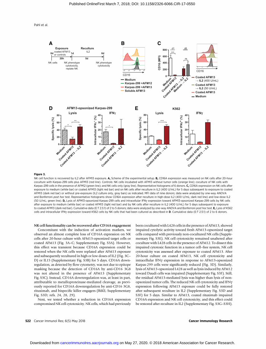
NK cell functionality can be recovered after CD16A engagementConcomitant with the induction of activation markers, we
observed an almost complete loss of CD16A expression on NKcells after 20-hour culture with AFM13-opsonized target cells orcoated AFM13 (Fig. 3A–C; Supplementary Fig. S3A). However,this effect was transient because CD16A expression could berestored when the NK cells were replated after AFM13 exposureand subsequently recultured in high or low doses of IL2 (Fig. 3C–D) or IL15 (Supplementary Fig. S3B) for 5 days. CD16A down-regulation, as detected by flow cytometry, was not due to epitopemasking because the detection of CD16A by anti-CD16 3G8was not altered in the presence of AFM13 (SupplementaryFig. S3C). Instead, CD16A downregulation was, at least in part,attributable to metalloproteinase-mediated cleavage, as previ-ously reported for CD16A downregulation by anti-CD16 3G8,rituximab, and bispecific killer engagers (BiKE; SupplementaryFig. S3D; refs. 24, 28, 29).
Next, we tested whether a reduction in CD16A expressioncompromisedNK cell cytotoxicity. NK cells, which had previously
been coculturedwith L428 cells in thepresence ofAFM13, showedimpaired cytolytic activity toward fresh AFM13-opsonized targetcells compared with previously non-cocultured NK cells (Supple-mentary Fig. S3E). NK cell cytotoxicity remained unaltered aftercoculture with L428 cells in the presence of AFM12. To dissect thisimpaired cytotoxic function in a tumor cell–free system, NK cellcytotoxicity was assessed after exposure to coated AFM13. After20-hour culture on coated AFM13, NK cell cytotoxicity andintracellular IFNg expression in response to AFM13-opsonizedKarpas-299 cells were significantly reduced (Fig. 3D). Similarly,lysis of AFM13-opsonized L428 aswell as lysis induced by AFM12toward Daudi cells was impaired (Supplementary Fig. S3F). Still,the residual AFM13-mediated lysis was higher than lysis of non-opsonized tumor cells. The reduced NK cell cytotoxicity and IFNgexpression following AFM13 exposure could be fully restoredafter subsequent reculture in IL2 (Supplementary Fig. S3D andS3F) for 5 days. Similar to AFM13, coated rituximab impairedCD16A expression and NK cell cytotoxicity, and this effect couldbe restored after reculture in IL2 (Supplementary Fig. S3G–S3H).
NK cell function is recovered by IL2 after AFM13 exposure. A, Scheme of the experimental setup. B, CD16A expression was measured on NK cells after 20-hourcoculture with Karpas-299 cells plus AFM13 (red line). Controls: NK cells incubated with AFM13 without tumor cells (orange line); coculture of NK cells withKarpas-299 cells in the presence of AFM12 (green line); and NK cells only (gray line). Representative histograms of 6 donors. C, CD16A expression on NK cells afterexposure to medium (white bar) or coated AFM13 (light red bar) and on NK cells after reculture in IL2 (400 U/mL) for 5 days subsequent to exposure to coatedAFM13 (dark red bar) or without pre-exposure (IL2 culture only, gray bars) as indicated. MFI data of nine donors; data were analyzed by one-way ANOVAand Bonferroni post hoc test. Representative histograms show CD16A expression after reculture in high-dose IL2 (400 U/mL; dark red line) and low-dose IL2(50 U/mL; green line). D, Lysis of AFM13-opsonized Karpas-299 cells and intracellular IFNg expression toward AFM13-opsonized Karpas-299 cells by NK cellsafter exposure to medium (white bar) or coated AFM13 (light red bar) and by NK cells after reculture in IL2 (400 U/mL) for 5 days subsequent to exposureto coated AFM13 (dark red bar). Cumulative data (E:T 2.5:1) of 2 to 5 donors; data were analyzed by one-way ANOVA and Bonferroni post hoc test. E, Lysis of K562cells and intracellular IFNg expression toward K562 cells by NK cells that had been cultured as described in D. Cumulative data (E:T 2.5:1) of 2 to 6 donors.
Pahl et al.
Cancer Immunol Res; 6(5) May 2018 Cancer Immunology Research522
Because the expression of perforin and granzyme B was unalteredafter exposure to coated AFM13 or rituximab (Supplementary Fig.S3I), the impairment of NK cell cytotoxicity was not due to theabsence of cytolytic mediators. However, expression of theCD16A and ITAM-signaling adaptor FceR1g (but not CD3z) wasreduced after exposure to coated AFM13 or rituximab, which wasrestored after IL2 reculture (Supplementary Fig. S3J). We addi-tionally tested NK cell responsiveness to PMA/ionomycin and tothe prototypical target cell line K562. IFNg expression upon PMA/ionomycin stimulation (Supplementary Fig. S3K), as well as"natural" cytotoxicity and IFNg expression in response to K562cells (Fig. 3E), was impaired but functional following IL2 recul-ture.Hence, althoughNKcell functionalitywas enhanced indirectresponse to AFM13-opsonized target cells, subsequently, CD16Aengagement led to lower NK cell reactivity toward tumor cells,which could be rescued by IL2 or IL15 reculture.
In a next step, we investigatedwhether preactivation of NK cellsvia CD16A influenced IFNg production in their subsequentresponse to cytokines or tumor cells. IFNg expression in responseto IL12/15/18 was increased directly after AFM13 exposure (day1; Fig. 4A). This coincided with higher expression of the high-affinity IL12Rb2 and IL18Ra (day 1: Fig. 4B). The higher expres-sion of IL12Rb2 was also observed after subsequent reculture inIL2 (day 6). When IL2-recultured NK cells had initially beenpreactivated by coated AFM13 or coated rituximab, IFNg produc-tion was significantly enhanced in response to restimulation withIL12/15 (Fig. 4C; Supplementary Fig. S4A). After IL2 reculture,IFNg production was enhanced after restimulation with PMA/ionomcyin and in response to K562, L428, or AFM13-opsonizedL428 cells (Fig. 4C). Thus, after IL2 reculture, NK cells preactivated
IL12/15restimulation600
500
400
300
200
Coat
ed A
FM13
→ IL
2
IFN
g (n
g/m
L)
100
0
Mediu
m →
IL2
K562restimulation70503030
20
Coat
ed A
FM13
→ IL
2
IFN
g (n
g/m
L)
10
0
Mediu
m →
IL2
L428restimulation
160100
4040
20Co
ated
AFM
13 →
IL2
IFNγ
(ng/
mL)
10
0
Mediu
m →
IL2
30
L428+AFM13restimulation1601004040
20
Coat
ed A
FM13
→ IL
2
IFN
g (n
g/m
L)
10
0
Mediu
m →
IL2
30
C0d
NK cells
1d
Exposurecoated AFM13or controls
RecultureIL2
20h 5d
6d
Replate NK Replate NK
IL12/15 ortumor cells
24hIFN-γ
Restimulation7d
IFNg toward IL12/15/18
10
30
20
40
Medium
Coated AFM13
Intr
acel
lula
r IFN
g+ (%
)0
BA
Day 1
Day 6
IL12Rβ2 IL18Rα
Coated AFM13
Medium
Coated AFM13→ IL2
Medium→ IL2
IL12Rβ2 IL18Rα
PMA/Ionorestimulation
50
40
30
20
IFN
g (n
g/m
L)
10
0
Coat
ed A
FM13
→ IL
2
Mediu
m →
IL2
100
101
102
103
104
100
101
102
103
104
100
101
102
103
104
100
101
102
103
104
Figure 4.
Preactivation of NK cells via CD16A byAFM13 enhances IFNg productionupon restimulation with IL12/15 orlymphoma cells. A, NK cells werecultured in medium (white bar) orexposed to coated AFM13 (light graybar). Intracellular IFNg expression ofNK cells in response to IL12/15/18 wasmeasured. Cumulative data of4 donors; data were analyzed bypaired Student t test. B, Expression ofthe high-affinity IL12Rb2 and IL18Raon NK cells previously activated bycoated AFM13 or cultured in mediumas indicated (top pair of histograms);and by NK cells recultured in IL2 for5 days subsequent to AFM13exposure or in IL2 withoutpreexposure as indicated (bottompair of histograms). Representativedata of 4 donors. C, Scheme ofthe experimental setup andIFNg production in response to24-hour restimulation by IL12/15;PMA/ionomycin; or K562 cells,L428 cells, or AFM13-opsonizedL428 cells by NK cells reculturedin IL2 (100–400 U/mL) for 5 dayssubsequent to the exposure to coatedAFM13 (dark gray squares), orcultured only in IL2 withoutpre-exposure (light gray circles).Cumulative data of 2 to 16 donors;data were analyzed by pairedStudent t tests.
CD16A Activation Promotes Memory-Like NK Cell Function
www.aacrjournals.org Cancer Immunol Res; 6(5) May 2018 523
through CD16A displayed enhanced IFNg production uponrestimulation with IL12/15 or different tumor cells.
CD16A preactivation enhances "natural" cytotoxicity to CD30þ
and CD30– tumor cellsThus far, we have demonstrated that the reduced NK cell
cytotoxicity following AFM13 exposure could be rescued througha 5-day reculture in IL2 (Fig. 3D; Supplementary Fig. S3E). In anext step, we explored whether preactivation of NK cells viaCD16A could modulate the "natural" cytolytic activity of IL2-cultured NK cells when restimulated with non-opsonized tumorcells, expressing ligands to the NK cell–activating receptorsNKG2D, DNAM-1, and NKp30. The "natural" lysis of CD30þ
Karpas-299 and HDLM-2 cells was enhanced, when IL2-culturedNK cells had initially been exposed to AFM13 (Fig. 5A,). Theselymphoma cells were highly resistant to lysis by IL2-cultured NKcells not preactivated by AFM13 and fully resistant to na€�ve NKcells. Even the lysis of the CD30– and CD30low lymphoma celllines Daudi and L1236, respectively, and to some extent to K562cells, was amplified by the initial AFM13 exposure (Fig. 5A;Supplementary Fig. S4B). Preactivation by AFM13 and reculturein IL2 enhanced not only the "natural" cytotoxicity but alsoAFM13- and AFM12-mediated target cell lysis involving CD16Aengagement (Supplementary Fig. S4B). This enhanced cytotoxic-ity of NK cells after IL2 reculture could not only be induced bypreactivationwith coated CD16A-recruiting bispecific antibodies,such as AFM13, but also by classical IgG antibodies such asrituximab (Supplementary Fig. S4C). The enhanced cytolyticactivity was associated with increased degranulation towardtumor cells. In parallel, perforin and granzyme B levels remainedunchanged (Supplementary Fig. S4D). After IL2 reculture,NKG2D surface expressionwas increased onNK cells preactivatedby AFM13 or rituximab, whereas the expression of NKp30 orDNAM-1 was not altered (Fig. 5B; Supplementary Fig. S4E).Therefore, to determine which NK cell receptors and pathwayscontributed to the enhanced cytolytic activity due to preactivationvia CD16A, lysis of murine Ba/F3 cells stably transfected with theNKG2D ligand MICA, the DNAM-1 ligand CD155, or the NKp30ligand B7H6 was examined. Lysis of Ba/F3-MICA was enhancedby AFM13-preactivatedNK cells, whereas lysis of Ba/F3-CD155 orBa/F3-B7H6 cells was not significantly altered (Fig. 5C). The lysisof Ba/F3-MICA cells by AFM13-preactivated NK cells was abro-gated in the presence of neutralizing antibodies against NKG2D(Supplementary Fig. S4F). In summary, antibody-experiencedNKcells (e.g., by AFM13) recultured in IL2 or IL15 exhibited ampli-fied cytotoxicity toward CD30þ and CD30– lymphoma cells, dueto improved killing through the activating NK cell receptorNKG2D.
DiscussionCD16A is the only activating receptor that can trigger the
cytotoxic activity of na€�ve human NK cells, even in the absenceof costimulatory signals (30, 31). In our study, we demonstratethat CD16A activation by tetravalent, bispecific AFM13improved the functionality of NK cells beyond their classicalantibody-dependent cytotoxicity and IFNg production elicitedin response to CD30þ lymphoma cells, which are otherwisepoorly killed by NK cells. We show that CD16A engagementincreased the sensitivity of NK cells to IL15 or low-dose IL2.This led to an amplification of IL15 and IL2-dependent NK cell
proliferation, resulting in increased numbers of functional NKcells. When preactivated via CD16A, CD16A-experienced NKcells displayed enhanced IFNg production and exertedenhanced cytotoxicity toward CD30þ and CD30– lymphomacells after IL2 reculture, which appeared to mainly involve theNK cell NKG2D pathway. The classic therapeutic antibodyrituximab largely recapitulated the results of the tetravalentbispecific antibody AFM13, supporting these observations ofCD16A-mediated priming of NK cells.
Our phenotypic analysis revealed that CD16A engagement byAFM13 leads to a defined signature of upregulated activating andinhibitory receptors on CD56dim CD16þ NK cells. As expected,CD56bright CD16dim/–NKcells remained largely unaffected. Inourstudy with healthy donors, only low numbers of CD56dim
CD16dim/– NK cells were detected, which has been previouslydescribed in leukemia patients (32). Increased frequencies ofthese subsets in cancer patients might be relevant for patientstratification before or during antibody therapy to predict optimalclinical responses. The signature of upregulated receptors onCD56dim CD16þ NK cells may be further developed as biomar-kers to monitor NK cell responses during AFM13 therapy andcould be targeted in combinatorial treatments. For instance,cotargeting of activating receptors like CD137 with agonisticantibodies, as previously described (33), or inhibitory receptors,such as CD96, with antagonistic antibodies (34) may potentiateNK cell antitumor function after AFM13 exposure.
The improved sensitivity of CD16A-preactivated NK cells toIL15 and low-dose IL2 coincided with the induction of CD25(IL2Ra), which, together with CD122 (IL2Rb) and CD132 (gc),assembles the trimeric high-affinity IL2 receptor, and the upre-gulation of CD132 together with CD122 assembles the dimericintermediate-affinity IL2 and IL15 receptors (35). In our study,CD25 induction by AFM13-opsonized target cells was muchstronger than previously shown upon CD16A cross-linkingusing anti-CD16 3G8with a secondary antibody (36). Similarly,NK cell activation by IL12/15/18 has been shown to induceCD25 expression, boosting IL2-dependent proliferation in vitroand in vivo in tumor-bearing mice (9, 37). Hence, the inductionof CD25 following AFM13 exposure may enable NK cells tocompete for low amounts of IL2 with regulatory T cells, whichstrictly limit the availability of IL2 due to their constitutiveCD25expression, restraining NK cell expansion after adoptive celltransfer (38–40).
Following CD16A engagement, we observed a transientimpaired function in CD16A-dependent as well as "natural" NKcell cytotoxicity and IFNg expression toward tumor cells. Theimpairment of CD16A-dependent activity may be explainedby the nearly complete loss of CD16A expression upon CD16Aengagement, which, at least in part, involved matrix metallopro-teinase-mediated cleavage, consistent with previous studies,or receptor internalization, as additionally described (24, 28,41–43). The transient lower responsiveness in "natural" NK cellantitumor reactivity towardK562 cells suggested a desensitizationof otherNK cell–activating receptors, such asNKp30 andNKG2D,that have previously been shown to be involved in K562 lysis(44, 45). The reduction in IFNg expression in response to PMA/ionomycin was indicative of a dysfunctional state that mayalso affect PKC activation and/or Ca2þ mobilization, which aredirectly activated by PMA/ionomycin (46). Mobilization of Ca2þ
is critically involved in the signaling of CD16A and other acti-vating receptors (31, 47). PKC activation can mediate IFNg
Pahl et al.
Cancer Immunol Res; 6(5) May 2018 Cancer Immunology Research524
Preactivation of NK cells via CD16A by AFM13 enhances "natural" NK cell cytotoxicity upon restimulation with lymphoma cells. A, Lysis of nonopsonized CD30þ
Karpas-299 and HDLM-2 cells, CD30– Daudi and CD30low L1236 cells by na€�ve NK cells (open squares), by NK cells recultured in IL2 for 5 days subsequentto the exposure to coated AFM13 (dark gray squares), or by NK cells cultured only in IL2 without preexposure (light gray circles). Representative data (top) andcumulative data (Karpas-299, HDLM-2, and L1236 E:T 2.5:1; Daudi E:T 0.6:1; bottom) of 7 to 10 donors; data were analyzed by paired Student t tests. B, Afterexposure to coated AFM13 and IL2 reculture (dark gray line) or after IL2 reculture only (light gray line), surface expression of NKG2D, DNAM-1, and NKp30 wasmeasured. Representative data of 6 donors. C, Lysis of murine Ba/F3 cells transfected with MICA, CD155, and B7H6 by NK cells recultured in IL2 subsequenttoAFM13 exposure (dark graybar) or byNKcells cultured in IL2 only (light graybar). Lysis of Ba/F3 cells transfectedwith an empty vectorwas subtracted. Cumulativedata (E:T 2.5:1) of 7 donors; data were analyzed by two-way ANOVA and Bonferroni post hoc test.
CD16A Activation Promotes Memory-Like NK Cell Function
www.aacrjournals.org Cancer Immunol Res; 6(5) May 2018 525
production and is important for K562 lysis but dispensable forADCC, which, in turn, requires PI3K activation (48, 49). Theobserved impairment in our study after 20-hour exposure toAFM13 or rituximab may exceed the inhibitory effect of short-term (1.5-hour) CD16A engagement, which was reported to notaffect IFNg production or responsiveness to PMA/ionomycin,while resulting in defective degranulation due to SHP-1 recruit-ment, inhibition of PLCg2/Vav-1/SLP-76 phosphorylation, andboth FceR1g and CD3z degradation (42, 43, 50).
We found that the IFNg response ofNK cells to IL12/15/18 afterCD16A engagement was conserved or even further enhanced,indicating that the impaired IFNg expression after AFM13 expo-sure was selective for the response to tumor cells. This may beexplained by the upregulation of the high-affinity IL12 and IL18receptors, which potently induce IFNg . Similarly, IFNg produc-tion in response to IL12 has recently been shown to be functionaland enhanced after Fc-mediated antibody engagement of CD16A(43). It has been reported that CD16A and IL12 receptor activa-tion can synergistically promote IFNg production (51). Becausethe signaling cascades for IFNg induction downstream of CD16Aand IL12/18 receptors differ, it is tempting to speculate that theimpaired IFNg production, as observed in our study, is selectivefor CD16A–ITAM–Syk–PI3K–ERK–AP-1–induced signaling,whereas IL12/18R–STAT4/AP-1–induced signaling remains com-petent (51–53).
The lower NK cell reactivity following AFM13 or rituximabexposurewas reverted after reculture in IL2or IL15 for 5days. AfterIL2 reculture, these CD16A-experienced NK cells displayed, inparticular, enhanced NKG2D-dependent cytotoxic activity, aswell as enhanced IFNg production, when restimulated withotherwise weakly susceptible lymphoma cells or cytokines. Pre-viously, IL12/15/18–preactivated, memory-like NK cells havebeen described to exert enhanced IFNg production and antitumorresponses after reculture in IL2 or IL15 (8–11). It is possible thatthe novel functionality of "CD16A-induced memory-like NKcells"might have similarities to the enhanced antitumor potentialof IL12/15/18-induced memory-like NK cells.
Taken together, our data imply that CD16A-mediated NK cellactivation by AFM13 results in a distinctive phenotype that may
provide parameters to monitor AFM13-mediated NK cell acti-vation in cancer patients. Our data support a strategy forrational treatment combinations of AFM13 with IL2 or IL15that may boost NK cell functionality upon repeated tumorencounters and expand the quantity of tumor-reactive NK cellsin lymphoma patients.
Disclosure of Potential Conflicts of InterestJ.H.W. Pahl and A. Cerwenka report receiving a commercial research grant
from Affimed. A. Cerwenka is a consultant/advisory board for SAB DragonflyTherapeutics. No potential conflicts of interest were disclosed by the otherauthors.
Authors' ContributionsConception anddesign: J.H.W. Pahl, J. Koch,U. Reusch,M. Treder, A. CerwenkaDevelopment of methodology: J.H.W. Pahl, A. ArnoldAcquisition of data (provided animals, acquired and managed patients,provided facilities, etc.): J.H.W. Pahl, A. Arnold, A. CerwenkaAnalysis and interpretation of data (e.g., statistical analysis, biostatistics,computational analysis): J.H.W. Pahl, J. Koch, J.-J. G€otz, A. Arnold, U. Reusch,T. Gantke, E. Rajkovic, M. TrederWriting, review, and/or revision of the manuscript: J.H.W. Pahl, J. Koch, U.Reusch, T. Gantke, E. Rajkovic, M. Treder, A. CerwenkaAdministrative, technical, or material support (i.e., reporting or organizingdata, constructing databases): J.H.W. Pahl, J.-J. G€otz, U. ReuschStudy supervision: M. Treder, A. Cerwenka
AcknowledgmentsJana-Julia G€otz is supported by the Cooperation Program inCancer Research
of the Deutsches Krebsforschungszentrum, Israel's Ministry of Science andTechnology (CA172).
We thank Margareta Correia and Ana Stojanovic for valuable input anddiscussions, Tim Machauer and Claudia Felbinger for technical assistance, andAnca Alexandru and Leonie Kohlhammer for careful reading of themanuscript.
The costs of publication of this article were defrayed in part by thepayment of page charges. This article must therefore be hereby markedadvertisement in accordance with 18 U.S.C. Section 1734 solely to indicatethis fact.
Received September 27, 2017; revised December 9, 2017; accepted March 1,2018; published first March 7, 2018.
References1. Spits H, Bernink JH, Lanier L. NK cells and type 1 innate lymphoid cells:
partners in host defense. Nat Immunol 2016;17:758–64.2. Koch J, Steinle A, Watzl C, Mandelboim O. Activating natural cytotoxicity
receptors of natural killer cells in cancer and infection. Trends Immunol2013;34:182–91.
3. Pahl J, Cerwenka A. Tricking the balance: NK cells in anti-cancer immunity.Immunobiology 2017;222:11–20.
4. Vivier E, Raulet DH, Moretta A, Caligiuri MA, Zitvogel L, Lanier LL, et al.Innate or adaptive immunity? The example of natural killer cells. Science2011;331:44–9.
5. Schuster IS, Coudert JD, Andoniou CE, Degli-Esposti MA. "Natural reg-ulators": NK cells as modulators of T cell immunity. Front Immunol2016;7:235.
6. Cerwenka A, Lanier LL. Natural killer cell memory in infection, inflam-mation and cancer. Nat Rev Immunol 2016;16:112–23.
7. Luetke-EverslohM, Hammer Q, Durek P, Nordstrom K, Gasparoni G, PinkM, et al. Human cytomegalovirus drives epigenetic imprinting of the IFNGlocus in NKG2Chi natural killer cells. PLoS Pathog 2014;10:e1004441.
9. Ni J, Miller M, Stojanovic A, Garbi N, Cerwenka A. Sustained effectorfunction of IL-12/15/18-preactivatedNK cells against established tumors. JExp Med 2012;209:2351–65.
10. Romee R, Schneider SE, Leong JW, Chase JM, Keppel CR, Sullivan RP, et al.Cytokine activation induces human memory-like NK cells. Blood 2012;120:4751–60.
11. Ni J, Holsken O, Miller M, Hammer Q, Luetke-Eversloh M, Romagnani C,et al. Adoptively transferred natural killer cells maintain long-term anti-tumor activity by epigenetic imprinting and CD4þ T cell help. Oncoim-munology 2016;5:e1219009.
12. Parkhurst MR, Riley JP, Dudley ME, Rosenberg SA. Adoptive transfer ofautologous natural killer cells leads to high levels of circulating naturalkiller cells but does not mediate tumor regression. Clin Cancer Res 2011;17:6287–97.
13. Reiners KS, Kessler J, Sauer M, Rothe A, Hansen HP, Reusch U, et al.Rescue of impaired NK cell activity in hodgkin lymphoma withbispecific antibodies in vitro and in patients. Mol Ther 2013;21:895–903.
14. Pahl JH, Ruslan SE, Buddingh EP, Santos SJ, Szuhai K, Serra M, et al. Anti-EGFR antibody cetuximab enhances the cytolytic activity of natural killercells toward osteosarcoma. Clin Cancer Res 2012;18:432–41.
Cancer Immunol Res; 6(5) May 2018 Cancer Immunology Research526
15. de Landazuri MO, Silva A, Alvarez J, Herberman RB. Evidence thatnatural cytotoxicity and antibody-dependent cellular cytotoxicity aremediated in humans by the same effector cell populations. J Immunol1979;123:252–8.
16. Lanier LL, Ruitenberg JJ, Phillips JH. Functional and biochemical analysisof CD16 antigen on natural killer cells and granulocytes. J Immunol1988;141:3478–85.
17. Vivier E, Morin P, O'Brien C, Druker B, Schlossman SF, Anderson P.Tyrosine phosphorylation of the Fc gamma RIII(CD16): zeta complex inhuman natural killer cells. Induction by antibody-dependent cytotoxicitybut not by natural killing. J Immunol 1991;146:206–10.
18. Bibeau F, Lopez-Crapez E, Di FF, Thezenas S, Ychou M, Blanchard F, et al.Impact of Fc{gamma}RIIa-Fc{gamma}RIIIa polymorphisms and KRASmutations on the clinical outcome of patients with metastatic colorectalcancer treatedwith cetuximabplus irinotecan. J ClinOncol 2009;27:1122–9.
19. Dall'Ozzo S, Tartas S, Paintaud G, Cartron G, Colombat P, Bardos P, et al.Rituximab-dependent cytotoxicity by natural killer cells: influence ofFCGR3A polymorphism on the concentration-effect relationship. CancerRes 2004;64:4664–9.
20. Nechansky A, Schuster M, Jost W, Siegl P, Wiederkum S, Gorr G, et al.Compensation of endogenous IgG mediated inhibition of antibody-dependent cellular cytotoxicity by glyco-engineering of therapeutic anti-bodies. Mol Immunol 2007;44:1815–7.
21. Iida S, Misaka H, Inoue M, Shibata M, Nakano R, Yamane-Ohnuki N,et al. Nonfucosylated therapeutic IgG1 antibody can evade the inhib-itory effect of serum immunoglobulin G on antibody-dependent cel-lular cytotoxicity through its high binding to FcgammaRIIIa. ClinCancer Res 2006;12:2879–87.
22. Arndt MA, Krauss J, Kipriyanov SM, Pfreundschuh M, Little M. A bispecificdiabody that mediates natural killer cell cytotoxicity against xenotrans-plantated human Hodgkin's tumors. Blood 1999;94:2562–8.
23. Kipriyanov SM, Cochlovius B, SchaferHJ,Moldenhauer G, Bahre A, LeGallF, et al. Synergistic antitumor effect of bispecific CD19 x CD3 and CD19 xCD16 diabodies in a preclinical model of non-Hodgkin's lymphoma. JImmunol 2002;169:137–44.
24. Wiernik A, Foley B, Zhang B, Verneris MR, Warlick E, Gleason MK, et al.Targeting natural killer cells to acute myeloid leukemia in vitro with aCD16 x 33 bispecific killer cell engager and ADAM17 inhibition. ClinCancer Res 2013;19:3844–55.
25. Reusch U, Burkhardt C, Fucek I, Le Gall F, Le Gall M, Hoffmann K, et al. Anovel tetravalent bispecific TandAb (CD30/CD16A) efficiently recruits NKcells for the lysis of CD30þ tumor cells. MAbs 2014;6:728–39.
26. Rothe A, Sasse S, ToppMS, Eichenauer DA, Hummel H, Reiners KS, et al. Aphase 1 study of the bispecific anti-CD30/CD16A antibody constructAFM13 in patients with relapsed or refractory Hodgkin lymphoma. Blood2015;125:4024–31.
27. Zingoni A, Sornasse T, Cocks BG, Tanaka Y, Santoni A, Lanier LL. Cross-talkbetween activated human NK cells and CD4þ T cells via OX40-OX40ligand interactions. J Immunol 2004;173:3716–24.
28. Romee R, Foley B, Lenvik T, Wang Y, Zhang B, Ankarlo D, et al. NK cellCD16 surface expression and function is regulated by a disintegrin andmetalloprotease-17 (ADAM17). Blood 2013;121:3599–608.
29. Borrego F, Lopez-Beltran A, Pena J, Solana R.Downregulation of Fc gammareceptor IIIA alpha (CD16-II) on natural killer cells induced by anti-CD16mAb is independent of protein tyrosine kinases and protein kinase C. CellImmunol 1994;158:208–17.
30. Bryceson YT, Ljunggren HG, Long EO. Minimal requirement for inductionof natural cytotoxicity and intersection of activation signals by inhibitoryreceptors. Blood 2009;114:2657–66.
31. Bryceson YT,MarchME, LjunggrenHG, Long EO. Synergy among receptorson resting NK cells for the activation of natural cytotoxicity and cytokinesecretion. Blood 2006;107:159–66.
32. Stabile H, Nisti P, Morrone S, Pagliara D, Bertaina A, Locatelli F, et al.Multifunctional human CD56 low CD16 low natural killer cells are theprominent subset in bone marrow of both healthy pediatric donors andleukemic patients. Haematologica 2015;100:489–98.
33. Kohrt HE, Houot R, Goldstein MJ, Weiskopf K, Alizadeh AA, Brody J, et al.CD137 stimulation enhances the antilymphoma activity of anti-CD20antibodies. Blood 2011;117:2423–32.
34. Blake SJ, Stannard K, Liu J, Allen S, YongMC,Mittal D, et al. Suppression ofmetastases using a new lymphocyte checkpoint target for cancer immu-notherapy. Cancer Discov 2016;6:446–59.
35. Waldmann TA. The biology of interleukin-2 and interleukin-15: impli-cations for cancer therapy and vaccine design. Nat Rev Immunol2006;6:595–601.
36. Marquez ME, Millet C, Stekman H, Conesa A, Deglesne PA, Toro F, et al.CD16 cross-linking induces increased expression of CD56 and productionof IL-12 in peripheral NK cells. Cell Immunol 2010;264:86–92.
37. Lee SH, Fragoso MF, Biron CA. Cutting edge: a novel mechanism bridginginnate and adaptive immunity: IL-12 induction of CD25 to form high-affinity IL-2 receptors on NK cells. J Immunol 2012;189:2712–6.
38. Gasteiger G, Hemmers S, Firth MA, Le Floc'h A, Huse M, Sun JC, et al. IL-2-dependent tuning of NK cell sensitivity for target cells is controlled byregulatory T cells. J Exp Med 2013;210:1167–78.
39. KimM, Kim TJ, Kim HM, Doh J, Lee KM. Multi-cellular natural killer (NK)cell clusters enhance NK cell activation through localizing IL-2 within thecluster. Sci Rep 2017;7:40623.
40. Knorr DA, Bachanova V, Verneris MR, Miller JS. Clinical utility of naturalkiller cells in cancer therapy and transplantation. Semin Immunol 2014;26:161–72.
41. Lajoie L, Congy-Jolivet N, Bolzec A, Gouilleux-Gruart V, Sicard E, SungHC,et al. ADAM17-mediated shedding of FcgammaRIIIA on human NK cells:identification of the cleavage site and relationship with activation. JImmunol 2014;192:741–51.
42. Capuano C, Romanelli M, Pighi C, Cimino G, Rago A, Molfetta R, et al.Anti-CD20 therapy acts via FcgammaRIIIA to diminish responsiveness ofhuman natural killer cells. Cancer Res 2015;75:4097–108.
43. Capuano C, Pighi C, Molfetta R, Paolini R, Battella S, Palmieri G, et al.Obinutuzumab-mediated high-affinity ligation of FcgammaRIIIA/CD16primes NK cells for IFNgamma production. Oncoimmunology 2017;6:e1290037.
44. Brandt CS, Baratin M, Yi EC, Kennedy J, Gao Z, Fox B, et al. The B7 familymember B7-H6 is a tumor cell ligand for the activating natural killer cellreceptor NKp30 in humans. J Exp Med 2009;206:1495–503.
45. Kuylenstierna C, Bjorkstrom NK, Andersson SK, Sahlstrom P, Bosnjak L,Paquin-Proulx D, et al. NKG2D performs two functions in invariantNKT cells: direct TCR-independent activation of NK-like cytolysisand co-stimulation of activation by CD1d. Eur J Immunol 2011;41:1913–23.
46. Chatila T, Silverman L,Miller R, Geha R.Mechanisms of T cell activation bythe calcium ionophore ionomycin. J Immunol 1989;143:1283–9.
47. Cassatella MA, Anegon I, Cuturi MC, Griskey P, Trinchieri G, Perussia B. Fcgamma R(CD16) interaction with ligand induces Ca2þmobilization andphosphoinositide turnover in human natural killer cells. Role of Ca2þ inFc gamma R(CD16)-induced transcription and expression of lymphokinegenes. J Exp Med 1989;169:549–67.
48. Bonnema JD, Karnitz LM, Schoon RA, Abraham RT, Leibson PJ. Fc receptorstimulation of phosphatidylinositol 3-kinase in natural killer cells isassociated with protein kinase C-independent granule release and cell-mediated cytotoxicity. J Exp Med 1994;180:1427–35.
49. Hara H, Ishihara C, Takeuchi A, Xue L, Morris SW, Penninger JM, et al. Celltype-specific regulation of ITAM-mediated NF-kappaB activation by theadaptors, CARMA1 and CARD9. J Immunol 2008;181:918–30.
50. Galandrini R, Tassi I, Mattia G, Lenti L, Piccoli M, Frati L, et al. SH2-containing inositol phosphatase (SHIP-1) transiently translocates to raftdomains and modulates CD16-mediated cytotoxicity in human NK cells.Blood 2002;100:4581–9.
51. Kondadasula SV, Roda JM, Parihar R, Yu J, Lehman A, Caligiuri MA, et al.Colocalization of the IL-12 receptor and FcgammaRIIIa to natural killer celllipid rafts leads to activation of ERK and enhanced production of inter-feron-gamma. Blood 2008;111:4173–83.
52. Zompi S, Colucci F. Anatomy of a murder–signal transduction pathwaysleading to activation of natural killer cells. Immunol Lett 2005;97:31–9.
53. Nakahira M, AhnHJ, ParkWR, Gao P, TomuraM, Park CS, et al. Synergy ofIL-12 and IL-18 for IFN-gamma gene expression: IL-12-induced STAT4contributes to IFN-gamma promoter activation by up-regulating the bind-ing activity of IL-18-induced activator protein 1. J Immunol 2002;168:1146–53.
www.aacrjournals.org Cancer Immunol Res; 6(5) May 2018 527
CD16A Activation Promotes Memory-Like NK Cell Function
2018;6:517-527. Published OnlineFirst March 7, 2018.Cancer Immunol Res Jens H.W. Pahl, Joachim Koch, Jana-Julia Götz, et al. Memory-Like Cytotoxicity against Cancer CellsCD16A Activation of NK Cells Promotes NK Cell Proliferation and
Updated version
10.1158/2326-6066.CIR-17-0550doi:
Access the most recent version of this article at: