cDNA and Genomic Cloning of Lacritin, a Novel Secretion Enhancing Factor from the Human Lacrimal Gland Sandhya Sanghi 1 , Rajesh Kumar 1 , Angela Lumsden 1 Douglas Dickinson 2 , Veronica Klepeis 3 , Vickery Trinkaus-Randall 3 Henry F. Frierson Jr 4 and Gordon W. Laurie 1 * 1 Department of Cell Biology University of Virginia Charlottesville, VA 22908 USA 2 Department of Oral Biology Medical College of Georgia Augusta, GA 30912 USA 3 Department of Biochemistry Boston University, Boston, MA 02118 USA 4 Department of Pathology University of Virginia Charlottesville, VA 22908 USA Multiple extracellular factors are hypothesized to promote the differen- tiation of unstimulated and/or stimulated secretory pathways in exocrine secretory cells, but the identity of differentiation factors, particularly those organ-specific, remain largely unknown. Here, we report on the identification of a novel secreted glycoprotein, lacritin, that enhances exo- crine secretion in overnight cultures of lacrimal acinar cells which other- wise display loss of secretory function. Lacritin mRNA and protein are highly expressed in human lacrimal gland, moderately in major and minor salivary glands and slightly in thyroid. No lacritin message or pro- tein is detected elsewhere among more than 50 human tissues examined. Lacritin displays partial similarity to the glycosaminoglycan-binding region of brain-specific neuroglycan C (32 % identity over 102 amino acid residues) and to the possibly mucin-like amino globular region of fibulin-2 (30 % identity over 81 amino acid residues), and localizes pri- marily to secretory granules and secretory fluid. The lacritin gene consists of five exons, displays no alternative splicing and maps to 12q13. Recom- binant lacritin augments unstimulated but not stimulated acinar cell secretion, promotes ductal cell proliferation, and stimulates signaling through tyrosine phosphorylation and release of calcium. It binds col- lagen IV, laminin-1, entactin/nidogen-1, fibronectin and vitronectin, but not collagen I, heparin or EGF. As an autocrine/paracrine enhancer of the lacrimal constitutive secretory pathway, ductal cell mitogen and stimulator of corneal epithelial cells, lacritin may play a key role in the function of the lacrimal gland-corneal axis. # 2001 Academic Press Keywords: exocrine secretion; lacrimal; mitogen; corneal; ECM *Corresponding author Introduction The polarized exocrine secretory cell is an ele- gant manifestation of epithelial differentiation, a complex process influenced by mesenchymal inter- actions 1 and adhesion to elements of the newly formed epithelial basement membrane. A well- studied example is the exocrine pancreas in which basement membrane-coincident 2 acquisition of cell polarity, protein synthetic organelles including subluminal secretory granules, calcium signaling machinery and constitutive secretion is followed by a delayed rab3D- 3,4 and rab4-dependent 5 com- pletion of stimulus-secretion coupling. More com- plex are salivary glands in which several different parenchymal cells are specified at varying rates in extracellular environments thought to differ subtly. 6 The regulation of epithelial differentiation in exo- crine glands, as examined in gene knockout exper- iments, appears multifactorial. There is evidence that the TGFb superfamily has an important role in acinar formation in the exocrine pancreas (type II TGFb receptor; 7 type II activin receptor 8 ) and mammary gland (activins and inhibins 9 ). Also involved (mammary gland) are ErbB4, 10 the pro- gesterone receptor, 11 the extracellular matrix glyco- protein osteopontin, 12 and the EGF receptor 13 S.S. and R.K. contributed equally to this work. E-mail address of the corresponding author: [email protected]doi:10.1006/jmbi.2001.4748 available online at http://www.idealibrary.com on J. Mol. Biol. (2001) 310, 127–139 0022-2836/01/010127–13 $35.00/0 # 2001 Academic Press
Transcript
doi:10.1006/jmbi.2001.4748 available online at http://www.idealibrary.com on J. Mol. Biol. (2001) 310, 127±139
cDNA and Genomic Cloning of Lacritin, a NovelSecretion Enhancing Factor from the HumanLacrimal Gland
Sandhya Sanghi1, Rajesh Kumar1, Angela Lumsden1
Douglas Dickinson2, Veronica Klepeis3, Vickery Trinkaus-Randall3
Henry F. Frierson Jr4 and Gordon W. Laurie1*
1Department of Cell BiologyUniversity of VirginiaCharlottesville, VA 22908 USA2Department of Oral BiologyMedical College of GeorgiaAugusta, GA 30912 USA3Department of BiochemistryBoston University, Boston, MA02118 USA4Department of PathologyUniversity of VirginiaCharlottesville, VA 22908USA
S.S. and R.K. contributed equallyE-mail address of the correspond
Multiple extracellular factors are hypothesized to promote the differen-tiation of unstimulated and/or stimulated secretory pathways in exocrinesecretory cells, but the identity of differentiation factors, particularlythose organ-speci®c, remain largely unknown. Here, we report on theidenti®cation of a novel secreted glycoprotein, lacritin, that enhances exo-crine secretion in overnight cultures of lacrimal acinar cells which other-wise display loss of secretory function. Lacritin mRNA and protein arehighly expressed in human lacrimal gland, moderately in major andminor salivary glands and slightly in thyroid. No lacritin message or pro-tein is detected elsewhere among more than 50 human tissues examined.Lacritin displays partial similarity to the glycosaminoglycan-bindingregion of brain-speci®c neuroglycan C (32 % identity over 102 aminoacid residues) and to the possibly mucin-like amino globular region of®bulin-2 (30 % identity over 81 amino acid residues), and localizes pri-marily to secretory granules and secretory ¯uid. The lacritin gene consistsof ®ve exons, displays no alternative splicing and maps to 12q13. Recom-binant lacritin augments unstimulated but not stimulated acinar cellsecretion, promotes ductal cell proliferation, and stimulates signalingthrough tyrosine phosphorylation and release of calcium. It binds col-lagen IV, laminin-1, entactin/nidogen-1, ®bronectin and vitronectin, butnot collagen I, heparin or EGF. As an autocrine/paracrine enhancer ofthe lacrimal constitutive secretory pathway, ductal cell mitogen andstimulator of corneal epithelial cells, lacritin may play a key role in thefunction of the lacrimal gland-corneal axis.
The polarized exocrine secretory cell is an ele-gant manifestation of epithelial differentiation, acomplex process in¯uenced by mesenchymal inter-actions1 and adhesion to elements of the newlyformed epithelial basement membrane. A well-studied example is the exocrine pancreas in whichbasement membrane-coincident2 acquisition of cellpolarity, protein synthetic organelles includingsubluminal secretory granules, calcium signalingmachinery and constitutive secretion is followed
to this work.ing author:
by a delayed rab3D-3,4 and rab4-dependent5 com-pletion of stimulus-secretion coupling. More com-plex are salivary glands in which several differentparenchymal cells are speci®ed at varying ratesin extracellular environments thought to differsubtly.6
The regulation of epithelial differentiation in exo-crine glands, as examined in gene knockout exper-iments, appears multifactorial. There is evidencethat the TGFb superfamily has an important role inacinar formation in the exocrine pancreas (type IITGFb receptor;7 type II activin receptor8) andmammary gland (activins and inhibins9). Alsoinvolved (mammary gland) are ErbB4,10 the pro-gesterone receptor,11 the extracellular matrix glyco-protein osteopontin,12 and the EGF receptor13
# 2001 Academic Press
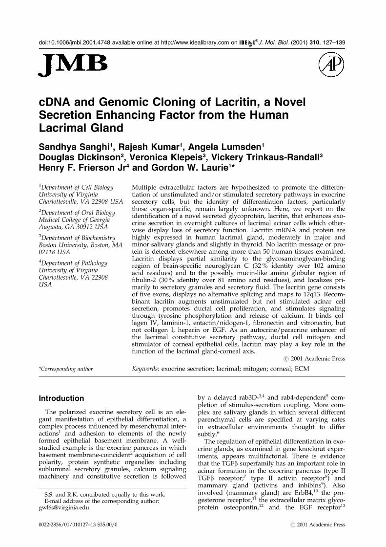
Figure 1. Lacritin cDNA, deduced protein sequence,and partial similarity to human neuroglycan C and ®bu-lin-2. (a) Lacritin consists of 138 amino acid residueswith a putative endoplasmic reticulum signal peptide(underlined, probability score 0.97; SignalP), oneN-glycosylation site (PeptideStructure), and six pre-dicted O-glycosylation sites (respective potential 0.62,0.99, 0.97, 0.88, 0.99 and 0.98; NetOGlyc 2.0), the lattergrouped between residues 52 and 64. Lacritin lacksidentity with other sequences, even with ESTs (in keep-ing with an absence of lacrimal-speci®c ESTs and anexpression pattern that is highly restricted). (b) Hom-ology is with the glycosaminoglycan attachment regionof neuroglycan C (af059274), and with the cysteine-freeN-terminal globular domain (Nb) of ®bulin-2 (x82494).BestFit quality of lacritin lacking signal peptide withneuroglycan C or ®bulin-2 is 83 (versus 37 � 5 whenlacritin sequence is randomized) and 81 (versus 38 � 5),respectively. Shown is lacritin without signal peptideversus residues 21 to 130 of neuroglycan C (539 aminoacid residues total length) and 217 to 336 of ®bulin-2(1184 amino acid residues total length).
128 Characterization of Lacritin
when knocked out with TGFa and amphiregulin.14
In a similar way, partial or complete knockout of®broblast growth factor receptor 2 (IIIb) inmammary15 and salivary gland16 is associated withimpaired acinar formation, or even partial (pan-creas), or incomplete (salivary gland)organogenesis.17 FGF-10 null mice display a com-plete absence of lacrimal glands, a result in keep-ing with ectopic lacrimal gland formation inregions of transgenic FGF-10 overexpression.18,19
Dissection of the extracellular matrix contri-bution has taken advantage of primary acinar cellculture in which the delicate and reversible natureof differentiation in polarized secretory cells canbecome fully apparent. Agonist responsiveness inisolated lacrimal acinar cells is, for example, depen-dent on adhesion to laminin-1, an effect enhancedby inclusion of a lower molecular mass extracellu-lar matrix fraction enriched in BM180.20 In a simi-lar way, constitutive casein secretion21 and insulin-dependent tyrosine phosphorylation22 by mam-mary epithelial cells are dependent on contact witha laminin-1 enriched basement membrane.Although a number of exocrine cell lines appear tobe fully functional without supplements, severalauthors have noted a ``differentiative'' morphogen-esis by the human submandibular ductal (HSG)cell line on laminin-1,23 a process reported to beinhibitable with anti-TGFb3 antibodies.24 Theseobservations are in keeping with a remarkable ten-dency of laminin-1 to selectively constitute fetalglandular basement membranes, particularly thoseof differentiating pancreatic, salivary25 andmammary26 glands. Other differentiation factorsare expected to exist, particularly those that mayconfer organ speci®city.
We report on the cDNA and genomic cloning,chromosome mapping and initial functionalcharacterization of a new human gene product,lacritin, that has a highly restricted glandular dis-tribution. Lacritin enhances unstimulated but notstimulated secretion, has mitogenic activity andpromotes signaling in both lacrimal acinar and cor-neal epithelial cells. Its highly restricted expressionpattern and functional attributes are evidence forits putative autocrine/paracrine differentiative rolein the lacrimal gland and neighboring ocularsystem.
Results
Multiple extracellular factors are hypothesized tomodulate the differentiation of stimulated andunstimulated tear secretion pathways in lacrimalacinar cells, a polarized exocrine secretory cell withsome mRNAs remarkably under-represented ingene data banks. In a systematic oligonucleotidescreen of a human lacrimal gland library, a novelgene product was identi®ed (Figure 1) with a levelof lacrimal gland speci®city (Figure 2) not pre-viously observed. We refer to this gene product aslacritin. Its 417 bp open reading frame (Figure 1(a))predicts a 14.3 kDa hydrophilic protein core with a
19 amino acid residue signal peptide giving rise toa mature secreted core protein of 12.3 kDa with anisoelectic point of 5. Lacritin displays a moderately
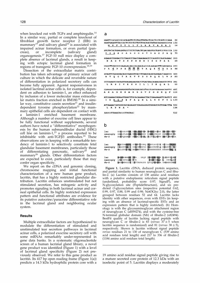
Figure 2. Organ expression of lacritin mRNA is highlyrestricted. Human dot-blot lacking lacrimal RNA butcontaining 50 other tissue poly(A)� RNAs reveals lacri-tin message in salivary (submandibular) gland and thyr-oid, but not elsewhere. Upper inset, abundant adulthuman lacrimal gland expression versus relatively lesssubmandibular gland and no detectable liver expression.Message size is 760 bp. Top right, no lacritin mRNA isapparent in human fetal brain, lung, liver or kidney.Bottom, slight expression in human adult thyroid, but
Characterization of Lacritin 129
high level of glycosylation with six putativeO-glycosylation sites between residues 52 and 64,and a single N-glycosylation site near the C termi-nus. In FASTA searches of the primate database,partial homology (Figure 1(b)) is detected with theglycosaminoglycan binding region of humanneuroglycan C (32 % identity over 102 amino acidresidues; BestFit quality � 83 versus 37 � 5 whenlacritin sequence was randomized) and with the``cysteine-free'', possibly mucin-like, amino globu-lar region of human ®bulin-2 (30 % identity over 81amino acid residues; BestFit quality � 81 versus38 � 5 for random). Although all three are rich inO-glycosylation, positioning of serine and threo-nine residue is not strictly shared (Figure 1(b)); andboth lacritin and ®bulin-2 lack glycosaminoglycan-binding sites. Neuroglycan C (af059274; 539 aminoacid residues) is a component of brain extracellularmatrix anchored by a transmembrane domain.27
Fibulin-2 (x89494; 1184 amino acid residues) iswidely dispersed in basement membranes andstroma of embryonic and adult tissues.28 Searchesof non-primate databases pointed to modest hom-ologies with Trypanosoma cruzi mucin-like protein(af036464; BestFit quality � 78 versus 46 � 10); Plas-modium falciparum merozoite surface antigen 2(u91656; BestFit quality � 76 versus 53 � 6) andPinus taeda putative arabinogalactan protein(af101791; BestFit quality � 74 versus 37 � 4).
No matching or homologous ESTs weredetected, in keeping with lacritin's abundance inhuman lacrimal gland and restricted expressionelsewhere (Figure 2). Northern analysis revealed astrong 760 bp lacrimal gland message, and weakersubmandibular and thyroid gland messages of thesame size (Figure 2, upper inset). No message wasdetected in human adult adrenal gland, testis, thy-mus, pancreas, small intestine or stomach; nor inhuman fetal brain lung, liver or kidney. Similarly,in a commercial dot-blot of 50 different human tis-sue poly(A)� RNAs that excluded lacrimal gland(Figure 2), lacritin expression was found only insubmandibular gland (salivary gland), and to a les-ser degree in thyroid. We subcloned lacritin codingsequence into pET-28b and pcDNA3.1/myc-His(�)C to generate recombinant bacterial(Figure 3(a)) and mammalian (293-T cell;
not in pancreas, adrenal medulla or cortex, testis, thy-mus, small intestine or stomach. Upper inset load is5 mg of total RNA/lane (top left); and 2 mg of poly(A)�
RNA/lane (right and bottom). Wash conditions were0.1 � SSC, 0.1 % (w/v) SDS at 55 �C. Lower inset, dotblot from which histogram was developed. No RNAwas dotted in B8, F5-F8 and G8. H1-H8 contain controlRNAs (yeast total RNA, yeast tRNA, E. coli rRNA,E. coli DNA, poly r(A), human Cot DNA, human DNA(1�) and human DNA (5�), respectively). Dot blotwash conditions were 2 � SSC, 0.1 % (w/v) SDS at55 �C. Exposure time for both dot blot and Northernwas two weeks.
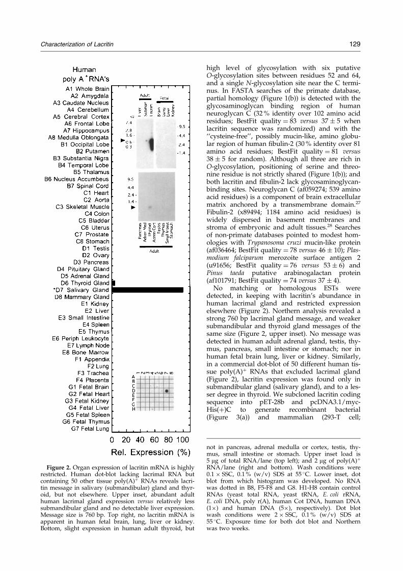
Figure 3. Preparation of recombinant lacritin andimmunolocalization. (a) Puri®cation of His-taggedrecombinant human lacritin (lac) from induced (�) butnot uninduced (ÿ) bacterial lysate (lys), as monitoredusing anti-His antibodies and after Coomassie blue (CB)staining. Detected with anti-recombinant lacritin (anti-lac) antibodies is puri®ed His-tagged lacritin from themedia of transfected 293T cells. Lacritin mobility inSDS-PAGE is anomalous, as con®rmed by mass spectro-scopic analysis of the excised lacritin band (18.3 kDamass spectroscopy peak size for His-tagged lacritin withsignal peptide from bacterial lysate). Anti-lacritin anti-bodies were prepared in rabbit. Lack of cross-reactivityis illustrated in Figure 7(a). (b) Preimmune and (c) antil-acritin immunostaining (brown) of human lacrimalgland sections. Lacritin is present in secretory granules.Inset, lower power image. (d) Antilacritin immunostain-
130 Characterization of Lacritin
Figure 3(a)) lacritin, respectively. Both forms oflacritin displayed anomalous migration in SDS-PAGE, as con®rmed by mass spectroscopic sequen-cing and molecular mass analysis of the gel-excisedband. Antibodies prepared against bacterial lacritinwere applied to sections of human lacrimal andsalivary glands (Figure 3) and to tissue microarrayscontaining formalin-®xed, paraf®n-embedded sec-tions of 75 different human tissues and organs(Table 1). Immunoreactivity was clearly observedin secretory granules of acinar cells in lacrimal andmajor and minor salivary glands, but was notapparent in other epithelia or stroma. The presenceof lacritin in thyroid was equivocal (Table 1). Fre-quency of acinar cell staining was high in lacrimalgland (Figure 3(c)), whereas only scattered salivaryacinar cells were reactive (Figure 3(d) and (e)).Immunoreactivity was also apparent in secretionswithin lumens of lacrimal and salivary ducts (notshown). By ELISA, lacritin was detected in humantears (Figure 3(f)) and to a lesser extent in saliva(not shown).
Lacritin's highly restricted expression patternand strong conservation with mouse (99 % aminoacid identity over two-thirds of mouse cDNA onhand; Walton & G.W.L., unpublished data)suggested an important physiological role(s). In theabsence of functional clues, advantage was takenof an assortment of well-established assays invol-ving each of the three main cell types in the acinar-corneal axis. Included were experiments askingwhether lacritin promoted intracellular signalingthrough tyrosine phosphorylation and intracellularcalcium release, and the consequences of signaling,including cellular secretion and proliferation.Accordingly, we chose to test the effect of lacritinon serum-free cultures of lacrimal acinar, salivaryductal and corneal epithelial cells using secretion(acinar), proliferation (ductal), tyrosine phos-phorylation (acinar, ductal) and calcium signaling(corneal epithelial) assays. Freshly isolated rat lacri-mal acinar cells were plated on increasing amountsof lacritin (with a constant small amount of lami-nin-1 to ensure adherence), or on laminin-1-coatedwells in which lacritin was added to the medium.Both coated and soluble lacritin enhanced unstimu-lated secretion in a dose-dependent manner(Figure 4(a)), but no effect was observed on thestimulated secretory pathways activated by theagonists carbachol and VIP (Figure 4(b)). Theseresults suggest an autocrine or paracrine role, poss-ibly via receptors on the luminal acinar cell surface.We next cultured quiescent HSG cells in serum-
ing of human submandibular gland, as compared to (e)human parotid. (f) ELISA detection of lacritin (®lledbars) in human tears versus a recombinant lacritin posi-tive control. Open bars, preimmune. Original magni®-cation 400� ((b) and (c)) and 200� (inset, (d) and (e)).Sections have been counterstained with hematoxylin(blue) and eosin (red).
Table 1. Restricted immunolocalization of lacritin in human organs
free media containing increasing amounts of lacri-tin and studied cell proliferation. The lacritin cul-tures looked healthier; after four days, a dose-dependent increase in ductal cell number wasapparent (Figure 5(a)) that reached a level morethan twofold that of the BSA (10 ng/ml) negativecontrol (Figure 5(b)). The same level of lacritin pro-moted the transient tyrosine phosphorylation of a48 kDa band in both HSG and rat lacrimal cells(Figure 5(c)). Next, we examined calcium transientsin human corneal epithelial cells. Whereas thebasal level of signaling was negligible, the additionof lacritin resulted in rapid and sustained calciumwaves that propagated throughout the cells(Figure 6). Wave onset preceded that of theusual response to epidermal growth factor(20-40 seconds), and the amplitude of the responsedepended on the concentration of lacritin. Toensure that bacterial lipopolysaccharide (a possiblecontaminant of recombinant protein preps) wasnot involved, samples were tested in the limulusamebocyte lysate assay, and no lipopolysaccharidewas detected (<0.05 EU/ml). Finally, we examinedthe ability of lacritin to bind (Figure 7) the tear ®lmcomponents ®bronectin or vitronectin; as well asconstituents of the periacinar basement membranethat might harbor small amounts of lacritin notdetectable by our immunohistochemical procedure.Also tested was binding to EGF, a common gland-ular growth factor that might synergize with lacri-tin to enhance acinar cell secretion. Lack of anti-lacritin antibody cross-reactivity with all ligands(Figure 7(a)) made feasible an ELISA-based bind-ing approach in which lacritin displayed a remark-able avidity for all constituents tested with theexception of collagen I, EGF or heparin(Figure 7(b)). Binding to the tear ®lm constituents®bronectin and vitronectin, and to the basementmembrane component laminin-1, was exceededsomewhat by collagen IV and nidogen/entactin.Lacritin is therefore capable of multiple differentbinding partners. Partially analogous is the af®nityof ®bulin-2 for collagen IV, entactin/nidogen andlaminin-1.29
The rather broad lacritin lacrimal gland message(Figure 2; upper inset) was suggestive of alterna-
tively spliced forms, or RNA degradation. Thesame was not true for submandibular gland inwhich a discrete, but much less intense signal wasapparent (Figure 2; upper inset). To address thisissue and to gain information on how the lacritingene is arranged, we sequenced a 12.4 kb genomicfragment, the largest lacritin-positive fragmentreadily obtainable from lacritin genomic clones.The gene consists of ®ve exons (Figure 8) precededby a predicted promoter sequence 109 to 59 bpupstream of the translation start site (promoterscore � 1.0; NNPP/Eukaryotic). Exon 1(Figure 8(a)) encodes the complete signal peptideand includes 38 bp of 50 untranslated sequence.Exon 3 contains sequence for all putative O-glyco-sylation sites. The predicted N-glycosylation site isformed at the exon 4/exon 5 splice junction. Exon5 includes 53 bp of 30 untranslated sequence. Threepotential polyadenylation sites are detected 367,474 and 534 bp downstream of exon 5, the ®rst ofwhich would be in keeping with a 760 bp tran-script. Sequences at exon-intron boundaries all con-form to predicted splice donors or acceptors(Table 2), with the exception of the exon 4 spliceacceptor. Intronic sequences revealed commonintronic repeat elements. Also independently dis-covered on a separate genomic fragment was alacritin pseudogene lacking 38 bp of 50 exon 1sequence. To examine possible alternative splicing,we used RT-PCR with submandibular or lacrimalgland cDNA as template (Figure 8(b)) and forwardand reverse primers from exons 1 and 5, respect-ively, each including untranslated ¯ankingsequence. A single PCR product was detected inboth organs whose size (449 bp) was in keepingwith transcription from all ®ve exons withoutalternative splicing. Fluorescent in situ hybridiz-ation (FISH) revealed that the lacritin gene islocated on chromosome 12 (Figure 9), a result con-®rmed by double labeling with a probe for 12q15.Measurement of ten speci®cally labeled chromo-somes located the lacritin gene approximately 16 %of the distance from the centromere to the telomereof 12q, an area that corresponds to 12q13. Alsofound on 12q13 is a rare genetic alacrimia knownas triple A syndrome.30 Attempted PCR using
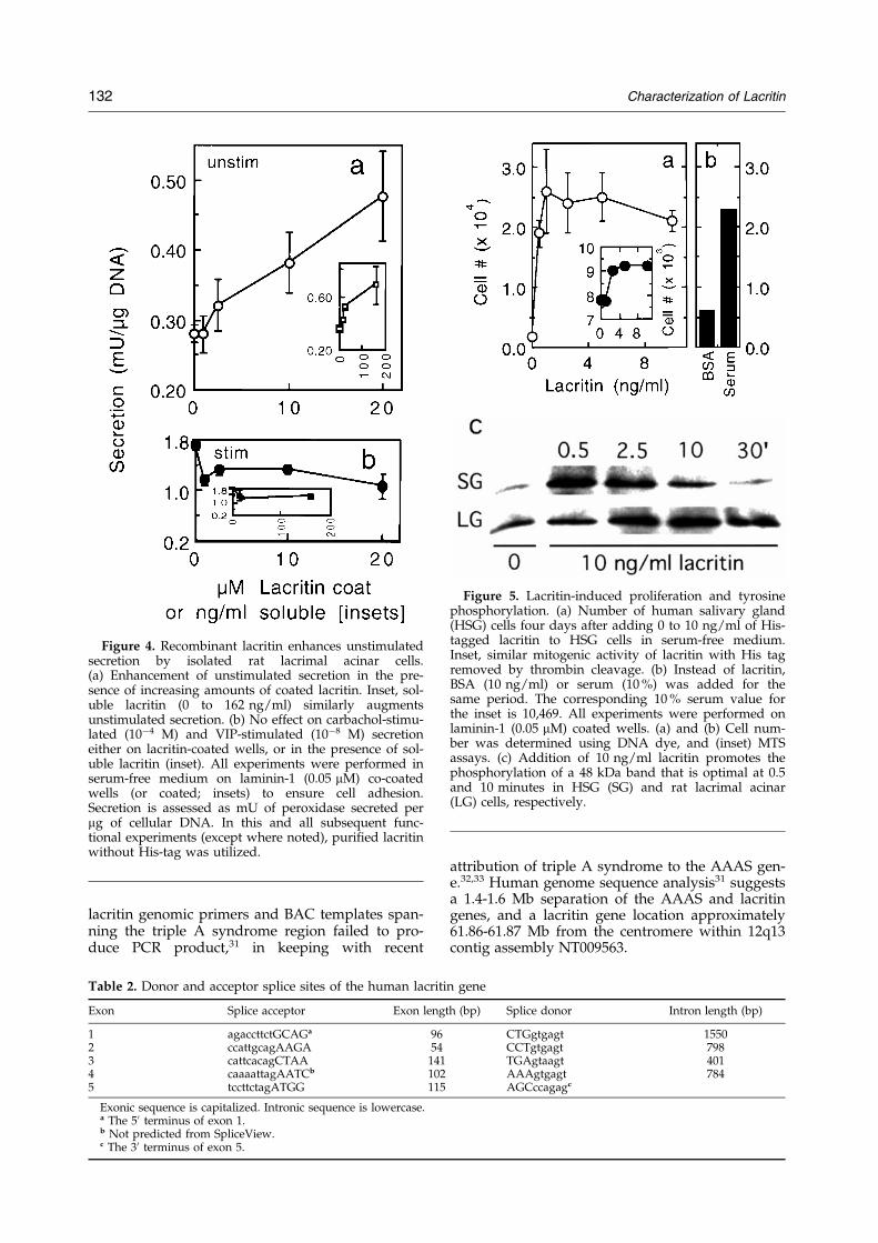
Figure 4. Recombinant lacritin enhances unstimulatedsecretion by isolated rat lacrimal acinar cells.(a) Enhancement of unstimulated secretion in the pre-sence of increasing amounts of coated lacritin. Inset, sol-uble lacritin (0 to 162 ng/ml) similarly augmentsunstimulated secretion. (b) No effect on carbachol-stimu-lated (10ÿ4 M) and VIP-stimulated (10ÿ8 M) secretioneither on lacritin-coated wells, or in the presence of sol-uble lacritin (inset). All experiments were performed inserum-free medium on laminin-1 (0.05 mM) co-coatedwells (or coated; insets) to ensure cell adhesion.Secretion is assessed as mU of peroxidase secreted permg of cellular DNA. In this and all subsequent func-tional experiments (except where noted), puri®ed lacritinwithout His-tag was utilized.
Figure 5. Lacritin-induced proliferation and tyrosinephosphorylation. (a) Number of human salivary gland(HSG) cells four days after adding 0 to 10 ng/ml of His-tagged lacritin to HSG cells in serum-free medium.Inset, similar mitogenic activity of lacritin with His tagremoved by thrombin cleavage. (b) Instead of lacritin,BSA (10 ng/ml) or serum (10 %) was added for thesame period. The corresponding 10 % serum value forthe inset is 10,469. All experiments were performed onlaminin-1 (0.05 mM) coated wells. (a) and (b) Cell num-ber was determined using DNA dye, and (inset) MTSassays. (c) Addition of 10 ng/ml lacritin promotes thephosphorylation of a 48 kDa band that is optimal at 0.5and 10 minutes in HSG (SG) and rat lacrimal acinar(LG) cells, respectively.
132 Characterization of Lacritin
lacritin genomic primers and BAC templates span-ning the triple A syndrome region failed to pro-duce PCR product,31 in keeping with recent
Table 2. Donor and acceptor splice sites of the human lacriti
Exonic sequence is capitalized. Intronic sequence is lowercase.a The 50 terminus of exon 1.b Not predicted from SpliceView.c The 30 terminus of exon 5.
attribution of triple A syndrome to the AAAS gen-e.32,33 Human genome sequence analysis31 suggestsa 1.4-1.6 Mb separation of the AAAS and lacritingenes, and a lacritin gene location approximately61.86-61.87 Mb from the centromere within 12q13contig assembly NT009563.
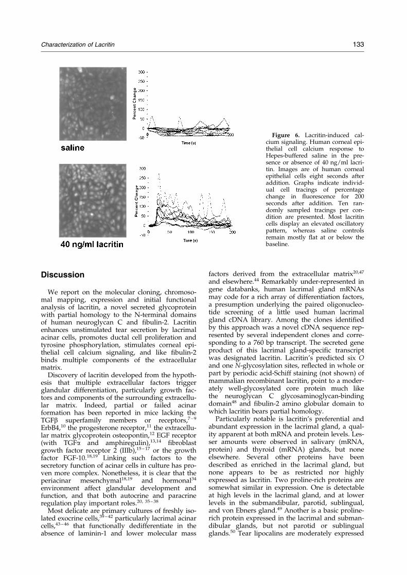
Figure 6. Lacritin-induced cal-cium signaling. Human corneal epi-thelial cell calcium response toHepes-buffered saline in the pre-sence or absence of 40 ng/ml lacri-tin. Images are of human cornealepithelial cells eight seconds afteraddition. Graphs indicate individ-ual cell tracings of percentagechange in ¯uorescence for 200seconds after addition. Ten ran-domly sampled tracings per con-dition are presented. Most lacritincells display an elevated oscillatorypattern, whereas saline controlsremain mostly ¯at at or below thebaseline.
Characterization of Lacritin 133
Discussion
We report on the molecular cloning, chromoso-mal mapping, expression and initial functionalanalysis of lacritin, a novel secreted glycoproteinwith partial homology to the N-terminal domainsof human neuroglycan C and ®bulin-2. Lacritinenhances unstimulated tear secretion by lacrimalacinar cells, promotes ductal cell proliferation andtyrosine phosphorylation, stimulates corneal epi-thelial cell calcium signaling, and like ®bulin-2binds multiple components of the extracellularmatrix.
Discovery of lacritin developed from the hypoth-esis that multiple extracellular factors triggerglandular differentiation, particularly growth fac-tors and components of the surrounding extracellu-lar matrix. Indeed, partial or failed acinarformation has been reported in mice lacking theTGFb superfamily members or receptors,7 ± 9
ErbB4,10 the progesterone receptor,11 the extracellu-lar matrix glycoprotein osteopontin,12 EGF receptor(with TGFa and amphiregulin),13,14 ®broblastgrowth factor receptor 2 (IIIb),15 ± 17 or the growthfactor FGF-10.18,19 Linking such factors to thesecretory function of acinar cells in culture has pro-ven more complex. Nonetheless, it is clear that theperiacinar mesenchymal18,19 and hormonal34
environment affect glandular development andfunction, and that both autocrine and paracrineregulation play important roles.20, 35 ± 38
Most delicate are primary cultures of freshly iso-lated exocrine cells,39 ± 42 particularly lacrimal acinarcells,43 ± 46 that functionally dedifferentiate in theabsence of laminin-1 and lower molecular mass
factors derived from the extracellular matrix20,47
and elsewhere.44 Remarkably under-represented ingene databanks, human lacrimal gland mRNAsmay code for a rich array of differentiation factors,a presumption underlying the paired oligonucleo-tide screening of a little used human lacrimalgland cDNA library. Among the clones identi®edby this approach was a novel cDNA sequence rep-resented by several independent clones and corre-sponding to a 760 bp transcript. The secreted geneproduct of this lacrimal gland-speci®c transcriptwas designated lacritin. Lacritin's predicted six Oand one N-glycosylation sites, re¯ected in whole orpart by periodic acid-Schiff staining (not shown) ofmammalian recombinant lacritin, point to a moder-ately well-glycosylated core protein much likethe neuroglycan C glycosaminoglycan-bindingdomain48 and ®bulin-2 amino globular domain towhich lacritin bears partial homology.
Particularly notable is lacritin's preferential andabundant expression in the lacrimal gland, a qual-ity apparent at both mRNA and protein levels. Les-ser amounts were observed in salivary (mRNA,protein) and thyroid (mRNA) glands, but noneelsewhere. Several other proteins have beendescribed as enriched in the lacrimal gland, butnone appears to be as restricted nor highlyexpressed as lacritin. Two proline-rich proteins aresomewhat similar in expression. One is detectableat high levels in the lacrimal gland, and at lowerlevels in the submandibular, parotid, sublingual,and von Ebners gland.49 Another is a basic proline-rich protein expressed in the lacrimal and subman-dibular glands, but not parotid or sublingualglands.50 Tear lipocalins are moderately expressed
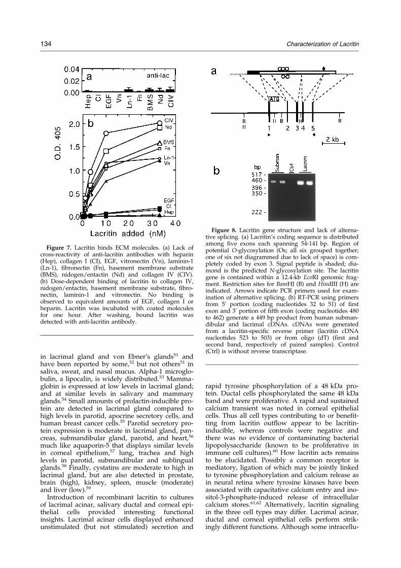
Figure 7. Lacritin binds ECM molecules. (a) Lack ofcross-reactivity of anti-lacritin antibodies with heparin(Hep), collagen I (CI), EGF, vitronectin (Vn), laminin-1(Ln-1), ®bronectin (Fn), basement membrane substrate(BMS), nidogen/entactin (Nd) and collagen IV (CIV).(b) Dose-dependent binding of lacritin to collagen IV,nidogen/entactin, basement membrane substrate, ®bro-nectin, laminin-1 and vitronectin. No binding isobserved to equivalent amounts of EGF, collagen I orheparin. Lacritin was incubated with coated moleculesfor one hour. After washing, bound lacritin wasdetected with anti-lacritin antibody.
Figure 8. Lacritin gene structure and lack of alterna-tive splicing. (a) Lacritin's coding sequence is distributedamong ®ve exons each spanning 54-141 bp. Region ofpotential O-glycosylation (Os; all six grouped together;one of six not diagrammed due to lack of space) is com-pletely coded by exon 3. Signal peptide is shaded; dia-mond is the predicted N-glycosylation site. The lacritingene is contained within a 12.4-kb EcoRI genomic frag-ment. Restriction sites for BamHI (B) and HindIII (H) areindicated. Arrows indicate PCR primers used for exam-ination of alternative splicing. (b) RT-PCR using primersfrom 50 portion (coding nucleotides 32 to 51) of ®rstexon and 30 portion of ®fth exon (coding nucleotides 480to 462) generate a 449 bp product from human subman-dibular and lacrimal cDNAs. cDNAs were generatedfrom a lacritin-speci®c reverse primer (lacritin cDNAnucleotides 523 to 503) or from oligo (dT) (®rst andsecond band, respectively of paired samples). Control(Ctrl) is without reverse transcriptase.
134 Characterization of Lacritin
in lacrimal gland and von Ebner's glands51 andhave been reported by some,52 but not others51 insaliva, sweat, and nasal mucus. Alpha-1 microglo-bulin, a lipocalin, is widely distributed.53 Mamma-globin is expressed at low levels in lacrimal gland;and at similar levels in salivary and mammaryglands.54 Small amounts of prolactin-inducible pro-tein are detected in lacrimal gland compared tohigh levels in parotid, apocrine secretory cells, andhuman breast cancer cells.55 Parotid secretory pro-tein expression is moderate in lacrimal gland, pan-creas, submandibular gland, parotid, and heart,56
much like aquaporin-5 that displays similar levelsin corneal epithelium,57 lung, trachea and highlevels in parotid, submandibular and sublingualglands.58 Finally, cystatins are moderate to high inlacrimal gland, but are also detected in prostate,brain (high), kidney, spleen, muscle (moderate)and liver (low).59
Introduction of recombinant lacritin to culturesof lacrimal acinar, salivary ductal and corneal epi-thelial cells provided interesting functionalinsights. Lacrimal acinar cells displayed enhancedunstimulated (but not stimulated) secretion and
rapid tyrosine phosphorylation of a 48 kDa pro-tein. Ductal cells phosphorylated the same 48 kDaband and were proliferative. A rapid and sustainedcalcium transient was noted in corneal epithelialcells. Thus all cell types contributing to or bene®t-ting from lacritin out¯ow appear to be lacritin-inducible, whereas controls were negative andthere was no evidence of contaminating bacteriallipopolysaccharide (known to be proliferative inimmune cell cultures).60 How lacritin acts remainsto be elucidated. Possibly a common receptor ismediatory, ligation of which may be jointly linkedto tyrosine phosphorylation and calcium release asin neural retina where tyrosine kinases have beenassociated with capacitative calcium entry and ino-sitol-3-phosphate-induced release of intracellularcalcium stores.61,62 Alternatively, lacritin signalingin the three cell types may differ. Lacrimal acinar,ductal and corneal epithelial cells perform strik-ingly different functions. Although some intracellu-
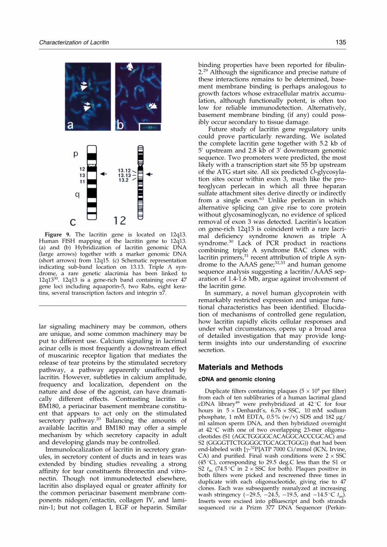
Figure 9. The lacritin gene is located on 12q13.Human FISH mapping of the lacritin gene to 12q13.(a) and (b) Hybridization of lacritin genomic DNA(large arrows) together with a marker genomic DNA(short arrows) from 12q15. (c) Schematic representationindicating sub-band location on 13.13. Triple A syn-drome, a rare genetic alacrimia has been linked to12q1330. 12q13 is a gene-rich band containing over 47gene loci including aquaporin-5, two Rabs, eight kera-tins, several transcription factors and integrin a7.
Characterization of Lacritin 135
lar signaling machinery may be common, othersare unique, and some common machinery may beput to different use. Calcium signaling in lacrimalacinar cells is most frequently a downstream effectof muscarinic receptor ligation that mediates therelease of tear proteins by the stimulated secretorypathway, a pathway apparently unaffected bylacritin. However, subtleties in calcium amplitude,frequency and localization, dependent on thenature and dose of the agonist, can have dramati-cally different effects. Contrasting lacritin isBM180, a periacinar basement membrane constitu-ent that appears to act only on the stimulatedsecretory pathway.20 Balancing the amounts ofavailable lacritin and BM180 may offer a simplemechanism by which secretory capacity in adultand developing glands may be controlled.
Immunolocalization of lacritin in secretory gran-ules, in secretory content of ducts and in tears wasextended by binding studies revealing a strongaf®nity for tear constituents ®bronectin and vitro-nectin. Though not immunodetected elsewhere,lacritin also displayed equal or greater af®nity forthe common periacinar basement membrane com-ponents nidogen/entactin, collagen IV, and lami-nin-1; but not collagen I, EGF or heparin. Similar
binding properties have been reported for ®bulin-2.29 Although the signi®cance and precise nature ofthese interactions remains to be determined, base-ment membrane binding is perhaps analogous togrowth factors whose extracellular matrix accumu-lation, although functionally potent, is often toolow for reliable immunodetection. Alternatively,basement membrane binding (if any) could poss-ibly occur secondary to tissue damage.
Future study of lacritin gene regulatory unitscould prove particularly rewarding. We isolatedthe complete lacritin gene together with 5.2 kb of50 upstream and 2.8 kb of 30 downstream genomicsequence. Two promoters were predicted, the mostlikely with a transcription start site 55 bp upstreamof the ATG start site. All six predicted O-glycosyla-tion sites occur within exon 3, much like the pro-teoglycan perlecan in which all three heparansulfate attachment sites derive directly or indirectlyfrom a single exon.63 Unlike perlecan in whichalternative splicing can give rise to core proteinwithout glycosaminoglycan, no evidence of splicedremoval of exon 3 was detected. Lacritin's locationon gene-rich 12q13 is coincident with a rare lacri-mal de®ciency syndrome known as triple Asyndrome.30 Lack of PCR product in reactionscombining triple A syndrome BAC clones withlacritin primers,31 recent attribution of triple A syn-drome to the AAAS gene;32,33 and human genomesequence analysis suggesting a lacritin/AAAS sep-aration of 1.4-1.6 Mb, argue against involvement ofthe lacritin gene.
In summary, a novel human glycoprotein withremarkably restricted expression and unique func-tional characteristics has been identi®ed. Elucida-tion of mechanisms of controlled gene regulation,how lacritin rapidly elicits cellular responses andunder what circumstances, opens up a broad areaof detailed investigation that may provide long-term insights into our understanding of exocrinesecretion.
Materials and Methods
cDNA and genomic cloning
Duplicate ®lters containing plaques (5 � 104 per ®lter)from each of ten sublibraries of a human lacrimal glandcDNA library49 were prehybridized at 42 �C for fourhours in 5 � Denhardt's, 6.76 � SSC, 10 mM sodiumphosphate, 1 mM EDTA, 0.5 % (w/v) SDS and 182 mg/ml salmon sperm DNA, and then hybridized overnightat 42 �C with one of two overlapping 23-mer oligonu-cleotides (S1 (AGCTGGGGCACAGGCACCCGCAC) andS2 (GGGGTTCTGGGGCTGCAGCTGGG)) that had beenend-labeled with [g-32P]ATP 7000 Ci/mmol (ICN, Irvine,CA) and puri®ed. Final wash conditions were 2 � SSC(45 �C), corresponding to 29.5 deg.C less than the S1 orS2 tm (74.5 �C in 2 � SSC for both). Plaques positive inboth ®lters were picked and rescreened three times induplicate with each oligonucleotide, giving rise to 47clones. Each was subsequently reanalyzed at increasingwash stringency (ÿ29.5, ÿ24.5, ÿ19.5, and ÿ14.5 �C tm).Inserts were excised into pBluescript and both strandssequenced via a Prizm 377 DNA Sequencer (Perkin-
136 Characterization of Lacritin
Elmer, Branchburg, NJ; University of Virginia Biomole-cular Research Facility). Of identical clones, most com-mon was a novel sequence lacking homology to BM180(BestFit quality � 16, versus random quality of 17 � 2)from which the poly(G)-rich S1 and S2 oligonucleotideswere derived.64 Predicted was a 417 bp open readingframe, whose expected protein product was designatedlacritin, in keeping with its lacrimal gland expression.Lacritin insert was subsequently used to screen a humanP1 genomic library (carried out by Genome Systems Inc;St. Louis, MO) and three identical clones were obtained,as determined by restriction digestion and Southernanalysis. The largest lacritin-positive fragment (12.4 kb)was subcloned intact into pBluescript and both strandswere completely sequenced. Alignment and analyses65 ofcDNA and genomic sequence was primarily with Unix-based (Gelstart, Gap) and web-based (FASTA, BestFit,Gap) Genetics Computer Group (Madison WI) softwareusing default settings and E values (FASTA) restricted to5 or less. Genomic exon searching and identi®cation ofsplice sites was facilitated by the Baylor College of Medi-cine Human Genome Sequencing Center web-site.
Northern analysis
Human lacrimal and submandibular glands wereobtained during autopsy through the Southern divisionof the Cooperative Human Tissue Network within18 hours of death and most within eight hours to mini-mize autolytic degradation. The tenets of the Declarationof Helsinki were followed and informed consent and fullIRB approval were obtained. Donors were withoutknown systemic bacterial or viral infections, and tissueswere normal as determined from cause of death, pathol-ogy reports and in most cases histological examination.Tissues were snap frozen in liquid nitrogen after removaland stored at ÿ85 �C until used for RNA preparation.Total RNA was extracted from 100-300 mg of tissueusing a commercial version of the acidi®ed guanidinethiocyanate/phenol method (RNazol B, Tel-Test, TheWoodlands, TX). Puri®ed RNA was dissolved in diethyl-pyrocarbonate-treated water, and the concentration andpurity determined from the A260/280 absorption values. Aratio close to 2.0 was considered acceptable. RNA integ-rity was initially determined by electrophoresis of ethi-dium bromide-complexed RNA samples in a gelcontaining 0.22 M formaldehyde. Samples that did notshow prominent 28 S and 18 S rRNA bands in a 1:1-2:1ratio under UV light were rejected. For blotting, RNA(5 mg/lane) was separated on a 0.8 % agarose gel underdenaturing conditions66 and transferred to nitrocellulose.Also assayed were two purchased (cat # 7756-1 and7751-1; Clontech Labs, Palo Alto, CA) Northern blotswith multiple human fetal and adult poly(A)� RNAsand a dot blot (cat # 7770-1; Clontech Labs) containing50 different human poly(A)� RNAs together with controlRNAs and DNAs. Blots were hybridized with 32P-labeledlacritin insert, washed in 0.1 � SSC, 0.1 % (w/v) SDS(Northern) or 2 � SSC, 0.1 % (w/v) SDS (dot blot) at55 �C, and exposed to X-ray ®lm. Dot blots were thenquanti®ed using NIH Image by measurement of pixelgray values of individual dots.
Preparation of recombinant lacritin and anti-lacritin antisera
Full-length lacritin cDNA was subcloned in-frame intopET-28b (Novagen, Madison, WI), with orientation
con®rmed by completely sequencing through the insert.Recombinant His-tagged lacritin was then generated byIPTG-induction of BL-21 transformed cells, and puri®edfrom cell lysate on Talon (Clontech; Palo Alto, CA) resinusing standard denaturing procedures. After elution,lacritin was extensively dialyzed versus PBS, and the Histag was removed by thrombin cleavage. Protein qualitywas assessed by SDS-PAGE and Western blotting withanti-His antibody (Santa Cruz Biotechnology; SantaCruz, CA). Lacritin displays anomalous mobility in SDS-PAGE. Lack of contaminating bacterial lipopolysacchar-ide was con®rmed by the limulus amebocyte lysateassay (MRL Reference Lab; Cypress, CA). For analyticalcomparison, small amounts of mammalian lacritin wereexpressed in 293T cells using pcDNA3.1/myc-His(�)(Invitrogen, Carlsbad, CA) containing lacritin insert, andthen puri®ed under native conditions. Preimmune serumand anti-bacterial lacritin antiserum were subsequentlyprepared in rabbits (Covance Research Products, Denver,PA), partially puri®ed by precipitation in 4 % caprylicacid67 and treatment with 293T cell acetone extract (toeliminate background), and assessed by ELISA (1/1000dilution) using recombinant bacterial lacritin (4 mg/ml)as coat. For immunohistochemistry, sections of zinc for-malin-®xed, paraf®n-embedded human tissues and ahuman tissue microarray were deparaf®nized and rehy-drated, and microwave heated (20 minutes in 10 mMcitrate buffer (pH 6.0)) to expose antigen. Endogenousperoxidase was blocked, and then immunodetection wasperformed using the avidin-biotin-peroxidase complexmethod (Vectastain Elite kit, Vector Laboratories, Burlin-game, CA) after incubation with anti-lacritin Ig or preim-mune Ig (1/1000; prepared as above) for one hour atroom temperature. Sections were counterstained withhematoxylin, placed in cupric sulfate, and thenimmersed in lithium carbonate.
Cell function analysis
Freshly isolated rat lacrimal acinar cells20,47,67 andHSG (human salivary gland) ductal23 and HCE (humancorneal epithelial)68 cell lines were used to study lacritinfunction. For secretion studies, rat acinar cells were pla-ted serum-free overnight on wells co-coated with0.05 mM laminin-1 (to ensure adhesion) and 0 to 20 mMlacritin, or alternatively with laminin-1 (0.05 mM) andtreated the next day with serum-free medium containing0 to 162 ng/ml of soluble lacritin for four hours. Unsti-mulated and stimulated (carbachol 10ÿ4 M/VIP 10ÿ8 M)secretions were then collected, assessed (peroxidaseassay) and normalized to mg cellular DNA.20,47 Cell pro-liferation was examined in serum-free HSG (human sali-vary gland ductal cell line) cultures grown for four daysin 0 to 10 ng/ml of soluble lacritin. Cell number wasassessed using DNA dye (Quantos kit; Stratagene, LaJolla, CA) and MTS (Promega, Madison, WI) assays inparallel with standard curves of increasing cell quantity.In controls, lacritin was replaced with BSA (10 ng/ml) or10 % (w/v) serum. To study tyrosine phosphorylation,overnight serum-free cultures of both rat lacrimal acinarand HSG cells were washed and treated with 10 ng/mlof soluble lacritin for 0.5, 2.5, 10 and 30 minutes. Py(20)anti-phosphotyrosine antibody immunoprecipitation ofcell lysates was then examined in Western blots ofSDS-7 % PAGE gels using Py(20) and ECL for detection.Calcium signaling in human corneal epithelial cells wassimilarly carried out in serum-free culture69 (Klepeis &V.T.-R., unpublished data). HCE cells were grown tocon¯uency on glass coverslips in keratinocyte media
Characterization of Lacritin 137
(Life Technologies, Rockville, MD) containing bovinepituitary extract (30 mg/ml), EGF (0.1 ng/ml) and peni-cillin/streptomycin, and rendered quiescent 18 hoursbefore loading with Fluo-3AM (2 mM; Molecular Probes,Eugene, OR) at 37 �C for 30 minutes. Using an invertedZeiss 510 LSM for visualization, 50 seconds baselineimages were ®rst recorded. While the laser was running,lacritin was added (®nal concentration 4 and 40 ng/ml)and the response continually monitored every 786 msfor a minimum of 200 seconds.
ECM-binding studies
Binding studies were carried out in 96-well platescoated with 10 mg/well of collagen IV, laminin-1, entac-tin/nidogen-1, collagen I, ®bronectin, vitronectin, EGF,heparin or BMS.70 Wells were washed, blocked (PBS-T),incubated with 0-30 nM lacritin (in PBS-T containing 1 %(w/v) BSA) for one hour at 4 �C, washed and detectedwith anti-lacritin antibody (1/1000) by ELISA.
PCR analysis and chromosome mapping
Alternative splicing was examined by RT-PCR usinghuman submandibular or lacrimal total RNA and initialpriming with oligo(dT), or in a gene-speci®c mannerwith lacritin reverse primer CGCTACAAGGGTATT-TAAGGC (corresponding to nucleotides 523 to 503 fromlacritin cDNA). Subsequent ampli®cation with lacritinforward primer ACTCACTCCTCATCCCAAAG (fromexon 1; lacritin cDNA nucleotides 32 to 51) and reverseprimer TTTTCAGCTTCTCATGCCC (from exon 5; lacri-tin cDNA nucleotides 480 to 462) involved denaturationfor two minutes at 94 �C, 30 cycles of ampli®cation(94 �C for 30 seconds, 52 �C for 30 seconds and 72 �C forone minute), and a ®nal cycle for ®ve minutes at 72 �C.PCR product was analyzed in agarose gels.
For FISH mapping (Genome Systems; St. Louis, MO),lacritin genomic DNA was labeled with digoxigenindUTP by nick translation and hybridized (50 % (v/v) for-mamide, 10 % (w/v) dextran, 2 � SSC) to metaphasechromosomes from PHA-stimulated human peripheralblood lymphocytes. Following washes, speci®c labelingwas detected with ¯uoresceinated antidigoxigenin anti-bodies and DAPI, and examined in a Nikon Labophotmicroscope. A total of 80 metaphase cells were analyzedwith 60 exhibiting speci®c labeling. Con®rmation wasachieved by double labeling using a 12q15 marker, andby comparison with human genome sequence. Photo-graphs were taken on a Nikon AFX at a ®nal magni®-cation of 1435�.
Statistical analysis
All values are expressed as the mean � SD.
Data Bank accession numbers
All nucleotide sequences have been submitted to theGenBank/EBI Data Bank with accession numbersaf238867 (cDNA) and ay005150 (genomic).
Acknowledgments
This work was supported by NIH grant EY09747(G.W.L.) and EY06000 (V.T.R.).
References
1. Golosow, N. & Grobstein, C. (1962). Epitheliome-senchymal interaction in pancreatic morphogenesis.Dev. Biol. 4, 242-255.
2. Jamieson, J. D. (1982). Plasmalemmal glycoproteinsand basal lamina: involvement in pancreatic mor-phogenesis. Prog. Clin. Biol. Res. 91, 413-427.
3. Valentijn, J. A., Gumkowski, F. D. & Jamieson, J. D.(1996). The expression pattern of rab3D in the devel-oping rat exocrine pancreas coincides with theacquisition of regulated exocytosis. Eur. J. Cell Biol.71, 129-136.
4. Ohnishi, H., Samuelson, L. C., Yule, D. I., Ernst,S. A. & Williams, J. A. (1997). Overexpression ofRab3D enhances regulated amylase secretion frompancreatic acini of transgenic mice. J. Clin. Invest.100, 3044-3052.
5. Valentijn, J. A., LaCivita, D. Q., Gumkowski, F. D.& Jamieson, J. D. (1997). Rab4 associates with theactin terminal web in developing rat pancreaticacinar cells. Eur. J. Cell Biol. 72, 1-8.
6. Denny, P. C., Ball, W. D. & Redman, R. S. (1997).Salivary glands: a paradigm for diversity of glanddevelopment. Crit. Rev. Oral Biol. Med. 8, 51-75.
7. BoÈ ttinger, E. P., Jakubczak, J. L., Roberts, I. S.,Mumy, M., Hemmati, P. & Bagnall, K. et al. (1997).Expression of a dominant-negative mutant TGF-betatype II receptor in transgenic mice reveals essentialroles for TGF-beta in regulation of growth anddifferentiation in the exocrine pancreas. EMBO J. 16,2621-2633.
8. Shiozaki, S., Tajima, T., Zhang, Y. Q., Furukawa, M.,Nakazato, Y. & Kojima, I. (1999). Impaired differen-tiation of endocrine and exocrine cells of the pan-creas in transgenic mouse expressing the truncatedtype II activin receptor. Biochim. Biophys. Acta, 1450,1-11.
9. Robinson, G. W. & Hennighausen, L. (1997). Inhi-bins and activins regulate mammary epithelial celldifferentiation through mesenchymal-epithelial inter-actions. Development, 124, 2701-2708.
10. Jones, F. E., Welte, T., Fu, X. Y. & Stern, D. F.(1999). ErbB4 signaling in the mammary gland isrequired for lobuloalveolar development and Stat5activation during lactation. J. Cell Biol. 147, 77-88.
11. Humphreys, R. C., Lydon, J., O'Malley, B. W. &Rosen, J. M. (1997). Mammary gland development ismediated by both stromal and epithelial progester-one receptors. Mol. Endocrinol. 11, 801-811.
12. Nemir, M., Bhattacharyya, D., Li, X., Singh, K.,Mukherjee, A. B. & Mukherjee, B. B. (2000). Tar-geted inhibition of osteopontin expression in themammary gland causes abnormal morphogenesisand lactation de®ciency. J. Biol. Chem. 275, 969-976.
13. Wiesen, J. F., Young, P., Werb, Z. & Cunha, G. R.(1999). Signaling through the stromal epidermalgrowth factor receptor is necessary for mammaryductal development. Development, 126, 335-344.
14. Luetteke, N. C., Qiu, T. H., Fenton, S. E., Troyer,K. L., Riedel, R. F., Chang, A. & Lee, D. C. (1999).Targeted inactivation of the EGF and amphiregulingenes reveals distinct roles for EGF receptor ligandsin mouse mammary gland development. Develop-ment, 126, 2739-2750.
15. Jackson, D., Bresnick, J., Rosewell, I., Crafton, T.,Poulsom, R., Stamp, G. & Dickson, C. (1997). Fibro-blast growth factor receptor signalling has a role in
138 Characterization of Lacritin
lobuloalveolar development of the mammary gland.J. Cell Sci. 110, 1261-1268.
16. De Moerlooze, L., Spencer-Dene, B., Revest, J.,Hajihosseini, M., Rosewell, I. & Dickson, C. (2000).An important role for the IIIb isoform of ®broblastgrowth factor receptor 2 (FGFR2) in mesenchymal-epithelial signalling during mouse organogenesis.Development, 127, 483-492.
17. Celli, G., LaRochelle, W. J., Mackem, S., Sharp, R. &Merlino, G. (1998). Soluble dominant-negative recep-tor uncovers essential roles for ®broblast growthfactors in multi-organ induction and patterning.EMBO J. 17, 1642-1655.
18. Makarenkova, H. P., Ito, M., Govindarajan, V.,Faber, S. C., Sun, L., McMahon, G., Overbeek, P. A.& Lang, R. A. (2000). FGF10 is an inducer and Pax6a competence factor for lacrimal gland development.Development, 127, 2563-2572.
19. Govindarajan, V., Ito, M., Makarenkova, H. P.,Lang, R. A. & Overbeek, P. A. (2000). Endogenousand ectopic gland induction by FGF-10. Dev. Biol.225, 188-200.
20. Laurie, G. W., Glass, J. D., Ogle, R. A., Stone, C. M.,Sluss, J. R. & Chen, L. (1996). ``BM180``: a novelbasement membrane protein with a role in stimulus-secretion coupling by lacrimal acinar cells. Am. J.Physiol. 270, C1743-C1750.
21. Streuli, C. H., Bailey, N. & Bissell, M. J. (1991). Con-trol of mammary epithelial differentiation: basementmembrane induces tissue-speci®c gene expression inthe absence of cell-cell interaction and morphologi-cal polarity. J. Cell Biol. 115, 1383-1395.
22. Lee, Y. J. & Streuli, C. H. (1999). Extracellular matrixselectively modulates the response of mammaryepithelial cells to different soluble signaling ligands.J. Biol. Chem. 274, 22401-22408.
23. Hoffman, M. P., Nomizu, M., Roque, E., Lee, S.,Jung, D. W., Yamada, Y. & Kleinman, H. K. (1998).Laminin-1 and laminin-2 G-domain synthetic pep-tides bind syndecan-1 and are involved in acinarformation of a human submandibular gland cellline. J. Biol. Chem. 273, 28633-28641.
24. Hoffman, M. P., Kibbey, M. C., Letterio, J. J. &Kleinman, H. K. (1996). Role of laminin-1 andTGF-beta 3 in acinar differentiation of a human sub-mandibular gland cell line (HSG). J. Cell Sci. 109,2013-2021.
25. Virtanen, I., Gullberg, D., Rissanen, J., Kivilaakso,E., Kiviluoto, T. & Laitinen, L. A., et al. (2000). Lami-nin alpha1-chain shows a restricted distribution inepithelial basement membranes of fetal and adulthuman tissues. Exp. Cell Res. 257, 298-309.
26. Klinowska, T. C., Soriano, J. V., Edwards, G. M.,Oliver, J. M., Valentijn, A. J., Montesano, R. &Streuli, C. H. (1999). Laminin and beta1 integrinsare crucial for normal mammary gland developmentin the mouse. Dev. Biol. 215, 13-32.
27. Yasuda, Y., Tokita, Y., Aono, S., Matsui, F., Ono, T.& Sonta, S. et al. (1998). Cloning and chromosomalmapping of the human gene of neuroglycan C(NGC), a neural transmembrane chondroitin sulfateproteoglycan with an EGF module. Neurosci. Res. 32,313-322.
28. Sasaki, T., Gohring, W., Miosge, N., Abrams, W. R.,Rosenbloom, J. & Timpl, R. (1999). Tropoelastinbinding to ®bulins, nidogen-2 and other extracellularmatrix proteins. FEBS Letters, 460, 280-284.
29. Sasaki, T., GoÈhring, W., Pan, T. C., Chu, M. L. &Timpl, R. (1995). Binding of mouse and human
®bulin-2 to extracellular matrix ligands. J. Mol. Biol.254, 892-899.
30. Huebner, A., Elias, L. L. & Clark, A. J. (1999).ACTH resistance syndromes. J. Pediatr. Endocrinol.Metab. 12, 277-293.
31. Kumar, R., Huebner, A. & Laurie, G. W. (2001).Genetic separation of the human lacritin gene andtriple A (Allgrove) syndrome on 12q13. Advan. Exp.Med. Biol. in the press.
32. Tullio-Pelet, A., Salomon, R., Hadj-Rabia, S.,Mugnier, C., de Laet, M. H. & Chaouachi, B., et al.(2000). Mutant WD-repeat protein in triple-A syn-drome. Nature Genet. 26, 332-335.
33. Handschug, K., Sperling, S., Yoon, S. J., Hennig, S.,Clark, A. J. & Huebner, A. (2001). Triple Asyndrome is caused by mutations in AAAS, a newWD-repeat protein gene. Hum. Mol. Genet. 10, 283-290.
34. Sullivan, D. A., Block, L. & Pena, J. D. (1996). In¯u-ence of androgens and pituitary hormones on thestructural pro®le and secretory activity of the lacri-mal gland. Acta Ophthalmol. Scand. 74, 421-435.
35. Morita, S., Fernandez-Mejia, C. & Melmed, S. (1989).Retinoic acid selectively stimulates growth hormonesecretion and messenger ribonucleic acid levels inrat pituitary cells. Endocrinology, 124, 2052-2056.
36. Gotti, C., Sher, E., Cabrini, D., Bondiolotti, G.,Wanke, E., Mancinelli, E. & Clementi, F. (1987).Cholinergic receptors, ion channels, neurotransmittersynthesis, and neurite outgrowth are independentlyregulated during the in vitro differentiation of ahuman neuroblastoma cell line. Differentiation, 34,144-155.
37. Logsdon, C. D., Moessner, J., Williams, J. A. &Gold®ne, I. D. (1985). Glucocorticoids increase amy-lase mRNA levels, secretory organelles, andsecretion in pancreatic acinar AR42 J cells. J. CellBiol. 100, 1200-1208.
38. Brunet-de Carvalho, N., Picart, R., Van de Moortele,S., Tougard, C. & Tixier-Vidal, A. (1989). Laminininduces formation of neurite-like processes andpotentiates prolactin secretion by GH3 rat pituitarycells. Differentiation, 40, 106-118.
39. Amsterdam, A. & Jamieson, J. D. (1974). Studies ondispersed pancreatic exocrine cells. II. Functionalcharacteristics of separated cells. J. Cell Biol. 63,1057-2073.
40. Arvan, P. & Castle, J. D. (1987). Phasic releaseof newly synthesized secretory proteins in theunstimulated rat exocrine pancreas. J. Cell Biol. 104,243-252.
41. Herzog, V., Sies, H. & Miller, F. (1976). Exocytosis insecretory cells of rat lacrimal gland. Peroxidaserelease from lobules and isolated cells upon cholin-ergic stimulation. J. Cell Biol. 70, 692-706.
42. Parod, R. J. & Putney, J. W., Jr. (1980). Stimulus-permeability coupling in rat lacrimal gland. Am. J.Physiol. 239, G106-G113.
43. Hann, L. E., Kelleher, R. S. & Sullivan, D. A. (1991).In¯uence of culture conditions on the androgen con-trol of secretory component production by acinarcells from the rat lacrimal gland. Invest. Ophthalmol.Vis. Sci. 32, 2610-2621.
44. Schonthal, A. H., Warren, D. W., Stevenson, D.,Schecter, J. E., Azzarolo, A. M., Mircheff, A. K. &Trousdale, M. D. (2000). Proliferation of lacrimalgland acinar cells in primary culture. Stimulation byextracellular matrix, EGF, and DHT. Exp. Eye Res.70, 639-649.
Characterization of Lacritin 139
45. Yoshino, K. (2000). Establishment of a human lacri-mal gland epithelial culture system with in vivomimicry and its substrate modulation. Cornea, 19,S26-S36.
46. Yoshino, K., Tseng, S. C. & P¯ugfelder, S. C. (1995).Substrate modulation of morphology, growth, andtear protein production by cultured human lacrimalgland epithelial cells. Exp. Cell Res. 220, 138-151.
47. Chen, L., Glass, J. D., Walton, S. C. & Laurie, G. W.(1998). Role of laminin-1, collagen IV, and an auto-crine factor(s) in regulated secretion by lacrimalacinar cells. Am. J. Physiol. 275, C278-C284.
48. Aono, S., Keino, H., Ono, T., Yasuda, Y., Tokita, Y.& Matsui, F. et al. (2000). Genomic organization andexpression pattern of mouse neuroglycan C in thecerebellar development. J. Biol. Chem. 275, 337-342.
49. Dickinson, D. P. & Thiesse, M. (1995). A majorhuman lacrimal gland mRNA encodes a new pro-line-rich protein family member. Invest. Ophthalmol.Vis. Sci. 36, 2020-2131.
50. Dickinson, D. P. & Thiesse, M. (1996). cDNA cloningof an abundant human lacrimal gland mRNAencoding a novel tear protein. Curr. Eye Res. 15, 377-386.
51. Glasgow, B. J. (1995). Tissue expression of lipocalinsin human lacrimal and von Ebner's glands: colocali-zation with lysozyme. Graefes Arch. Clin. Exp.Ophthalmol. 233, 513-522.
52. Redl, B., Holzfeind, P. & Lottspeich, F. (1992).cDNA cloning and sequencing reveals human tearprealbumin to be a member of the lipophilic-ligandcarrier protein superfamily. J. Biol. Chem. 267, 20282-20287.
53. Logdberg, L. E., Akerstrom, B. & Badve, S. (2000).Tissue distribution of the lipocalin alpha-1 microglo-bulin in the developing human fetus. J. Histochem.Cytochem. 48, 1545-1552.
54. Zhao, C., Nguyen, T., Yusifov, T., Glasgow, B. J. &Lehrer, R. I. (1999). Lipophilins: human peptideshomologous to rat prostatein. Biochem. Biophys. Res.Commun. 256, 147-155.
55. Myal, Y., Iwasiow, B., Cosby, H., Yarmill, A.,Blanchard, A. & Tsuyuki, D. et al. (1998). Analysisof tissue- and hormone-speci®c regulation of thehuman prolactin-inducible protein/gross cystic dis-ease ¯uid protein-15 gene in transgenic mice. J. Mol.Endocrinol. 21, 217-223.
56. Robinson, C. P., Bounous, D. I., Alford, C. E.,Nguyen, K. H., Nanni, J. M., Peck, A. B. &Humphreys-Beher, M. G. (1997). PSP expression inmurine lacrimal glands and function as a bacteriabinding protein in exocrine secretions. Am. J. Physiol.272, G863-G871.
57. Hamann, S., Zeuthen, T., La Cour, M., Nagelhus,E. A., Ottersen, O. P., Agre, P. & Nielsen, S. (1998).Aquaporins in complex tissues: distribution of aqua-porins 1-5 in human and rat eye. Am. J. Physiol. 274,C1332-C1345.
58. Raina, S., Preston, G. M., Guggino, W. B. & Agre, P.(1995). Molecular cloning and characterization of anaquaporin cDNA from salivary, lacrimal, and respir-atory tissues. J. Biol. Chem. 270, 1908-1912.
59. Hakansson, K., Huh, C., Grubb, A., Karlsson, S. &Abrahamson, M. (1996). Mouse and rat cystatin C:Escherichia coli production, characterization and tis-sue distribution. Comp. Biochem. Physiol. ser. B, 114,303-311.
60. Ogata, H., Su, I., Miyake, K., Nagai, Y., Akashi, S. &Mecklenbrauker, I. et al. (2000). The toll-like receptorprotein RP105 regulates lipopolysaccharide signalingin B cells. J. Exp. Med. 192, 23-29.
61. Zhou, W. L., Sugioka, M., Sakaki, Y. & Yamashita,M. (1999). Regulation of capacitative Ca2� entry bytyrosine phosphorylation in the neural retina ofchick embryo. Neurosci. Letters, 272, 123-126.
62. Strauss, O., Steinhausen, K., Mergler, S., Stumpff, F.& Wiederholt, M. (1999). Involvement of proteintyrosine kinase in the InsP3-induced activation ofCa2�-dependent Cl-currents in cultured cells of therat retinal pigment epithelium. J. Membr. Biol. 169,141-153.
63. Cohen, I. R., Grassel, S., Murdoch, A. D. & Iozzo,R. V. (1993). Structural characterization of the com-plete human perlecan gene and its promoter. Proc.Natl Acad. Sci. USA, 90, 10404-10408.
64. Laurie, G. W., Ciclitira, P. J., Ellis, H. J. & Pogany,G. (1995). Immunological and partial sequenceidentity of mouse BM180 with wheat alpha-gliadin.Biochem. Biophys. Res. Commun. 217, 10-15.
65. Kumar, R., Lumsden, A., Ciclitira, P. J., Ellis, H. J. &Laurie, G. W. (2000). Human genome search inceliac disease using gliadin cDNA as probe. J. Mol.Biol. 300, 1157-1169.
66. Laurie, G. W., Horikoshi, S., Killen, P. D., Segui-Real, B. & Yamada, Y. (1989). In situ hybridizationreveals temporal and spatial changes in cellularexpression of mRNA for a laminin receptor, laminin,and basement membrane (type IV) collagen in thedeveloping kidney. J. Cell Biol. 109, 1351-1362.
67. Sanghi, S., Kumar, R., Walton, S. & Laurie, G. W.(2000). Quantitation of rat lacrimal secretion: a novelsandwich ELISA with high sensitivity. Exp. Eye Res.70, 651-658.
68. Araki-Sasaki, K., Ohashi, Y., Sasabe, T., Hayashi, K.,Watanabe, H., Tano, Y. & Handa, H. (1995). AnSV40-immortalized human corneal epithelial cell lineand its characterization. Invest. Ophthalmol. Vis. Sci.36, 614-621.
69. Trinkaus-Randall, V., Kewalramani, R., Payne, J. &Cornell-Bell, A. (2000). Calcium signaling inducedby adhesion mediates protein tyrosine phosphoryl-ation and is independent of pHi. J. Cell Physiol. 184,385-399.
70. Matter, M. L. & Laurie, G. W. (1994). A novel lami-nin E8 cell adhesion site required for lung alveolarformation in vitro. J. Cell Biol. 124, 1083-1090.
Edited by J. Karn
(Received 22 December 2000; received in revised form 10 May 2001; accepted 10 May 2001)