Cell adhesion to protein-micropatterned-supported lipidbilayer membranes
Lance Kam, Steven G. BoxerDepartment of Chemistry, Stanford University, Stanford, California 94305-5080
Received 12 June 2000; revised 14 November 2000; accepted 16 November 2000
Abstract: A new method for constructing controlled inter-faces between cells and synthetic supported lipid bilayermembranes is reported. Microcontact printing is used to de-fine squares and grid lines of fibronectin onto glass, whichsubsequently direct the self-assembly of fluid lipid bilayersonto the complementary, uncoated regions of the surface.Features of fibronectin as small as 5 mm effectively controlthe lateral organization of the lipid bilayers. These fibronec-tin barriers also facilitate the adhesion of endothelial cells,which exhibit minimal adhesion to fluid supported lipid bi-layers alone. Cells selectively adhere to the features of fibro-nectin, spanning over and exposing the cells to the interven-ing regions of supported lipid bilayer. Cell spreading is cor-related with both the geometry and dimensions of thefibronectin barriers. Importantly, lipids underlying adherent
Cell–cell communication is mediated in large partby membrane-associated proteins. Substrate-supported lipid bilayers offer a unique system withwhich to study the interaction of cells with membraneproteins, and to potentially utilize these biomoleculesto modulate and investigate cellular function. Sup-ported lipid bilayers consist of two leaflets of phos-pholipids in close association with a hydrophilic sur-face such as glass. A thin layer of water severalnanometers thick separates the membrane from thesupport.1–3 Consequently, molecular components insupported lipid bilayers of appropriate compositionfreely diffuse within the plane of the membrane, mim-icking a property of cellular membranes that is essen-tial for many cellular functions.4–6 Furthermore, thecomposition and fluid properties of supported lipid
bilayers are easily controlled, providing a robust toolfor the study of systems, ranging from membrane-associated biomolecules (e.g., integrins, gap junctions,ion channels, GPI-anchored proteins, and syntheticpeptides) to cells of the immune system.7–11
Interfacing anchorage-dependent cells such as he-patocytes, endothelial cells, and neurons with syn-thetic surfaces using lipid bilayers may also be usefulin bioengineering applications, including biomaterialdesign, cell isolation, and high throughput screeningof small molecules that modulate cell–membrane in-teractions. However, anchorage-dependent cells ex-hibit minimal adhesion onto fluid phospholipid struc-tures; both the fluidity of lipid bilayers and the abilityof specific lipids to resist protein adsorption contrib-ute to the inability of cells to form stable attachmentsto supported membranes.12 In this report, we explorethe use of recently developed micropatterning tech-niques to facilitate adhesion of anchorage-dependentcells onto surfaces containing supported lipid bilayers.Previously, our laboratory has described the microfab-rication of barriers that direct and corral lipid diffu-sion on surfaces by using materials such as TiOx andphotoresist,13,14 or by selectively removing regions ofthe assembled bilayer through either scratching15,16 orblotting.17 Our laboratory has also demonstrated that
Correspondence to: S.G. BoxerContract grant sponsor: NIH Genome Training Grant (LK)Contract grant sponsor: NSF Biophysics ProgramContract grant sponsor: MRSEC Program of the NSF; Con-
the protein bovine serum albumin, patterned ontosubstrates by microcontact printing,18–21 can be usedas a barrier material.22 This development suggestedthat by creating barriers of biologically active proteins,it may be possible to introduce added functionality tomicropatterned lipid bilayers. As outlined schemati-cally in Figure 1(A), we micropatterned supportedlipid bilayer membranes using the cell adhesive pro-tein fibronectin. These protein barriers not only pat-tern and corral the supported bilayers, but also pro-vide stable anchorages for cells, thereby promotingand directing the interaction between adherent cellsand supported membranes.
MATERIALS AND METHODS
Substrate modification
Vesicle preparation
Stock solutions of small unilamellar vesicles (SUV) wereprepared by extrusion using standard techniques. Briefly,egg phosphatidylcholine (egg PC; Avanti Polar Lipids, Ala-baster, AL) was dried from chloroform in glass flasks, thendesiccated under vacuum for at least 90 min. These lipids
were reconstituted in deionized water at a concentration of5 mg/mL, then extruded through 50-nm pore size polycar-bonate membranes (Avanti) using a LiposoFast unit (Aves-tin, Inc., Ottowa, ON, Canada). For visualization of lipidbilayers, vesicles of egg PC were prepared with either 1 mol% of a negatively charged, Texas Redt-labeled phosphati-dylethanolamine (TR-PE; Texas Redt 1,2-dihexadecanoyl-sn-glycero-3-phosphoethanolamine; Molecular Probes,Eugene, OR), or 2 mol % of a neutral, NBD-labeled phos-phatidylethanolamine (NBD-PE; 1-palmitoyl-2-[12-[(7-nitro-2-1,3-benzoxadiazol-4-yl)amino]dodecanoyl]-sn-glycero-3-phosphoethanolamine; Avanti). The inclusion of either ofthese fluorescently labeled lipids into bilayers of egg PC didnot influence subsequent cellular response.
Surface micropatterning
Protein-micropatterned lipid bilayer surfaces were pre-pared as outlined in Figure 1(A). Borosilicate glass cover-slips (VWR Scientific, Media, PA) were cleaned by immer-sion into Linbro 7X detergent (ICN Biomedicals, Inc., Au-rora, OH), diluted 1: 3 (v/v) in deionized water, baked at450°C for 4 h, and then micropatterned with fibronectin bymicrocontact printing.18,20–22 Polydimethylsiloxane (PDMS;Sylgard 184; Dow Corning, Midland, MI) elastomer stampscontaining topological representations of a microscale pat-tern, detailed in the section entitled “Micropattern Geom-etry,” were oxidized for 20 s in air plasma using a cleaning/
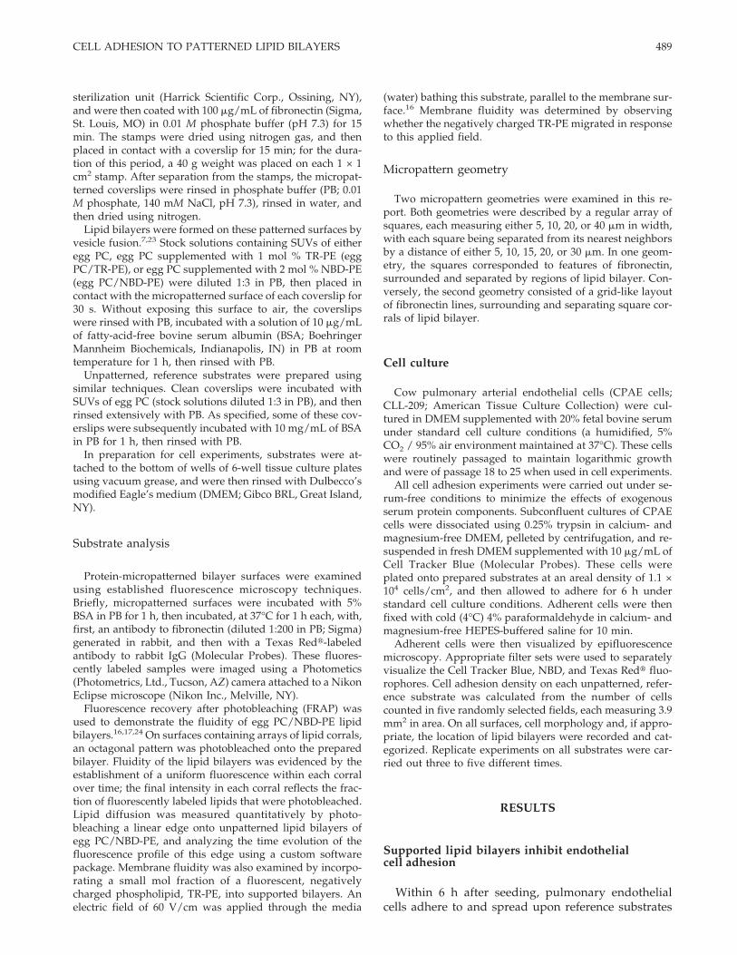
Figure 1. Micropatterning of substrates with fibronectin and phospholipid bilayers. (A) Schematic outlining the microcon-tact printing process used to create fibronectin barriers on a supported lipid bilayer surface. (B) A surface containing gridlinesof fibronectin measuring 5 mm in width and spaced 40 mm apart, shown here as light gray lines (and in red in the online, colorversion), surrounding corrals of supported lipid bilayers that contain a fluorescently labeled lipid, NBD-PE, and appear as anarray of dark gray (green) squares. (C) An octagonal pattern was photobleached onto an array of 16 lipid corrals. This imagewas taken immediately after photobleaching. Different fractions of NBD-PE in adjacent corrals underwent photodamage. (D)The same region shown in panel (C), 10 min after photobleaching. Lipids within each corral mixed completely, demonstratingboth that the lipid bilayers were fluid and that neighboring corrals were isolated from each other. The scale bar in each image= 50 mm. [Color figure can be viewed in the online issue, which is available at www.interscience.wiley.com.]
488 KAM AND BOXER
sterilization unit (Harrick Scientific Corp., Ossining, NY),and were then coated with 100 mg/mL of fibronectin (Sigma,St. Louis, MO) in 0.01 M phosphate buffer (pH 7.3) for 15min. The stamps were dried using nitrogen gas, and thenplaced in contact with a coverslip for 15 min; for the dura-tion of this period, a 40 g weight was placed on each 1 × 1cm2 stamp. After separation from the stamps, the micropat-terned coverslips were rinsed in phosphate buffer (PB; 0.01M phosphate, 140 mM NaCl, pH 7.3), rinsed in water, andthen dried using nitrogen.
Lipid bilayers were formed on these patterned surfaces byvesicle fusion.7,23 Stock solutions containing SUVs of eitheregg PC, egg PC supplemented with 1 mol % TR-PE (eggPC/TR-PE), or egg PC supplemented with 2 mol % NBD-PE(egg PC/NBD-PE) were diluted 1:3 in PB, then placed incontact with the micropatterned surface of each coverslip for30 s. Without exposing this surface to air, the coverslipswere rinsed with PB, incubated with a solution of 10 mg/mLof fatty-acid-free bovine serum albumin (BSA; BoehringerMannheim Biochemicals, Indianapolis, IN) in PB at roomtemperature for 1 h, then rinsed with PB.
Unpatterned, reference substrates were prepared usingsimilar techniques. Clean coverslips were incubated withSUVs of egg PC (stock solutions diluted 1:3 in PB), and thenrinsed extensively with PB. As specified, some of these cov-erslips were subsequently incubated with 10 mg/mL of BSAin PB for 1 h, then rinsed with PB.
In preparation for cell experiments, substrates were at-tached to the bottom of wells of 6-well tissue culture platesusing vacuum grease, and were then rinsed with Dulbecco’smodified Eagle’s medium (DMEM; Gibco BRL, Great Island,NY).
Substrate analysis
Protein-micropatterned bilayer surfaces were examinedusing established fluorescence microscopy techniques.Briefly, micropatterned surfaces were incubated with 5%BSA in PB for 1 h, then incubated, at 37°C for 1 h each, with,first, an antibody to fibronectin (diluted 1:200 in PB; Sigma)generated in rabbit, and then with a Texas Redt-labeledantibody to rabbit IgG (Molecular Probes). These fluores-cently labeled samples were imaged using a Photometics(Photometrics, Ltd., Tucson, AZ) camera attached to a NikonEclipse microscope (Nikon Inc., Melville, NY).
Fluorescence recovery after photobleaching (FRAP) wasused to demonstrate the fluidity of egg PC/NBD-PE lipidbilayers.16,17,24 On surfaces containing arrays of lipid corrals,an octagonal pattern was photobleached onto the preparedbilayer. Fluidity of the lipid bilayers was evidenced by theestablishment of a uniform fluorescence within each corralover time; the final intensity in each corral reflects the frac-tion of fluorescently labeled lipids that were photobleached.Lipid diffusion was measured quantitatively by photo-bleaching a linear edge onto unpatterned lipid bilayers ofegg PC/NBD-PE, and analyzing the time evolution of thefluorescence profile of this edge using a custom softwarepackage. Membrane fluidity was also examined by incorpo-rating a small mol fraction of a fluorescent, negativelycharged phospholipid, TR-PE, into supported bilayers. Anelectric field of 60 V/cm was applied through the media
(water) bathing this substrate, parallel to the membrane sur-face.16 Membrane fluidity was determined by observingwhether the negatively charged TR-PE migrated in responseto this applied field.
Micropattern geometry
Two micropattern geometries were examined in this re-port. Both geometries were described by a regular array ofsquares, each measuring either 5, 10, 20, or 40 mm in width,with each square being separated from its nearest neighborsby a distance of either 5, 10, 15, 20, or 30 mm. In one geom-etry, the squares corresponded to features of fibronectin,surrounded and separated by regions of lipid bilayer. Con-versely, the second geometry consisted of a grid-like layoutof fibronectin lines, surrounding and separating square cor-rals of lipid bilayer.
Cell culture
Cow pulmonary arterial endothelial cells (CPAE cells;CLL-209; American Tissue Culture Collection) were cul-tured in DMEM supplemented with 20% fetal bovine serumunder standard cell culture conditions (a humidified, 5%CO2 / 95% air environment maintained at 37°C). These cellswere routinely passaged to maintain logarithmic growthand were of passage 18 to 25 when used in cell experiments.
All cell adhesion experiments were carried out under se-rum-free conditions to minimize the effects of exogenousserum protein components. Subconfluent cultures of CPAEcells were dissociated using 0.25% trypsin in calcium- andmagnesium-free DMEM, pelleted by centrifugation, and re-suspended in fresh DMEM supplemented with 10 mg/mL ofCell Tracker Blue (Molecular Probes). These cells wereplated onto prepared substrates at an areal density of 1.1 ×10⁴ cells/cm2, and then allowed to adhere for 6 h understandard cell culture conditions. Adherent cells were thenfixed with cold (4°C) 4% paraformaldehyde in calcium- andmagnesium-free HEPES-buffered saline for 10 min.
Adherent cells were then visualized by epifluorescencemicroscopy. Appropriate filter sets were used to separatelyvisualize the Cell Tracker Blue, NBD, and Texas Redt fluo-rophores. Cell adhesion density on each unpatterned, refer-ence substrate was calculated from the number of cellscounted in five randomly selected fields, each measuring 3.9mm2 in area. On all surfaces, cell morphology and, if appro-priate, the location of lipid bilayers were recorded and cat-egorized. Replicate experiments on all substrates were car-ried out three to five different times.
Within 6 h after seeding, pulmonary endothelialcells adhere to and spread upon reference substrates
489CELL ADHESION TO PATTERNED LIPID BILAYERS
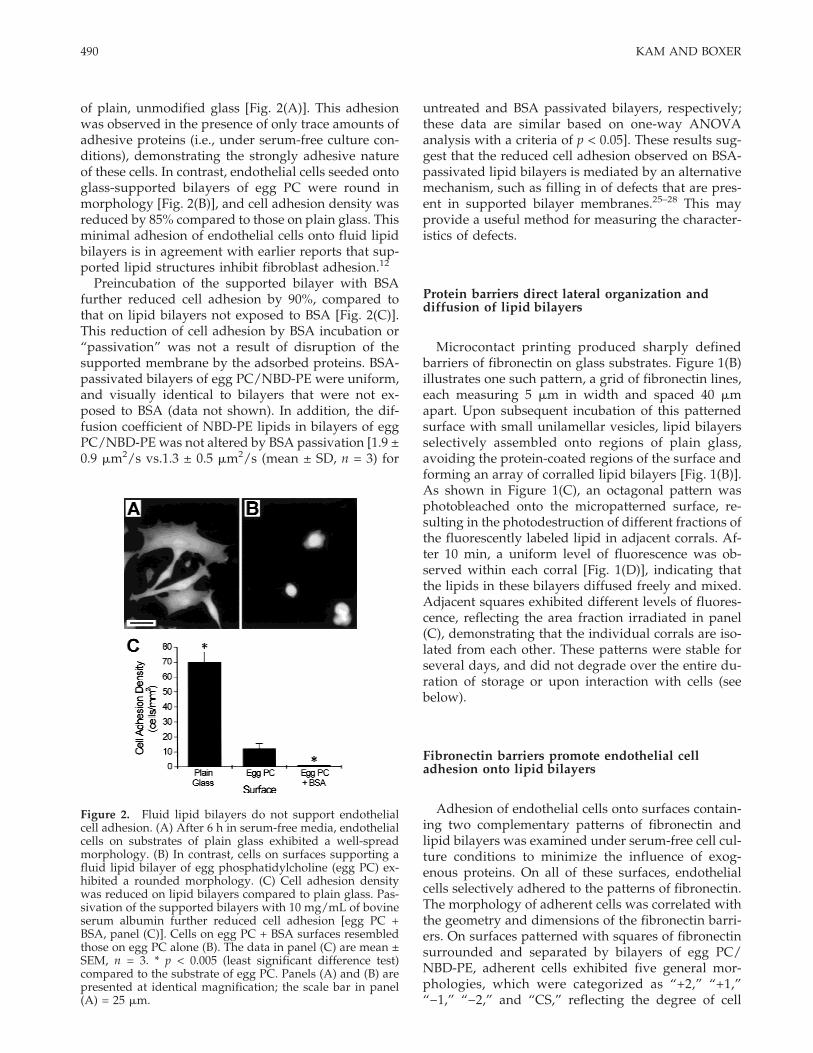
of plain, unmodified glass [Fig. 2(A)]. This adhesionwas observed in the presence of only trace amounts ofadhesive proteins (i.e., under serum-free culture con-ditions), demonstrating the strongly adhesive natureof these cells. In contrast, endothelial cells seeded ontoglass-supported bilayers of egg PC were round inmorphology [Fig. 2(B)], and cell adhesion density wasreduced by 85% compared to those on plain glass. Thisminimal adhesion of endothelial cells onto fluid lipidbilayers is in agreement with earlier reports that sup-ported lipid structures inhibit fibroblast adhesion.12
Preincubation of the supported bilayer with BSAfurther reduced cell adhesion by 90%, compared tothat on lipid bilayers not exposed to BSA [Fig. 2(C)].This reduction of cell adhesion by BSA incubation or“passivation” was not a result of disruption of thesupported membrane by the adsorbed proteins. BSA-passivated bilayers of egg PC/NBD-PE were uniform,and visually identical to bilayers that were not ex-posed to BSA (data not shown). In addition, the dif-fusion coefficient of NBD-PE lipids in bilayers of eggPC/NBD-PE was not altered by BSA passivation [1.9 ±0.9 mm2/s vs.1.3 ± 0.5 mm2/s (mean ± SD, n = 3) for
untreated and BSA passivated bilayers, respectively;these data are similar based on one-way ANOVAanalysis with a criteria of p < 0.05]. These results sug-gest that the reduced cell adhesion observed on BSA-passivated lipid bilayers is mediated by an alternativemechanism, such as filling in of defects that are pres-ent in supported bilayer membranes.25–28 This mayprovide a useful method for measuring the character-istics of defects.
Protein barriers direct lateral organization anddiffusion of lipid bilayers
Microcontact printing produced sharply definedbarriers of fibronectin on glass substrates. Figure 1(B)illustrates one such pattern, a grid of fibronectin lines,each measuring 5 mm in width and spaced 40 mmapart. Upon subsequent incubation of this patternedsurface with small unilamellar vesicles, lipid bilayersselectively assembled onto regions of plain glass,avoiding the protein-coated regions of the surface andforming an array of corralled lipid bilayers [Fig. 1(B)].As shown in Figure 1(C), an octagonal pattern wasphotobleached onto the micropatterned surface, re-sulting in the photodestruction of different fractions ofthe fluorescently labeled lipid in adjacent corrals. Af-ter 10 min, a uniform level of fluorescence was ob-served within each corral [Fig. 1(D)], indicating thatthe lipids in these bilayers diffused freely and mixed.Adjacent squares exhibited different levels of fluores-cence, reflecting the area fraction irradiated in panel(C), demonstrating that the individual corrals are iso-lated from each other. These patterns were stable forseveral days, and did not degrade over the entire du-ration of storage or upon interaction with cells (seebelow).
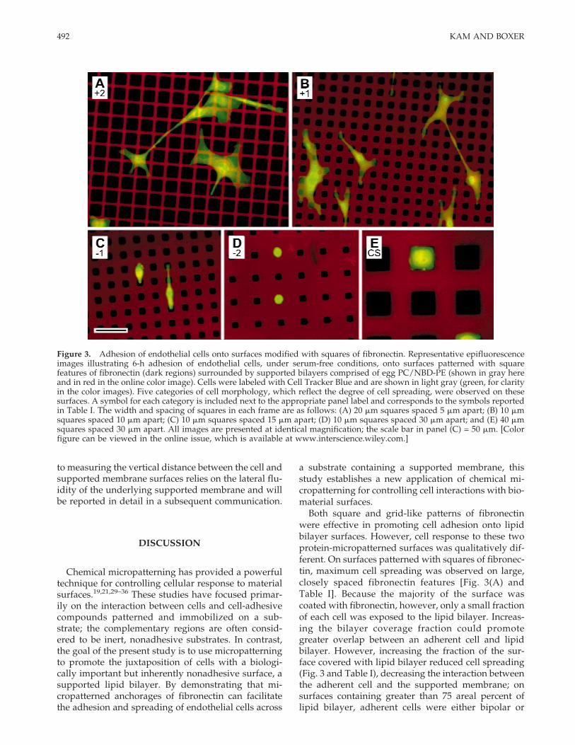
Adhesion of endothelial cells onto surfaces contain-ing two complementary patterns of fibronectin andlipid bilayers was examined under serum-free cell cul-ture conditions to minimize the influence of exog-enous proteins. On all of these surfaces, endothelialcells selectively adhered to the patterns of fibronectin.The morphology of adherent cells was correlated withthe geometry and dimensions of the fibronectin barri-ers. On surfaces patterned with squares of fibronectinsurrounded and separated by bilayers of egg PC/NBD-PE, adherent cells exhibited five general mor-phologies, which were categorized as “+2,” “+1,”“−1,” “−2,” and “CS,” reflecting the degree of cell
Figure 2. Fluid lipid bilayers do not support endothelialcell adhesion. (A) After 6 h in serum-free media, endothelialcells on substrates of plain glass exhibited a well-spreadmorphology. (B) In contrast, cells on surfaces supporting afluid lipid bilayer of egg phosphatidylcholine (egg PC) ex-hibited a rounded morphology. (C) Cell adhesion densitywas reduced on lipid bilayers compared to plain glass. Pas-sivation of the supported bilayers with 10 mg/mL of bovineserum albumin further reduced cell adhesion [egg PC +BSA, panel (C)]. Cells on egg PC + BSA surfaces resembledthose on egg PC alone (B). The data in panel (C) are mean ±SEM, n = 3. * p < 0.005 (least significant difference test)compared to the substrate of egg PC. Panels (A) and (B) arepresented at identical magnification; the scale bar in panel(A) = 25 mm.
490 KAM AND BOXER
spreading; the predominant morphology observed oneach pattern is presented in Table I. On surfaces modi-fied with large, closely spaced squares of fibronectin,adherent cells were well spread [Fig. 3(A)] and re-sembled cells on unpatterned, adhesive surfaces suchas plain glass [Fig. 2(A)]; this well-spread morphologywas categorized as +2. Adherent cells spanned mul-tiple features of fibronectin, bridging over the narrow,intervening regions of supported lipid bilayer. De-creasing the width of each fibronectin square and/orincreasing the spacing between squares resulted in areduction in cell spreading; adherent cells exhibited abranched morphology, as illustrated in Figure 3(B).This morphology was categorized as +1. As was ob-served for the +2 morphology, cellular processesspanned multiple fibronectin squares, juxtaposing re-gions of the adherent cells to the lipid bilayers. Furtherreduction of the width of each square and/or increaseof the spacing between squares resulted in continuedreduction in cell spreading. Adherent cells exhibitedthe bipolar cell morphology illustrated in Figure 3(C),which was categorized as −1. On these surfaces, ad-herent cells extended only thin processes across theintervening lipid bilayer regions. On surfaces pat-terned with the smallest fibronectin squares (whichmeasured 5 mm in width), adherent cells wererounded, occasionally elaborating thin processes thatextended to a neighboring square. This morphology isillustrated in Figure 3(D), and was categorized as −2.For these four classes of cell morphology, the best pre-dictor of cell spreading was the fraction of surfaceoccupied by lipid bilayer, a parameter referred to as
the “bilayer coverage fraction,” which is reported inTable I for each surface. Specifically, the transitionfrom the +2 morphology to +1, −1, and finally −2,which represents a decrease in cell spreading, was di-rectly correlated to the bilayer coverage fraction, inde-pendent of the width or spacing of the fibronectinsquares (Table I).
The final morphology observed on surfaces modi-fied with squares of fibronectin is illustrated in Figure3(E), and is characterized by adhesion and spreadingof cells on individual features of fibronectin. Adherentcells did not extend processes across intervening re-gions of lipid bilayer. This confined spreading (CS)morphology was observed on fibronectin squaresmeasuring 40 mm in width and spaced 20 mm orgreater from neighboring squares.
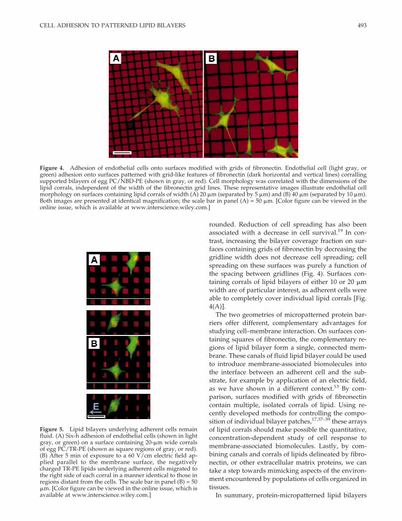
On surfaces patterned with grids of fibronectin sur-rounding corrals of lipid bilayers, adherent cells ex-hibited two morphologies. Cells adherent to surfacespatterned with lipid corrals measuring either 10 or 20mm in width exhibited a well-spread morphology,covering either large fractions or entire areas of mul-tiple corrals; this morphology is illustrated in Figure4(A), which shows cells on a surface containing anarray of 20-mm wide corrals. In contrast, cells on sur-faces containing lipid corrals measuring 40 mm inwidth extended multiple, long processes selectivelyalong the fibronectin grids [Fig. 4(B)]. Cell bodies alsoselectively adhered to fibronectin, but rarely extendedacross the lipid corrals. Cell morphology on these sur-faces was only correlated with the width of the lipidbilayer corrals and was independent of the width ofthe fibronectin gridlines.
Cell adhesion over a region of lipid bilayer did notinfluence the fluid properties of the supported mem-brane. Figure 5(A) illustrates 6-h adhesion of endothe-lial cells onto a surface containing an array of 20-mmwide lipid corrals (surrounded by fibronectin) com-posed of egg PC/TR-PE. After the cells were fixed, anelectric field of 60 V/cm was applied parallel to themembrane surface, inducing migration of the nega-tively charged, fluorescently labeled TR-PE lipids tothe right side of each corral [Fig. 5(B)]. Gradients wereformed in corrals underlying an adherent cell thatwere identical to corrals not covered by a cell, indicat-ing that the mobility of lipids in these supported mem-branes was not affected by cell adhesion over the bi-layer regions. This lateral mobility of lipids under ad-herent cells suggests that the distance between the cellsurface and the supported membrane is greater thanabout 10 Å, the likely extent of the dye headgroup ofTR-PE above the membrane surface. In preliminaryexperiments we have found that 40-nm diameterbeads attached to the headgroups of lipids in sup-ported bilayers do not diffuse under adherent cells,suggesting an upper limit to the distance between thecell surface and supported membrane. This approach
TABLE ICell Morphology as a Function of Pattern Dimensions
Width of Squares (mm)
40 20 10 5
Spac
ing
betw
een
squa
res
(mm
)
5 +2 (0.21) +2 (0.36) +2 (0.56)
10 +2 (0.36) +2 (0.56) +1 (0.75) −2 (0.89)
15 +2,+1 (0.67) −1 (0.84) −2 (0.94)
20 CS (0.56) +1 (0.75) −1 (0.89) −2 (0.96)
30 −2 (0.84) −2 (0.94) −2 (0.98)
Six-h adhesion of endothelial cells onto surfaces patternedwith squares of fibronectin. The predominant cell morphol-ogy (illustrated in Fig. 3) observed on each pattern is pre-sented as a function of the width of and spacing between thefibronectin squares. The fraction of each surface occupied bylipid bilayer is indicated in parentheses. The inclusion oftwo symbols for 20-mm wide squares spaced 15 mm apartreflects the observation of large populations of both mor-phologies.
491CELL ADHESION TO PATTERNED LIPID BILAYERS
to measuring the vertical distance between the cell andsupported membrane surfaces relies on the lateral flu-idity of the underlying supported membrane and willbe reported in detail in a subsequent communication.
DISCUSSION
Chemical micropatterning has provided a powerfultechnique for controlling cellular response to materialsurfaces.19,21,29–36 These studies have focused primar-ily on the interaction between cells and cell-adhesivecompounds patterned and immobilized on a sub-strate; the complementary regions are often consid-ered to be inert, nonadhesive substrates. In contrast,the goal of the present study is to use micropatterningto promote the juxtaposition of cells with a biologi-cally important but inherently nonadhesive surface, asupported lipid bilayer. By demonstrating that mi-cropatterned anchorages of fibronectin can facilitatethe adhesion and spreading of endothelial cells across
a substrate containing a supported membrane, thisstudy establishes a new application of chemical mi-cropatterning for controlling cell interactions with bio-material surfaces.
Both square and grid-like patterns of fibronectinwere effective in promoting cell adhesion onto lipidbilayer surfaces. However, cell response to these twoprotein-micropatterned surfaces was qualitatively dif-ferent. On surfaces patterned with squares of fibronec-tin, maximum cell spreading was observed on large,closely spaced fibronectin features [Fig. 3(A) andTable I]. Because the majority of the surface wascoated with fibronectin, however, only a small fractionof each cell was exposed to the lipid bilayer. Increas-ing the bilayer coverage fraction could promotegreater overlap between an adherent cell and lipidbilayer. However, increasing the fraction of the sur-face covered with lipid bilayer reduced cell spreading(Fig. 3 and Table I), decreasing the interaction betweenthe adherent cell and the supported membrane; onsurfaces containing greater than 75 areal percent oflipid bilayer, adherent cells were either bipolar or
Figure 3. Adhesion of endothelial cells onto surfaces modified with squares of fibronectin. Representative epifluorescenceimages illustrating 6-h adhesion of endothelial cells, under serum-free conditions, onto surfaces patterned with squarefeatures of fibronectin (dark regions) surrounded by supported bilayers comprised of egg PC/NBD-PE (shown in gray hereand in red in the online color image). Cells were labeled with Cell Tracker Blue and are shown in light gray (green, for clarityin the color images). Five categories of cell morphology, which reflect the degree of cell spreading, were observed on thesesurfaces. A symbol for each category is included next to the appropriate panel label and corresponds to the symbols reportedin Table I. The width and spacing of squares in each frame are as follows: (A) 20 mm squares spaced 5 mm apart; (B) 10 mmsquares spaced 10 mm apart; (C) 10 mm squares spaced 15 mm apart; (D) 10 mm squares spaced 30 mm apart; and (E) 40 mmsquares spaced 30 mm apart. All images are presented at identical magnification; the scale bar in panel (C) = 50 mm. [Colorfigure can be viewed in the online issue, which is available at www.interscience.wiley.com.]
492 KAM AND BOXER
rounded. Reduction of cell spreading has also beenassociated with a decrease in cell survival.19 In con-trast, increasing the bilayer coverage fraction on sur-faces containing grids of fibronectin by decreasing thegridline width does not decrease cell spreading; cellspreading on these surfaces was purely a function ofthe spacing between gridlines (Fig. 4). Surfaces con-taining corrals of lipid bilayers of either 10 or 20 mmwidth are of particular interest, as adherent cells wereable to completely cover individual lipid corrals [Fig.4(A)].
The two geometries of micropatterned protein bar-riers offer different, complementary advantages forstudying cell–membrane interaction. On surfaces con-taining squares of fibronectin, the complementary re-gions of lipid bilayer form a single, connected mem-brane. These canals of fluid lipid bilayer could be usedto introduce membrane-associated biomolecules intothe interface between an adherent cell and the sub-strate, for example by application of an electric field,as we have shown in a different context.13 By com-parison, surfaces modified with grids of fibronectincontain multiple, isolated corrals of lipid. Using re-cently developed methods for controlling the compo-sition of individual bilayer patches,17,37–39 these arraysof lipid corrals should make possible the quantitative,concentration-dependent study of cell response tomembrane-associated biomolecules. Lastly, by com-bining canals and corrals of lipids delineated by fibro-nectin, or other extracellular matrix proteins, we cantake a step towards mimicking aspects of the environ-ment encountered by populations of cells organized intissues.
In summary, protein-micropatterned lipid bilayers
Figure 5. Lipid bilayers underlying adherent cells remainfluid. (A) Six-h adhesion of endothelial cells (shown in lightgray, or green) on a surface containing 20-mm wide corralsof egg PC/TR-PE (shown as square regions of gray, or red).(B) After 5 min of exposure to a 60 V/cm electric field ap-plied parallel to the membrane surface, the negativelycharged TR-PE lipids underlying adherent cells migrated tothe right side of each corral in a manner identical to those inregions distant from the cells. The scale bar in panel (B) = 50mm. [Color figure can be viewed in the online issue, which isavailable at www.interscience.wiley.com.]
Figure 4. Adhesion of endothelial cells onto surfaces modified with grids of fibronectin. Endothelial cell (light gray, orgreen) adhesion onto surfaces patterned with grid-like features of fibronectin (dark horizontal and vertical lines) corrallingsupported bilayers of egg PC/NBD-PE (shown in gray, or red). Cell morphology was correlated with the dimensions of thelipid corrals, independent of the width of the fibronectin grid lines. These representative images illustrate endothelial cellmorphology on surfaces containing lipid corrals of width (A) 20 mm (separated by 5 mm) and (B) 40 mm (separated by 10 mm).Both images are presented at identical magnification; the scale bar in panel (A) = 50 mm. [Color figure can be viewed in theonline issue, which is available at www.interscience.wiley.com.]
493CELL ADHESION TO PATTERNED LIPID BILAYERS
introduce a new level of sophistication into investigat-ing how membrane-associated biomolecules, whichcan be incorporated into the synthetic supported bi-layers, mediate cellular function. These surfaces po-tentially facilitate the study of how anchorage-dependent cells recognize and respond to membranebiomolecules, free of the complexity introduced by thepresence of other membrane components or internalcellular structures. Reorganization of either endog-enous or engineered40 molecular species incorporatedinto membranes of adherent cells using electric fields,a concept that has already proven useful in severalcontexts,41,42 may provide additional insight into themechanisms that regulate cell–membrane interactions.Finally, we can speculate that incorporation of cell–cell communication proteins, such as gap junctions,and electronics integrated into the solid support,could be used to probe the internal state of a cell,leading to advanced, cell-based devices.
The authors would like to thank Steven S. Andrews andCaroline M. Ajo for the fitting program and algorithm usedto extract diffusion coefficients from electrophoresis data,and Professor Chaitan Khosla for providing the cell culturefacilities utilized for this work. The Stanford Nanofabrica-tion Facility (SNF) is gratefully acknowledged for support infabrication.
References
1. Bayerl TM, Bloom M. Physical-properties of single phospho-lipid-bilayers adsorbed to micro glass-beads : a new vesicularmodel system studied by 2H nuclear magnetic-resonance. Bio-phys J 1990;58:357–362.
2. Johnson SJ, Bayerl TM, McDermott DC, Adam GW, Rennie AR,Thomas RK, Sackmann E. Structure of an adsorbed dimyris-toylphosphatidylcholine bilayer measured with specular re-flection of neutrons. Biophys J 1991;59:289–294.
3. Koenig BW, Kruger S, Orts WJ, Majkrzak CF, Berk NF, Silver-ton JV, Gawrisch K. Neutron reflectivity and atomic force mi-croscopy studies of a lipid bilayer in water adsorbed to thesurface of a silicon single crystal. Langmuir 1996;12:1343–1350.
4. Schoenwaelder SM, Burridge K. Bidirectional signaling be-tween the cytoskeleton and integrins. Curr Opinion Cell Biol1999;11:274–286.
5. Giancotti FG, Ruoslahti E. Transduction : integrin signaling.Science 1999;285:1028–1032.
6. Viola A, Lanzavecchia A. T-cell activation and the dynamicworld of rafts. Apmis 1999;107:615–623.
7. Sackmann E. Supported membranes: scientific and practicalapplications. Science 1996;271:43–48.
8. Watts TH, McConnell HM. Biophysical aspects of antigen rec-ognition by T cells. Ann Rev Immunol 1987;5:461–475.
9. McConnell HM, Watts TH, Weis RM, Brian AA. Supportedplanar membranes in studies of cell-cell recognition in the im-mune system. Biochim Biophys Acta 1986;864:95–106.
10. Grakoui A, Bromley SK, Sumen C, Davis MM, Shaw AS, AllenPM, Dustin ML. The immunological synapse: a molecular ma-chine controlling T cell activation. Science 1999;285:221–227.
11. Dori Y, Bianco-Peled H, Satija SK, Fields GB, McCarthy JB,Tirrell M. Ligand accessibility as means to control cell response
to bioactive bilayer membranes. J Biomed Mater Res 2000;50:75–81.
13. van Oudenaarden A, Boxer SG. Brownian ratchets: molecularseparations in lipid bilayers supported on patterned arrays.Science 1999;285:1046–1048.
14. Groves JT, Ulman N, Boxer SG. Micropatterning of fluid lipidbilayers on solid supports. Science 1997;275:651–653.
15. Cremer PS, Groves JT, Kung LA, Boxer SG. Writing and eras-ing barriers to lateral mobility into fluid phospholipid bilayers.Langmuir 1999;15:3893–3896.
16. Groves JT, Boxer SG. Electric field-induced concentration gra-dients in planar supported bilayers. Biophys J 1995;69:1972–1975.
17. Hovis JS, Boxer SG. Patterning barriers to lateral diffusion insupported lipid bilayer membranes by blotting and stamping.Langmuir 2000;16:894–897.
18. Kumar A, Biebuyck HA, Whitesides GM. Patterning self-assembled monolayers: applications in material science. Lang-muir 1994;10:1498–1511.
19. Chen CS, Mrksich M, Huang S, Whitesides GM, Ingber DE.Geometric control of cell life and death. Science 1997;276:1425–1428.
20. James CD, Davis RC, Kam L, Craighead HG, Isaacson M,Turner JN, Shain W. Patterned protein layers on solid sub-strates by thin stamp microcontact printing. Langmuir 1998;14:741–744.
21. Kam L, James CD, Withers G, Banker G, Craighead HG, Isaac-son M, Turner JN, Shain W. Neuron attachment and outgrowthon microcontact-printed polylysine-conjugated laminin. J Neu-rosci Meth 2000, in press.
22. Kung LA, Kam L, Hovis JS, Boxer SG. Patterning hybrid sur-faces of proteins and supported lipid bilayers. Langmuir 2000;16:6773–6776.
23. Brian AA, McConnell HM. Allogeneic stimulation of cytotoxicT cells by supported planar membranes. Proc Nat Acad SciUSA 1984;81:6159–6163.
24. Stelzle M, Miehlich R, Sackmann E. 2-Dimensional microelec-trophoresis in supported lipid bilayers. Biophys J 1992;63:1346–1354.
25. Hui SW, Viswanathan R, Zasadzinski JA, Israelachvili JN. Thestructure and stability of phospholipid bilayers by atomic forcemicroscopy. Biophys J 1995;68:171–178.
26. Mou J, Yang J, Shao Z. Tris(hydroxymethyl)aminomethane(C4H11NO3) induced a ripple phase in supported unilamellarphospholipid bilayers. Biochem 1994;33:4439–4443.
27. Mou J, Yang J, Shao Z. Atomic force microscopy of choleratoxin B-oligomers bound to bilayers of biologically relevantlipids. J Mol Biol 1995;248:507–512.
28. Radler J, Radmacher M, Gaub HE. Velocity-dependent forcesin atomic-force microscopy imaging of lipid films. Langmuir1994;10:3111–3115.
29. Bhatia SN, Yarmush ML, Toner M. Controlling cell interactionsby micropatterning in co-cultures: hepatocytes and 3T3 fibro-blasts. J Biomed Mater Res 1997;34:189–199.
30. Healy KE, Thomas CH, Rezania A, Kim JE, McKeown PJ, LomB, Hockberger PE. Kinetics of bone cell organization and min-eralization on materials with patterned surface chemistry. Bio-materials 1996;17:195–208.
31. James CD, Davis R, Meyer M, Turner A, Turner S, Withers G,Kam L, Banker G, Craighead H, Isaacson M, Turner J, Shain W.Aligned microcontact printing of micrometer-scale poly-L-lysine structures for controlled growth of cultured neurons onplanar microelectrode arrays. IEEE Trans Biomed Eng 2000;47:17–21.
32. Kam L. Modulation of neuron and astroglial cell function by
494 KAM AND BOXER
micropatterning and immobilization of select biomolecules onbiomaterial surfaces. Troy, NY: Dep Biomed Eng RensselaerPolytechnic Institute; 1999. p 205.
34. Saneinejad S, Shoichet MS. Patterned glass surfaces direct celladhesion and process outgrowth of primary neurons of thecentral nervous system. J Biomed Mater Res 1998;42:13–19.
35. St. John PM, Kam L, Turner SW, Craighead HG, Isaacson M,Turner JN, Shain W. Preferential glial cell attachment to mi-crocontact-printed surfaces. J Neurosci Meth 1997;75:171–177.
36. Singhvi R, Kumar A, Lopez GP, Stephanopoulos GN, WangDIC, Whitesides GM, Ingber DE. Engineering cell shape andfunction. Science 1994;264:696–698.
37. Cremer PS, Yang T. Creating spatially addressed arrays of pla-
nar supported fluid phospholipid membranes. J Am Chem Soc1999;121:8130–8131.
38. Kung LA, Groves JT, Ulman N, Boxer SG. Printing via photo-lithography on micropartitioned fluid lipid membranes. AdvMat 2000;12:731–734.
39. Kam L, Boxer SG. Formation of supported lipid bilayer com-position arrays by controlled mixing and surface capture. J AmChem Soc, to appear.
40. Saxon E, Bertozzi CR. Cell surface engineering by a modifiedStaudinger reaction. Science 2000;287:2007–2010.
41. Lin-Liu S, Adey WR, Poo MM. Migration of cell surface con-canavalin A receptors in pulsed electric fields. Biophys J 1984;45:1211–1217.