University of Wisconsin Milwaukee UWM Digital Commons Cell and Molecular Biology 3e: What We Know and How We Found Out - All Versions Biological Sciences 7-25-2016 Cell and Molecular Biology : What We Know & How We Found Out (Second Edition, Sample Chapter) Gerald Bergtrom University of Wisconsin - Milwaukee, [email protected]Follow this and additional works at: hps://dc.uwm.edu/biosci_facbooks_bergtrom is Book is brought to you for free and open access by UWM Digital Commons. It has been accepted for inclusion in Cell and Molecular Biology 3e: What We Know and How We Found Out - All Versions by an authorized administrator of UWM Digital Commons. For more information, please contact [email protected]. Recommended Citation Bergtrom, Gerald, "Cell and Molecular Biology : What We Know & How We Found Out (Second Edition, Sample Chapter)" (2016). Cell and Molecular Biology 3e: What We Know and How We Found Out - All Versions. 7. hps://dc.uwm.edu/biosci_facbooks_bergtrom/7
Transcript
University of Wisconsin MilwaukeeUWM Digital CommonsCell and Molecular Biology 3e: What We Know andHow We Found Out - All Versions Biological Sciences
7-25-2016
Cell and Molecular Biology : What We Know &How We Found Out (Second Edition, SampleChapter)Gerald BergtromUniversity of Wisconsin - Milwaukee, [email protected]
Follow this and additional works at: https://dc.uwm.edu/biosci_facbooks_bergtrom
This Book is brought to you for free and open access by UWM Digital Commons. It has been accepted for inclusion in Cell and Molecular Biology 3e:What We Know and How We Found Out - All Versions by an authorized administrator of UWM Digital Commons. For more information, pleasecontact [email protected].
Recommended CitationBergtrom, Gerald, "Cell and Molecular Biology : What We Know & How We Found Out (Second Edition, Sample Chapter)" (2016).Cell and Molecular Biology 3e: What We Know and How We Found Out - All Versions. 7.https://dc.uwm.edu/biosci_facbooks_bergtrom/7
Most introductory science courses start with a discussion of scientific method. The 2nd edition of
this interactive Cell & Molecular Biology electronic textbook, or iText is no exception. A key
feature of CMB 2e is still a focus on experimental support for what we know about cell and
molecular biology. A sense of how science is practiced and how investigators think about
experimental results is essential to understanding the relationship of cell structure and function,
not to mention the rest of the world around us. Rather than trying to be a comprehensive
reference book, CMB 2e selectively details essential methods and experiments that are the
basis of our current understanding of the biochemical and molecular basis of cell structure and
function. This focus is nowhere more obvious than in the list of learning objectives and in the
Voice-Over PowerPoint (VOP) presentations provided for each chapter. Learning objectives
align with chapter content and serve as an aid and guide to learning. They ask students to use
new-found knowledge to make connections and demonstrate deeper concept understanding and
critical thinking skills. The VOPs are freely available on Youtubetm (with optional closed
captioning), as are most of the videos linked elsewhere in the iText.
There are two versions of CMB 2e iText (all versions of the first edition are still available). The
Annotated CMB-2e iText, contains many embedded just-in-time links to external resources
including links to animations of cell process, relevant current research summaries, etc.
Challenge text boxes raise provocative questions about the iText content, and may be used to
provoke class or online discussion (assessed or not!). A CMB-2e iText For Instructors
(available on request) includes these features and adds writing assessments that the author has actually assigned for course credit. These appear in the right margin of the text and are 25
Words or Less writing assignments that aim to strengthen critical thinking and writing skills.
Some of these features are modeled in the CMB 2e Sample Chapter, such as online
discussions and low-stakes formative objective quizzes (note that hyperlinks to assessments in
the Sample and Instructor iText versions require student/instructor login to a course
management system and are therefore inactive).
While not comprehensive, this iText was written with the goal of creating content that is
engaging, free and comparable in quality to very expensive commercial textbooks. To that end,
illustrations created especially for the iText are supplemented by online open sources (with
appropriate attribution). So, whichever CMB 2e version you are use, we encourage instructors
to use the interactive features in this iText to challenge students. For their part, we
encourage students to think about how great experiments were inspired and designed, how alternative experimental results were predicted, how actual data was interpreted, and finally, and what questions the investigators (and we!) might want to ask next. Although the online iText is the most efficient way to access links and complete online assignments, students are free to download, read, study, and add your own annotations off-line... or print it out and write in the margins the old fashioned way! Your instructor will undoubtedly provide more detailed instructions for using your iText.
CMB2e Page v
Special Note to Instructors from the Author
All features of the Annotated version of the CMB 2e iText are freely available to you and your
students. The Complete version of the iText is available after filling out a short form identifying
you as an instructor. Feel free to add, subtract, modify or embellish any part of any version of
the text or interactive content to suit your purposes… and then provide your customized version
of the text to your students. Feel free also to ask your students participate in the improvement of
the iText… for fun or for credit and then…, share the results your efforts with others!
One final bit of advice: where I provide content updates e.g., in links to very current sources,
please be aware (and let the students know) that I refer to the content as new, interesting and
not necessarily definitive (i.e., it is subject to confirmation). I hope that you (and perhaps your
students!) will enjoy creating and customizing interactive elements in the iText. Above all, I hope
that your students will achieve a better understanding of how scientists use skills of inductive
and inferential logic to ask questions and formulate hypotheses…, and to how they apply
concept and method to testing those hypotheses.
Acknowledgements
First and foremost, credit for my efforts has to go to the University of Wisconsin-Milwaukee and
the 35-plus years of teaching and research experience that inform the content, concept and
purpose of this digital Open Education Resource (OER). I want to thank my colleagues in the
Center for Excellence in Teaching and Learning (CETL) and the Golda Meir Library at UW-M for
the opportunity and the critical input that led to what I have defined as an iText (interactive text).
Many thanks to Matthew Russell, Megan Haak, Melissa Davey Castillo, Jessica Hutchings,
Dylan Barth for help and the inspiration to suggest at least a few ways to model how open
course content can be made interactive and engaging. Thanks also to Tim Gritten and Kristen
Woodward for putting competent editorial eyes on the iText. Finally special thanks to Tim
Gritten for walking me through the intricacies of publication of the iText on the UW-M Digital
Commons… with uncommon patience!
CMB2e Page vi
About the Author Dr. Bergtrom is Professor (Emeritus) of Biological Sciences and a Learning Technology Consultant (formerly in the Center for Excellence in Teaching and Learning at the University of Wisconsin-Milwaukee. Scientific interests are cell and molecular biology and evolution. Pedagogic interests include blended and online instruction and the use of technology in the service of better teaching and learning. Dedicated to an active learning approach, he has taught face-to-face, blended and so called “flipped” classes, as well as fully online undergraduate and graduate courses in cell and molecular biology. He also developed and co-instructed Teaching with Technology, an interdisciplinary course aimed at graduate students that might someday find themselves struggling to teach others. With more than 40 years of experience in teaching and research, he has frequently tested and incorporated pedagogically proven teaching technologies into his courses. In addition to many research publications in cell biology and evolution, he has published on aspects of active blended, online and flipped classroom methods1-3. In 2015 Dr. Bergtrom published Cell and Molecular Biology – What We Know & How We Found Out, an Open Access/Creative Commons (i.e., no-cost) electronic textbook4. The updated second edition (CMB 2e) of this textbook was published in 20164. Access to the older editions remain available on the UWM Digital Commons website. 1. Bergtrom, G. (2006) Clicker Sets as Learning Objects. Int. J. Knowl. & Learn. Obj. 2:105-110.
(http://www.ijello.org/Volume2/v2p105-110Bergtrom.pdf) 2. Bergtrom, G. (2009) On Offering a Blended Cell Biology Course. J. Res. Center Ed. Tech.
5(1) (http://www.rcetj.org/?type=art&id=91609&). 3. Bergtrom, G. (2011) Content vs. Learning: An Old Dichotomy in Science Courses. J.
Asynchr. Learning Networks 15:33-44 (http://jaln_v15n1_bergtrom.pdf) 4. Bergtrom, G. Cell and Molecular Biology: What We Know & How We Found Out [CMB1e,
(2015) and CMB2e (2016), all versions] (http://dc.uwm.edu/biosci_facbooks_bergtrom/)
Scientific Laws are even closer to ‘fact’ than theories! These Laws are thought of as universal and are most common in math and physics. In life sciences, we recognize Mendel’s Law of Segregation and Law of Independent Assortment as much in his
honor as for their universal and enduring explanation of genetic inheritance in living
things. But we do not call these Laws facts. They are always subject to experimental
test. Astrophysicists are actively testing universally accepted laws of physics even
Mendel’s Law of Independent Assortment should not be called law (strictly speaking)
since it is not true as he stated it (go back and see how chromosomal crossing over was
found to violate this law!).
In describing how we do science, the Wikipedia entry suggests that the goal of a scientific
inquiry is to obtain knowledge in the form of testable explanations (hypotheses) that
can predict the results of future experiments. This allows scientists to gain an
understanding of reality, and later use that understanding to intervene in its causal
mechanisms (such as to cure disease). The better an hypothesis is at making predictions,
the more useful it is, and the more likely it is to be correct.
In the last analysis, think of hypotheses as educated guesses and think of Theories and/or
Laws as one or more experimentally supported hypothesis that everyone agrees should
serve as guideposts to help us evaluate new observations and hypotheses.
Here is how Wikipedia presents the protocol of the Scientific Method:
The cycle of formulating hypotheses, testing and analyzing the results, and formulating new
hypotheses, will resemble the cycle described below:
Characterizations: observations, definitions, and measurements of the subject of inquiry
Hypotheses: possible explanations of observations and measurements Predictions: reasoning by deductive and inferential logic from the hypothesis (note that
even widely accepted theories are subject to testing in this way)
Experiments (tests of predictions)
New Characterizations: observations, definitions, and measurements of the subject of
inquiry
CHALLENGE: Since “An hypothesis is a declarative sentence that sounds
like a fact…”, and since both theories and hypotheses are stated as
declarative sentences, articulate in your own words the difference
between an hypothesis and a theory.
CMB2e Page 12
A linearized, pragmatic scheme of the five points above is sometimes offered as a guideline
for proceeding:
1. Define a question
2. Gather information and resources (observe)
3. Form an explanatory hypothesis 4. Test the hypothesis by performing an experiment and collecting data in
a reproducible manner
5. Analyze the data
6. Interpret the data and draw conclusions that serve as a starting point for new hypothesis
…To which we would add the requirement that the work of the scientist be disseminated by
publication!
Why did philosophers (not scientists!) come up with systems of deductive and inductive logic so essential to the scientific method? Perhaps because experimental science only became common in the 19th century, when the term scientist began to define one who investigated natural phenomena by doing experiments. But long before this, philosophers developed formal rules of logic to try to understand nature, humanity’s relationship to nature, and the relationship of humans to each other. The scientific method grew along with increasing empirical observation and experimentation. We recognize these origins when we award the Ph.D. (Doctor of Philosophy), our highest academic degree!
III. Domains of Life
We believe with good reason (as you shall see) that all life on earth evolved from the
progenote, a cell that existed soon after the origin of life on the planet. Prokaryotes lack
nuclei (pro meaning before and karyon meaning kernel, or nucleus). Prokaryotic cells,
among the first descendants of the progenote, fall into two groups, archaea and eubacteria
(including bacteria and cyanobacteria, or blue-green algae). Prokaryotes were long defined
as a major life grouping, alongside eukaryotes. But the recent discovery of archaea
changed all that! Cells that thrive in inhospitable environments like boiling hot springs or
arctic ice were the first to be characterized as archaea, but now we know that these
unusual organisms inhabit more temperate environments. As of 1990, eubacteria, archaea
and eukaryotes characterize the three domains of life. That all living organisms can be
shown to belong to one of these three domains has dramatically changing our
understanding of evolution.
A. The Prokaryotes (eubacteria = bacteria and cyanobacteria)
Compared to eukaryotes, prokaryotic cells typically lack a nucleus as well as
mitochondria, chloroplasts, internal membranes and other organelles (e.g., endoplasmic
reticulum, assorted vesicles and internal membranes). They are typically
CMB2e Page 13
unicellular, although a few live colonial lives at least some of the time (e.g.,
cyanobacteria). Typical rod-shaped bacteria are shown (below left). A schematic
diagram of typical bacterial structure is also shown (below right).
1. Bacterial Reproduction
Without the compartments afforded by the internal membrane systems common to
eukaryotic cells, all intracellular events, from DNA replication to transcription and
translation to the biochemistry of life all happen in the cytoplasm of the cell. DNA is
a circular double helix that duplicates as the cell grows. While not enclosed in a
nucleus, bacterial DNA is concentrated in a region of the cell called the nucleoid.
Bacteria replicate their DNA throughout the life of the cell, ultimately dividing by
binary fission. The result is the equal partition of duplicated bacterial “chromosomes”
into new cells. The bacterial chromosome is essentially naked DNA, unassociated
with chromosomal proteins. In contrast, eukaryotic cells divide by mitosis, a time
when their DNA is organized into tightly packed chromosomes associated with many
different proteins (see below). Just to make life more interesting, we should note
that one group of prokaryotes (the Planctomycetes) have surrounded their nucleoid
DNA with a membrane!
2. Cell Motility and the Possibility of a Cytoskeleton
Movement of bacteria is typically by chemotaxis, a response to environmental
chemicals. They can move to or away from nutrients or noxious/toxic substances.
Bacteria exhibit one of several modes of motility. For example, many move using
CHALLENGE: How do you imagine these cells would divide their DNA
equally between daughter cells during cell division?
4. Bacterial Ribosomes do the Same Thing as Eukaryotic Ribosomes… and look
like them!
Ribosomes are the protein synthesizing machines of life. The ribosomes of prokaryotes are smaller than those of eukaryotes, but in vitro they can be made to translate eukaryotic messenger RNA (mRNA). Underlying this common basic function is the fact that the ribosomal RNAs of all species share base sequence and structural similarities indicating an evolutionary relationship. It was these similarities that revealed the closer relationship of archaea to eukaryotes than prokaryotes. Clearly, prokaryotes are a diverse group of organisms, occupying almost every wet or dry or hot or cold nook and cranny of our planet. But despite of this diversity, all prokaryotic cells share many structural and functional metabolic properties with each other… and with the archaea and eukaryotes! As we have seen with ribosomes, shared structural and functional properties support the common ancestry of all life. Finally, we not only share common ancestry with prokaryotes, we even share living arrangements with them. Our gut bacteria represent up to 10X more cells than our own! Read more at The NIH Human Microbiome Project. Also check out the following link for A Relationship Between Microbiomes, Diet and Disease.
B. The Archaebacteria (Archaea)
Allessandro Volta, a physicist for whom the Volt is named, discovered methane
producing bacteria (methanogens) way back in 1776! He found them living in the
extreme environment at the bottom of Lago Maggiore, a lake shared by Italy and
Switzerland. These unusual bacteria are cheomoautotrophs that get energy from H2
and CO2 and generate methane gas in the process. It was not until the 1960s that
Thomas Brock (from the University of Wisconsin-Madison) discovered thermophilic
bacteria living at temperatures approaching 100oC in Yellowstone National Park in
Wyoming. The nickname extremophiles was soon applied to describe organisms living
in any extreme environment. One of the thermophilic bacteria, now called Thermus
aquaticus, became the source of Taq polymerase, the heat-stable DNA polymerase that
made the polymerase chain reaction (PCR) a household name in labs around the world!
Extremophile and “normal” bacteria both lack nuclei are similar in size and shape(s), which initially suggested that they were closely related to bacteria and were therefore prokaryotes (see the electron micrograph of Methanosarcina and Pyrolobus, below). But Carl Woese [Woese CR (2004) A new biology for a new century. Microbiol. Mol. Biol. Rev. 68:173-186] compared the sequences of genes for ribosomal RNAs in normal bacteria and an increasing number of extremophiles, including the methanogens. Based on sequence similarities and differences, the extremophiles seemed to form a separate group from the rest of the bacteria as well as from eukaryotes. They were named archaebacteria, or archaea because these organisms were thought to have evolved even before bacteria.
Woese concluded that Archaea were a separate group, or domain of life from bacteria and eukaryotes profoundly changing our understanding of phylogenetic relationships. The three domains of life (Archaea, Eubacteria and Eukarya) quickly supplanted the older division of living things into Five Kingdoms (Monera, Protista, Fungi, Plants, and Animals). Another big surprise from rRNA gene sequence comparisons was that the archaea were more closely related to eukaryotes than bacteria! The evolution of the three domains is illustrated below.
Archaea contain genes and proteins as well as metabolic pathways found in eukaryotes
but not in bacteria, speaking to their closer evolutionary relationship to eukaryotes. They
also contain genes and proteins as well as metabolic pathways unique to the group,
testimony to their domain status.
While some bacteria and eukaryotes can live in extreme environments, the archaea
include the most diverse extremophiles:
Acidophiles: grow at acidic (low) pH.
Alkaliphiles: grow at high pH.
Halophiles: require high salt concentrations of salt for growth; Halobacterium
salinarium is shown below (at the left).
Methanogens: produce methane; a cross section of Methanosarcina acetivorans is
shown, above right. Note the absence of significant internal structure.
Barophiles: grow best at high hydrostatic pressure.
From: Halobacterium
Credit: James Ferry/Penn State University; Methanogen.
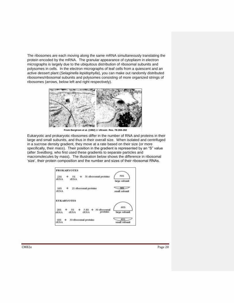
The ribosomes are each moving along the same mRNA simultaneously translating the
protein encoded by the mRNA. The granular appearance of cytoplasm in electron
micrographs is largely due to the ubiquitous distribution of ribosomal subunits and
polysomes in cells. In the electron micrographs of leaf cells from a quiescent and an
active dessert plant (Selaginella lepidophylla), you can make out randomly distributed
ribosomes/ribosomal subunits and polysomes consisting of more organized strings of
ribosomes (arrows, below left and right respectively).
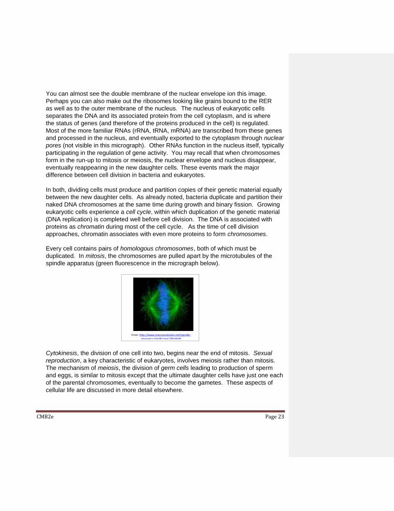
Eukaryotic and prokaryotic ribosomes differ in the number of RNA and proteins in their large and small subunits, and thus in their overall size. When isolated and centrifuged in a sucrose density gradient, they move at a rate based on their size (or more specifically, their mass). Their position in the gradient is represented by an “S” value (after Svedborg, who first used these gradients to separate particles and macromolecules by mass). The illustration below shows the difference in ribosomal ‘size’, their protein composition and the number and sizes of their ribosomal RNAs.
From Bergtrom et al. (1982) J. Ultrastr. Res. 78:269-282
CMB2e Page 21
B. Internal membranes and the Endomembrane System
Many of the vesicles and vacuoles in cells are part of an endomembrane system, or are
produced by it. The endomembrane system participates in synthesizing and packaging
proteins dedicated to specific uses into organelles. Proteins synthesized on the
ribosomes of the rough endoplasmic reticulum and the outer nuclear envelope
membrane will enter the interior space or lumen, or become part of the RER membrane
itself. Proteins incorporated into the RER bud off into transport vesicles that then fuse
with Golgi bodies. See some Golgi bodies (G) in the electron micrograph below.
Packaged proteins move through the endomembrane system where they undergo
different maturation steps before becoming biologically active, as illustrated below.
Adapted from Bergtrom and Robinson (1977) J. Ultrastr. Res. 60:395-405
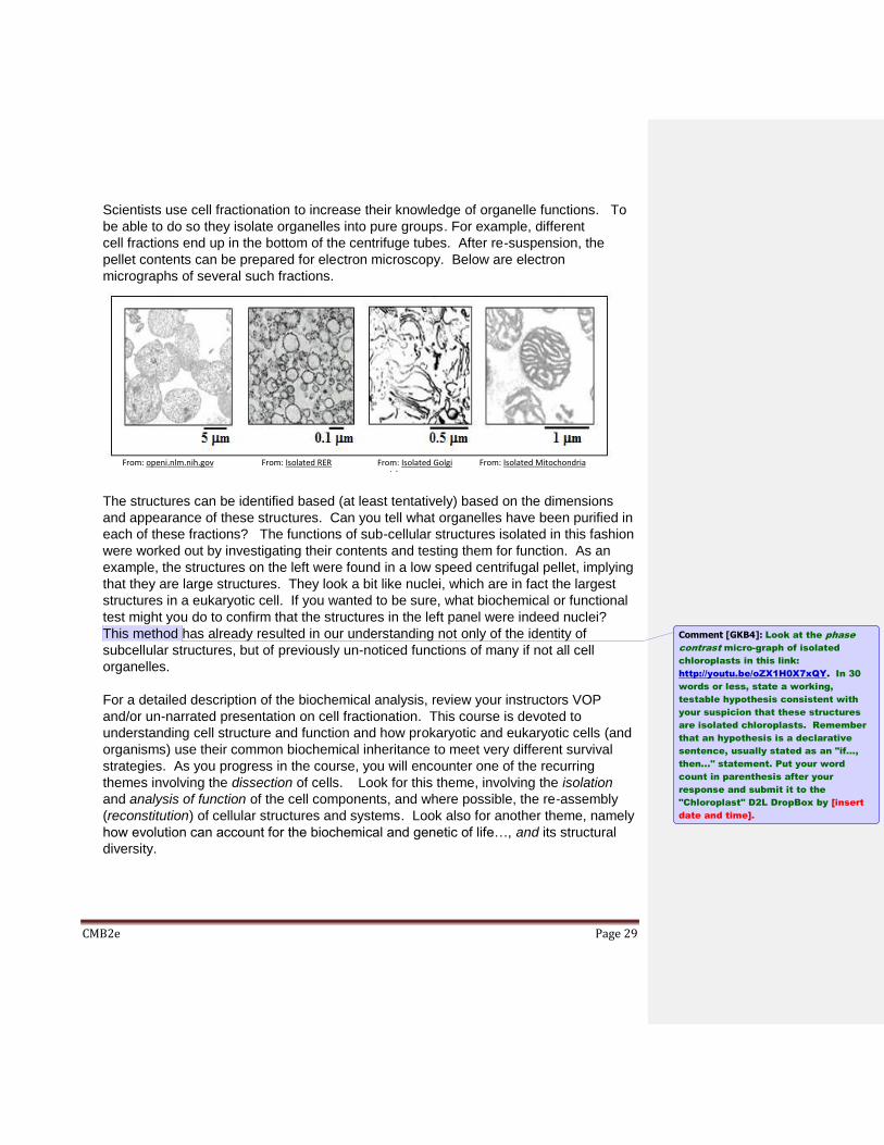
Nearly all eukaryotic cells contain mitochondria, seen in the electron micrograph below.
These organelles are surrounded by a double membrane and contain (and replicate)
their own DNA, with genes for some mitochondrial proteins. In the illustration above,
note that the surface area of the inner membrane is increased by being folded into
cristae, the site of cellular respiration (the oxidation of nutrients in aerobic organisms).
Mitochondria most likely evolved from aerobic bacteria (or protobacteria) engulfed by an early eukaryotic cell that later survived to become endosymbionts in the cell cytoplasm. The Endosymbiotic Theory was first proposed by Lynn Margulis [Sagan, L (1967) On the origin of mitosing cells. Journal of Theoretical Biology 14 (3): 225–274. (available at: Margulis L. Endosymbiotic theory)]. She also proposed an endosymbiotic origin of chloroplasts (see below).
The few protozoa that lack mitochondria have been found to contain mitochondrion-
derived organelles, such as hydrogenosomes and mitosomes; and thus probably lost
the mitochondria secondarily. Like mitochondria, the plastids of plants and some algae
have their own DNA and evolved from cyanobacteria that were are engulfed by primitive
eukaryotic cells. These endosymbionts became chloroplasts and other plastids.
V. Evolution, Speciation and the Diversity of Life
Natural selection was Charles Darwin’s theory for how evolution led to the structural
diversity of species. New species arise when beneficial traits are naturally selected from
genetically different individuals in a population, with the concomitant culling of less fit
individuals from populations over time. If natural selection acts on individuals, evolution
results from the persistence and spread of selected, heritable changes through successive
generations in a population. Evolution is reflected as an increase in diversity and
complexity at all levels of biological organization, from species to individual organisms to
molecules like DNA and proteins. For an easy read about the evolution of eyes (whose
very existence according to creationists could only have formed by intelligent design by a
creator), see the article in National Geographic by E. Yong (Feb., 2016, with its beautiful
photography by D. Littschwager).
We say that life on earth originated and then evolved from the progenote some 3.7-4.1
billion years ago. But the progenote may have been only one of many experimental cells
formed when conditions on earth were permissive to origins of life. Evolution began with
these first cells; by definition, all cells had all of the properties of life. Therefore, the
descendants of “first cells” with their separate origins, would have found different genetic
and biochemical solutions to achieving and maintaining life’s properties. But all cells and
organisms alive today also share the same genetics and biochemistries, suggesting that all
life forms other than the progenote never gained a foothold on the planet. At the same
time, the descendants of the progenote were evolving, diversifying and generating new
species. Since, it is possible that many lineages of its progeny (species) also went
extinct…, except for one, which we now call the last Universal Common Ancestor, or
LUCA. Repeated speciation, the continual divergence of life forms from this LUCA through
natural selection and evolution, is supported by the shared cellular structures, nucleic acid,
protein and metabolic chemistries (the ‘unity’ of life). Since the revolution in molecular
biology, shared gene and other DNA sequences have confirmed the shared common
ancestry of diverse organisms across all three of life’s domains.
These relationships largely confirm what we have learned from the species represented in
the fossil record. Morphological, biochemical and genetic traits that are shared across
species are defined as homologous, and can be used to reconstruct evolutionary histories.
The biodiversity that scientists (and environmentalists in particular) try to protect has
resulted from millions of years of speciation and extinction. It needs protection from the
unwanted evolutionary acceleration from human activities, including blatant extinctions
(think passenger pigeon), near extinction (think American bison by the late 1800s), the
introduction of invasive aquatic and terrestrial species, and the effects of climate change.
CMB2e Page 31
Let’s take a closer look at the biochemical and genetic unity among livings things. Albert
Kluyver first recognized that cells and organisms vary in form appearance in spite of the
essential biochemical unity of all organisms (http://en.wikipedia.org/wiki/Albert Kluyver).
We’ve already considered some of the consequences cells getting larger in evolution
when we tried to explain how larger cells divided their labors among smaller intracellular
structure (organelles). When eukaryotic cells evolved into multicellular organisms, it
became necessary for the different cells to communicate with each other and to respond to
environmental cues. Some cells evolved mechanisms to “talk” directly to adjacent cells and
others evolved to transmit electrical (neural) signals to other cells and tissues. Still other
cells produced hormones to communicate with cells to which they had no physical
attachment. As species diversified to live in very different habitats, they also evolved very
different nutritional requirements, along with more extensive and elaborate biochemical
pathways to digest their nutrients and capture their chemical energy. Nevertheless,
Kluyver and many others eventually recognized that despite billions of years of obvious
evolution and astonishing diversification, the underlying genetics and biochemistry of living
things on this planet is remarkably unchanged. This unity amidst the diversity of life is an
apparent paradox of life that we will probe in this course.
A. Genetic Variation, the Basis of Natural Selection
DNA contains the genetic instructions for the structure and function of cells and
organisms. When and where a cell or organism’s genetic instructions are used (i.e., to
make RNA and proteins) is regulated. Genetic variation results from random
mutations. Genetic diversity arising from mutations is in turn, the basis of natural
selection during evolution.
B. The Genome: an organisms complete genetic instructions
The genome of an organism is the entirety of its genetic material (DNA, or for some viruses, RNA). The genome of a common experimental strain of E. coli was sequenced by 1997. For details, see Blattner FR et al. (1997) The complete genome sequence of Escherichia coli K-12. Science 277:1452-1474. That of humans was completed by 001, well ahead of the predicted schedule! For more details, see Venter JC (2001) The sequence of the human genome. Science 291:1304-1351. Through mutation, genomes exhibit genetic variation, not only between species, but between individuals of the same species.
C. Genomic ‘Fossils’ Can Confirm Evolutionary relationships.
It has been known for some time that gene and protein sequencing can reveal evolutionary relationships and even familial relationships. Read about an early demonstration of such relationships based on amino acid sequence comparisons
across evolutionary time in Zuckerkandl E and Pauling L. (1965) Molecules as documents of evolutionary theory. J. Theor. Biol. 8:357-366. It is now possible to extract DNA from fossil bones and teeth, allowing comparisons of extant, ancient and even extinct species. Thus, DNA has been extracted from the fossil remains of humans, other hominids, and many animals. Sequencing this DNA (see the chapter on DNA Technologies) has revealed our relationship to some of our hominid ancestors and some of these ancient species. The reality though, is that DNA from organisms much older than 10,000 years is typically so damaged or simply absent that relationship building beyond that time is not possible. Now in a clever twist, using what we know of extant gene sequences, investigators recently ‘constructed’ a genetic phylogeny suggesting the sequences of some of our long-gone progenitors, including bacteria (click here to learn more: http://www.eurekalert.org/pub_releases/2010-12/miot-sd3121510.php). The comparison of these ‘reconstructed’ ancestral DNA sequences suggests when photosynthetic organisms diversified and when our oxygenic planet became a reality.
D. Origins of Life
Living things were once divided into 5 kingdoms. This classification has been replaced
by 3 domains of life. For more detail, check out Woese CR (1998) The universal
ancestor. Proc. Nat. Acad. Sci. 95:6854-6859. The molecular analyses discussed
above lead to the conclusion that all organisms alive today descended from a last
universal common ancestor, the LUCA. It is now accepted that there was a time,
however brief or long, when the earth was a lifeless (prebiotic) planet. But the question
of how life began has been with us since the beginnings or recorded history. We will
consider how we approach and suggest answers to questions about the origins of life in
a later chapter.
VI. Microscopy Reveals Life’s Diversity of Structure and Form
For a gallery of light, fluorescence and transmission and scanning electron micrographs,
check out this site (compare these with PowerPoint lecture images): Gallery of
Micrographs. The following is a brief description of different microscopic techniques and
what they can reveal.
Light microscopy reveals much of cellular diversity (The Optical Microscope). Check
this site through the section on fluorescence microscopy. Click on links to
different kinds of light microscopy to see sample micrographs of cell and tissue
samples. Also check micrographs and corresponding Drawings of Mitosis section for a
A 100 year-old variant of light microscopy, Lattice Light-Sheet Microscopy, was recently updated to allow us to follow subcellular structures and macromolecules moving about in living cells. It was recently applied to follow the movement and sub-cellular cellular location of RNA molecules associated with proteins in structures called RNA granules (check it out at RNA Organization in a New Light).
Confocal microscopy is a special form of fluorescence microscopy that enables imaging
through thick samples and sections. The result is often 3D-like, with much greater
depth of focus than other light microscope methods. Click at Gallery of Confocal
Microscopy Images to see a variety of confocal micrographs and related images; look
mainly at the specimens.
Transmission electron microscopy (TEM) achieves more power and resolution than any
form of light microscopy (Transmission Electron Microscopy). Together with
biochemical and molecular biological studies continues to reveal how different cell
components work with each other (see cell fractionation, below). The higher voltage in
High Voltage Electron microscopy is an adaptation that allows TEM through thicker
sections than regular (low voltage) TEM. The result is micrographs with greater
resolution and contrast.
Scanning Electron Microscopy (SEM) allows us to examine the surfaces of tissues,
small organisms like insects, and even of cells and organelles (Scanning Electron
Microscopy; check this web site through Magnification for a description of scanning EM,
and look at the gallery of SEM images at the end of the entry).