Applications :• quadruple labelling (sequential acquisition)• spectral resolution of 4 GFP variants• in-depth analysis of thick tissues and in vivo organs• time-lapse • FRAP• FRET
Accessibility : • free training on individual basis (Patrick Van Der Smissen)
referenced users with private login• protected data back-up (NAS)• first come / first served• 30 EUR /h in 2008
Confocal/multiphoton Zeiss LSM510+ thermostated chamber (25-40°C) with CO2
20
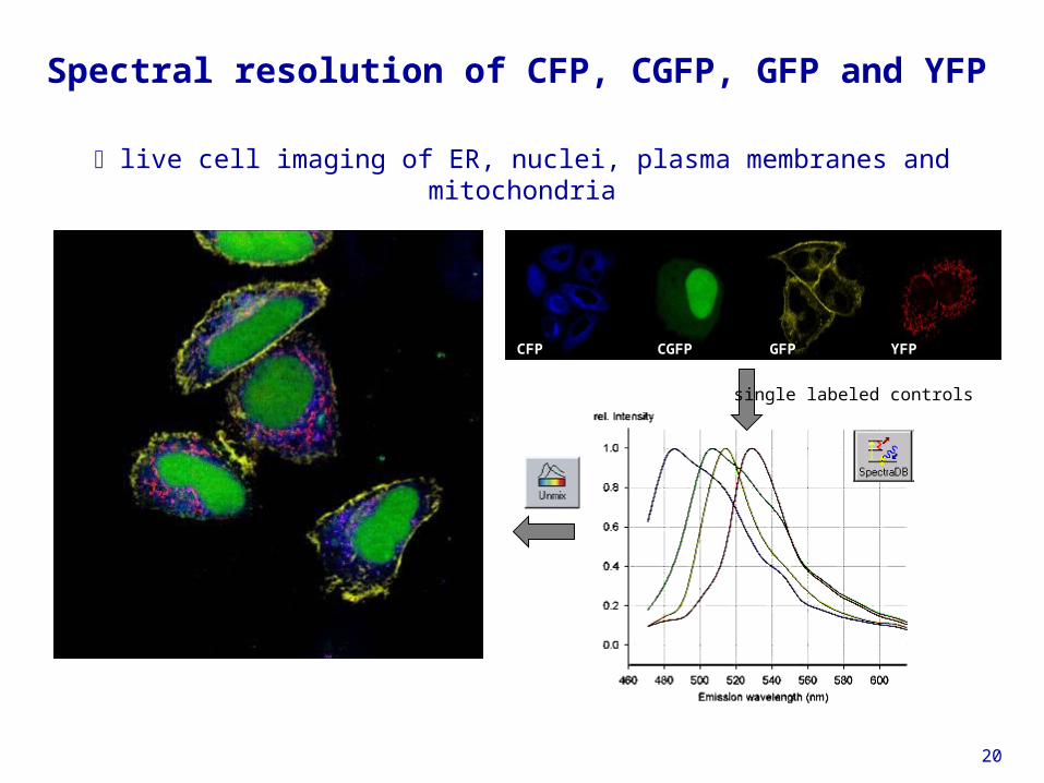
Spectral resolution of CFP, CGFP, GFP and YFP
live cell imaging of ER, nuclei, plasma membranes and mitochondria
CFP CGFP
GFP YFP
CFP CGFP YFPGFP
single labeled controls
21
Three-dimensional cell migration
brain slices; neurons expressing Thy1-YFP
50 µmStack 450 µm x 450 µm x 150 µm
22
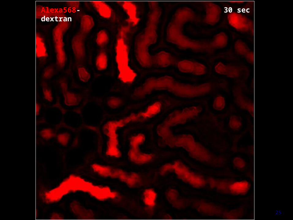
Functional imaging of kidney tubular function
23
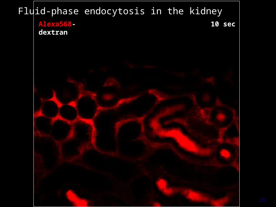
10 secAlexa568-dextran
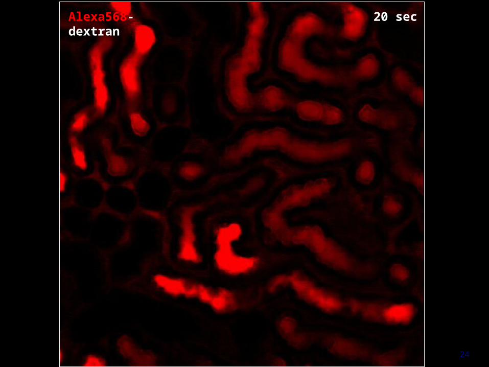
Fluid-phase endocytosis in the kidney
24
20 secAlexa568-dextran
25
30 secAlexa568-dextran
26
3 minAlexa568-dextran
27
20 minAlexa568-dextran
28
17 minFITC-OVA + TxRed-OVA
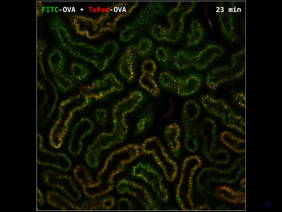
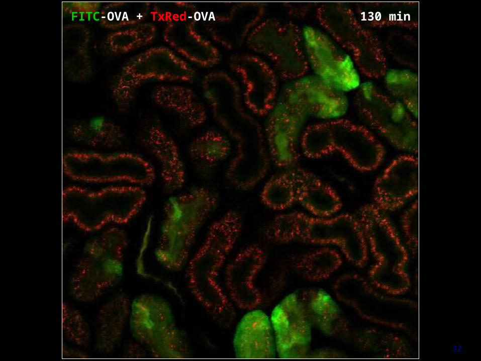
Receptor-mediated endocytosis and proteolysis
29
23 minFITC-OVA + TxRed-OVA
30
30 minFITC-OVA + TxRed-OVA
31
43 minFITC-OVA + TxRed-OVA
32
130 minFITC-OVA + TxRed-OVA
33
injected BCECF- dextran
Acidification in the kidney by ratio-imaging
plasma, pH 7.4
lysosomes,pH ~ 5.4
distal urine,pH < 5
34
Test of association : 1. co-localization ( ~ 500 nm)
mergeCD8 TC-R
2 µm
anergic CTL
competent CTL
P. Van Der Smissen (CELL) in collaboration with N. Demotte and P. Van der Bruggen (LICR)
35
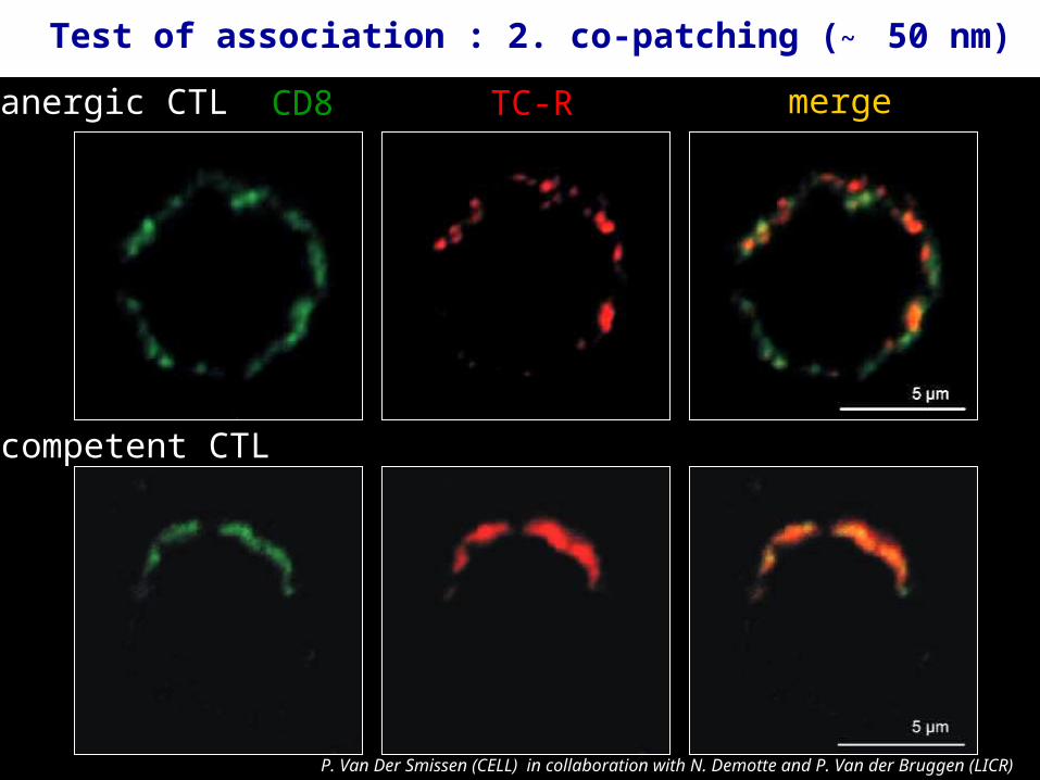
mergeCD8anergic CTL
competent CTL
TC-R
P. Van Der Smissen (CELL) in collaboration with N. Demotte and P. Van der Bruggen (LICR)
Test of association : 2. co-patching (~ 50 nm)
36
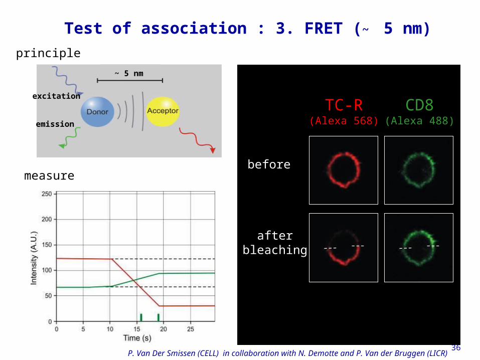
principle
measurebefore
afterbleaching
CD8(Alexa 488)
TC-R(Alexa 568)
P. Van Der Smissen (CELL) in collaboration with N. Demotte and P. Van der Bruggen (LICR)
Test of association : 3. FRET (~ 5 nm)
~ 5 nm
excitation
emission
37
Cell and tissue imaging platform
• Cell observer Zeiss Axiovert 200M
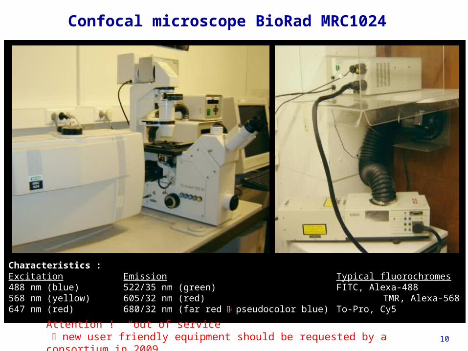
• "Old" confocal microscope BioRad MRC1024
• Confocal/multiphoton microscope Zeiss LSM510 Meta
• Transmission/scanning electron microscope Philips CM12
38
Transmission/scanning electron microscope Philips CM12
Accessibility : Collaborations
39
Transmission electron microscopy
receptor-mediated endocytosis in kidney PTC + HRP cytochemistry
B. K. Kishore et al (1996), Lab.Invest. 74 : 1013-1023
40
Scanning electron microscopy :thermoactivation of v-Src tsLA31 induces circular apical ruffling
Mettlen et al (2006), Traffic 7 : 589-603
2 µm
41
Immunogoldsurface labelling
ASGP receptors on hepatocytes
Immunogoldsurface labelling
ASGP receptors on hepatocytes
Van Der Smissen et al (1992), Eur. J. Cell Biol. 60 : 122-130
no ligand : random
+ ligand : clustered
42
Forthcoming equipments and applications :
• Stereodissection microscope with fluorescence (GFP transgenic mice)